Article

Solar Light-Assisted Oxidative Degradation of Ciprofloxacin in Aqueous Solution by Iron(III) Chelated Cross-Linked Chitosan Immobilized on a Glass Plate

1
Department of Chemistry, Jahangirnagar University, Savar 1342, Bangladesh
2
Department of Biotechnology and Genetic Engineering, Jahangirnagar University, Savar 1342, Bangladesh
3
Institute of Inorganic Chemistry, University of Göttingen, Tammannstrasse 4, D-37077 Göttingen, Germany
4
Institute of Organic and Biomolecular Chemistry, University of Göttingen, Tammannstrasse 2, D-37077 Göttingen, Germany
*
Authors to whom correspondence should be addressed.
Catalysts 2022, 12(5), 475; https://doi.org/10.3390/catal12050475
Submission received: 22 February 2022 / Revised: 8 April 2022 / Accepted: 12 April 2022 / Published: 23 April 2022
(This article belongs to the Special Issue Catalysis for the Removal of Water Pollutants)

Abstract

:
The massive worldwide use of antibiotics leads to water pollution and increasing microbial resistance. Hence, the removal of antibiotic residues is a key issue in water remediation. Here, we report the solar light-assisted oxidative degradation of ciprofloxacin (CPF), using H2O2 in aqueous solution, catalyzed by iron(III) chelated cross-linked chitosan (FeIII-CS-GLA) immobilized on a glass plate. The FeIII-CS-GLA catalyst was characterized by FTIR and 57Fe-Mössbauer spectroscopies as well as X-ray diffraction, revealing key structural motifs and a high-spin ferric character of the metal. Catalytic degradation of CPF was investigated as a function of solar light irradiation time, solution pH, concentration of H2O2 and CPF, as well as cross-linker dosage and iron(III) content in FeIII-CS-GLA. The system was found to serve as an efficient catalyst with maximum CPF degradation at pH 3. The specific ·OH scavenger mannitol significantly reduces the degradation rate, indicating that hydroxyl radicals play a key role. The mechanism of catalytic CPF degradation was evaluated in terms of pseudo-first-order and Langmuir-Hinshelwood kinetic models; adsorption of CPF onto the FeIII-CS-GLA surface was evidenced by field emission scanning electron microscopy coupled with energy dispersive X-ray spectroscopy. FeIII-CS-GLA can be reused multiple times with only minor loss of catalytic efficiency. Antimicrobial activity tests performed against both Gram-negative (Escherichia coli DH5α, Salmonella typhi AF4500) and Gram-positive bacteria (Bacillus subtilis RBW) before and after treatment confirmed complete degradation of CPF. These results establish the immobilized FeIII-CS-GLA as a rugged catalyst system for efficient photo-Fenton type degradation of antibiotics in aqueous solutions.

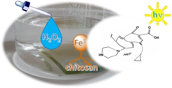
Graphical Abstract

1. Introduction

Antibiotics are used to treat microbial infections but it is well documented that the excess use of antibiotics leads to water pollution and antibiotic resistant microorganisms, which translates into higher medical costs, prolonged hospital stays, and increased mortality [1,2,3]. The reasons behind the high level of antibiotic residues in wastewater are the widespread use of pharmaceuticals around the world, including animal farms, agricultural land, hospital wastes etc. [4,5,6,7,8,9,10]. Several methods such as adsorption [11,12], advanced oxidation processes [13], sonochemistry [14], ionizing radiation [15], photodegradation [16], ozonation [17], ultraviolet irradiation [18], reverse osmosis [19], and coagulation [20], have been proposed to remove antibiotics from aqueous wastewater. Adsorption is found to be one of the most common and easiest methods in this context, but the removal process is very slow and the efficiency is low. Therefore, researchers are seeking alternative ways of removing antibiotics from wastewater [21,22].
In this study, one of the most commonly used antibiotics, ciprofloxacin hydrochloride (CPF) (Figure 1a), was considered as an exemplary substrate. CPF is a member of the broad-spectrum fluoroquinolones (FQs), which actively work against both Gram-positive and Gram-negative bacteria, and it is used worldwide for the treatment of bacterial infections in humans and animals [23,24]. The presence of this broad-spectrum antibiotic in aquatic systems may cause serious threats to the ecosystem and human health by inducing proliferation of bacterial drug resistance [25]. CPF is not biodegradable and is a highly recalcitrant pollutant, and even minute amounts of this antibiotic are problematic. Moreover, several reports confirmed the presence of CPF residues in fish tissues, which can have serious consequences for human health [26,27]. Thus, the removal of CPF from aquatic environments has already become a matter of concern. Various studies have addressed the removal of CPF from aqueous solutions by adsorption [28], membrane filtration [29], ultraviolet-activated persulfate [30], electrochemically activated persulfate [31,32], sonolysis [33], heterogeneous catalytic ozonation [34], advanced oxidation processes [35], pillared iron catalyst [36] Fenton’s oxidation [37], photo-Fenton degradation [38], FeS2/SiO2 microspheres [39], photodegradation [40], and photocatalytic processes using ZnO nanoparticles [41], graphene oxide-BiVO4 composites [42], titanium dioxide (TiO2) [43,44], silver/iron oxide/zinc oxide heterostructures [45], etc. Here we report the use of iron(III) chelated cross-linked chitosan, FeIII-CS-GLA, in the presence of hydrogen peroxide and solar light as a novel approach for the elimination of CPF from aqueous solutions. Lee and Lee previously investigated oxidative degradation of trichloroethylene in water by using FeII chelated with cross-linked chitosan [46], and Neto et al. [47] evaluated the removal of AsIII and AsV by employing crosslinked chitosan-FeIII (Ch-FeCL) beads as an adsorbent.
In the present study, the solar light-assisted oxidative degradation of CPF mediated by hydrogen peroxide and FeIII-CS-GLA immobilized on a glass plate was investigated in aqueous solutions under various pH conditions. Chitosan (CS) is a linear polysaccharide composed of randomly distributed β-(1–4)-linked D-glucosamine (deacetylated unit) and N-acetyl-D-glucosamine (acetylated unit) (Figure 1b). Chitosan has many useful characteristics such as hydrophilicity, biocompatibility, biodegradability and antibacterial properties. It can also selectively bind with various compounds and materials such as cholesterol, fats, metal ions, proteins, and tumor cells [48]. Chitosan is a cheap and commercially available biodegradable polymer. Thus, the process costs are low, and the present technique can potentially be used for removing antibiotics from aqueous solutions on a large scale.
The objectives of this comprehensive study were (i) to establish and assess robust protocols for preparing FeIII-CS-GLA immobilized on a glass plate by varying the concentration of glutaraldehyde (GLA) and iron(III); (ii) to characterize the FeIII-CS-GLA catalyst using Fourier transform infrared (FTIR) and 57Fe Mössbauer spectroscopy as well as X-ray diffraction (XRD); (iii) to perform batch experiments for investigating the effects of various parameters including solar light irradiation time, solution pH, cross-linker GLA and iron(III) dosages in FeIII-CS-GLA, and concentrations of H2O2 and CPF on the kinetics of catalytic degradation of CPF, and to examine the effect of a hydroxyl radical scavenger on the catalysis; (iv) to monitor both adsorption and degradation of CPF over FeIII-CS-GLA by performing field emission scanning electron microscopy (FE-SEM) and energy dispersive X-ray (EDX) investigations; (v) to analyze the degradation product of CPF by HPLC-UV-ESI-MS; (vi) to evaluate antimicrobial activities of CPF against both Gram-negative (i.e., Escherichia coli DH5α and Salmonella typhi AF450) and Gram-positive (i.e., Bacilius subtilis RBW) bacteria before and after catalytic degradation; and (vii) to investigate the reusability of the FeIII-CS-GLA catalyst for the oxidative degradation of CPF in aqueous solutions.

2. Results and Discussion

2.1. Characterization Results

2.1.1. FTIR Spectra

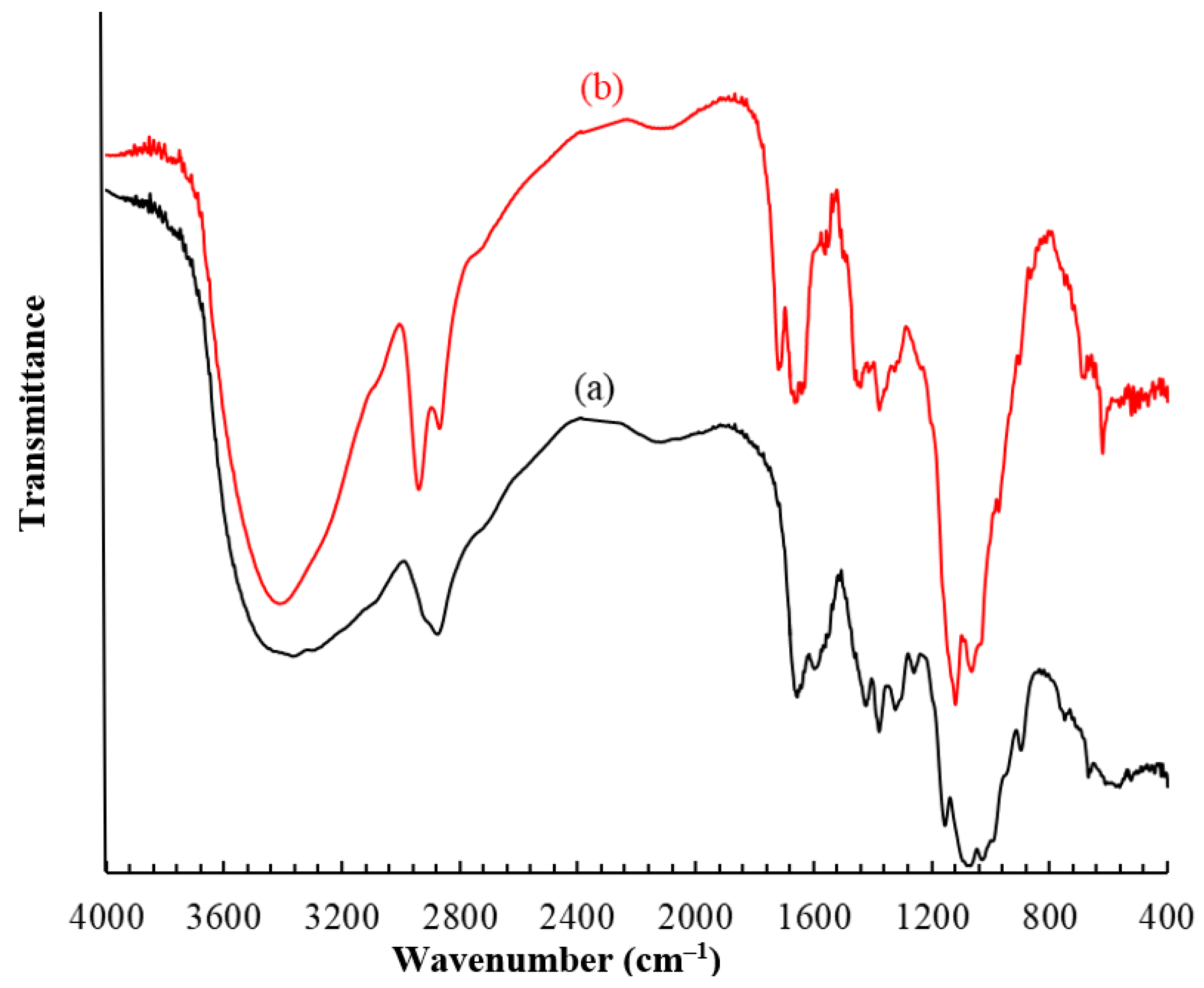
The FeIII-CS-GLA was prepared from CS, FeCl2·4H2O and GLA under aerobic conditions, following the method developed by Lee and Lee [46] with slight modifications as detailed in the experimental section. Figure 2 shows the FTIR spectra of CS and FeIII-CS-GLA recorded as KBr pellets in the range of 400–4000 cm−1. In the spectrum of CS (Figure 2a), the broad and intense peak in the region 3292–3361 cm−1 represents N–H and O–H stretching with intramolecular hydrogen bonds. The bands at around 2875 cm−1 with a shoulder at 2921 cm−1 correspond to the asymmetric and symmetric C–H stretching, respectively. The peaks located at 1654, 1597, 1550 and 1321 cm−1 are assigned to the stretching vibration of the C = O bond (amide I), the bending vibration of the N–H bond (primary amine), the bending vibration of the N–H bond (amide II) and the stretching vibration of the C–N bond (amide III), respectively [49]. The CH2 bending and CH3 symmetrical deformations were identified by the presence of peaks at around 1422 and 1377 cm–1, respectively. The peak at 1153 cm–1 represents the asymmetric stretching vibration of the C–O–C bridge. The peaks at 1066 and 1028 cm–1 are attributed to C–O stretching vibrations. The characteristic signal for the C–H deformation of the β-glycosidic bond is observed at 898 cm–1.
The spectrum of FeIII-CS-GLA (Figure 2b) is overall similar to the one of pure CS but shows some characteristic changes. Specifically, an additional absorption band is observed at 1660 cm−1 and assigned to the stretching vibration of C = N bonds, indicating the formation of Schiff bases as a result of the reaction between the aldehyde group of GLA and the amino group of CS during the crosslinking process. Furthermore, a sharp band appearing at 619 cm–1 is assigned to Fe-N stretching, reflecting coordination of the metal ion [47,50].

2.1.2. Mössbauer Spectroscopy

To determine the oxidation state and to gain insight into the electronic structure of the immobilized iron species in Fe-CS-GLA, 57Fe Mössbauer spectroscopy was applied. Various samples of Fe-CS-GLA prepared by varying the initial concentration of iron at constant CS and GLA concentrations were analyzed, as described in the materials and methods section. All Mössbauer spectra were recorded at 80 K, and a typical Mössbauer spectrum of Fe-CS-GLA is shown in Figure 3 (in that case the iron concentration was 7.5 mM). It is noted that all Mössbauer spectra were rather similar, featuring one quadrupole doublet with isomer shift δ = 0.48–0.49 mm/s and quadrupole splitting ΔEQ = 0.77–0.79 mm/s. These values are characteristic of high-spin iron(III) ions [51], as was previously observed in other Fe-chitosan complexes [52,53]. Webster et al. [54] proposed that iron(III) complexed with GLA cross-linked CS has an octahedral coordination sphere composed of two N-donors and two alcohol groups from the opposite glucosamine units in the equatorial plane, with two additional alcohol hydroxyl groups from C-6 in apical positions. The present isomer shift values (δ = 0.48–0.49 mm/s) and the characteristic Fe–N stretching vibration in the IR spectrum (Figure 2b) are in accordance with the proposed {N2O4} ligation of the iron ions in FeIII-CS-GLA.

2.1.3. X-ray Diffraction (XRD)

XRD patterns of pure CS and FeIII-CS-GLA are shown in Figure S1. The XRD pattern of (Figure S1a) CS shows its typical broad peaks for crystalline fragments at 2θ values of 11° for segments with –NH2 groups and 20° for the hydrophilic pockets in the chitosan, respectively [54]. The former peak disappeared in the XRD pattern of FeIII-CS-GLA and only a broad feature at the 2θ = 17° is present (Figure S1b). These changes upon cross-linking and iron coordination reflect the formation of Schiff bases as a result of the reaction between the GLA and the –NH2 groups of CS as well as the formation of FeIII-CS-GLA complexes through –NH2 binding sites and the hydrophilic pockets in the CS [50,54].

2.1.4. Surface Morphology and Elemental Composition of FeIII-CS-GLA

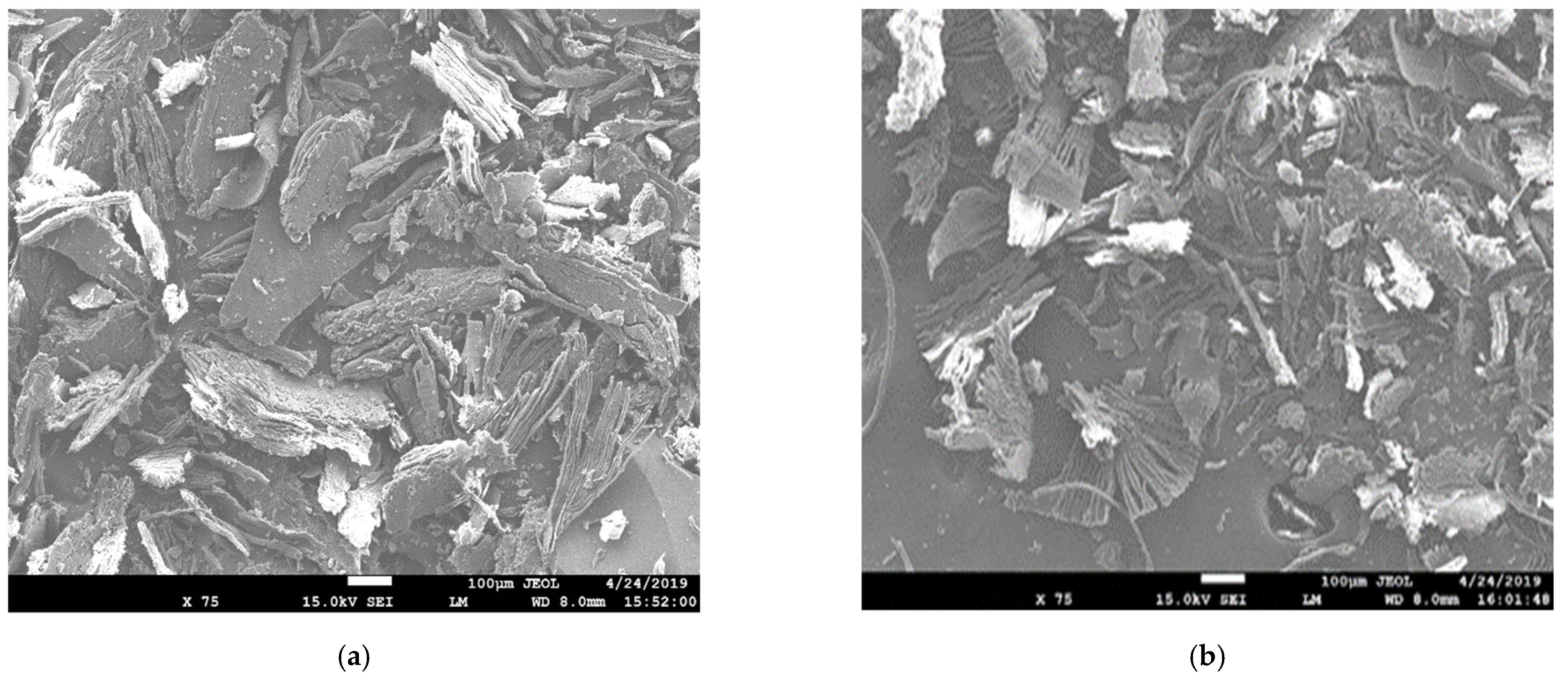
The surface morphology and elemental composition of the synthesized FeIII-CS-GLA immobilized on a glass plate were investigated using field emission scanning electron microscopy (FE-SEM) and energy-dispersive X-ray spectroscopy (EDX) analysis before and after its use for the catalytic degradation of CPF. A smooth surface structure of FeIII-CS-GLA is clearly observed in the FE-SEM image (Figure 4a); the corresponding EDX data are shown in Figure S2a. After CPF degradation, the FE-SEM image of the surface of FeIII-CS-GLA becomes brighter (Figure 4b) and the presence of precipitates suggests that a layer of CPF is present on the surface of FeIII-CS-GLA. A peak for fluorine was not detected in the EDX data of pristine FeIII-CS-GLA (Figure S2a), but it is clearly present in the EDX analysis after using FeIII-CS-GLA in the CPF degradation reaction (Figure S2b). Moreover, the mass percentage of nitrogen and chlorine becomes higher after degradation of CPF (Figure S2b), which indicates that CPF adsorption on the surface of FeIII-CS-GLA takes place under the catalytic conditions. The EDX data also show that the mass percentage of Fe slightly decreases after FeIII-CS-GLA has been used for the CPF degradation reaction, indicative of some leaching of FeIII during the catalytic process in aqueous solution at pH 3 [45].

2.2. Catalysis Results

2.2.1. Effect of Solar Light Irradiation

In order to explore suitable conditions for the catalytic degradation of CPF mediated by FeIII-CS-GLA, H2O2 and light, a number of control experiments omitting one or several components such as
(i)
50 µM CPF + CS-GLA immobilized on a glass plate + solar light;
(ii)
50 µM CPF + FeIII-CS-GLA immobilized on a glass plate + solar light;
(iii)
50 µM CPF + FeIII-CS-GLA immobilized on a glass plate + H2O2 + dark;
(iv)
50 µM CPF + FeIII-CS-GLA immobilized on a glass plate + H2O2 + solar light
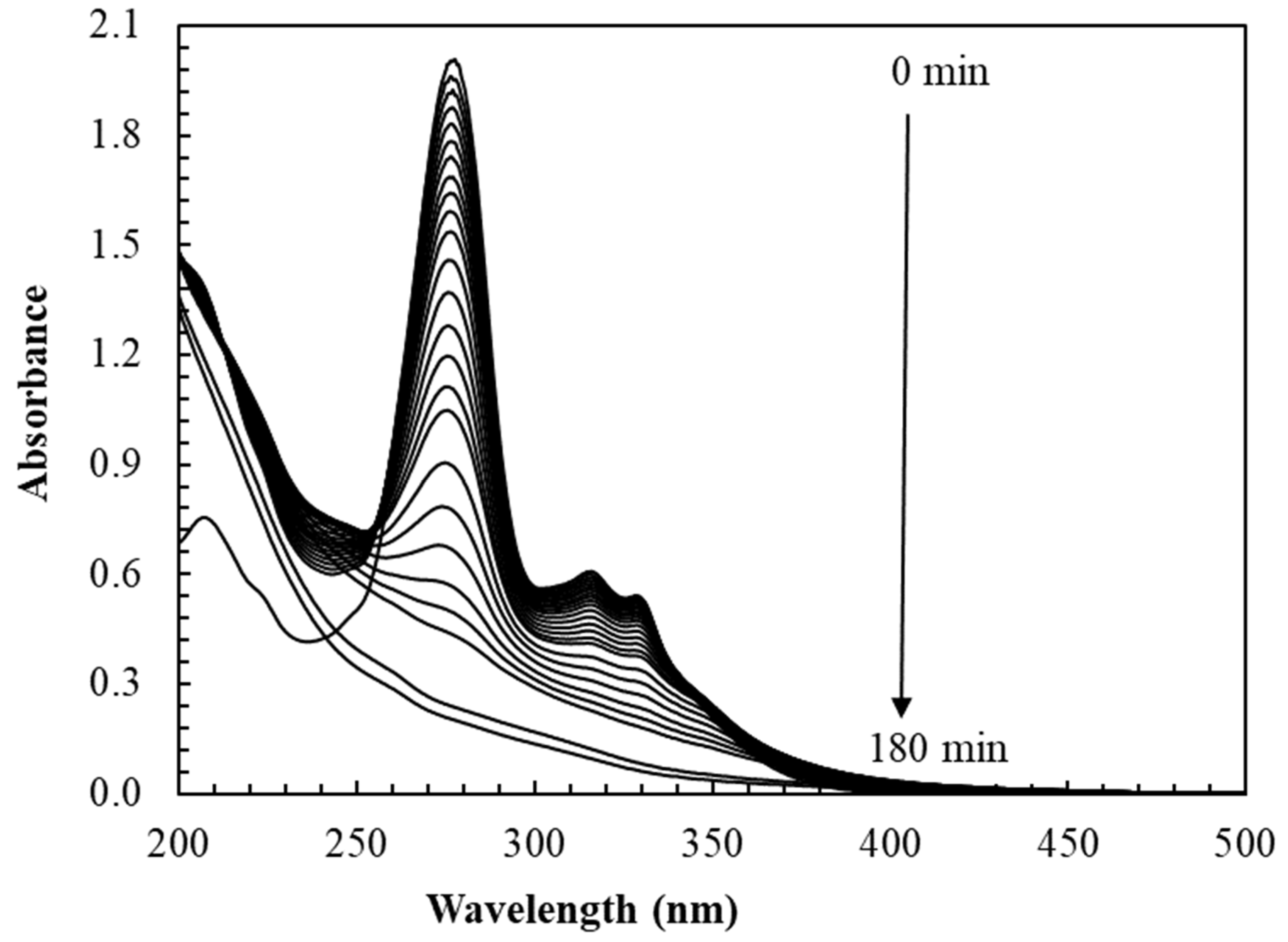
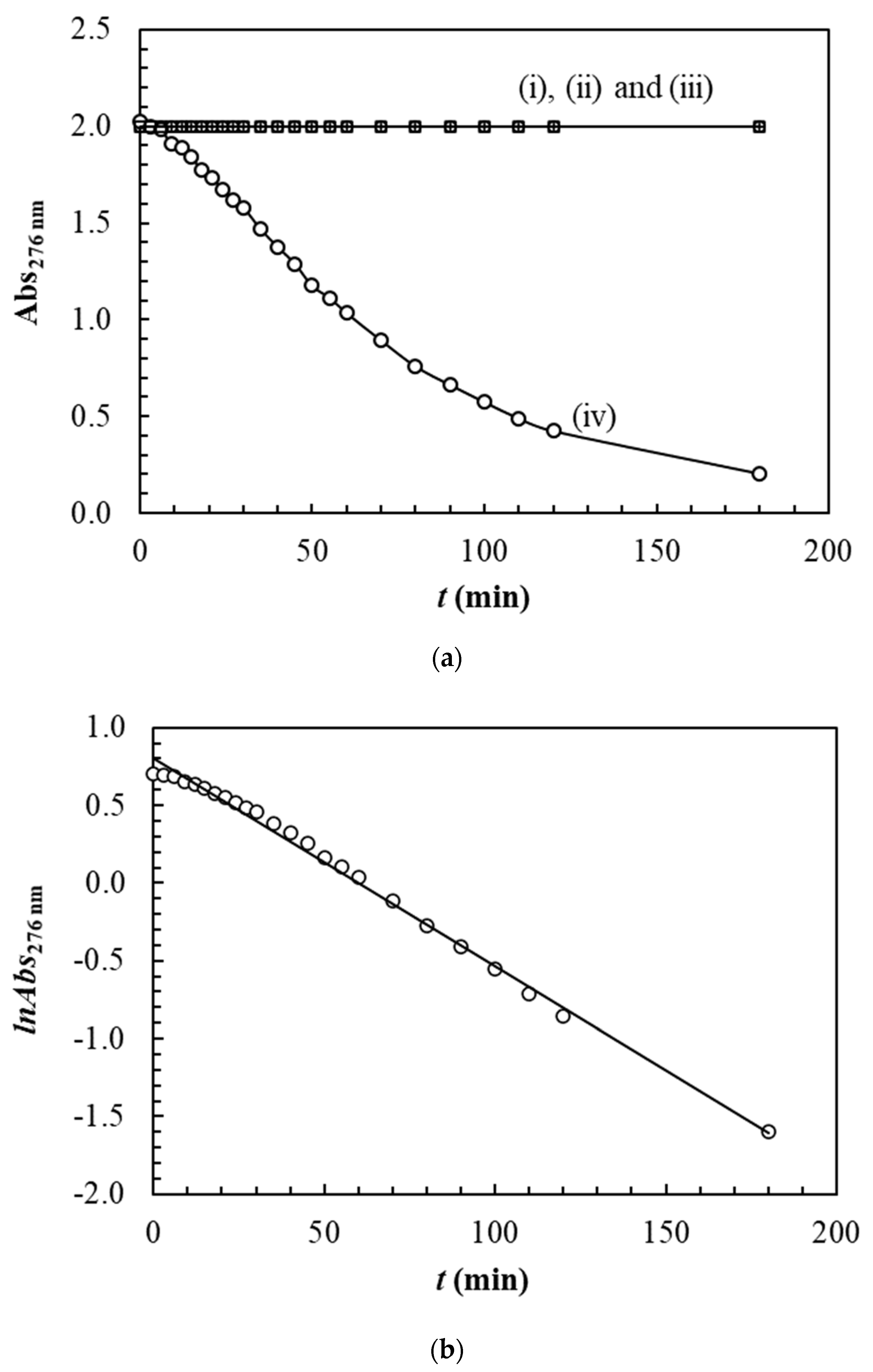
were performed in aqueous solution (pH 3) as described in the materials and methods section. The reaction progress was assessed by UV-vis spectroscopic monitoring of the characteristic absorption peak of CPF at 276 nm [32]. It is noted that the UV-vis spectra remained nearly constant in aqueous solution (pH 3) over 180 min for the control experiments (i), (ii) and (iii), indicating that CPF is not degraded under those conditions. Indeed, the spectrophotometric data confirm that the presence of both solar light and H2O2 is required for achieving FeIII-CS-GLA catalyzed oxidative degradation of CPF in water. The typical time-resolved spectral changes during oxidative degradation of CPF in experiment (iv) are depicted in Figure 5, evidencing that the characteristic absorption peak of CPF at 276 nm gradually decreases with time and completely disappears within 180 min.
Changes in absorbance of the characteristic peak (λmax: 276 nm) with time are depicted in Figure 6 for the various control experiments. The kinetic trace of CPF degradation in experiment (iv) follows a single exponential decay with time (Figure 6a), which was then used to determine a pseudo-first-order rate constant (kobs) for the solar light-assisted oxidative degradation of CPF catalyzed by FeIII-CS-GLA/H2O2. A typical pseudo-first-order kinetic plot lnAt vs. t is shown in Figure 6b. The value of the observed rate constant (kobs: 0.0134 min–1; Table 1) was determined from the slope of the straight line, and the half-life time was estimated to be 51.7 min. Similar reaction kinetics (kobs: 0.0117 min–1) were reported for the photocatalytic degradation of ciprofloxacin using ZnO nanoparticles in aqueous solution (pH 4) [41]. Other research groups also investigated the oxidative degradation of CPF by using sonolysis and various catalysts in aqueous solution; the reported kobs values were 0.0210 min–1 for the sonolysis process at pH 3 [33], 0.0253 min–1 for CPF degradation using a pillared iron catalyst (Fe-Lap-RD) at pH 3 [36], 0.0210 min–1 using TiO2 at pH 3 [43], and 0.0051 min–1 for Ag/Fe2O3/ZnO heterostructures at pH 4 [45], respectively (Table 1).

2.2.2. Effect of pH

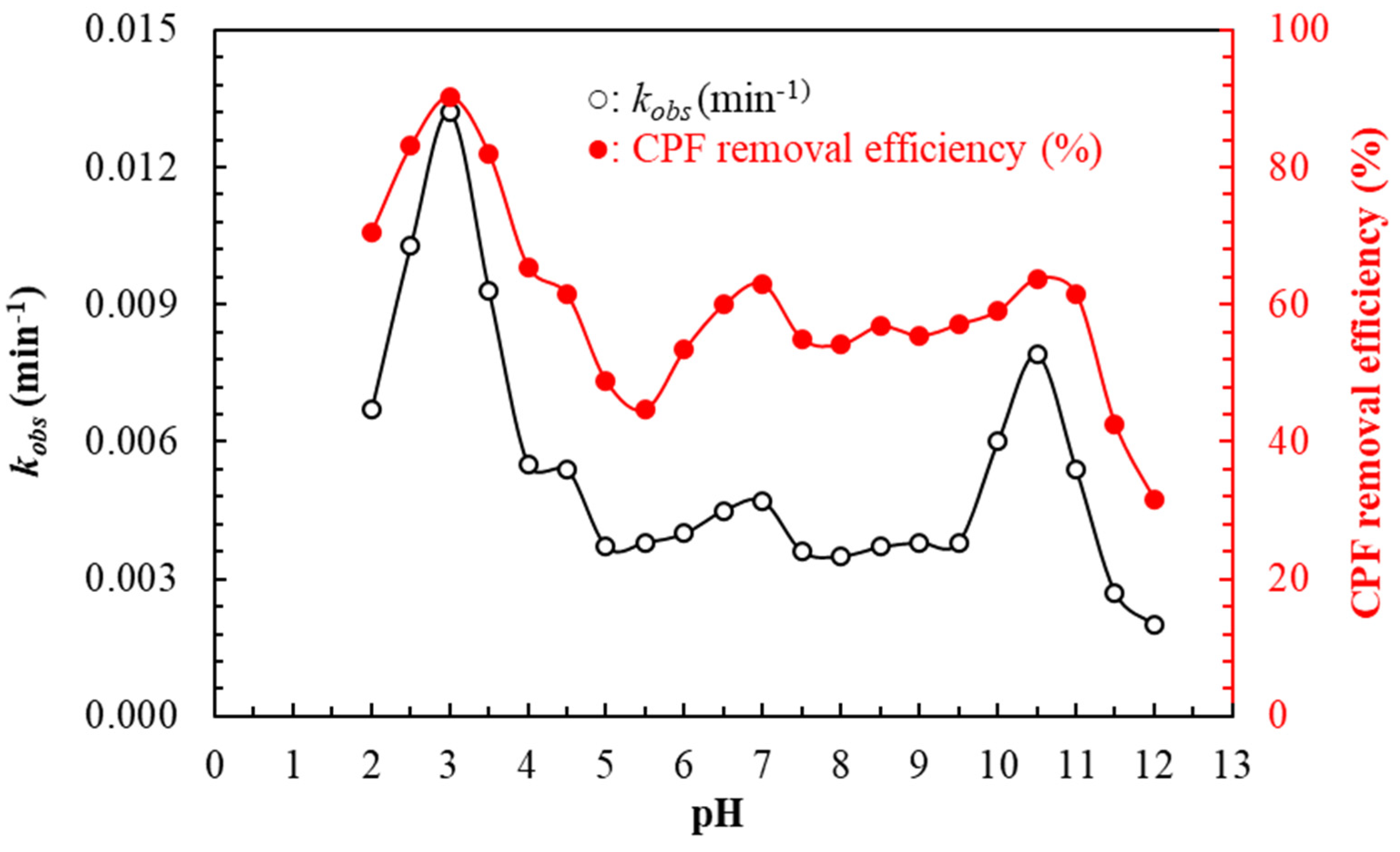
The solution pH is an important control parameter for the CPF degradation process. The solar light-assisted oxidative degradation of CPF catalyzed by FeIII-CS-GLA immobilized on a glass plate in aqueous solution in the range pH 2–12 at fixed initial concentration of CPF (50 μM) and H2O2 (6 mM) was investigated for 180 min. The effects of pH on kobs and CPF removal efficiency are shown in Figure 7. Starting at pH 2 and gradually increasing the pH, the values of kobs and CPF removal efficiency significantly increase to reach their maximum values at pH 3 (kobs: 0.0134 min−1 and CPF removal efficiency: 90.25%), but then decrease upon raising the pH up to pH ≤ 5.5 (kobs: 0.0038 min−1 and CPF removal efficiency: 44.8%). Further increase of pH leads to variations that peak at pH 7 for CPF removal efficiency (kobs: 0.0047 min−1 and CPF removal efficiency: 63.0%) and for both parameters at pH 10.5 (kobs: 0.0079 min−1 and CPF removal efficiency: 63.8%). Both kobs and CPF removal efficiency dramatically drop at pH ≥ 11. The pH dependence can likely be explained by the stability of hydroxyl radicals (OH) and the degree of protonation of CPF molecules depending on the solution pH. CPF exists as CPF3+ at pH < 3.64, CPF2+ at pH ≤ 5.05, CPF+ at pH ≤ 6.95, CPF at pH 6.95 to 8.95 and CPF at pH > 8.95 [33]. Bel et al. reported a similar correlation between the pseudo-first-order degradation rate constants and solution pH for the sonolysis of ciprofloxacin [33]. It has also been reported that the best photo-assisted degradation of CPF catalyzed by a modified laponite clay-based iron(III) nanocomposite (Fe-Lap-RD: 1 g/L) in the presence of H2O2 (60 mM) occurs in aqueous solution at pH 3 [36]. The present results are in accordance with those earlier observations. Thus, the solar light-assisted oxidative degradation of CPF catalyzed by FeIII-CS-GLA immobilized on a glass plate is most efficient in aqueous solution at pH 3 and hence, all further experiments were carried out at solution pH 3.

2.2.3. Effect of GLA Dosage in FeIII-CS-GLA

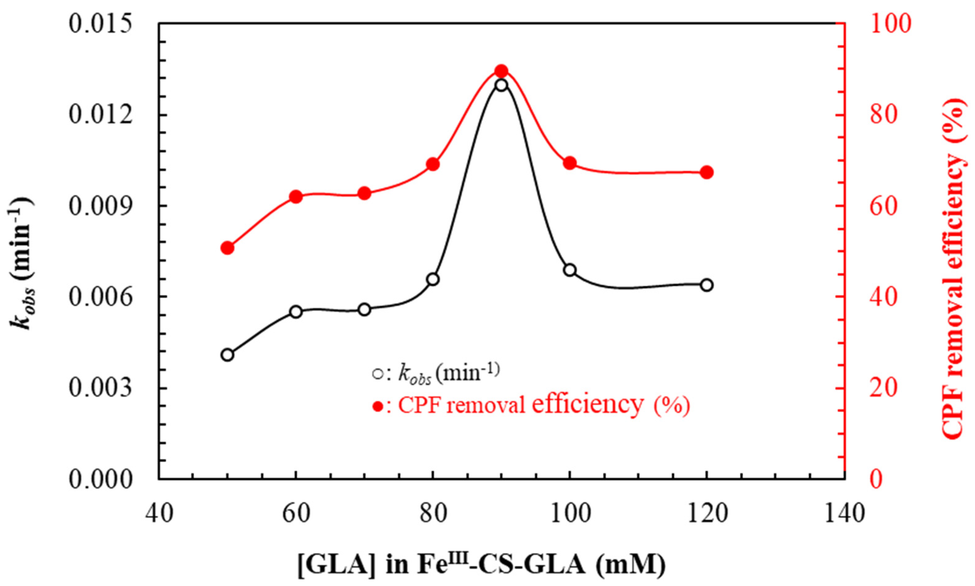
CS is frequently crosslinked with glutaraldehyde to enrich its mechanical strength, heat resistance, and chemical stability [55]. Usually, natural CS dissolves under acidic pH conditions, and cross-linking could help its application at low pH. The effects of cross-linking density in FeIII-CS-GLA immobilized on a glass plate were investigated by measuring the CPF degradation rate constants (kobs) and CPF removal efficiency in aqueous solution (pH 3) with a fixed initial concentration of CPF (50 μM) and H2O2 (6 mM) for 180 min. The effects of cross-linking density in FeIII-CS-GLA on kobs and CPF removal efficiency are shown in Figure 8. It is evident that the values of kobs and CPF removal efficiency increase with increasing concentration of GLA up to 90 mM in FeIII-CS-GLA, which may be due to the increase of FeIII content on the surface of the FeIII-CS-GLA material, leading to more production of the OH reactive species [46]. However, the kinetic rate constant (kobs) and CPF removal efficiency decrease significantly as the GLA dosage in FeIII-CS-GLA is continuously increased to 120 mM. This may be due to the increase in Schiff base formation as a result of the reaction between the aldehyde group of glutaraldehyde and the –NH2 groups of CS, leading to diminished complexation of FeIII through the –NH2 binding sites of CS. For CS microspheres loaded with gadolinium diethylenetriaminopentaacetic acid, some of us previously found that the size distribution and gadolinium content were influenced by the cross-linking density of the CS [56]. Therefore, all further solar light-assisted oxidative CPF degradation experiments at pH 3 were carried out with FeIII-CS-GLA (immobilized on a glass plate) synthesized using 90 mM of GLA.

2.2.4. Effect of Iron(III) Content in FeIII-CS-GLA

Figure 9 shows the change in CPF degradation rate constant (kobs) and CPF removal efficiency at various FeIII contents in FeIII-CS-GLA immobilized on a glass plate with a fixed initial concentration of CPF (50 μM) and H2O2 (6 mM) after 180 min. It is noticed that the values of kobs and CPF removal efficiency increase with increasing FeIII content, from 0.0134 min−1 and 90.3% for 1.25 mM to 0.0230 min−1 and 98.0% for 7.5 mM FeIII content in the FeIII-CS-GLA catalyst. These may be due to the increase of OH radical production with increasing FeIII content, where OH radicals control the rate of the CPF degradation reaction. Hence, a high concentration of FeIII in FeIII-CS-GLA is beneficial for high degradation rates and high CPF removal efficiency. Similar results were also observed in the photo-assisted mineralization of ciprofloxacin by using a modified laponite clay-based Fe nanocomposite (Fe-Lap-RD) as a heterogeneous catalyst in the presence of H2O2 and UV light [36], in the oxidative degradation of trichloroethylene via a modified Fenton reaction using FeII chelated with cross-linked CS [46], and in the degradation of alachlor with FeII-citrate in a photo-Fenton reaction [57]. For all further CPF degradation experiments of the present study, the iron concentration in the FeIII-CS-GLA catalyst immobilized on a glass plate was kept at 1.25 mM.

2.2.5. Effect of H2O2 Concentration

It has been reported that the H2O2 concentration is directly related to the number of hydroxyl radicals generated in the photo-Fenton reaction for the degradation of organic compounds [36]. In the present study, it is found that the H2O2 concentration is one of the dominant control parameters for the solar light-assisted oxidative degradation of CPF by FeIII-CS-GLA in aqueous solution (pH 3). The effect on kobs and on the CPF removal efficiency was studied by varying the H2O2 concentrations in the range 1–12 mM while keeping fixed the initial concentration of CPF (50 μM) and the FeIII content in the FeIII-CS-GLA catalyst (1.25 mM); the results are shown in Figure 10. Both the values of kobs and the CPF removal efficiency increase with increasing concentration of H2O2 from 1 mM H2O2 (kobs = 0.0090 min–1, 79.7% CPF removal efficiency) to 6 mM H2O2 (kobs = 0.0134 min–1, 90.3%). This is likely due to the formation of more hydroxyl radicals at higher H2O2 concentrations, which increases the degradation rate of CPF [36]. However, the values of kobs and CPF removal efficiency then level off at even higher H2O2 concentrations, reaching kobs = 0.0150 min–1 and 91.8% for 12 mM H2O2. This phenomenon might be explained by the scavenging effect of excess H2O2, which attenuates the number of OH radicals in the solution. These results are in accordance with previous findings for the degradation of ciprofloxacin [36] and azo dyes such as Orange II [58], reactive red HE-3B [59] and acid black 1 [60] catalyzed by Fe-Lap-RD in the presence of H2O2 and UV light. Therefore, the H2O2 concentration was kept at 6 mM for further CPF degradation experiments using the FeIII-CS-GLA immobilized on a glass plate (at pH 3).

2.2.6. Effect of CPF Concentration

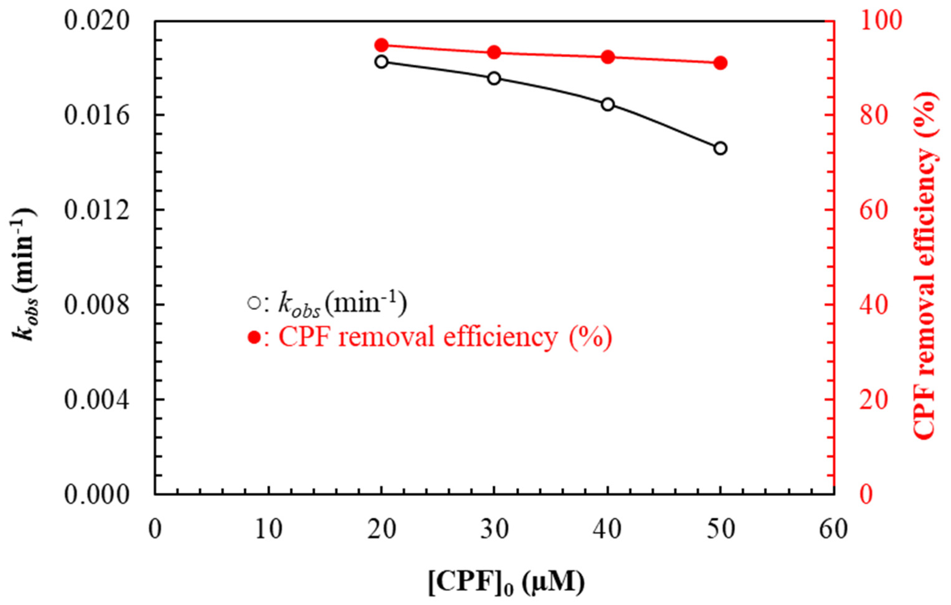
To examine the effect of substrate concentrations on the degradation rate, the CPF concentration was varied in the range 20–50 µM under standard conditions established in the experiments described above (1.25 mM FeIII content in FeIII-CS-GLA immobilized on a glass plate, 6 mM H2O2, pH 3, and 180 min reaction time). The CPF degradation rate constant (kobs) and the CPF removal efficiency decrease with increasing initial CPF concentration (Figure 11), from kobs = 0.0183 min–1 and 95.0% for a 20 µM CPF solution to kobs = 0.0134 min–1 and 90.3% for a 50 µM CPF solution. However, the overall CPF degradation rate increases with increasing initial CPF concentration. A likely reason for these observations is that the concentration of hydroxyl radicals remains constant for all CPF molecules when the CPF concentration is increased, leading to a net decrease of kobs and the removal efficiency [61]. It was also reported that the CPF degradation rate constant (kobs) gradually decreases with increasing initial CPF concentration because of the increase in Cl concentration, since Cl scavenges the hydroxyl radicals and results in prolonged reaction times [43]. Formation of inorganic radical anions ( Cl + OH ClOH ) is possible under these conditions. Although the reactivity of the latter radical may be relevant, it is not as reactive as OH, and thus the observed retardation effect is thought to be caused by strong adsorption of the anions on the FeIII-CS-GLA surface [43].
It has previously been proposed that the kinetics for the heterogeneous catalytic antibiotic degradation processes follow a Langmuir-Hinshelwood (LH) model [43,62], which implies that both molecules adsorb on the catalyst surface where they undergo a bimolecular reaction, in this case between the hydroxyl radical (OH) and CPF. The Langmuir-Hinshelwood model of heterogeneous catalytic CPF degradation systems is given by:
R a t e = d [ C P F ] d t = k K C P F [ C P F ] 1 + K C P F [ C P F ] 0 = k o b s [ C P F ]
1 k o b s = 1 k K C P F + [ C P F ] 0 k
In Equations (1) and (2), [CPF]0 (μM) is the initial concentration of ciprofloxacin, KCPF (L·μmol−1) is the Langmuir-Hinshelwood adsorption equilibrium constant, k (μmol·L−1·min−1) is the rate constant of the surface reaction, and kobs (min−1) is the pseudo-first-order rate constant. According to Equation (2), a plot of 1 k o b s vs. [CPF]0 is a straight line with slope ( 1 k ) and intercept ( 1 k K C P F ) . Applying this model to the present system gives k = 2.2060 μmol·L−1·min−1 and KCPF = 0.0102 L·μmol−1 (R2 = 0.9238; see Figure 12). This result suggests that the solar-light assisted degradation of CPF catalyzed by FeIII-CS-GLA immobilized on a glass plate is well described by the Langmuir-Hinshelwood kinetics model, and the substrate degradation mechanism occurs via adsorption steps on the catalyst [43,62].

2.2.7. Effect of a Hydroxyl Radical Scavenger

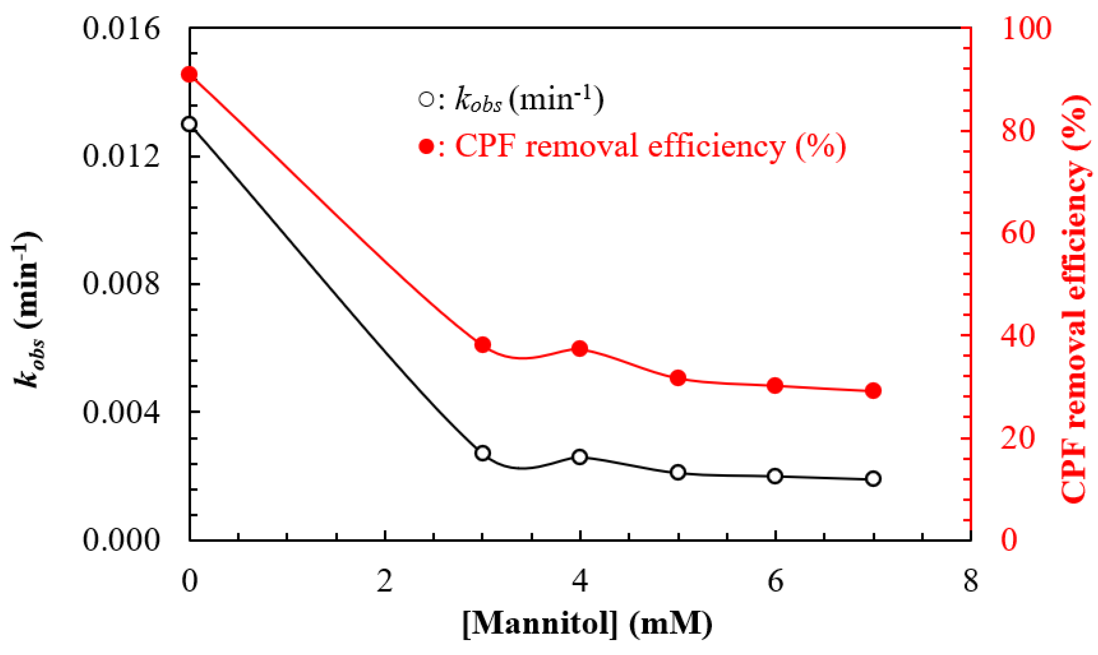
To further investigate the degradation mechanism, the oxidative decomposition of CPF catalyzed by FeIII-CS-GLA was evaluated in aqueous solution under the optimized reaction conditions in presence of the specific hydroxyl radical scavenger mannitol [63]. The changes in kobs values and CPF removal efficiency as a function of mannitol concentration are depicted in Figure 13. It is noticed that the values of kobs and the removal efficiency of CPF significantly decrease upon addition of mannitol, corroborating that hydroxyl radicals (•OH) are involved in the CPF degradation process. However, the values of kobs and the removal efficiency of CPF remain at 0.0019 min–1 and 29%, respectively, at the highest concentration of mannitol (Figure 13), suggesting that the CPF initially adsorbs onto the catalyst surface followed by oxidative degradation via hydroxyl radicals (•OH), as was also observed in photocatalytic oxidation of CPF under simulated sunlight [43].

2.2.8. Identification of the Oxidative Degradation Products of CPF

HPLC-UV-ESI-MS investigations were performed to identify residual amounts of CPF together with the degradation products obtained from the solar-light assisted oxidative degradation of CPF catalyzed by FeIII-CS-GLA in the presence of H2O2 in aqueous solution, using the standard conditions established in this work; the resulting chromatograms are shown in Figure S3. Three major peaks with different retention times are detected in the ESI-MS(−)- (Figure S3a) and UV-based (Figure S3b) chromatograms. The degradation products identified in the reaction solution are specified in Table 2. Compounds 1 (C5H8O5) and 3 (C5H8O4) are hydroxy glutaric acid and glutaric acid, respectively, as confirmed by the measurement of those compounds as reference substances (see supporting information); these two products likely originate from the oxidative degradation of the Schiff base crosslinking units formed in the reaction between glutaraldehyde and chitosan. The identity of degradation product 2 as succinic acid was also confirmed by measurement of a reference sample. Applying HPLC-UV-ESI(+)-MS analysis in the positive ion mode, CPF as a reference compound is detected with a retention time of 13.18 min (see supporting information). However, no such signal for intact CPF was found in the HPLC-UV-ESI-MS traces after oxidative treatment, neither in the positive nor in the negative ion mode, confirming complete degradation of the substrate.

2.2.9. Antibacterial Activity of CPF before and after Catalytic Degradation

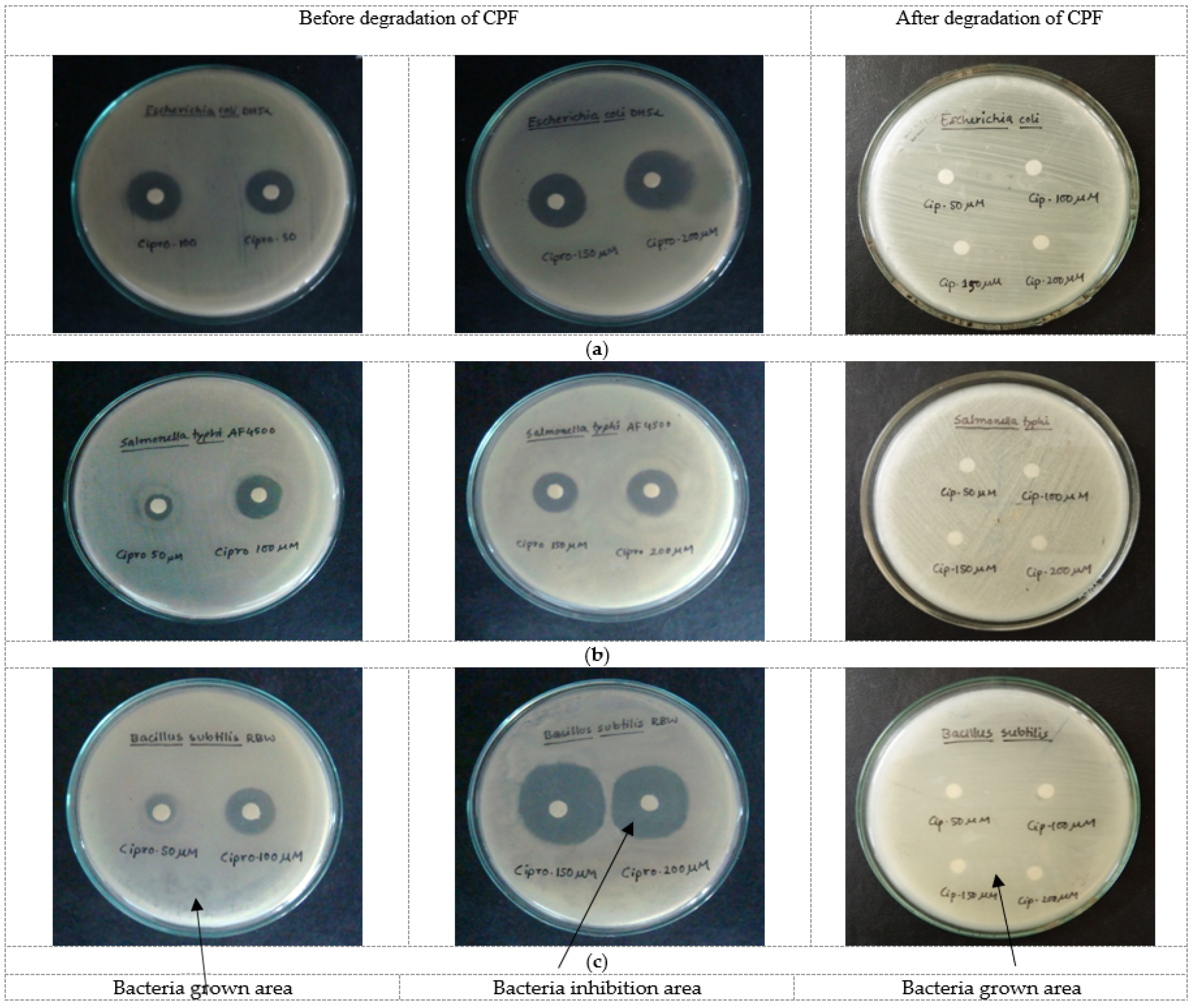
The antibacterial activity of CPF in the concentration range 50–200 μM was investigated against both Gram-negative (i.e., Escherichia coli DH5α and Salmonella typhi AF450) and Gram-positive (i.e., Bacilius subtilis RBW) bacteria before and after treatment with FeIII-CS-GLA/H2O2 under the standard conditions developed above. The typical zone of inhibition observed for each bacterium for various CPF solutions before and after catalytic degradation is shown in Figure 14, and the antibacterial activities are listed in Table 3. The pristine CPF solutions, before catalytic CPF degradation, significantly inhibit the growth of both Gram-negative (i.e., Escherichia coli DH5α and Salmonella typhi AF450) and Gram-positive (i.e., Bacilius subtilis RBW) bacteria. However, no zone of inhibition was observed for any of the bacteria after treatment with FeIII-CS-GLA/H2O2, which indicates that the CPF was completely degraded during the process. This is in line with the absence of any CPF in the HPLC-UV-ESI-MS analyses of samples that had undergone the oxidative treatment, and the findings suggest complete mineralization of CPF with FeIII-CS-GLA/H2O2 in the presence of solar light in aqueous solution [36].

2.2.10. Reuse of the FeIII-CS-GLA Catalyst for Degradation of CPF in Aqueous Solution

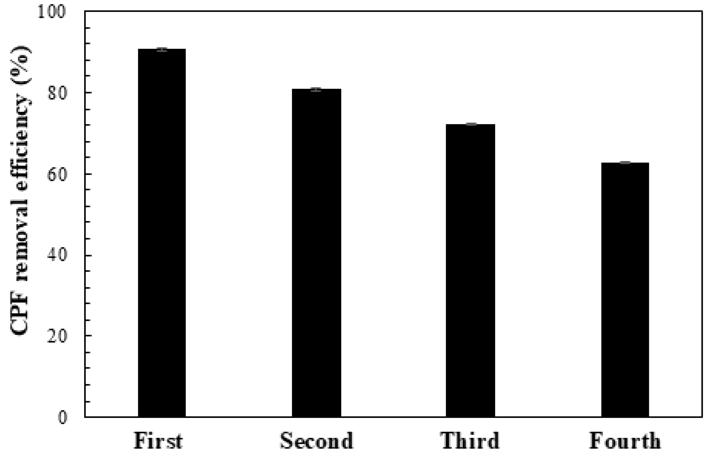
In order to examine the reusability of the catalyst, the CPF degradation using FeIII-CS-GLA immobilized on a glass plate was studied for four consecutive cycles (Figure 15). The CPF removal efficiency decreases slightly after repeated use of the FeIII-CS-GLA catalyst: after three and four runs, the CPF removal decreased from 90% to 80%, 72% and finally 63%, respectively. This might be due to the gradual leaching of iron ions as well as the loss of cross-linking density during the degradation process. The leaching of iron from FeIII-CS-GLA is corroborated by the changes in iron mass percentage found in the EDX analysis of the immobilized catalyst before and after CPF degradation (Figure S2a,b). Similarly, observation of the degradation products glutaric acid and hydroxy glutaric acid (Table 2) evidence the loss of cross-linking density in the FeIII-CS-GLA during the CPF degradation process. Overall, however, the synthesized FeIII-CS-GLA system was found to exhibit reasonable catalytic stability with only moderate loss of catalytic activity during repeated use for oxidative CPF degradation, and hence it shows good promise as a relatively stable and efficient photo-Fenton catalyst.

3. Materials and Methods

3.1. Materials and Reagents

Ciprofloxacin hydrochloride was purchased from Sigma-Aldrich, Germany and used as received. Chitosan (Sigma-Aldrich, Stuttgart, Germany), acetic acid (MERCK, Darmstadt, Germany), ferrous chloride tetrahydrate (FeCl2·4H2O; Sigma-Aldrich), glutaraldehyde (GLA; MERCK), analytical-grade H2O2 (35% in solution, Merck), mannitol (Sigma–Aldrich), hydrochloric acid (MERCK, Germany), sodium hydroxide (MERCK), deionized water, Müller-Hinton agar powder (Becon, Courbevoie, France), peptone (Becon), yeast extract (UNI-CHEM, Wuxi, China) etc. were used for this study in extremely pure form and used as received without further purification.

3.2. Preparation of Iron(III) Chelated Cross-Linked Chitosan, FeIII-CS-GLA, Immobilized on a Glass Plate

The FeIII-CS-GLA immobilized on a glass plate was prepared according to the method reported by Lee and Lee [46] with minor modifications. To prepare the chitosan solution, an exact amount (0.2 g) of chitosan was dissolved in 100 mL of 5% (v/v) acetic acid in a volumetric flask, and the solution was stirred with a magnetic stirrer at 250 rpm overnight. The required amount (0.7455 g) of FeCl2·4H2O was dissolved in 25 mL of 5% (v/v) acetic acid to prepare a 150 mM iron containing stock solution. Then, the required volume of this iron stock solution was mixed with the chitosan solution under magnetic stirring at 250 rpm for 1 h at room temperature (30 °C) to prepare solution A. Glutaraldehyde (GLA) was added to solution A with stirring to prepare the final solution B. To prepare the catalyst samples, an equal number of droplets of solution B was then deposited on the previously weighted glass slide (76 mm × 30 mm × 3 mm; previously cleaned with dilute HCl solution and ethanol), and the glass slides were heated at 100 °C for 2 h in an electronic oven to anneal the chitosan film. After that, the glass side with the catalyst films were cooled to room temperature and washed with deionized water. Finally the glass plates with immobilized FeIII-CS-GLA were dried again and stored in a desiccator to avoid the absorption of moisture. Different catalyst samples for the experiments described in this publication were prepared by varying the concentration of GLA (50–120 mM) and FeIII content (1.25–7.50 mM) in the FeIII-CS-GLA, respectively.

3.3. Characterization of FeIII-CS-GLA

The FeIII-CS-GLA catalyst was characterized by Fourier transform infra-red (FTIR) and [57] Fe Mössbauer spectroscopies, field emission scanning electron microscopy (FE-SEM) analysis coupled with energy dispersive X-ray spectroscopy (EDX), as well as X-ray diffraction (XRD). The FTIR spectra of chitosan and FeIII-CS-GLA were recorded in KBr in the range of 400–4000 cm−1 using a SHIMADZU Prestige-21 spectrometer (Japan). Mössbauer spectra of FeIII-CS-GLA were recorded with a [57] Co source in a Rh matrix using an alternating constant acceleration Wissel Mössbauer spectrometer operated in the transmission mode and equipped with a Janis closed-cycle helium cryostat. Isomer shifts are given relative to iron metal at ambient temperature. Simulation of the experimental data was performed with the Mfit program using Lorentzian line doublets [64]. FE-SEM analysis of the surface morphology of the materials was performed using a JSM-7610F Schottky field emission scanning electron microscope (JEOL Ltd., Japan) equipped with EDX at an accelerating voltage of 15 kV. The crystallinity of the samples was investigated by using an X-ray diffractometer (GNR-EXPLORER, GNR Analytical Instruments Group, Macerata, Italy) with Cu-Kα radiation (λ = 1.54056 Å) at 30 kV and 20 mA.

3.4. FeIII-CS-GLA Catalyzed Batch Oxidative Degradation of CPF in Aqueous Solution

The degradation of CPF was carried out in a batch reaction system under open air on the roof of the Department of Chemistry at Jahangirnagar University between 11 am to 2 pm on sunny days. In order to explore the suitable condition for CPF elimination from aqueous solution, the following control experiments were executed in aqueous solution at pH 3:
(i)
50 µM CPF + CS-GLA immobilized on a glass plate + solar light;
(ii)
50 µM CPF + Fe(III)-CS-GLA immobilized on a glass plate + solar light;
(iii)
50 µM CPF + Fe(III)-CS-GLA immobilized on a glass plate + H2O2 + dark;
(iv)
50 µM CPF + Fe(III)-CS-GLA immobilized on a glass plate + H2O2 + solar light.
In batch degradation experiments, 250 mL of 50 µM CPF solution was taken in a 1000 mL beaker. The pH of antibiotic solution was adjusted with 1 M HCl or NaOH solution and measured by using a pH meter (Adwa AD8000). The Fe(III)-CS-GLA immobilized on the glass plate was placed in the beaker containing 6 mM H2O2 with 50 µM CPF solution and was irradiated directly with sunlight of intensity between 70,000 to 90,000 Lux (URCERI MT-912 digital Lux meter) for oxidative degradation. After a definite time interval, a certain portion of the irradiated solution (3 mL) was collected and returned to the beaker after it was scanned in the range from 500 nm to 190 nm using a Shimadzu UV-1800 spectrophotometer (Shimadzu, Japan). The volume of the reaction mixture was kept constant during the experiment. The absorbance of CPF at λmax = 276 nm was monitored and used in the kinetics analysis [32]. The apparent molar absorptivity of CPF was estimated to be 0.0399 L·µM−1.cm−1 at 276 nm.
The CPF removal efficiency (%) was calculated by the following equation (3): [42]
CPF   removal   efficiency   ( % ) = A 0 A t A 0 × 100
where A0 and At are the absorbance of the CPF solution at 276 nm at initial time and at any time t, respectively.
The observed pseudo-first-order rate constant (kobs) was determined from the slope of the straight line Equation (4): [65]
l n A t = l n A 0 k o b s t
where A0 and At are same as in Equation (3).
The CPF degradation kinetics were also determined after varying the solution pH (2–12), concentration of GLA in FeIII-CS-GLA (50–120 mM), FeIII content in FeIII-CS-GLA (1.25–7.50 mM), concentration of H2O2 (1–12 mM), concentration of CPF (20–50 μM), and concentration of the added hydroxyl radical (OH) scavenger mannitol (0–7 mM), respectively. After catalytic degradation of CPF, the FeIII-CS-GLA catalyst was withdrawn from the reaction mixture and washed several times with deionized water to remove remaining substrate and its degradation products. Then, the FeIII-CS-GLA catalyst was reused for the degradation of CPF in aqueous solution, and the amount of CPF degradation was determined in the same way as described above. All data presented in this paper are the average at least twofold measurements.

3.5. Investigation of the Products of Oxidative CPF Degradation by HPLC-UV-ESI-MS

HPLC-MS measurements were performed on a Thermo Fisher Scientific (Waltham, MA, USA) HPLC-MS system consisting of a Accela HPLC with a Finnigan Surveyor PDA Detector and coupled to a LTQ Orbitrap XL high resolution mass spectrometer (R~60,000) equipped with an electrospray ionization (ESI) source. Chromatographic separation was carried out using a Synergi Hydro-RP column (150 mm length, 2 mm inner diameter, 4 µm particle size, Phenomenex). An aliquot of the aqueous reaction solution (10 µL) and reference substance solutions were injected by the autoinjector. Water with 0.05 % formic acid (A) and methanol with 0.05 % formic acid (B) were used as eluents with a flow rate of 0.2 mL·min−1 applying the following gradient elution: 0–2 min 90/10 A/B, 2–15 min to 50/50 A/B, 15–20 min to 10/90 A/B, 20–25 min 10/90 A/B. The oven temperature was set to 45 °C. UV spectra were acquired in the range of 200–390 nm. Mass spectrometric analyses were performed by applying electrospray ionization with a spray voltage of 5.0 kV (negative ion mode) resp. 4.0 kV (positive ion mode), with a sheath gas flow of 50 (arb units) and a capillary temperature of 275 °C in the m/z range from 100 to 1000.

3.6. Assay for Antibacterial Activity Measurement

All the test samples were investigated for their antimicrobial activity against both Gram-negative (i.e., Escherichia coli DH5α and Salmonella typhi AF450) and Gram-positive (i.e., Bacillus subtilis RBW) bacteria by a simple disc diffusion method [66]. Qualitative assays were performed to evaluate the antibacterial activity of ciprofloxacin (CPF) before and after catalytic degradation against the fully susceptible test organisms including pathogenic (Salmonella typhi AF4500) and non-pathogenic Escherichia coli DH5α (Gram-negative) and Bacillus subtilis RBW (Gram-positive). All plates were prepared in sterile petri dishes with Luria Bertani (LB) agar. LB agar was prepared with 1% peptone, 0.5% yeast extract, 1% sodium chloride and 1.5% agar maintaining pH 7. LB broth and LB agar were prepared for inoculation and the assay, respectively. All of them were sterilized properly using an autoclave at 121 °C for 15 min. After autoclaving, agar plates were prepared before the LB agar became solid. LB broth was cooled to room temperature for inoculation. An inoculation loop was then taken from the rack placed in a laminar air flow cabinet. The loop was heated in the blue flame of the Bunsen burner until it became hot, and it was then allowed to cool for some time. The petri dish containing the microbial culture was then taken and an isolated colony was picked using the sterile inoculation loop and mixed in a tube containing 5 mL of LB broth. The tube was allowed to shake in a shaking water bath overnight at 120 rpm at 37 °C for optimum growth of the microorganisms. An amount of 100 μL of the bacterial suspension was then taken and spread properly in the agar plate. Sterile antimicrobial disks were taken and were made wet with the corresponding samples. Then, the disks were placed on the prepared culture plates and placed into an incubator upside down at 37 °C for overnight growth of the microorganisms. After incubation, the plate was taken from the incubator and the diameter of the inhibition zones (in mm scale) was measured and recorded.

4. Conclusions

An iron(III) chelated cross-linked chitosan catalyst immobilized on a glass plate, FeIII-CS-GLA, was developed for the elimination of CPF from aqueous solutions using hydrogen peroxide as an oxidant in combination with solar light. The immobilized FeIII-CS-GLA was characterized by FTIR, 57Fe Mössbauer and EDX spectroscopies as well as X-ray diffraction, which evidenced the presence of iron(III) and Schiff base functional groups of the crosslinked CS-GLA matrix. The oxidative degradation of CPF in aqueous solution using this catalyst was investigated as a function of solar light irradiation time, solution pH, cross-linker dosages and FeIII content in the FeIII-CS-GLA hybrid material, as well as concentrations of H2O2 and CPF. The degradation of CPF was found to be more rapid and efficient in acidic medium than alkaline medium, with an optimum in aqueous solution at pH 3. The CPF removal rate constant (kobs) increases at higher iron content in the FeIII-CS-GLA catalyst but decreases at a higher initial concentration of CPF, while both kobs and the CPF removal efficiency increase at the higher of H2O2 concentrations. The kinetics data were found to follow pseudo-first-order and Langmuir-Hinshelwood models, and the adsorption of CPF onto the surface of FeIII-CS-GLA was confirmed in FE-SEM and EDX analyses. The specific hydroxyl radical scavenger mannitol significantly inhibits the degradation of CPF in aqueous solution, suggesting that hydroxyl radicals play a major role in the catalytic activity of the FeIII-CS-GLA/H2O2/solar light system. The oxidative degradation of CPF was proven by HPLC-UV-ESI-MS analyses and by evaluating the antimicrobial activity against both Gram-negative and Gram-positive bacteria before and after treatment; no zone of inhibition was observed for any bacteria after treating the CPF solution with the newly developed FeIII-CS-GLA/H2O2/solar light catalyst system. Furthermore, this readily available catalyst immobilized on a glass plate can be repeatedly used for at least four times without significant loss of catalytic efficiency. Thus, FeIII-CS-GLA is a very promising hybrid material that may serve as a stable and efficient photo-Fenton catalyst for future applications towards the removal of CPF (and potentially other antibiotics) from aqueous solutions.

Supplementary Materials

The following are available online at https://www.mdpi.com/article/10.3390/catal12050475/s1, Figure S1. X-ray diffraction patterns of CS (a) and FeIII-CS-GLA (b); Figure S2. EDX analysis of FeIII-CS-GLA before (a) and after (b) its use in the catalytic degradation of CPF; Figure S3. HPLC-UV-ESI(-)-MS chromatograms of the products obtained from solar light-assisted oxidative degradation of CPF catalyzed by FeIII-CS-GLA in aqueous solution. (a) ESI(-)-MS base peak chromatogram and (b) UV chromatogram (total scan). Experimental conditions: [CPF]0: 50 µM, [FeIII]0 in FeIII-CS-GLA: 1.25 mM, [GLA]0 in FeIII-CS-GLA: 90 mM, [H2O2]0: 6 mM, volume of CPF solution: 250 mL, pH: 3, light intensity: 70000-90000 Lux, solar light irradiation time: 180 min.

Author Contributions

Conceptualization, T.K.S. and S.K.; methodology, S.S., T.K.S., S.K., Z.I., S.D. and H.F.; software, S.S., T.K.S., Z.I., S.D. and H.F.; validation, S.S., T.K.S., Z.I., S.D. and H.F.; formal analysis, S.S., T.K.S., Z.I., S.D. and H.F.; investigation, S.S., T.K.S., Z.I., S.D. and H.F.; resources, T.K.S. and F.M.; data curation, S.S., T.K.S., Z.I., S.D. and H.F.; writing—original draft preparation, S.S.; writing—review and editing, T.K.S., S.K., H.F. and F.M.; visualization, T.K.S. and F.M.; supervision, T.K.S., S.K. and F.M.; project administration, T.K.S. and F.M.; funding acquisition, T.K.S. and F.M. All authors have read and agreed to the published version of the manuscript.

Funding

This research was funded by the Ministry of Science and technology, Government of the People’s Republic of Bangladesh (grant number FY 2020-2021) and by the Alexander von Humboldt Foundation. The APC was funded by the Open Access Publication Funds of the University of Göttingen and the Alexander von Humboldt Foundation.

Acknowledgments

T.K.S. is grateful to the Ministry of Science and technology, Government of the People’s Republic of Bangladesh for a research grant (FY 2020-2021) and thanks the Alexander von Humboldt Foundation for supporting his research stays in Göttingen.

Conflicts of Interest

The authors declare no conflict of interest.

References

  1. Aslam, B.; Wang, W.; Arshad, M.I.; Khurshid, M.; Muzammil, S.; Rasool, M.H.; Nisar, M.A.; Alvi, R.F.; Aslam, M.A.; Qamar, M.U.; et al. Antibiotic resistance: A rundown of a global crisis. Infect. Drug Resist. 2018, 11, 1645–1658. [Google Scholar] [CrossRef] [PubMed] [Green Version]
  2. Rather, I.A.; Kim, B.C.; Bajpai, V.K.; Park, Y.-H. Self-medication and antibiotic resistance: Crisis, current challenges, and prevention. Saudi J. Biol. Sci. 2017, 24, 808–812. [Google Scholar] [CrossRef] [PubMed]
  3. Harrower, J.; McNaughtan, M.; Hunter, C.; Hough, R.; Zhang, Z.; Helwig, K. Chemical fate and partitioning behavior of antibiotics in the aquatic environment—A review. Environ. Toxicol. Chem. 2021, 40, 3275–3298. [Google Scholar] [CrossRef] [PubMed]
  4. Beek, T.A.D.; Weber, F.-A.; Bergmann, A.; Hickmann, S.; Ebert, I.; Hein, A.; Küster, A. Pharmaceuticals in the environment—Global occurrences and perspectives. Environ. Toxicol. Chem. 2016, 35, 823–835. [Google Scholar] [CrossRef]
  5. Gothwal, R.; Shashidhar, T. Antibiotic pollution in the environment: A review. Clean-Soil Air Water 2015, 43, 479–489. [Google Scholar] [CrossRef]
  6. Kuppusamy, S.; Kakarla, D.; Venkateswarlu, K.; Megharaj, M.; Yoon, Y.-E.; Lee, Y.B. Veterinary antibiotics (VAs) contamination as a global agro-ecological issue: A critical view. Agric. Ecosyst. Environ. 2018, 257, 47–59. [Google Scholar] [CrossRef]
  7. Zhu, Y.-G.; Johnson, T.A.; Su, J.-Q.; Qiao, M.; Guo, G.-X.; Stedtfeld, R.-D.; Hashsham, S.A.; Tiedje, J.M. Diverse and abundant antibiotic resistance genes in Chinese swine farms. Proc. Natl. Acad. Sci. USA 2013, 110, 3435–3440. [Google Scholar] [CrossRef] [Green Version]
  8. Munir, M.; Xagoraraki, I. Levels of antibiotic resistance genes in manure, biosolids, and fertilized soil. J. Environ. Qual. 2011, 40, 248–255. [Google Scholar] [CrossRef] [Green Version]
  9. Wang, Q.; Wang, P.; Yang, Q. Occurrence and diversity of antibiotic resistance in untreated hospital wastewater. Sci. Total Environ. 2018, 621, 990–999. [Google Scholar] [CrossRef]
  10. Lorenzo, P.; Adriana, A.; Jessica, S.; Carles, B.; Marinella, F.; Marta, L.; Luis, B.J.; Pierre, S. Antibiotic resistance in urban and hospital wastewaters and their impact on a receiving freshwater ecosystem. Chemosphere 2018, 206, 70–82. [Google Scholar] [CrossRef]
  11. Zhang, M.; Meng, J.; Liu, Q.; Gu, S.; Zhao, L.; Dong, M.; Zhang, J.; Hou, H.; Guo, Z. Corn stover–derived biochar for efficient adsorption of oxytetracycline from wastewater. J. Mater. Res. 2019, 34, 3050–3060. [Google Scholar] [CrossRef] [Green Version]
  12. Fu, H.; Li, X.; Wang, J.; Lin, P.; Chen, C.; Zhang, X.; Suffet, I.H. Activated carbon adsorption of quinolone antibiotics in water: Performance, mechanism, and modeling. J. Environ. Sci. 2017, 56, 145–152. [Google Scholar] [CrossRef] [PubMed]
  13. Cuerda-Correa, E.M.; Alexandre-Franco, M.F.; Fernandez-Gonzalez, C. Advanced oxidation process for the removal of antibiotics from water. An overview. Water 2020, 12, 102. [Google Scholar] [CrossRef] [Green Version]
  14. Liu, P.; Wu, Z.; Abramova, A.V.; Cravotto, G. Sonochemical processes for the degradation of antibiotics in aqueous solution: A review. Ultrason. Sonochem. 2021, 74, 105566. [Google Scholar] [CrossRef] [PubMed]
  15. Wang, J.; Zhuan, R.; Chu, L. The occurrence, distribution and degradation of antibiotics by ionizing radiation: An overview. Sci. Total Environ. 2019, 646, 1385–1397. [Google Scholar] [CrossRef]
  16. Xu, Y.; Liu, J.; Xie, M.; Jing, L.; Xu, H.; She, X.; Li, H.; Xie, J. Construction of novel CNT/LaVO4 nanostructures for efficient antibiotic photodegradation. Chem. Eng. J. 2019, 357, 487–497. [Google Scholar] [CrossRef]
  17. Iakovides, I.C.; Michael-Kordatou, I.; Moreira, N.F.F.; Ribeiro, A.R.; Fernandes, T.; Pereira, M.F.R.; Nunes, O.C.; Manaia, C.M.; Silva, A.M.T.; Fatta-Kassinos, D. Continuous ozonation of urban wastewater: Removal of antibiotics, antibiotic-resistant Escherichia coli and antibiotic resistance genes and phytotoxicity. Water Res. 2019, 159, 333–347. [Google Scholar] [CrossRef]
  18. Guo, M.-T.; Yuan, Q.-B.; Yang, J. Distinguishing effects of ultraviolet exposure and chlorination on the horizontal transfer of antibiotic resistance genes in municipal wastewater. Environ. Sci. Technol. 2015, 49, 5771–5778. [Google Scholar] [CrossRef]
  19. Lan, L.; Kong, X.; Sun, H.; Li, C.; Liu, D. High removal efficiency of antibiotic resistance genes in swine wastewater via nanofiltration and reverse osmosis processes. J. Environ. Manag. 2019, 231, 439–445. [Google Scholar] [CrossRef]
  20. Li, N.; Sheng, G.-P.; Lu, Y.-Z.; Zeng, R.J.; Yu, H.-Q. Removal of antibiotic resistance genes from wastewater treatment plant effluent by coagulation. Water Res. 2017, 111, 204–212. [Google Scholar] [CrossRef]
  21. Parvulescu, V.I.; Epron, F.; Garcia, H.; Granger, P. Recent progress and prospects in catalytic water treatment. Chem. Rev. 2022, 122, 2981–3121. [Google Scholar] [CrossRef] [PubMed]
  22. Mathur, P.; Sanyal, D.; Callahan, D.L.; Conlan, X.A.; Pfeffer, F.M. Treatment technologies to mitigate the harmful effects of recalcitrant fluoroquinolone antibiotics on the environment and human health. Environ. Pollut. 2021, 291, 118233. [Google Scholar] [CrossRef] [PubMed]
  23. Fantin, B.; Duval, X.; Massias, L.; Alavoine, L.; Chau, F.; Retout, S.; Andremont, A.; Mentre, F. Ciprofloxacin dosage and emergence of resistance in human commensal bacteria. J. Infect. Dis. 2009, 200, 390–398. [Google Scholar] [CrossRef] [PubMed]
  24. Khan, G.J.; Khan, R.A.; Majeed, I.; Siddiqui, F.A.; Khan, S. Ciprofloxacin; the frequent use in poultry and its consequences on human health. Professional Med. J. 2015, 22, 001–005. [Google Scholar] [CrossRef]
  25. Fasugba, O.; Gardner, A.; Mitchell, B.G.; Mnatzaganian, G. Ciprofloxacin resistance in community- and hospital-acquired Escherichia coli urinary tract infection: A systematic review and meta-analysis of observational studies. BMC Infect. Dis. 2015, 15, 545. [Google Scholar] [CrossRef] [Green Version]
  26. Canada-Canada, F.; de la Peña, A.M.; Espinosa-Mansilla, A. Analysis of antibiotics in fish samples. Anal. Bioanal. Chem. 2009, 395, 987–1008. [Google Scholar] [CrossRef]
  27. Zhang, Y.; Teng, Y.; Qin, Y.; Ren, Z.; Wang, Z. Determination of ciprofloxacin in fish by surface-enhanced raman scattering using a liquid-liquid self-assembled gold nanofilm. Anal. Lett. 2020, 53, 660–670. [Google Scholar] [CrossRef]
  28. Jiang, W.-T.; Chang, P.-H.; Wang, Y.-S.; Tsai, Y.; Jean, J.-S.; Li, Z.; Krukowski, K. Removal of ciprofloxacin from water by birnessite. J. Hazard. Mater. 2013, 250, 362–369. [Google Scholar] [CrossRef]
  29. Zaviska, F.; Drogui, P.; Grasmick, A.; Azais, A.; Heran, M. Nanofiltration membrane bioreactor for removing pharmaceutical compounds. J. Membr. Sci. 2013, 429, 121–129. [Google Scholar] [CrossRef]
  30. Ye, J.-S.; Liu, J.; Ou, H.-S.; Wang, L.-L. Degradation of ciprofloxacin by 280 nm ultraviolet-activated persulfate: Degradation pathway and intermediate impact on proteome of Escherichia coli. Chemosphere 2016, 165, 311–319. [Google Scholar] [CrossRef]
  31. Matzek, L.W.; Tipton, M.J.; Farmer, A.T.; Steen, A.D.; Carter, K.E. Understanding electrochemically activated persulfate and its application to ciprofloxacin abatement. Environ. Sci. Technol. 2018, 52, 5875–5883. [Google Scholar] [CrossRef] [PubMed]
  32. Malakootian, M.; Ahmadian, M. Removal of ciprofloxacin from aqueous solution by electro-activated persulfate oxidation using aluminum electrodes. Water Sci. Technol. 2019, 80, 587–596. [Google Scholar] [CrossRef] [PubMed]
  33. Bel, E.D.; Dewulf, J.; Witte, B.D.; Langenhove, H.V.; Janssen, C. Influence of pH on the sonolysis of ciprofloxacin: Biodegradability, ecotoxicity and antibiotic activity of its degradation products. Chemosphere 2009, 77, 291–295. [Google Scholar] [CrossRef] [PubMed]
  34. Luo, L.; Zou, D.; Lu, D.; Xin, B.; Zhou, M.; Zhai, X.; Ma, J. Heterogeneous catalytic ozonation of ciprofloxacin in aqueous solution using a manganese-modified silicate ore. RSC Adv. 2018, 8, 33534. [Google Scholar] [CrossRef] [Green Version]
  35. Yang, H.; Li, Y.; Chen, Y.; Ye, G.; Sun, X. Comparison of ciprofloxacin degradation in reclaimed water by UV/chlorine and UV/persulfate advanced oxidation processes. Water Environ. Res. 2019, 91, 1576–1588. [Google Scholar] [CrossRef]
  36. Bobu, M.; Yediler, A.; Siminiceanu, I.; Schulte-Hostede, S. Degradation studies of ciprofloxacin on a pillared iron catalyst. Appl. Catal. B-Environ. 2008, 83, 15–23. [Google Scholar] [CrossRef]
  37. Gupta, A.; Garg, A. Degradation of ciprofloxacin using Fenton’s oxidation: Effect of operating parameters, identification of oxidized by-products and toxicity assessment. Chemosphere 2018, 193, 1181–1188. [Google Scholar] [CrossRef]
  38. de Lima Perini, J.A.; Perez-Moya, M.; Nogueira, R.F.P. Photo-Fenton degradation kinetics of low ciprofloxacin concentration using different iron sources and pH. J. Photochem. Photobiol. A 2013, 259, 53–58. [Google Scholar] [CrossRef]
  39. Diao, Z.-H.; Xu, X.-R.; Jiang, D.; Li, G.; Liu, J.-J.; Kong, L.-J.; Zuo, L.-Z. Enhanced catalytic degradation of ciprofloxacin with FeS2/SiO2 microspheres as heterogeneous Fenton catalyst: Kinetics, reaction pathways and mechanism. J. Hazard. Mater. 2017, 327, 108–115. [Google Scholar] [CrossRef]
  40. Torniainen, K.; Tammilehto, S.; Ulvi, V. The effect of pH, buffer type and drug concentration on the photodegradation of ciprofloxacin. Int. J. Pharm. 1996, 132, 53–61. [Google Scholar] [CrossRef]
  41. El-Kemary, M.; El-Shamy, H.; El-Mehasseb, I. Photocatalytic degradation of ciprofloxacin drug in water using ZnO nanoparticles. J. Lumin. 2010, 130, 2327–2331. [Google Scholar] [CrossRef]
  42. Yan, Y.; Sun, S.; Song, Y.; Yan, X.; Guan, W.; Liu, X.; Shi, W. Microwave-assisted in situ synthesis of reduced graphene oxide-BiVO4 composite photocatalysts and their enhanced photocatalytic performance for the degradation of ciprofloxacin. J. Hazard. Mater. 2013, 250, 106–114. [Google Scholar] [CrossRef] [PubMed]
  43. Gad-Allah, T.A.; Ali, M.E.M.; Badawy, M.I. Photocatalytic oxidation of ciprofloxacin under simulated sunlight. J. Hazard. Mater. 2011, 186, 751–755. [Google Scholar] [CrossRef] [PubMed]
  44. Malakootian, M.; Nasiri, A.; Gharaghani, M.A. Photocatalytic degradation of ciprofloxacin antibiotic by TiO2 nanoparticles immobilized on a glass plate. Chem. Eng. Commun. 2020, 207, 56–72. [Google Scholar] [CrossRef]
  45. Kaur, A.; Anderson, W.A.; Tanvir, S.; Kansal, S.K. Solar light active silver/iron oxide/zinc oxide heterostructure for photodegradation of ciprofloxacin, transformation products and antibacterial activity. J. Colloid Interface Sci. 2019, 557, 236–253. [Google Scholar] [CrossRef]
  46. Lee, Y.; Lee, W. Degradation of trichloroethylene by Fe(II) chelated with cross-linked chitosan in a modified Fenton reaction. J. Hazard. Mater. 2010, 178, 187–193. [Google Scholar] [CrossRef]
  47. Marques Neto, J.d.O.; Bellato, C.R.; Milagres, J.L.; Pessoa, K.D.; de Alvarenga, E.S. Preparation and evaluation of chitosan beads immobilized with Iron(III) for the removal of As(III) and As(V) from water. J. Braz. Chem. Soc. 2013, 24, 121–132. [Google Scholar] [CrossRef] [Green Version]
  48. Trung, T.S.; Thein-Han, W.W.; Qui, N.T.; Ng, C.-H.; Stevens, W.F. Functional characteristics of shrimp chitosan and its membranes as affected by the degree of deacetylation. Bioresour. Technol. 2006, 97, 659–663. [Google Scholar] [CrossRef]
  49. Queiroz, M.F.; Melo, K.R.T.; Sabry, D.A.; Sassaki, G.L.; Rocha, H.A.O. Does the use of chitosan contribute to oxalate kidney stone formation? Mar. Drugs 2015, 13, 141–158. [Google Scholar] [CrossRef]
  50. Hernandez, R.B.; Franco, A.P.; Yola, O.R.; Lopez-Delgado, A.; Felcman, J.; Recio, M.A.L.; Merce, A.L.R. Coordination study of chitosan and Fe3+. J. Mol. Struct. 2008, 877, 89–99. [Google Scholar] [CrossRef]
  51. Gütlich, P. Fifty Years of Mossbauer Spectroscopy in Solid State Research—Remarkable Achievements, Future Perspectives. Z. Anorg. Allg. Chem. 2012, 638, 15–43. [Google Scholar] [CrossRef]
  52. Chiessi, E.; Paradossi, G.; Venanzzi, M.; Pispisa, B. Association complexes between Fe(III) or Cu(II) ions and chitosan derivatives. A thermodynamic and spectroscopic investigation. Int. J. Biol. Macromol. 1993, 15, 145–151. [Google Scholar] [CrossRef]
  53. Bhatia, S.C.; Ravi, N. A magnetic study of a Fe-chitosan complex and its relevance to other biomolecules. Biomacromolecules 2000, 1, 413–417. [Google Scholar] [CrossRef] [PubMed]
  54. Webster, A.; Halling, M.D.; Grant, D.M. Metal complexation of chitosan and its glutaraldehyde cross-linked derivative. Carbohydr. Res. 2007, 342, 1189–1201. [Google Scholar] [CrossRef] [PubMed]
  55. Zhu, H.-Y.; Xiao, L.; Jiang, R.; Zeng, G.-M.; Liu, L. Efficient decolorization of azo dye solution by visible light-induced photocatalytic process using SnO2/ZnO heterojunction immobilized in chitosan matrix. Chem. Eng. J. 2011, 172, 746–753. [Google Scholar] [CrossRef]
  56. Saha, T.K.; Ichikawa, H.; Fukumori, Y. Gadolinium diethylenetriaminopentaacetic acid-loaded chitosan microspheres for gadolinium neutron-capture therapy. Carbohydr. Res. 2006, 341, 2835–2841. [Google Scholar] [CrossRef]
  57. Katsumata, H.; Kaneco, S.; Suzuki, T.; Ohta, K.; Yobiko, Y. Photo-Fenton degradation of alachlor in the presence of citrate solution. J. Photochem. Photobiol. A 2006, 180, 38–45. [Google Scholar] [CrossRef]
  58. Feng, J.; Hu, X.; Yue, P.L.; Zhu, H.Y.; Lu, G.Q. Degradation of Azo-dye orange II by a photoassisted Fenton reaction using a novel composite of iron oxide and silicate nanoparticles as a catalyst. Ind. Eng. Chem. Res. 2003, 42, 2058–2066. [Google Scholar] [CrossRef]
  59. Feng, J.; Hu, X.; Yue, P.L.; Zhu, H.Y.; Lu, G.Q. Discoloration and mineralization of reactive red HE-3B by heterogeneous photo-Fenton reaction. Water Res. 2003, 37, 3776–3784. [Google Scholar] [CrossRef]
  60. Sum, O.S.N.; Feng, J.; Hu, X.; Yue, P.L. Pillared laponite clay-based Fe nanocomposites as heterogeneous catalysts for photo-Fenton degradation of acid black 1. Chem. Eng. Sci. 2004, 59, 5269–5275. [Google Scholar]
  61. Tamimi, M.; Qoural, S.; Barka, N.; Assabbane, A.; Ait-Ichou, Y. Methomyl degradation in aqueous solutions by Fenton’s reagent and the photo-Fenton system. Sep. Purif. Technol. 2008, 61, 103–108. [Google Scholar] [CrossRef]
  62. Pouretedal, H.R.; Kadkhodaie, A. Synthetic CeO2 nanoparticle catalysis of methylene blue photodegradation: Kinetics and mechanism. Chin. J. Catal. 2010, 31, 1328–1334. [Google Scholar] [CrossRef]
  63. Desesso, J.M.; Scialli, A.R.; Goeringer, G.C. D-Mannitol, a specific hydroxyl free radical scavenger, reduces the developmental toxicity of hydroxyurea in rabbits. Teratology 1994, 49, 248–259. [Google Scholar] [CrossRef] [PubMed]
  64. Bill, E. Mfit; Max–Planck Institute for Chemical Energy Conversion: Mülheim/Ruhr, Germany, 2008. [Google Scholar]
  65. Saha, T.K.; Frauendorf, H.; John, M.; Dechert, S.; Meyer, F. Efficient oxidative degradation of azo dyes by a water-soluble manganese porphyrin catalyst. ChemCatChem 2013, 5, 796–805. [Google Scholar] [CrossRef]
  66. Sarker, S.R.; Polash, S.A.; Boath, J.; Kandjani, A.E.; Poddar, A.; Dekiwadia, C.; Shukla, R.; Sabri, Y.; Bhargava, S.K. Functionalization of elongated tetrahexahedral Au nanoparticles and their antimicrobial activity assay. ACS Appl. Mater. Interfaces 2019, 11, 13450–13459. [Google Scholar] [CrossRef]
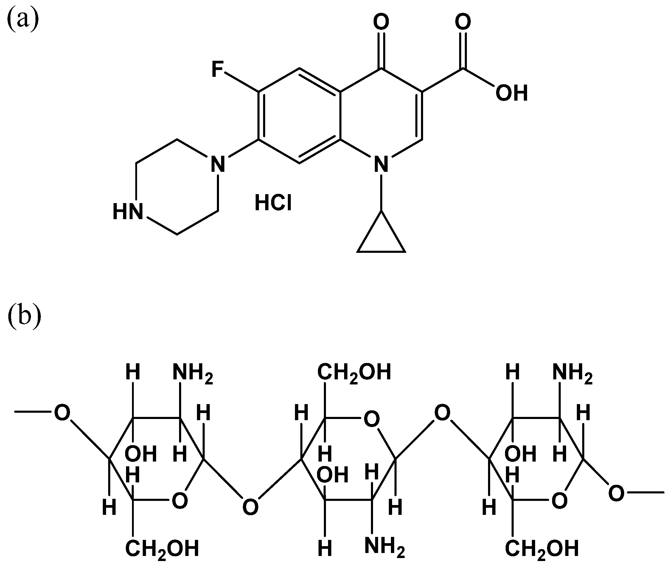
Figure 1. Structures of (a) CPF and (b) CS.
Catalysts 12 00475 g001
Figure 2. FTIR spectra of pure CS (a) and FeIII-CS-GLA (b). The spectra were recorded as KBr pellets.
Catalysts 12 00475 g002
Figure 3. Typical zero-field 57Fe Mössbauer spectrum of FeIII-CS-GLA recorded at 80 K (the iron concentration during preparation was 7.5 mM). The solid line represents the best fit with parameters δ = 0.48 mm/s, ΔEQ = 0.79 mm/s and line width Γ = 0.46 mm/s.
Catalysts 12 00475 g003
Figure 4. FE-SEM photograph of FeIII-CS-GLA before (a) and after (b) its use in the catalytic degradation of CPF.
Catalysts 12 00475 g004
Figure 5. Typical time-resolved spectral changes observed in solar light-assisted oxidative degradation of CPF catalyzed by FeIII-CS-GLA in aqueous solution. Experimental conditions: [CPF]0: 50 µM; [FeIII]0 in FeIII-CS-GLA: 1.25 mM; [GLA]0 in FeIII-CS-GLA: 90 mM; [H2O2]0: 6 mM; volume of CPF solution: 250 mL; pH: 3; light intensity: 70,000–90,000 Lux; solar light irradiation time: 180 min.
Catalysts 12 00475 g005
Figure 6. (a) Plots of A276nm vs. t for CPF degradation in aqueous solution for various control experiments: (i) 50 µM CPF + CS-GLA immobilized on a glass plate + solar light; (ii) 50 µM CPF + FeIII-CS-GLA immobilized on a glass plate + solar light; (iii) 50 µM CPF + FeIII-CS-GLA immobilized on a glass plate + H2O2 + dark; (iv) 50 µM CPF + FeIII-CS-GLA immobilized on a glass plate + H2O2 + solar light. (b) Plot of lnA276nm vs. t for oxidative degradation of CPF in aqueous solution at optimum condition (control (iv)). Experimental conditions: [CPF]0: 50 µM; [FeIII]0 in FeIII-CS-GLA: 1.25 mM; [GLA]0 in FeIII-CS-GLA: 90 mM; [H2O2]0: 6 mM; volume of CPF solution: 250 mL; pH: 3; light intensity: 70,000–90,000 Lux; solar light irradiation time: 180 min.
Catalysts 12 00475 g006
Figure 7. The effects of pH on kobs (min−1) and CPF removal efficiency (%) observed in solar light-assisted oxidative degradation of CPF catalyzed by FeIII-CS-GLA in aqueous solution. Experimental conditions: [CPF]0: 50 µM; [FeIII]0 in FeIII-CS-GLA: 1.25 mM; [GLA]0 in FeIII-CS-GLA: 90 mM; [H2O2]0: 6 mM; volume of CPF solution: 250 mL; pH: 2–12; light intensity: 70,000–90,000 Lux; solar light irradiation time: 180 min.
Catalysts 12 00475 g007
Figure 8. The effects of [GLA]0 in FeIII-CS-GLA on kobs (min−1) and CPF removal efficiency (%) observed in solar light-assisted oxidative degradation of CPF catalyzed by FeIII-CS-GLA in aqueous solution. Experimental conditions: [CPF]0: 50 µM; [FeIII]0 in FeIII-CS-GLA: 1.25 mM; [GLA]0 in FeIII-CS-GLA: 50–120 mM; [H2O2]0: 6 mM; volume of CPF solution: 250 mL; pH: 3; light intensity: 70,000–90,000 Lux; solar light irradiation time: 180 min.
Catalysts 12 00475 g008
Figure 9. The effects of [FeIII] content in FeIII-CS-GLA on kobs (min−1) and CPF removal efficiency (%) observed in solar light-assisted oxidative degradation of CPF catalyzed by Fe(III)-CS-GLA in aqueous solution. Experimental conditions: [CPF]0: 50 µM; [FeIII]0 in FeIII-CS-GLA: 1.25–7.50 mM; [GLA]0 in FeIII-CS-GLA: 90 mM; [H2O2]0: 6 mM; volume of CPF solution: 250 mL; pH: 3; light intensity: 70,000–90,000 Lux; solar light irradiation time: 180 min.
Catalysts 12 00475 g009
Figure 10. The effects of H2O2 concentration on kobs (min−1) and CPF removal efficiency (%) observed in the solar light-assisted oxidative degradation of CPF catalyzed by FeIII-CS-GLA in aqueous solution. Experimental conditions: [CPF]0: 50 µM, [FeIII]0 in FeIII-CS-GLA: 1.25 mM; [GLA]0 in FeIII-CS-GLA: 90 mM; [H2O2]0: 1–12 mM; volume of CPF solution: 250 mL; pH: 3; light intensity: 70,000–90,000 Lux; solar light irradiation time: 180 min.
Catalysts 12 00475 g010
Figure 11. The effect of CPF concentration on kobs (min−1) and CPF removal efficiency (%) observed in the solar light-assisted oxidative degradation of CPF catalyzed by FeIII-CS-GLA in aqueous solution. Experimental conditions: [CPF]0: 20–50 µM; [FeIII]0 in FeIII-CS-GLA: 1.25 mM; [GLA]0 in FeIII-CS-GLA: 90 mM; [H2O2]0: 6 mM; volume of CPF solution: 250 mL; pH: 3; light intensity: 70,000–90,000 Lux; solar light irradiation time: 180 min.
Catalysts 12 00475 g011
Figure 12. Plot of 1/kobs vs. initial concentration of CPF.
Catalysts 12 00475 g012
Figure 13. The effects of mannitol concentration on kobs (min−1) and CPF removal efficiency (%) observed in solar light-assisted oxidative degradation of CPF catalyzed by FeIII-CS-GLA in aqueous solution. Experimental conditions: [CPF]0: 50 µM, [FeIII]0 in FeIII-CS-GLA: 1.25 mM, [GLA]0 in FeIII-CS-GLA: 90 mM, [H2O2]0: 6 mM, [Mannitol]: 0–7 mM, volume of CPF solution: 250 mL, pH: 3, light intensity: 70,000–90,000 Lux, solar light irradiation time: 180 min.
Catalysts 12 00475 g013
Figure 14. Antimicrobial activity of CPF (from left to right: 50, 100, 150, and 200 µM) against (a) Escherichia coli DH5α, (b) Salmonella typhi AF4500 and (c) Bacillus subtilis RBW bacteria before and after solar light-assisted oxidative treatment with FeIII-CS-GLA/H2O2 in aqueous solution.
Catalysts 12 00475 g014
Figure 15. Efficiency of the FeIII-CS-GLA catalyst during repeated use for solar light-assisted oxidative degradation of CPF in aqueous solution. Experimental conditions: [CPF]0: 50 µM; [FeIII]0 in FeIII-CS-GLA: 1.25 mM; [GLA]0 in FeIII-CS-GLA: 90 mM; [H2O2]0: 6 mM; volume of CPF solution: 250 mL; pH: 3, light intensity: 70,000–90,000 Lux; solar light irradiation time: 180 min.
Catalysts 12 00475 g015
Table 1. Pseudo-first order rate constants (kobs) of CPF degradation by different processes.
Degradation ProcesspHkobs (min1)References
Sonolysis30.0210[33]
Pillared iron catalyst (Fe-Lap-RD)30.0253[36]
ZnO nanopartices40.0117[41]
Commercial anatase TiO230.0210[43]
Ag/Fe2O3/ZnO heterostructures40.0051[45]
FeIII-CS-GLA/H2O230.0134This work
Table 2. Compounds detected by HPLC-ESI(-)-MS after solar light-assisted oxidative degradation of CPF in aqueous solution catalyzed by FeIII-CS-GLA in presence of H2O2.
No.t(R) minm/zElemental CompositionProposed Compound
12.5147.0300 [M-H]
129.0194 [M-H-H2O]
C5H7O5
C5H5O4
C5H8O5
Hydroxy-pentanedioic acid
23.0117.0195 [M-H]C4H5O4C4H6O4
Butanedioic acid
34.0131.0350 [M-H]C5H7O4C5H8O4
Pentanedioic acid
Table 3. Antibacterial activities of CPF before and after catalytic degradation with FeIII-CS-GLA/H2O2 against Escherichia coli DH5α, Salmonella typhi AF4500 and Bacillus subtilis RBW bacteria.
Concentration of CPF (μM)Zone of Inhibition (mm) for Test Bacteria
Escherichia coli DH5αSalmonella typhi AF4500Bacillus subtilis RBW
Before catalytic degradation of CPF
5016.20 ± 0.4010.00 ± 0.729.30 ± 1.10
10018.10 ± 0.7213.40 ± 0.6816.60 ± 0.20
15019.60 ± 0.5015.10 ± 0.5622.20 ± 0.10
20020.40 ± 0.7124.30 ± 0.4025.10 ± 0.98
After catalytic degradation of CPF
50Not detectedNot detectedNot detected
100Not detectedNot detectedNot detected
150Not detectedNot detectedNot detected
200Not detectedNot detectedNot detected
Publisher’s Note: MDPI stays neutral with regard to jurisdictional claims in published maps and institutional affiliations.