Article

Rutile-TiO2/PtO2 Glass Coatings Disinfects Aquatic Legionella pneumophila via Morphology Change and Endotoxin Degradation under LED Irradiation

1
Laboratory of Global Infectious Diseases Control Science, Graduate School of Agricultural and Life Sciences, The University of Tokyo, 1-1-1 Yayoi, Bunkyo-ku, Tokyo 113-8657, Japan
2
Laboratory of Global Animal Resource Science, Graduate School of Agricultural and Life Sciences, The University of Tokyo, 1-1-1 Yayoi, Bunkyo-ku, Tokyo 113-8657, Japan
3
Kaltech Co., Ltd., 3-3-7 Bakuromachi, Chuo-ku, Osaka 541-0059, Japan
*
Author to whom correspondence should be addressed.
Catalysts 2022, 12(8), 856; https://doi.org/10.3390/catal12080856
Submission received: 30 June 2022 / Revised: 1 August 2022 / Accepted: 2 August 2022 / Published: 3 August 2022
(This article belongs to the Special Issue Innovative Functional Materials in Photocatalysis)

Abstract

:
Legionella pneumophila (L. pneumophila) is the causative agent of Legionnaires’ disease and Pontiac fever, collectively known as legionellosis. L. pneumophila infection occurs through inhalation of contaminated aerosols from water systems in workplaces and institutions. The development of disinfectants that can eliminate L. pneumophila in such water systems without evacuating people is needed to prevent the spread of L. pneumophila. Photocatalysts are attractive disinfectants that do not harm human health. In particular, the TiO2 photocatalyst kills L. pneumophila under various conditions, but its mode of action is unknown. Here, we confirmed the high performance of TiO2 photocatalyst containing PtO2 via the degradation of methylene blue (half-value period: 19.2 min) and bactericidal activity against Escherichia coli (half-value period: 15.1 min) in water. Using transmission electron microscopy, we demonstrate that the disinfection of L. pneumophila (half-value period: 6.7 min) by TiO2 photocatalyst in water is accompanied by remarkable cellular membrane and internal damage to L. pneumophila. Assays with limulus amebocyte lysate and silver staining showed the release of endotoxin from L. pneumophila due to membrane damage and photocatalytic degradation of this endotoxin. This is the first study to demonstrate the disinfection mechanisms of TiO2 photocatalyst, namely, via morphological changes and membrane damage of L. pneumophila. Our results suggest that TiO2 photocatalyst might be effective in controlling the spread of L. pneumophila.

1. Introduction

Since the 1970s, many kinds of emerging infectious diseases, such as acquired immunodeficiency syndrome (AIDS), enterohaemorrhagic Escherichia coli infection, Ebola virus disease, and coronavirus disease 2019 (COVID-19), have been reported, which has led to an increasing interest in public health measures to control their spread. Various methods for disinfection including drugs, antibiotics, and sterilization using light (e.g., ultraviolet [UV]) have been actively researched. Photocatalysts are attractive choices as disinfectants because they do not harm human health. For example, although the International Agency for Research on Cancer classified TiO2 as possibly carcinogenic to humans (group 2B), it noted that there was limited evidence of carcinogenicity in experimental animals, and two large epidemiological studies of workers employed in the TiO2 production industry in the United States and Europe clearly demonstrated that no increased risks were found for lung cancer, total cancers, or other causes of death [1,2]. In addition, porous TiO2 synthesized from Ti and ammonium hydrogen carbonate (NH4HCO3) was found to have no cytotoxic effects on human HeLa and Vero cells [3]. Irradiation with light activates the photocatalytic reaction of TiO2, the most common photocatalyst, generating reactive oxygen species (ROS), such as hydroxyl (·OH) and superoxide radicals (O2·) on the surface of the TiO2 [4]. The band gap energy and peak photoexcitation wavelength of rutile-type TiO2 have been determined to be 3.05 eV and 406.5 nm, respectively [5]. Rutile-type TiO2 can be excited by a 405 nm LED, which is an inexpensive light source and harmless compared to UVC [6]. ROS have strong oxidizing power and mineralize organic compounds, and this mineralization leads to the degradation of membranes, proteins, and genetic material, killing microorganisms and viruses [4]. Indeed, TiO2 photocatalysts can kill various microorganisms [4], including Bacillus cereus [7,8], Escherichia coli (E. coli) [9,10], Enterobacter spp. [11,12], Porphyromonas gingivalis [13], Salmonella spp. [7,11,14,15,16], and Vibrio parahaemolyticus [14,17]. TiO2 photocatalysts can decompose the E. coli membrane during the process of sterilization [9,10]. TiO2 photocatalysts can also degrade E. coli endotoxin [18,19], which is a component of the outer membrane of Gram-negative bacteria and a causative agent of sepsis. Therefore, killing the microorganism and degrading endotoxin are both important functions of photocatalysts. In the previous studies, various types of TiO2, such as TiO2 synthesized from TiCl4, HCl, 2(NH4)HCO3, and H2O2 [7]; commercial TiO2 (P-25, Degussa–Huels AG, Frankfurt am Main, Germany; Yakuri Pure Chemicals Co., Osaka, Japan) [8,11,14,15]; Ag core and TiO2 shelled nanoparticles [9,10]; TiO2 mixed with 5-, 10-, 15-, 20-tetraphenyl-21H, 23H-porphine nickel [12]; TiO2 made from a titanium peroxo complex [13]; and TiO2-coated glass by plasma-enhanced chemical vapor deposition [16] were used. Moreover, in those studies, TiO2 was coated on various materials, such as quartz, silver, glass, and stainless-steel wires [7,9,10,12,13,16]. This suggests that TiO2 photocatalysts might have broad applicability in many different settings.
Legionellosis, which includes Legionnaires’ disease and Pontiac fever, is an emerging infectious disease. Legionella pneumophila (L. pneumophila), the causative agent of legionellosis, is a Gram-negative, aerobic, rod-shaped bacteria. Legionella spp. was first reported in 1976 when many people attending an American Legion event succumbed to legionellosis [18]. The following year, L. pneumophila was recognized as the major causative agent of legionellosis [19]. L. pneumophila damages human health, and the fatality rate of Legionnaires’ disease reported by the Centers for Disease Control and Prevention is approximately 9% [20]. The incidence of Legionella infection has increased in both the United States and Europe [20,21]. Infection of L. pneumophila occurs through inhalation of contaminated aerosols, and L. pneumophila infects alveolar macrophages [22]. L. pneumophila is naturally ubiquitous in aquatic and damp environments such as lakes, rivers, composted materials, and moist soil as intracellular bacteria living inside eukaryotes such as amebae [21]. In addition, many sources of L. pneumophila have been reported around workplaces [23] and in potable water [24,25], construction area sinks [26], showerheads [27], car air conditioners [28], humidifiers [29], dental unit waterlines [30], and especially hot springs [31,32,33,34,35]. Human–human transmission of L. pneumophila is non-existent or very rare [20,36]. There has been much interest in the development of disinfectants that can sterilize water from workplaces without evacuating people in order to prevent the spread of L. pneumophila, with various disinfectants under consideration.
It was previously reported that a TiO2 photocatalyst can kill L. pneumophila [37,38,39,40]. Indeed, L. pneumophila in aerosols [37], under laminar flow [38], under semi-dry conditions [39], and in rainwater [40] have been killed by the TiO2 photocatalyst. These previous reports suggest that the TiO2 photocatalyst has potentially widespread applications in the control of L. pneumophila. However, damage to the membrane and endotoxin of L. pneumophila by the TiO2 photocatalyst has not yet been reported. Moreover, the mechanisms by which the TiO2 photocatalyst kills L. pneumophila remain unknown.
To investigate whether rutile-type TiO2/PtO2 (hereafter “TiO2”) -coated glass exerts photocatalytic activity in water by excitation of light with a wavelength of 405 nm, we first evaluated the performance of TiO2-coated glass in water by degradation of methylene blue and killing of E. coli. Furthermore, we demonstrated the susceptibility to disinfection in water of L. pneumophila, and then clarified the disinfection mechanism of TiO2 by observing the morphology of L. pneumophila using transmission electron microscopy (TEM). Finally, we performed the limulus amebocyte lysate (LAL) assay [41,42] and silver staining to detect the degradation of the L. pneumophila endotoxin.

2. Results

2.1. Photocatalytic Degradation of Methylene Blue in Water by TiO2 Photocatalyst

To clarify whether TiO2-coated glass (5 cm × 5 cm) exerts photocatalytic activity in water by excitation of light with a wavelength of 405 nm, we first tested the decomposition of methylene blue in water purified by ion-exchange. As shown in Figure 1A, TiO2-coated glass decomposed methylene blue by excitation of light in a time-dependent manner. The half-value period was 19.2 min (R2 = 0.9915) and the curve flattened between 90 and 120 min after the light emitting diode (LED)-TiO2 photocatalytic reaction (Figure 1B). In addition, reaction rate was estimated as followed equation:
r   nM / min = 0.107   m e t h l e n e   b l u e 1.06
where r is the reaction rate; 0.107 is reaction rate constant; [methylene blue] is the concetration of methylene blue; and 1.06 is the order of reaction. This equation suggested that this degradation reaction was first order reaction. By contrast, neither TiO2-coated glass under dark conditions (TiO2 + Dark) nor glass alone under dark conditions (Glass + Dark) decreased absorbance. As expected, glass under the light condition (Glass + Light) decreased absorbance because it is known that methylene blue is photodegradable. However, degradation of methylene blue by TiO2 under light conditions (TiO2 + Light) was more efficient than that by the glass under light conditions. These results demonstrate that the photocatalytic activity of TiO2-coated glass degrades methylene blue in water.

2.2. Disinfection of E. coli in Water by TiO2 Photocatalyst

The TiO2 photocatalyst can kill many kinds of microorganisms [4]. Therefore, we examined the bactericidal effect of TiO2-coated glass in water using E. coli, the most studied lab bacterium. As shown in Figure 2A, TiO2-coated glass was placed in a 10-cm diameter dish on the shaker, 30 mL of phosphate-buffered saline (PBS) containing E. coli with a titer of 1 × 10⁸ colony forming unit (CFU)/mL was added, and the dish was then exposed to LED light (placed above the dish) with a wavelength of 405 nm. The E. coli was killed such that its titer reached below the detection limit in a time-dependent manner within 8 h (Figure 2B). In addition, the Glass + Light group also killed E. coli in a time-dependent manner. This is because light with a wavelength of 405 nm has a bactericidal effect. Notably, the disinfection effect for E. coli in the TiO2 + Light group was stronger than that in the Glass + Light group (Figure 2C,D), and the titer was significantly (p < 0.05) lower at 6 h after light excitation (Figure 2C). In addition, the half-value periods of the TiO2 + Light group and Glass + Light group were 15.1 min (R2 = 0.97), and 25.8 min (R2 = 0.95), respectively, suggesting that the TiO2 + Light group disinfects E. coli 1.71 times faster than the Glass + Light group. These results suggested that TiO2-coated glass has a bactericidal effect in water and that the photocatalytic reaction can kill E. coli.

2.3. Disinfection of L. pneumophila in Water by TiO2 Photocatalyst

L. pneumophila grows in aquatic environments and can cause serious health problems in humans [20,21]. Therefore, it is essential to confirm sterilization ability of TiO2-coated glass against L. pneumophila in water. Thus, 30 mL of solution including L. pneumophila with a titer of 1 × 10⁷ CFU/mL and TiO2-coated glass was irradiated by an excitation light from 0 to 8 h. As shown in Figure 3A,C, the titer of L. pneumophila drastically decreased in a time-dependent manner for the TiO2-coated glass with excitation light and reached undetectable levels at 4 h. Although the Glass + Light group also decreased the titer of L. pneumophila, the TiO2 + Light group showed a stronger bactericidal effect, and the titer was significantly lower at 1 h (p < 0.05) and 2 h (p < 0.001) (Figure 3B). In addition, the half-value periods of the TiO2 + Light group and the Glass + Light group were 6.7 min (R2 = 0.99) and 12.0 min (R2 = 0.99), respectively, suggesting that the TiO2 + Light group disinfected water with L. pneumophila 1.80 times faster when compared with the Glass + Light group. These results show that TiO2-coated glass can sterilize water with both E. coli and L. pneumophila, suggesting that TiO2-coated glass might be an effective sterilizing agent for all Gram-negative bacteria.

2.4. Morphological Changes in L. pneumophila Induced by TiO2 Photocatalytic Disinfection

It has been previously reported that photocatalytic reactions disrupt the bacterial cell membrane [9,10]. To clarify the mechanism of L. pneumophila disinfection by a photocatalytic reaction, we observed the morphology of L. pneumophila using TEM. L. pneumophila with a titer of 2.91 × 10⁷ CFU/mL was irradiated by excitation light with TiO2-coated glass for 24 h; TEM images of L. pneumophila before and after the photocatalytic reaction were obtained (Figure 4). Under this condition, 99.6% of L. pneumophila in the TEM image was killed (Figure 4E). As shown in Figure 4A (left panel) and Figure 4B (left panel), untreated L. pneumophila before the photocatalytic reaction had a normal membrane, and 70.5% of the cells were stained black with uranyl acetate (Figure 4D). By contrast, after 24 h of the photocatalytic reaction, membranes had broken down (lacking the fluffy edge) and cells were not stained, as shown in Figure 4A (right panel) and Figure 4B (right panel). These results suggest that disinfection by photocatalytic reaction is based on significant damage to cell membranes and interiors. Thus, after 24 h of the photocatalytic reaction, 80.2% of cells in low magnification TEM images were dead L. pneumophila (shown by the blue arrows in Figure 4C, right panel). The proportion of dead L. pneumophila was significantly higher (p < 0.001) in the treatment group compared with that in the non-treatment group (Figure 4D). These results suggest that damage to membranes is one mechanism for the photocatalytic sterilization of L. pneumophila in water.

2.5. L. pneumophila Endotoxin Degradation by TiO2 Photocatalyst

Endotoxin is a structural component of the outer membrane of Gram-negative bacteria and is a pyrogenic substance [43,44]. Since the TiO2 photocatalytic reaction damaged the membrane of L. pneumophila, it was considered likely that endotoxin would also be released. To test for release of endotoxin in water, 30 mL of L. pneumophila with a titer of 107 CFU/mL was irradiated with excitation light and TiO2-coated glass from 0 to 12 h, and the concentration of endotoxin in the water was measured. Interestingly, in the TiO2 + Light group, the concentration of endotoxin increased in a time-dependent manner (Figure 5A). This corroborates our TEM results showing the morphological changes in L. pneumophila by TiO2 photocatalytic disinfection. Likewise, in the Glass + Light group, the concentration of endotoxin also increased in a time-dependent manner. These increases in endotoxin concentration resemble those seen when Gram-negative bacteria are treated with drugs such as antibiotics [43]. However, the increase in endotoxin concentration in the TiO2 + Light group was limited compared with that in the Glass + Light group. This might be because of endotoxin degradation by the photocatalytic reaction but not by LED light.
To determine whether the TiO2 photocatalyst induces the degradation of endotoxin from L. pneumophila in water, we extracted endotoxin from L. pneumophila, dropped samples onto the TiO2-coated glass sheet (1 cm × 1cm) and irradiated the sheets with 405 nm light for 24 h (Figure 5B). Then, a sample was collected by washing with 10 mM Tris-HCl (pH = 8.0), and the concentration was measured using the LAL method. As shown in Figure 5C, the concentration of endotoxin was significantly decreased (by 81%; p < 0.05) during the 24 h photocatalytic reaction (Figure 5C). Untreated endotoxin extracted from L. pneumophila was detected by silver staining as any band between 15 and 20 kDa; however, these bands disappeared after the 24 h photocatalytic reaction (Figure 5D). Moreover, band density of endotoxin was significantly decreased (by 78%; p < 0.05) in the treated sample after 24 h photocatalytic reaction as compared to that in the sample at 0 h (Figure 5E). These results suggest that the TiO2 photocatalyst degrades endotoxin released from L. pneumophila.

2.6. Durability of TiO2 Photocatalyst

To check the integrity of the TiO2-coated glass before and after photocatalysis, we used SEM to compare the surfaces of the glass plate without TiO2, the fresh TiO2-coated glass plate and the TiO2-coated glass plate after they had been used for the photocatalytic tests (Figure 6A). The SEM image of the frost glass plate without TiO2 exhibited a uniformly uneven surface (Figure 6A). In contrast, the SEM image of the glass plate coated with TiO2 revealed the surface to be covered with small particles and uniform unevenness was not observed (Figure 6A). These results suggested that TiO2 completely covers the surface of the ground glass. In addition, TiO2 did not detach from TiO2-coated glass plate used for photocatalytic disinfection for 24 h (Figure 6A). Moreover, to confirm the amounts of TiO2 on the glass, elemental analysis was performed using energy dispersive X-ray analyzer (EDS) (Figure 6B). The elemental analysis results showed that there was no significant difference between the amounts of TiO2 (wt%) on the glass before and after use (Figure 6C). This result also strongly suggested that TiO2 did not detach during the sterilization process. To confirm the activity of the TiO2 photocatalyst before and after use for sterilization, methylene blue degradation assay was performed (Figure 6D). There was no difference between the rate of the TiO2 photocatalyst-mediated degradation of methylene blue before and after use for sterilization. These results suggested that the TiO2-coated glass in this study could be re-used and would retain activity for a long time.

3. Discussion

In this study, we first confirmed the efficient disinfection of both E. coli and L. pneumophila in water by a TiO2 photocatalyst coated on glass. Our results confirm the sterilization ability of TiO2-coated glass against two kinds of Gram-negative bacteria, suggesting that TiO2-coated glass might be effective for all Gram-negative bacteria in water. This supports a previous study that showed that TiO2 photocatalysts can kill many kinds of microorganisms [4]. However, the present study is the first to confirm by TEM imaging that there was membrane damage to L. pneumophila, suggesting that this is the likely mechanism of disinfection during the TiO2 photocatalytic reaction. Moreover, our results also demonstrate endotoxin release from L. pneumophila. Photocatalytic degradation of the endotoxin, which is a novel finding, was confirmed by LAL assay and silver stain. Degradation of the endotoxin accompanied by morphological changes to L. pneumophila following the TiO2 photocatalyst reaction was confirmed by TEM imaging. The morphological changes to L. pneumophila included membrane separation from the cytoplasm, membrane damage (e.g., lacking the fluffy edge), and no stained cytoplasm. As a result, the density of staining of the cytoplasm was reduced. Previous reports showed similar membrane damage in E. coli and Pseudomonas aeruginosa induced by TiO2 photocatalytic reactions [45,46]. In addition, the membrane of E. coli was degraded by ROS produced by the TiO2 photocatalytic reaction, leading to the leakage of intracellular molecules, and causing cell death [44,47].
The concentration of endotoxin increased in a time-dependent manner following exposure to excitation light with TiO2-coated glass. Endotoxin, which is a component of the outer membrane of Gram-negative bacteria, is a causative agent of sepsis. It was previously reported that antibiotics induce endotoxin release during disinfection [43]. Furthermore, endotoxin release is also caused by an increase in the permeability of the membrane [48]. Moreover, TiO2 photocatalysts can increase the permeability of cancer cell membranes [49]. Indeed, our TEM results clearly demonstrate morphological changes of L. pneumophila induced by TiO2 photocatalytic disinfection. These previous reports and our results suggest that increase in endotoxin concentration was due to endotoxin release from L. pneumophila via membrane damage by the TiO2 photocatalyst in water. However, we found that the increase in endotoxin concentration by the TiO2 photocatalyst was limited compared with that induced by disinfection by 405 nm LED without the TiO2 photocatalyst. This suggests the possibility for TiO2 photocatalytic degradation of endotoxin released from L. pneumophila in water. Until now, no report has shown the degradation of endotoxin of L. pneumophila by photocatalyst; however, it has been reported that endotoxin extracted from E. coli was degraded by a TiO2 photocatalyst [9,10]. Here, we directly confirmed the degradation of endotoxin extracted from L. pneumophila by LAL assay and silver staining. Since the oxidation of high molecular weight compounds is complicated and the photocatalytic degradation order of reaction of the endotoxin has not yet been clearly revealed, it remains unclear whether L. pneumophila endotoxin treated with a TiO2 photocatalyst loses virulence with degradation. Therefore, further study (for example, experimental inoculation of mice with endotoxin treated with or without the TiO2 photocatalyst) is needed to clarify the TiO2 photocatalytic effect on the endotoxin and loss of virulence.
The 405 nm LED without the TiO2 photocatalyst also decreased the titer of both E. coli and L. pneumophila. This suggests that 405 nm LED also has an anti-bacterial ability. It is known that 405 nm light stimulates porphyrin molecules to produce ROS and damages cells leading to microbial death [49,50]. Likewise, it has been reported that some bacteria, such as E. coli and Legionella rubrilucens, which is closely related to L. pneumophila, were disinfected by 405 nm light [50]. In this study, endogenous porphyrin was excited by the 405 nm LED. This is an advantage of the TiO2 photocatalyst, as it can disinfect microorganism by two mechanisms.
Numerous studies have considered the disinfection of L. pneumophila, with methods including UV, chlorination, heat, and filtration [46]. Each method has advantages and disadvantages. For example, UV is harmful to the human body. L. pneumophila infection can have various sources such as contaminated showerheads [22], humidifiers [29], dental unit waterlines [30], or hot springs [31,32,33,34,35]; therefore, chemical compounds such as sodium hypochlorite and antibiotics are not suitable for disinfection. Physical treatment such as heat and filtration offer only short-term benefits; L. pneumophila propagates again after sterilization. In the case of TiO2 photocatalyst, TiO2 is not cytotoxic and is regarded as low risk for causing cancer [1,2,3]. In addition, 405 nm LED which can excite rutile-type TiO2 photocatalyst is also harmless, unlike UVC, and can be applied even in places where sunlight does not reach. Moreover, in this study, the TiO2 was retained on the glass plate after the photocatalytic reaction, suggesting that the TiO2 photocatalyst can be re-used. Due to the safety and durability of the TiO2 photocatalyst and 405 nm LED, TiO2 photocatalyst can be applied for continuous disinfection of living and working spaces, without needing to evacuate people. It is expected that photocatalysts will be especially useful when applied to humidifiers, which are one of the main sources of L. pneumophila, and to hot spring pipes, in which L. pneumophila grows and where it is difficult for people to clean regularly. Thus, we suggest that TiO2 photocatalysts offer great promise in effectively controlling L. pneumophila transmission.

4. Materials and Methods

4.1. Preparation of TiO2-Coated Glass or Glass Fiber Sheet

TiO2 was coated to the frosted glass plate (SAG-003, Saint-Gobain S.A., Courbevoie, France) or glass fiber sheet (XU1310010, Osaka Lighting Corp., Osaka, Japan) by the following method. Commercial powder of fine (~20 nm) rutile-type TiO2 containing approximately 1% platinum dioxide to improve the photocatalytic reaction (MPT-623, Ishihara Sangyou Kaisha, Ltd., Osaka, Japan) [51,52] was dispersed in ion-exchanged water, and then, a frosted glass plate or a glass fiber sheet was immersed. To fuse the TiO2 and glass, the glass plate or fiber was dried in air at room temperature before being calcined in air at 400 °C for 90 min. Typically, 26 mg TiO2 was coated on the glass plate.

4.2. Methylene Blue Degradation

A glass plate (5 cm x 5 cm) coated with TiO2 was put in 100 mL of 12.5 nM methylene blue solution, and an LED light (405 nm) source was placed 1.5 cm above the TiO2-coated glass. The TiO2 photocatalyst was excited by the 405 nm LED light, for 0, 10, 20, 30, 40, 50, 60, 90, and 120 min. To confirm the effect of TiO2-LED on the methylene blue, methylene blue was collected at each time point, and absorbance at 660 nm was measured using an ASV11D-H spectrophotometer (AS ONE CORPORATION, Osaka, Japan). As a control, methylene blue was incubated with TiO2-coated glass without light, with glass and LED light, and with glass without light.

4.3. Scanning Electron Microscopy of TiO2-Coated Glass

To observe the surface of the TiO2-coated glass plate, a scanning electron microscopy (SEM) image was obtained using a VHX-D510 electron microscope (KEYENCE CORPORATION, Osaka, Japan) at 1.2 kV.

4.4. Elemental Analysis

To confirm the amounts of the TiO2 on the surface of glass, elemental analysis was performed using SEM with a dispersive X-ray analyzer (EDS) (JSM-IT100, JEOL Ltd., Akishima, Japan) at an accelerating voltage of 0.0 to 9.0 kV.

4.5. Microorganisms

L. pneumophila was obtained from RIKEN BRC (Tsukuba, Japan) and grown in buffered charcoal yeast extract (BCYE; 1% ACE buffer (pH = 6.9), 1% yeast extract, 0.2% charcoal, 0.1% α-ketoglutaric acid, 0.04% L-cysteine, and 0.025% ferric pyrophosphate) medium at 37 °C. As a control, Escherichia coli (E. coli) XL10 gold strain was obtained from Agilent Technologies, Inc. (Santa Clara, CA, USA) and grown in Lysogeny Broth (LB; 1% Tryptone, 1% NaCl, and 0.5% yeast extract) at 37 °C. To measure colony forming units (CFU) as a titer of L. pneumophila and E. coli, 10 to 100 μL of each suspension were transferred onto an LB agar plate or BCYE agar plate using a plate spreading technique and then incubated for 1 and 3 days at 37 °C, respectively.

4.6. Treatment of E. coli and L. pneumophila by the TiO2 Photocatalytic Reaction

TiO2-coated glass (5 cm × 5 cm) was placed in a dish with a diameter of 10 cm, and an LED light (405 nm) source was placed above the dish. E. coli in an LB medium was centrifuged at 3000 rpm for 10 min, and the pellet was resuspended in PBS. The L. pneumophila was centrifuged at 3000 rpm for 10 min, and the pellet was resuspended in PBS. Then, 30 mL of the suspension of either E. coli with 108 CFU/mL or L. pneumophila with 107 CFU/mL was added to a 10-cm diameter dish with TiO2 photocatalyst coated glass and was excited by a 405 nm LED for 0, 1, 2, 4, 6, 8, 10, and 12 h. During excitation, 1 mL of the suspensions of E. coli and L. pneumophila were collected at each time point. As a control, E. coli and L. pneumophila were incubated with TiO2-coated glass without light, with glass and LED light, and with glass without light. During irradiation, the dish was shaken at 40 rpm. A portion of the collected suspension was used to measure CFU as the titer of E. coli or L. pneumophila. The rest of the suspension of L. pneumophila was used to measure the concentration of endotoxin by the LAL assay.

4.7. LAL Assay

The concentration of endotoxin was measured using Limulus color KY (FUJIFILM Wako Pure Chemical Corporation, Osaka, Japan) according to the manufacturer’s instructions. Briefly, 50 μL of each sample was mixed with 50 μL of the Limulus Amebocyte lysate reagent in the Limulus Color KY into a 96-well plate, and absorbance was measured at 405 nm every 5 min. The levels of endotoxin were measured by comparing with a standard endotoxin solution

4.8. TEM

We placed 30 mL of L. pneumophila with 2.91 × 10⁷ CFU/mL in a 10-cm diameter dish with the TiO2-coated glass (5 cm × 5 cm). The photocatalytic reaction was activated by exposure to LED light (405 nm) for 24 h. As a control group, L. pneumophila was collected immediately before irradiation. After incubation, 100 μL of L. pneumophila was mixed with 100 μL of 2.5% glutaraldehyde for TEM negative staining. For TEM sample preparation, a droplet of L. pneumophila sample was loaded on a carbon-film grid and incubated for 10 s. Next, the grid was partially dried, and a droplet of 2% uranyl acetate staining solution was added, followed by incubation for 10 s. Finally, the excess liquid was removed with filter paper, and the grid was dried at room temperature before obtaining images using a HITACHI H-7600 electron microscope (Hitachi Global Life Solutions, Inc., Tokyo, Japan) at 100 kV.

4.9. Extraction of Endotoxin from L. pneumophila

Endotoxin from L. pneumophila with 8 × 10¹⁰ CFU was extracted by the hot phenol-water method using a lipopolysaccharide extraction kit (iNtRON Biotechnology; Seongnam, Korea) according to the manufacturer’s instructions. Extracted endotoxin was redissolved in 10 mM Tris-HCl (pH = 8.0), and its concentration was measured by the LAL assay.

4.10. Inactivation of Endotoxin from L. pneumophila by the TiO2 Photocatalytic Reaction

To confirm the degradation of extracted endotoxin from L. pneumophila by TiO2 photocatalyst, extracted endotoxin was treated by a TiO2-coated glass sheet as follows. Filter paper was placed at the bottom of the 10-cm diameter dish and moistened with 3 mL sterilized water. To avoid directly touching the filter paper, the plastic tube was placed on the filter paper, and the glass sheet coated (1 cm × 1 cm) with TiO2 photocatalyst was put on it. Then, 100 μL of extracted endotoxin from L. pneumophila with a titer of 100 EU/mL was dropped onto the glass sheet with TiO2 photocatalyst and covered with glass. The sample then was illuminated with 405 nm LED for 24 h. After illumination, the sample was immersed and washed by 100 μL 10 mM Tris-HCl (pH = 8.0).

4.11. Silver Stain

We mixed 12 µl of endotoxin treated with the photocatalyst with 4 µl of sample buffer (0.15 M Tris-HCl, 10% sodium dodecyl sulfate [SDS], 30% glycerol, 5% beta-mercaptoethanol, and 0.5% bromophenol blue) and heated at 100 °C for 5 min. Then, 15 µl of denatured endotoxin was loaded on a 15% SDS-polyacrylamide gel and electrophoresed with a running buffer containing 0.3% Tris, 0.1% SDS, and 1.44% glycine. The endotoxin was then detected by the silver staining method using a silver stain MS kit (FUJIFILM Wako Pure Chemical Corporation) according to the manufacturer’s instructions.

4.12. Statistical Analysis

Two-way ANOVA with Dunnett’s test was used to compare all samples with the sample obtained at 0 min for statistical determination. To compare each group, ANOVA followed by Tukey’s test was performed. p values < 0.05 were considered statistically significant. For TEM data analysis and endotoxin concentration analysis, the Student’s t-test was used to compare the 0 and 24 h samples. In addition, Student’s t-test was used to compare the amounts of TiO2 on the glass before and after use in the elemental analysis. Exponential regression analysis and linear expression analysis was performed to determine the relationship between absorbance of methylene blue and irradiation time and between disinfection effect of E. coli and L. pneumophila and irradiation time. All calculations were performed using the R software (version 3.6.3, R Foundation for Statistical Computing, Vienna, Austria).

5. Conclusions

To the best of our knowledge, this is the first report to show that TiO2 photocatalyst can degrade the membrane and endotoxin of L. pneumophila. TEM imaging showed TiO2 photocatalyst caused morphological changes in L. pneumophila, including separation of the membrane from the cytoplasm, and reduced staining of the cytoplasm, suggesting that the primary disinfection mechanism of L. pneumophila is membrane degradation and leakage of cellular components. In summary, the TiO2 photocatalyst can efficiently disinfect L. pneumophila (half-value period: 6.7 min), without harm to people. In addition, the TiO2 photocatalyst degrades the endotoxin of L. pneumophila in water. In conclusion, the TiO2 photocatalyst could be an effective tool for controlling the spread of L. pneumophila.

Author Contributions

Conceived and designed the experiments: Y.A. and R.M. Conducted and performed the experiments: A.K, R.M., T.F., K.F. and Y.A. Analyzed the data: R.M., A.K. and Y.A. Supervised this experiment: Y.A. and Y.M. Contributed reagents/materials/analysis tools: T.F., K.F., H.O., J.S. and Y.A. Wrote the paper: R.M. and Y.A. All authors have read and agreed to the published version of the manuscript.

Funding

This research was supported by a grant-in-aid from Kaltech Co., Ltd.

Data Availability Statement

Not applicable.

Acknowledgments

We are grateful to thank all members of Laboratory of Global Infectious Diseases Control Science, Graduate School of Agricultural and Life Sciences, The University of Tokyo, for their technical assistance, help, and suggestions. We would like to thank Hanaichi Ultrastructure Research Institute for TEM analysis.

Conflicts of Interest

The funders had no role in the design of the study; in the collection, analyses, or interpretation of data; in the writing of the manuscript, or in the decision to publish the results.

References

  1. Boffetta, P.; Soutar, A.; Cherrie, J.W.; Granath, F.; Andersen, A.; Anttila, A.; Blettner, M.; Gaborieau, V.; Klug, S.J.; Langard, S.; et al. Mortality among workers employed in the titanium dioxide production industry in Europe. Cancer Causes Control 2004, 15, 697–706. [Google Scholar] [CrossRef] [PubMed]
  2. Fryzek, J.P.; Chadda, B.; Marano, D.; White, K.; Schweitzer, S.; McLaughlin, J.K.; Blot, W.J. A cohort mortality study among titanium dioxide manufacturing workers in the United States. J. Occup. Environ. Med. 2003, 45, 400–409. [Google Scholar] [CrossRef] [PubMed] [Green Version]
  3. Koly, F.A.; Rahman, M.A.; Islam, M.S.; Rahman, M.M. Fabrication of porous TiO2 foams by powder metallurgy technique and study of bulk crushing strength for biomedical application. Prog. Biomater. 2021, 10, 299–308. [Google Scholar] [CrossRef] [PubMed]
  4. Foster, H.A.; Ditta, I.B.; Varghese, S.; Steele, A. Photocatalytic disinfection using titanium dioxide: Spectrum and mechanism of antimicrobial activity. Appl. Microbiol. Biotechnol. 2011, 90, 1847–1868. [Google Scholar] [CrossRef] [PubMed]
  5. Fonseca-Cervantes, O.R.; Pérez-Larios, A.; Romero Arellano, V.H.; Sulbaran-Rangel, B.; Guzmán González, C.A. Effects in Band Gap for Photocatalysis in TiO2 Support by Adding Gold and Ruthenium. Processes 2020, 8, 1032. [Google Scholar] [CrossRef]
  6. Matsuura, R.; Lo, C.W.; Wada, S.; Somei, J.; Ochiai, H.; Murakami, T.; Saito, N.; Ogawa, T.; Shinjo, A.; Benno, Y.; et al. SARS-CoV-2 Disinfection of Air and Surface Contamination by TiO2 Photocatalyst-Mediated Damage to Viral Morphology, RNA, and Protein. Viruses 2021, 13, 942. [Google Scholar] [CrossRef] [PubMed]
  7. Cho, M.; Choi, Y.; Park, H.; Kim, K.; Woo, G.J.; Park, J. Titanium dioxide/UV photocatalytic disinfection in fresh carrots. J. Food Prot. 2007, 70, 97–101. [Google Scholar] [CrossRef]
  8. Armon, R.; Weltch-Cohen, G.; Bettane, P. Disinfection of Bacillus spp. spores in drinking water by TiO2 photocatalysis as a model for Bacillus anthracis. Waterborne Pathog. 2004, 4, 7–14. [Google Scholar] [CrossRef]
  9. Sreeja, S.; Vidya Shetty, K. Microbial disinfection of water with endotoxin degradation by photocatalysis using Ag@TiO2 core shell nanoparticles. Environ. Sci. Pollut. Res. Int. 2016, 23, 18154–18164. [Google Scholar] [CrossRef]
  10. Gadgil, D.J.; Shetty Kodialbail, V. Suspended and polycaprolactone immobilized Ag @TiO2/polyaniline nanocomposites for water disinfection and endotoxin degradation by visible and solar light-mediated photocatalysis. Environ. Sci. Pollut. Res. Int. 2021, 28, 12780–12791. [Google Scholar] [CrossRef]
  11. Ibáñez, J.A.; Litter, M.I.; Pizarro, R.A. Photocatalytic bactericidal effect of TiO2 on Enterobacter cloacae. Comparative study with other Gram (−) bacteria. J. Photochem. Photobiol. A 2003, 157, 81–85. [Google Scholar] [CrossRef]
  12. Yao, K.S.; Wang, D.Y.; Chang, C.Y.; Weng, K.W.; Yang, L.Y.; Lee, S.J.; Cheng, T.C.; Hwang, C.C. Photocatalytic disinfection of phytopathogenic bacteria by dye-sensitized TiO2 thin film activated by visible light. Surf. Coat. Technol. 2007, 202, 1329–1332. [Google Scholar] [CrossRef]
  13. Chun, M.J.; Shim, E.; Kho, E.H.; Park, K.J.; Jung, J.; Kim, J.M.; Kim, B.; Lee, K.H.; Cho, D.L.; Bai, D.H.; et al. Surface modification of orthodontic wires with photocatalytic titanium oxide for its antiadherent and antibacterial properties. Angle Orthod. 2007, 77, 483–488. [Google Scholar] [CrossRef]
  14. Kim, B.; Kim, D.; Cho, D.; Cho, S. Bactericidal effect of TiO2 photocatalyst on selected food-borne pathogenic bacteria. Chemosphere 2003, 52, 277–281. [Google Scholar] [CrossRef]
  15. Cushnie, T.P.T.; Robertson, P.K.J.; Officer, S.; Pollard, P.M.; McCullagh, C.; Robertson, J.M.C. Variables to be considered when assessing the photocatalytic destruction of bacterial pathogens. Chemosphere 2009, 74, 1374–1378. [Google Scholar] [CrossRef]
  16. Cho, D.L.; Min, H.; Kim, J.H.; Cha, G.S.; Kim, G.S.; Kim, B.H.; Ohk, S.H. Photocatalytic characteristics of TiO2 thin films deposited by PECVD. J. Ind. Eng. Chem. 2007, 13, 434–437. [Google Scholar]
  17. Hara-Kudo, Y.; Segawa, Y.; Kimura, K. Sanitation of seawater effluent from seaweed processing plants using a photo-catalytic TiO2 oxidation. Chemosphere 2006, 62, 149–154. [Google Scholar] [CrossRef]
  18. Fraser, D.W.; Tsai, T.R.; Orenstein, W.; Parkin, W.E.; Beecham, H.J.; Sharrar, R.G.; Harris, J.; Mallison, G.F.; Martin, S.M.; McDade, J.E.; et al. Legionnaires’ disease: Description of an epidemic of pneumonia. N. Engl. J. Med. 1977, 297, 1189–1197. [Google Scholar] [CrossRef]
  19. Brenner, D.J.; Steigerwalt, A.G.; McDade, J.E. Classification of the Legionnaires’ disease bacterium: Legionella pneumophila, genus novum, species nova, of the family Legionellaceae, familia nova. Ann. Intern. Med. 1979, 90, 656–658. [Google Scholar] [CrossRef]
  20. Dooling, K.L.; Toews, K.; Hicks, L.A.; Garrison, L.E.; Bachaus, B.; Zansky, S.; Carpenter, L.R.; Schaffner, B.; Parker, E.; Petit, S.; et al. Active Bacterial Core Surveillance for Legionellosis—United States, 2011–2013. MMWR 2015, 64, 42. [Google Scholar] [CrossRef]
  21. Gattuso, G.; Rizzo, R.; Lavoro, A.; Spoto, V.; Porciello, G.; Montagnese, C.; Cinà, D.; Cosentino, A.; Lombardo, C.; Mezzatesta, M.L.; et al. Overview of the Clinical and Molecular Features of Legionella Pneumophila: Focus on Novel Surveillance and Diagnostic Strategies. Antibiotics 2022, 11, 370. [Google Scholar] [CrossRef]
  22. Ziltener, P.; Reinheckel, T.; Oxenius, A. Neutrophil and Alveolar Macrophage-Mediated Innate Immune Control of Legionella pneumophila Lung Infection via TNF and ROS. PLoS Pathog. 2016, 12, e1005591. [Google Scholar] [CrossRef]
  23. Orkis, L.T.; Harrison, L.H.; Mertz, K.J.; Brooks, M.M.; Bibby, K.J.; Stout, J.E. Environmental sources of community-acquired legionnaires’ disease: A review. Int. J. Hyg. Environ. Health 2018, 221, 764–774. [Google Scholar] [CrossRef]
  24. Stout, J.E.; Yu, V.L.; Muraca, P.; Joly, J.; Troup, N.; Tompkins, L.S. Potable water as a cause of sporadic cases of community-acquired legionnaires’ disease. N. Engl. J. Med. 1992, 326, 151–155. [Google Scholar] [CrossRef] [Green Version]
  25. Stout, J.E.; Yu, V.L.; Yee, Y.C.; Vaccarello, S.; Diven, W.; Lee, T.C. Legionella pneumophila in residential water supplies: Environmental surveillance with clinical assessment for Legionnaires’ disease. Epidemiol. Infect. 1992, 109, 49–57. [Google Scholar]
  26. Rampling, A.; Butt, C.J.; West, A.A.; Tully, M.; Palmer, K.T. Community-acquired Legionnaires’ disease following minimal exposure to a contaminated source. J. Infect. 1997, 35, 300–302. [Google Scholar] [CrossRef]
  27. Sax, H.; Dharan, S.; Pittet, D. Legionnaires’ disease in a renal transplant recipient: Nosocomial or home-grown? Transplantation 2002, 74, 890–892. [Google Scholar] [CrossRef]
  28. Pinar, A.; Ramirez, J.A.; Schindler, L.L.; Miller, R.D.; Summersgill, J.T. The use of heteroduplex analysis of polymerase chain reaction products to support the possible transmission of Legionella pneumophila from a malfunctioning automobile air conditioner. Infect. Control Hosp. Epidemiol. 2002, 23, 145–147. [Google Scholar] [CrossRef]
  29. Moran-Gilad, J.; Lazarovitch, T.; Mentasti, M.; Harrison, T.; Weinberger, M.; Mordish, Y.; Mor, Z.; Stocki, T.; Anis, E.; Sadik, C.; et al. Humidifier-associated paediatric Legionnaires’ disease, Israel, February 2012. Eurosurveillance 2012, 17, 20293. [Google Scholar] [CrossRef]
  30. Ricci, M.L.; Fontana, S.; Pinci, F.; Fiumana, E.; Pedna, M.F.; Farolfi, P.; Sabattini, M.A.; Scaturro, M. Pneumonia associated with a dental unit waterline. Lancet 2012, 379, 684. [Google Scholar] [CrossRef]
  31. Miyamoto, H.; Jitsurong, S.; Shiota, R.; Maruta, K.; Yoshida, S.; Yabuuchi, E. Molecular determination of infection source of a sporadic Legionella pneumonia case associated with a hot spring bath. Microbiol. Immunol. 1997, 41, 197–202. [Google Scholar] [CrossRef] [PubMed] [Green Version]
  32. Ito, I.; Naito, J.; Kadowaki, S.; Mishima, M.; Ishida, T.; Hongo, T.; Ma, L.; Ishii, Y.; Matsumoto, T.; Yamaguchi, K. Hot spring bath and Legionella pneumonia: An association confirmed by genomic identification. Intern. Med. 2002, 41, 859–863. [Google Scholar] [CrossRef] [PubMed] [Green Version]
  33. Nozue, T.; Chikazawa, H.; Miyanishi, S.; Shimazaki, T.; Oka, R.; Shimazaki, S.; Miyamoto, S. Legionella pneumonia associated with adult respiratory distress syndrome caused by Legionella pneumophila serogroup 3. Intern. Med. 2005, 44, 73–78. [Google Scholar] [CrossRef] [PubMed] [Green Version]
  34. Kurosawa, H.; Fujita, M.; Kobatake, S.; Kimura, H.; Ohshima, M.; Nagai, A.; Kaneko, S.; Iwasaki, Y.; Kozawa, K. A case of Legionella pneumonia linked to a hot spring facility in Gunma Prefecture, Japan. Jpn. J. Infect. Dis. 2010, 63, 78–79. [Google Scholar] [CrossRef]
  35. Matsui, M.; Fujii, S.I.; Shiroiwa, R.; Amemura-Maekawa, J.; Chang, B.; Kura, F.; Yamauchi, K. Isolation of Legionella rubrilucens from a pneumonia patient co-infected with Legionella pneumophila. J. Med. Microbiol. 2010, 59 Pt 10, 1242–1246. [Google Scholar] [CrossRef]
  36. Correia, A.M.; Ferreira, J.S.; Borges, V.; Nunes, A.; Gomes, B.; Capucho, R.; Gonçalves, J.; Antunes, D.M.; Almeida, S.; Mendes, A.; et al. Probable Person-to-Person Transmission of Legionnaires’ Disease. N. Engl. J. Med. 2016, 374, 497–498. [Google Scholar] [CrossRef] [Green Version]
  37. Josset, S.; Hajiesmaili, S.; Begin, D.; Edouard, D.; Pham-Huu, C.; Lett, M.C.; Keller, N.; Keller, V. UV-A photocatalytic treatment of Legionella pneumophila bacteria contaminated airflows through three-dimensional solid foam structured photocatalytic reactors. J. Hazard. Mater. 2010, 175, 372–381. [Google Scholar] [CrossRef]
  38. Oder, M.; Koklič, T.; Umek, P.; Podlipec, R.; Štrancar, J.; Dobeic, M. Photocatalytic biocidal effect of copper doped TiO2 nanotube coated surfaces under laminar flow, illuminated with UVA light on Legionella pneumophila. PLoS ONE 2020, 15, e0227574. [Google Scholar] [CrossRef]
  39. Ishiguro, H.; Yao, Y.; Nakano, R.; Hara, M.; Sunada, K.; Hashimoto, K.; Kajioka, J.; Fujishima, A.; Kubota, Y. Photocatalytic activity of Cu2+/TiO2-coated cordierite foam inactivates bacteriophages and Legionella pneumophila. Appl. Catal. B 2013, 129, 56–61. [Google Scholar] [CrossRef]
  40. Oana, K.; Kobayashi, M.; Yamaki, D.; Sakurada, T.; Nagano, N.; Kawakami, Y. Applicability assessment of ceramic microbeads coated with hydroxyapatite-binding silver/titanium dioxide ceramic composite earthplus™ to the eradication of Legionella in rainwater storage tanks for household use. Int. J. Nanomed. 2015, 10, 4971–4979. [Google Scholar] [CrossRef] [Green Version]
  41. Holzheimer, R.G. Antibiotic induced endotoxin release and clinical sepsis: A review. J. Chemother. 2001, 13, 159–172. [Google Scholar] [CrossRef]
  42. Kiwi, J.; Nadtochenko, V. Evidence for the mechanism of photocatalytic degradation of the bacterial wall membrane at the TiO2 interface by ATR-FTIR and laser kinetic spectroscopy. Langmuir 2005, 21, 4631–4641. [Google Scholar] [CrossRef]
  43. Hu, C.; Lan, Y.; Qu, J.; Hu, X.; Wang, A. Ag/AgBr/TiO2 visible light photocatalyst for destruction of azodyes and bacteria. J. Phys. Chem. B 2006, 110, 4066–4072. [Google Scholar] [CrossRef]
  44. Leive, L. Release of lipopolysaccharide by EDTA treatment of E. coli. Biochem. Biophys. Res. Commun. 1965, 21, 290–296. [Google Scholar] [CrossRef]
  45. Wu, P.; Imlay, J.A.; Shang, J.K. Mechanism of Escherichia coli inactivation on palladium-modified nitrogen-doped titanium dioxide. Biomaterials 2010, 31, 7526–7533. [Google Scholar] [CrossRef] [Green Version]
  46. Amézaga-Madrid, P.; Silveyra-Morales, R.; Córdoba-Fierro, L.; Nevárez-Moorillón, G.V.; Miki-Yoshida, M.; Orrantia-Borunda, E.; Solís, F.J. TEM evidence of ultrastructural alteration on Pseudomonas aeruginosa by photocatalytic TiO2 thin films. J. Photochem. Photobiol. B 2003, 70, 45–50. [Google Scholar] [CrossRef]
  47. Sakai, H.; Ito, E.; Cai, R.X.; Yoshioka, T.; Kubota, Y.; Hashimoto, K.; Fujishima, A. Intracellular Ca2+ concentration change of T24 cell under irradiation in the presence of TiO2 ultrafine particles. Biochim. Biophys. Acta 1994, 1201, 259–265. [Google Scholar] [CrossRef]
  48. Hoenes, K.; Stangl, F.; Gross, A.; Hessling, M. Improved contact lens disinfection by exposure to violet radiation. Technol. Health Care 2016, 24, 145–151. [Google Scholar] [CrossRef]
  49. Schmid, J.; Hoenes, K.; Vatter, P.; Hessling, M. Antimicrobial Effect of Visible Light-Photoinactivation of Legionella rubrilucens by Irradiation at 450, 470, and 620 nm. Antibiotics 2019, 8, 187. [Google Scholar] [CrossRef] [Green Version]
  50. Maclean, M.; Macgregor, S.J.; Anderson, J.G.; Woolsey, G.A. The role of oxygen in the visible-light inactivation of Staphylococcus aureus. J. Photochem. Photobiol. B 2008, 92, 180–184. [Google Scholar] [CrossRef]
  51. Ishihara Sangyou Kaisha, Ltd. Safety data sheet of MPT-427; Version: 2.0.; Ishihara Sangyou Kaisha, Ltd.: Osaka, Japan, 2021. [Google Scholar]
  52. Photopaque (R) Visible Light Activation Type MPT-623 (Powder) STS-427 (Water Dispesion). Available online: https://www.iskweb.co.jp/eng/products/pdf/MPT-623.pdf (accessed on 29 July 2022).
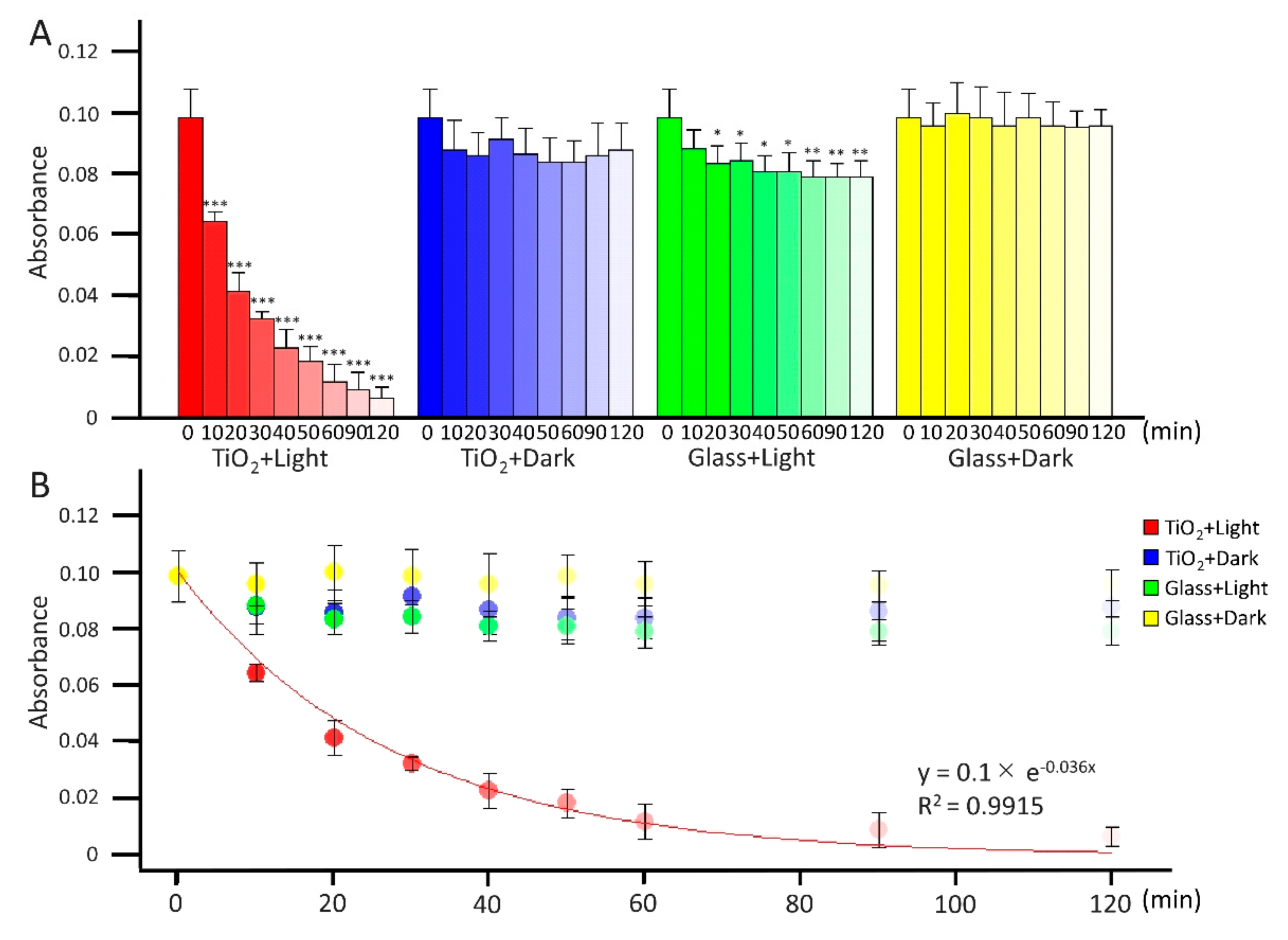
Figure 1. Photocatalytic degradation of methylene blue in water. In the “TiO2 + LED light” group, TiO2-coated glass in 100 mL of 12.5 nM methylene blue in water purified by ion-exchange was irradiated with a LED with a wavelength of 405 nm; absorbance was calculated at 660 nm from 0 to 120 min. As a control, methylene blue was incubated with either TiO2-coated glass without LED light (“TiO2 + Dark” group) or both glass and LED light (“Glass + Light” group), or glass without LED light (“Glass + Dark” group). (A) Each column and error bar represent the mean ± standard deviation (SD) for three experiments. All values in each group were compared with the 0 min sample by two-way analysis of variance (ANOVA) with Dunnett’s test. The asterisk indicates the statistical difference (* p < 0.05; ** p < 0.01; *** p < 0.001). (B) Exponential regression analysis between absorbance of methylene blue and irradiation time from 0 to 60 min before flattening out. R2 indicates the coefficient of determination.
Catalysts 12 00856 g001
Figure 2. Disinfection of E. coli by TiO2 photocatalyst. (A) Schematic diagram of disinfection of bacteria by the TiO2 photocatalyst. (B) In the “TiO2 + Light” group, 30 mL of E. coli with a titer of 1 × 10⁸ CFU/mL was added to a 10-cm diameter dish with TiO2-coated glass; then, 1 mL samples were taken at the marked hourly intervals. As a control, E. coli was incubated with TiO2-coated glass without LED light (“TiO2 + Dark” group), glass and LED light (“Glass + Light” group), or glass without LED light (“Glass + Dark” group). Each column and error bar represent the mean ± standard deviation (SD) of three experiments. Significance between 0 min and other time points in each group was determined using two-way analysis of variance (ANOVA) followed by Dunnett’s multiple comparisons test. (C) Significance between each condition in each time point was determined using two-way ANOVA followed by Tukey’s multiple comparisons test. Asterisks indicate statistical difference (* p < 0.05; ** p < 0.01; *** p < 0.001). (D) The line graph shows the time-dependent changes in disinfection effects of E. coli. Decrement of titer of E. coli calculated according to the following equation: Decrement of titer of E. coli = Titer of E. coli at each time point − Titer of E. coli at 0 h. (E) Linear regression analysis between disinfection effect of E. coli and irradiation time before levelling out. R2 indicates the coefficient of determination.
Catalysts 12 00856 g002
Figure 3. Disinfection of L. pneumophila by the TiO2 photocatalyst. (A) In the “TiO2 + Light” group, 30 mL of L. pneumophila with a titer of 1 × 10⁷ CFU/mL was added to a 10-cm diameter dish with TiO2-coated glass and exposed to light from a LED with a wavelength of 405 nm. Samples (1 mL) were taken at the indicated intervals. During irradiation, the dish was shaken at 40 rpm. As a control, L. pneumophila was incubated with either TiO2-coated glass without LED light (“TiO2 + Dark” group), glass and LED light (“Glass + Light” group), or glass without LED light (“Glass + Dark” group). Significance between 0 min and other time points in each group was determined using two-way analysis of variance (ANOVA) followed by Dunnett’s multiple comparisons test. Asterisks indicate statistical difference (* p < 0.05; ** p < 0.01; *** p < 0.001). (B) Significance between each condition in each time point determined using two-way ANOVA followed by Tukey’s multiple comparisons test. Asterisks indicate statistical difference (* p < 0.05; ** p < 0.01; *** p < 0.001). (C) The line graph shows the time-dependent changes in disinfection effects of L. pneumophila. Decrement of titer of L. pneumophila calculated according to the following equation: Decrement of titer of L. pneumophila = Titer of L. pneumophila at each time point − Titer of L. pneumophila at 0 h. (D) Linear regression analysis between disinfection effect of L. pneumophila and irradiation time before levelling out. R2 indicates the coefficient of determination.
Catalysts 12 00856 g003
Figure 4. Morphological changes in L. pneumophila owing to TiO2 photocatalytic disinfection. A 30 mL sample of L. pneumophila with a titer of 1 × 10⁸ CFU/mL was added to a 10-cm diameter dish with TiO2-coated glass and exposed to a LED with a wavelength of 405 nm for 0 or 24 h. The sample treated by photocatalytic disinfection for 24 h (Treated) and the untreated sample (Untreated) were fixed with 2.5% glutaraldehyde for transmission electron microscopy (TEM) negative staining. TEM images showing the sagittal plane (A) and transverse plane (B) of L. pneumophila. Bar = 500 nm. (C) Low magnification TEM images for counting living and dead L. pneumophila. Red arrows show living L. pneumophila, and blue arrows show dead L. pneumophila. Bar = 2.0 μm. (D) Dead and live L. pneumophila were counted by eye from three low magnification TEM images. The black and white columns represent the numbers of living and dead L. pneumophila for the three TEM images, respectively. Significance was analyzed by the chi-square test. Asterisks indicates the statistical difference (*** p < 0.001). (E) Tier of L. pneumophila in each group.
Catalysts 12 00856 g004
Figure 5. Release and degradation of L. pneumophila endotoxin by the TiO2 photocatalyst. (A) In the “TiO2 + Light” group, 30 mL of L. pneumophila with a titer of 1 × 10⁷ CFU/mL was added to a 10-cm diameter dish with TiO2-coated glass and exposed to a LED light with a wavelength of 405 nm. Samples (1 mL) were taken at the indicated intervals. During irradiation, the dish was shaken at 40 rpm. As a control, L. pneumophila was incubated with either TiO2-coated glass without LED light (“TiO2 + Dark” group), glass and LED light (“Glass + Light” group), or glass without LED light (“Glass + Dark” group). The concentration of endotoxin in each collected sample was measured using Limulus color KY. Significance between 0 min and other time points in each group was determined using two-way analysis of variance (ANOVA) followed by Dunnett’s multiple comparisons test. (B) Schematic diagram of degradation of endotoxin by the TiO2 photocatalyst. (C) Concentration of endotoxin was measured using Limulus color KY. Significance between 0 and 24 h was determined using the Student’s t-test. (D) Endotoxin was detected by silver staining. Positions of endotoxin and corresponding molecular weights are indicated. (E) Intensities of bands were analyzed using the ImageJ software. Significance between 0 and 24 h was determined using the Student’s t-test. The asterisk indicates the statistical difference (* p < 0.05).
Catalysts 12 00856 g005
Figure 6. SEM images, elemental analysis, and methylene blue degradation of TiO2-coated glass before and after use. (A) The SEM images shows surface of frosted glass plate, TiO2-coated glass before use and after use. Bar = 500 nm. (B) Elemental analysis on the surface of the TiO2-coated glass before use and after use was performed using an energy dispersive X-ray analyzer (EDS). Histogram show counts of positive pixels at each voltage. The peak of each chemical elements is indicated. (C) The amounts of TiO2 on the glass (wt%) was calculated from the EDS result. Each column and error bar represent the mean ± standard deviation (SD) for two images in two independent experiments. Significance of the difference between the amounts of TiO2 on the glass before use and after use was determined using the Student’s t-test. NS indicates not significant. (D) TiO2-coated glass before and after use in 100 mL of 12.5 nM methylene blue in water purified by ion-exchange was irradiated with a light emitting diode (LED) with a wavelength of 405 nm; absorbance was calculated at 660 nm from 0 to 120 min. Each column and error bar represent the mean ± SD for three experiments.
Catalysts 12 00856 g006
Publisher’s Note: MDPI stays neutral with regard to jurisdictional claims in published maps and institutional affiliations.