Article

Fabrication and Characterization of Dextranase Nano-Entrapped Enzymes in Polymeric Particles Using a Novel Ultrasonication–Microwave Approach

1
Food Science and Human Nutrition Department, College of Applied and Health Sciences, A’Sharqiyah University, P.O. Box 42, Ibra 400, Oman
2
School of Pharmaceutical Science, Nanchang University, Nanchang 330006, China
3
Therapeutic Nutrition Department, Faculty of Nursing and Health Sciences, Misurata University, Misrata 2478, Libya
4
Department of Food Science, College of Agriculture and Veterinary Medicine, United Arab Emirates University, Al Ain 15551, United Arab Emirates
5
School of Food and Health, Beijing Technology and Business University (BTBU), 11 Fucheng Road, Beijing 100048, China
6
Department of Biological Sciences, Faculty of Sciences, King Abdulaziz University, Jeddah 21589, Saudi Arabia
7
Najla Bint Saud Al Saud Center for Distinguished Research in Biotechnology, Jeddah 21577, Saudi Arabia
8
Department of Biochemistry, College of Sciences, University of Jeddah, Jeddah 21577, Saudi Arabia
9
Biology Department, Faculty of Science, University of Hail, Hail 55221, Saudi Arabia
10
Department of Pharmaceutics, Faculty of Pharmacy, Umm Al-Qura University, Makkah 21955, Saudi Arabia
11
Medicinal Chemistry Department, Faculty of Pharmacy, Beni-Suef University, Beni-Suef 62514, Egypt
12
Department of Pharmaceutical Chemistry, College of Pharmacy, Taif University, Taif 21944, Saudi Arabia
13
Molecular Physiology Division, Zoology Department, Faculty of Science, Beni-Suef University, Beni-Suef 62514, Egypt
14
Oncology Division, Department of Biomedical and Clinical Science, Faculty of Medicine, Linköping University, 58215 Linköping, Sweden
*
Author to whom correspondence should be addressed.
Catalysts 2023, 13(1), 125; https://doi.org/10.3390/catal13010125
Submission received: 19 November 2022 / Revised: 14 December 2022 / Accepted: 25 December 2022 / Published: 5 January 2023
(This article belongs to the Section Biocatalysis)

Abstract

:
In the current study, a novel method to improve the nano-entrapment of enzymes into Ca-alginate gel was investigated to determine the synergistic effects of ultrasound combined with microwave shock (UMS). The effects of UMS treatment on dextranase enzymes’ loading effectiveness (LE) and immobilization yield (IY) were investigated. By using FT-IR spectra and SEM, the microstructure of the immobilized enzyme (IE) was characterized. Additionally, the free enzyme was used as a control to compare the reusability and enzyme-kinetics characteristics of IEs produced with and without UMS treatments. The results demonstrated that the highest LE and IY were obtained when the IE was produced with a US of 40 W at 25 kHz for 15 min combined with an MS of 60 W at a shock rate of 20 s/min for 20 min, increasing the LE and the IY by 97.32 and 78.25%, respectively, when compared with an immobilized enzyme prepared without UMS treatment. In comparison with the control, UMS treatment dramatically raised the Vmax, KM, catalytic, and specificity constant values for the IE. The outcomes suggested that a microwave shock and ultrasound combination would be an efficient way to improve the immobilization of enzymes in biopolymer gel.

1. Introduction

Recent innovations in nanotechnology have transferred to several scientific and industrial areas including enzymology. Applications of nanotechnology have emerged with an increasing need of nanoparticle uses in enzyme immobilization approaches. Immobilization is one of the several cost-reduction techniques used to improve the efficiency of enzyme utilization in biotechnological processes [1]. Besides an easier handling of the enzyme, the immobilization process also makes manipulating the biocatalyst and managing the reaction process significantly simpler [2], while also improving the stability of the enzyme under both storage and use circumstances. Immobilization makes it simple to separate the enzyme from the product, which reduces or completely eliminates the protein contamination of the product and increases the recyclability [3]. On the other hand, enzyme immobilization significantly lowers the cost of the enzyme and the enzymatic products in addition to making it simple to separate the enzyme from the reaction mixture [4].
Dextranase (1,6-α-D-glucan-6-glucanohydrolase; E.C. 3.2.1.11) is an inducible enzyme that cleaves the α (1–6) glycosidic linkages in the interior of dextran to produce linear oligosaccharides. Dextranases are widely used for theoretical and practical purposes. Dextranases reduce the molecular mass of dextran and therefore the viscosity of juices [5]. More recently, dextranase has gained much interest in the directed synthesis of isomaltooligosaccharides, which have been shown to exhibit prebiotic effects [6].
The aqueous enzymatic process during oil extraction has been successfully improved by ultrasonic–microwave treatment [7,8]. In a previous study, it was shown that MS enhanced the microenvironment of an enzyme and accelerated the hydrolysis of polysaccharides with glucoamylase. MS does this by changing the spatial orientation of the active sites and important substrate groups in an enzyme’s catalytic system [9]. This reorientation causes the reactive groups in substrate molecules and the active core of the IE to be closer together and strictly coordinate, increasing the efficiency and specificity of enzymatic activities [10].
The impact of ultrasonication and microwave shock on the industrial efficacy of enzymes has recently been studied [11]. Dextranase enzyme activity was much higher when ultrasound (US) and microwave shock (MS) were combined than when US, MS, and traditional thermal incubation were independently used. However, when immobilizing enzymes in alginate gel, ultrasound increased the rate of immobilization and the catalytic kinetic activity [12]. Therefore, we have concentrated on the development of new techniques by fusing US with other cutting-edge process technologies to enhance the enzyme immobilization process in order to lower the cost of enzymatic reaction in many industries. In this regard, we recently showed that the immobilization of dextranase in alginate gel may be improved with low intensity ultrasonication combined with high pressure [13].
To date, there has not been any research conducted on how the combination of ultrasonication and microwave shock affects the process of immobilizing enzymes. The synergistic effects of UMS on the nano-entrapment of dextranase enzymes into alginate polymers were therefore examined in this study. We calculated the immobilized enzyme’s loading efficiency (LE) and immobilization yield (IY). FT-IR spectra and SEM were used to describe the microstructures of the IEs. Additionally, the IE generated with the UMS treatment’s kinetic activity reusability and thermodynamic properties was also investigated.

2. Results and Discussions

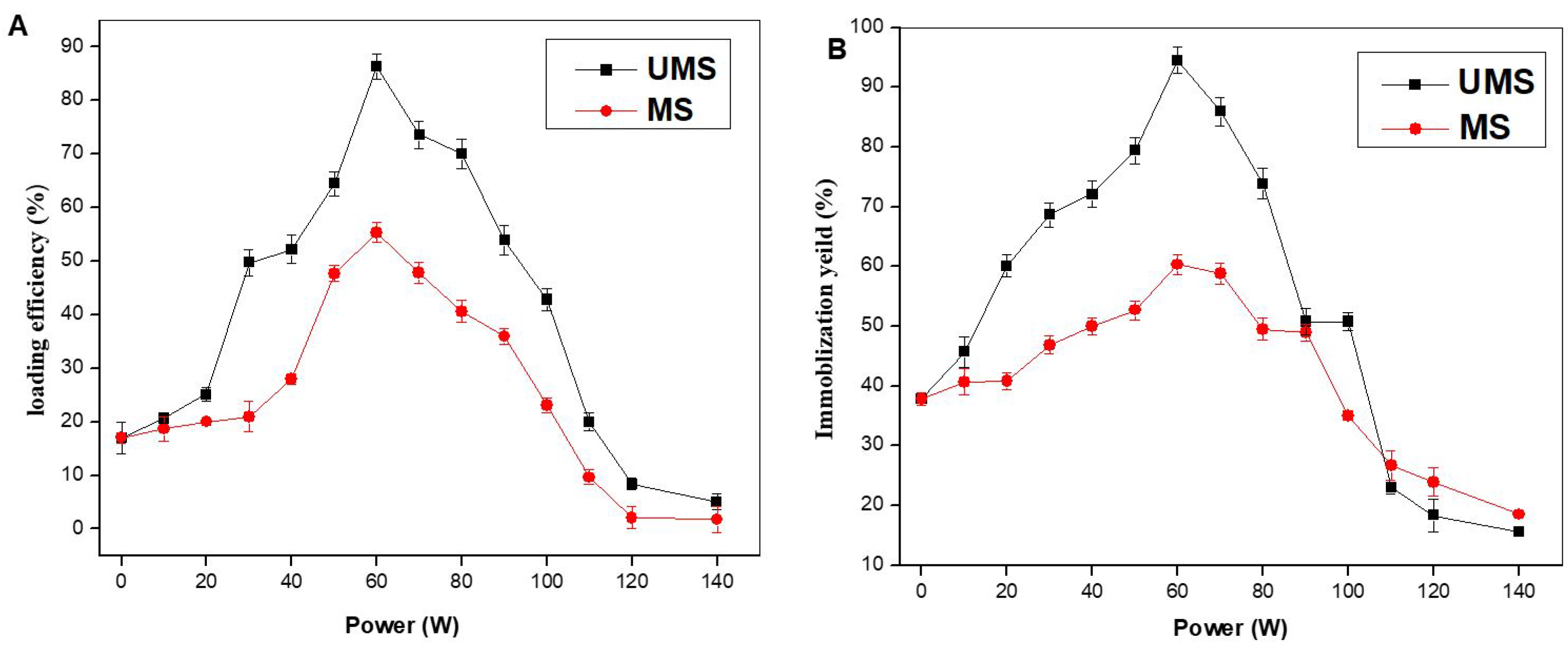
2.1. Effluence of UMS Treatment on the LE and IY of the Immobilized Enzyme

The LE and IY obtained with UMS were compared with those acquired by MS treatment in order to examine the impact of UMS on the immobilization of enzymes in Ca-alginate nanoparticles. The impact of various UMS treatments for 20 min on the LE and EY is depicted in Figure 1. The LE and IY increased by 84.15% and 60.15%, respectively, in comparison with the IE produced with MS when US (50 W at 25 kHz for 15 min) was utilized in combination with 60 W of MS delivered at a shock rate of 20 s/min, respectively. The LE of the IE created by UMS and MS treatments gradually reduced with an increase in the MS power when the MS was greater than 60 W. According to reports, US treatments delivered enough radiation to cause a cavitation effect and a homogenous system, which enhanced the IE’s activity, and a homogeneous system was possible because of the low-intensity ultrasound [12]. The cavitation, on the other hand, increased a mass transfer and strong acoustic streaming, which in turn encouraged collision frequencies between the enzyme molecules and the cross-linked Ca-alginate beads [14,15].
Additionally, microwave irradiation may alter the hydrophobic and hydrogen bonds that stabilize the secondary structure of the enzyme protein, which could alter the conformation and activity of the enzyme as well as the orientation of the initial forces [6,7,8]. The collision rate between the enzyme and Ca-alginate may also improve the nano-entrapment of the enzyme into Ca-alginate nanoparticles [16]. Additionally, microwave treatment improved pronase’s and chymotrypsin’s affinities to lactoglobulin [17]. As a result, the synergistic actions of US and MS increased the dextranase enzyme’s affinity for Ca-alginate nanoparticles, resulting in the increased nano-entrapment of the enzyme in the Ca-alginate gel.
As shown in Figure 1, the LE and IY of the immobilized enzyme generated with UMS and MS treatments gradually reduced as the MS power exceeded 100 W. It was evident that the synergistic effect of US increased the enzyme activity at a higher microwave power when the LE and IY of the UMS treatment were still higher than those seen for the MS treatment when the MS power surpassed 110 W [12]. This may have happened as a result of the reaction’s production of hydroxyl or hydrogen radicals, which interacted with the protein’s backbone [15]. By inhibiting the active sites, this in turn could cause enzyme aggregation and decrease protein stability [16]. Additionally, it has been claimed that the time-dependent activation and inactivation of various enzymes might explain the obtained product yields and the absence of the product hydrolysis of the immobilized enzyme [12,18].

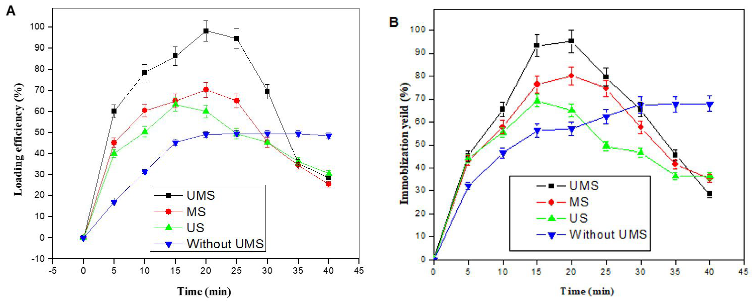
2.2. Effects of UMS Time on the LE and IY of the IE

The effects of UMS at a constant US of 40 W at 25 kHz for 15 min and an MS of 60 W, sequentially and at various incubation times, on the LE and IY of the immobilized enzyme are shown in Figure 2A,B. In comparison with the immobilized enzyme prepared without UMS treatment, both the UMS and MS treatments for 20 min enhanced the maximum LE and IY by 97.32% and 78.25%, respectively. The maximal LE and IY of the US-prepared IE, however, were noted at 15 min. The percentages of the LE and IY of the IE generated with the UMS treatment steadily reduced when the length of the MS treatment exceeded 20 min, as can be observed in Figure 2A. Furthermore, compared with the IE created with MS and US, the LE and IY of the immobilized enzyme prepared by UMS were still greater than those of the control after 30 min of treatment. However, when the time length was more than 30 min, the LE and IY of the IE generated with UMS treatment only started to fall below those of the control. According to the previous reports, short-term sonication can increase enzyme activity while long-term sonication can inactivate the enzyme [12,15]. Additionally, by sonicating for a prolonged period of time, a significant increase in transient cavitation was seen [19]. As a result of the mechanical impact of transient cavitation, the amount of nano-entrapped enzymes that leaked out could have increased over time as a result of the increased opening of the Ca-alginate beads’ surface. These large pores of the matrix in the immobilization support materials were caused by the MS. However, a significant impact of the reaction time on reducing the catalytic activity was observed, which was interpreted as enzyme deactivation due to microwave exposure. On the other hand, Nogueira et al. reported an increase in the enzyme activity with MS during enzymatic transesterification reactions for the production of biodiesel [20].

2.3. Characterization of the Immobilized Enzyme

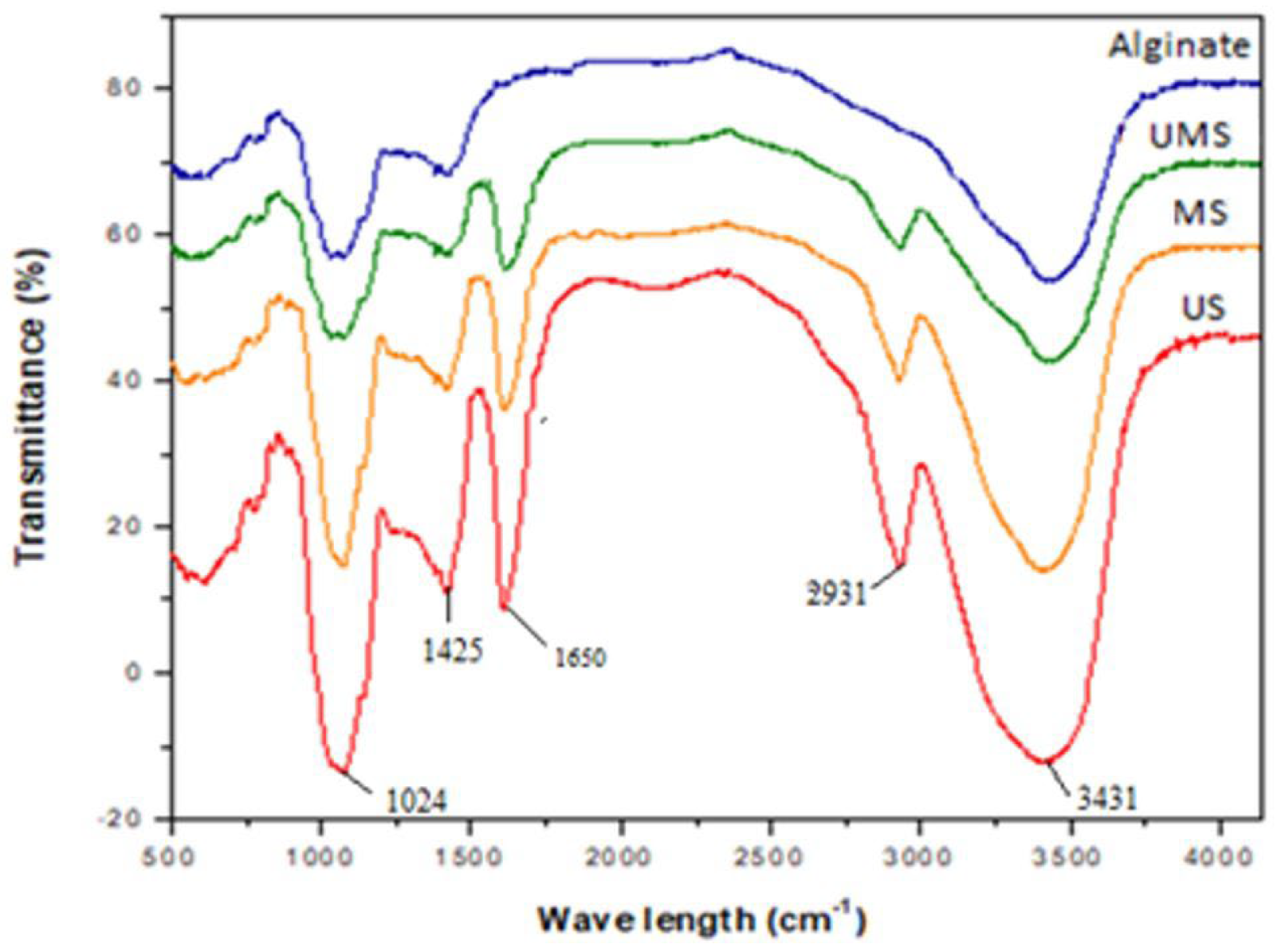
2.3.1. Fourier Transform Infrared Spectra

The FT-IR study proved that the dextranase was nano-entrapped in the alginate gel. A Ca-alginate FTIR spectrum and the FT-IR spectra for the immobilized enzyme obtained with UMS, MS, and US were compared (Figure 3). The distinctive peaks in the Ca-alginate FTIR spectra were found at 3431 cm−1 for OH stretching, 1599 and 1427 cm−1 for COO asymmetric and symmetric stretching, respectively, and 1081–1024 cm−1 for C–O–C antisymmetric stretching [21]. The spectra of the IEs produced with UMS, MS, and US revealed a strong peak at a wavelength of 1650 cm−1 that corresponded to the C=O bond of the amino acid, which is a characteristic peak of the second helix of the dextranase enzyme and correlated with the amide I and II groups [22]. N–H stretching vibrations were said to be the cause of the peaks at 1425 cm−1 [23]. These findings proved and validated dextranase’s successful nano-entrapment in alginate beads. Dextranase’s immobilization was also confirmed by FT-IR by Ding et al. and Agrawal et al., who reported a 1648 cm−1 absorbance of the enzyme’s amino acid group [22,24].

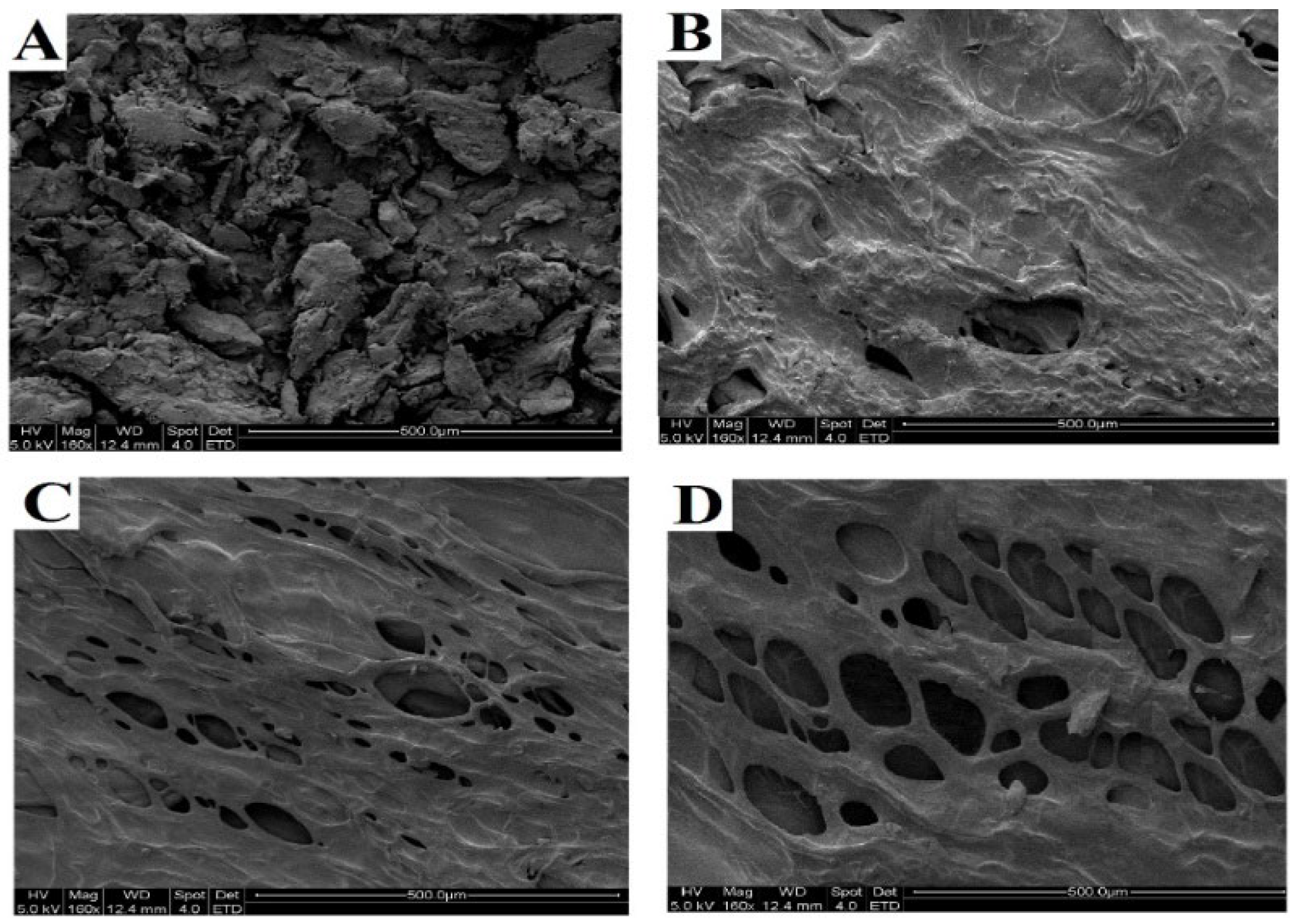
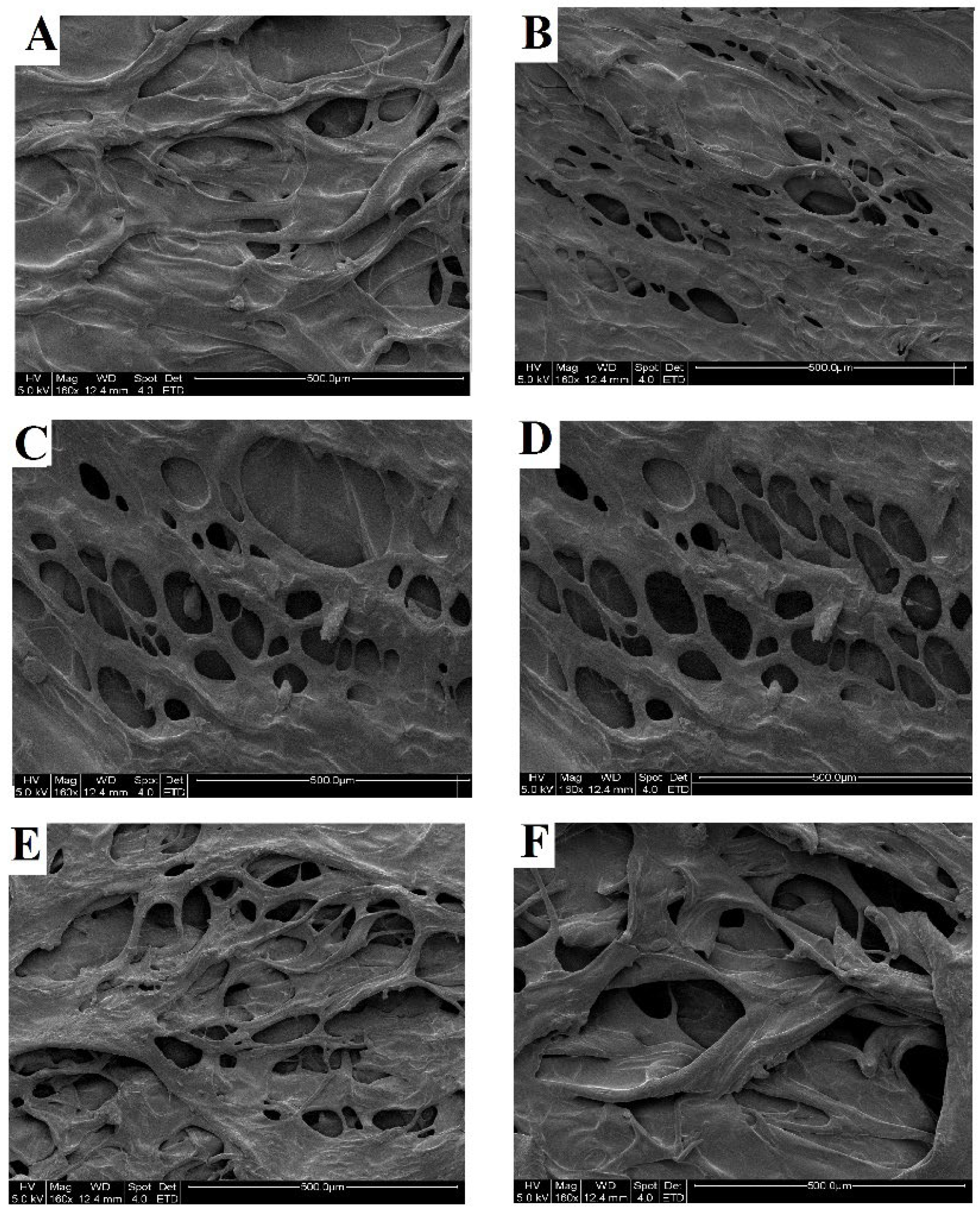
2.3.2. Scanning Electron Microscopy (SEM) Analysis

All samples were examined under a scanning electron microscope to investigate the IE’s microstructure. In contrast to the IEs created using US and alginate beads created without UMS, as illustrated in Figure 4A–D, the IEs created using UMS and MS treatments had porous surfaces. Additionally, the number of pores in alginate beads increased due to the US compensation for MS (Figure 4D). Additionally, the wide holes of the matrix brought on by the high ultrasonic power increased the amount of leaking of the entrapped enzyme over the course of time [12,18]. This came from the mechanical impact of transitory cavitation by US and the effective in situ heating with an even distribution of temperature throughout the sample with MS [16], which exposed the surface of the Ca-alginate nanoparticles. Figure 5 depicts the microstructure of the IE created with UMS treatment at various MS powers. The alginate beads’ pore diameters grew as the MS power increased. The alginate gel’s best opening was noted at 60 W, where consistent pore sizes were seen (Figure 5F). The arranged pores could ensure that there was enough room for the dextranase enzyme to be nano-entrapped in the alginate beads, confirming the synergistic effects of US combined with MS to increase the enzyme’s affinity for Ca-alginate. This decreased both the resistance of the substrate to diffuse into the Ca-alginate nanoparticles and the resistance of the products to diffuse out, increasing the mass-transfer characteristics [25].

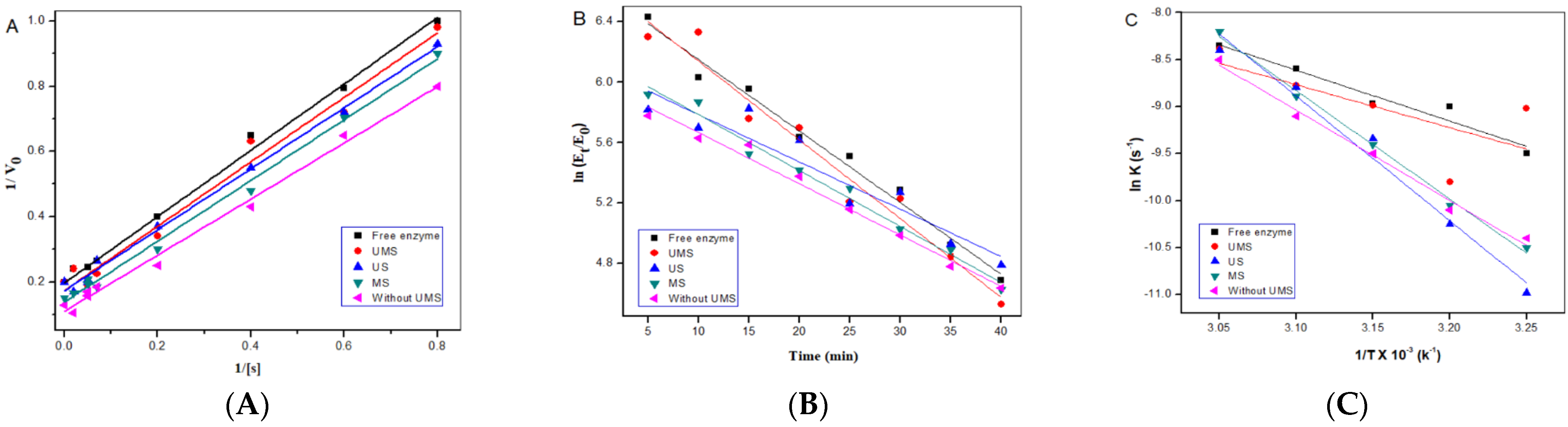
2.4. Study of the IE Kinetics

The kinetic investigations of the IE using the Michaelis–Menten equation are depicted in Figure 6A. In contrast to the free enzyme, the IE produced with UMS, MS, and US treatments displayed low Michaelis constant (KM) values and high reaction rates (Vmax). The low values of KM were brought on by the substrate’s resistance to mass transfer into the porous carrier and the substrate’s limited access to the enzyme-active site [12,13]. Similar to the low value of the Vmax of the IE, the low value of the Vmax of the IE may have been caused by the high molecular substrate’s inability to quickly diffuse into the Ca-alginate nanoparticles, increasing the concentration of the substrate around the gel and ultimately reducing the availability of the entrapped enzyme’s active site to the substrate [26]. At the same time, the product was inhibited for the IE due to the product’s high-level concentration close to the gel’s center, which created resistance to the molecules’ ability to diffuse. By trapping an alkaline protease inside Ca-alginate beads, Sharma et al. showed a similar trend [27].
The Vmax and KM values of the IE created with UMS treatment were higher than those seen for the IE prepared with US and MS, as shown in Table 1, indicating a change in the affinity between the substrate and the IE brought on by the UMS treatment. Similar to how the UMS treatment increased the values for both the catalytic (Kcat) and specificity constants (Kcat/KM) compared with the IE prepared with US and MS, this result showed that the UMS treatment had a positive impact on the IE’s catalytic efficiency, which in turn increased the rate at which the product was produced from the substrate. For immobilized araujiain and cysteine phytoprotease in Ca-alginate gel beads, Quiroga et al. observed a similar outcome [28]. Additionally, specific modifications to the structure and morphology of the immobilized tyrosinase layer on the surface of carbon felt occurred during tyrosinase immobilization with US treatment [29].

2.5. Thermodynamic Studies of IEs

The plots of the ln (Et/E0) of the IE at various incubation times are displayed in Figure 6B. In comparison with MS and US, UMS had higher relative IE activity at 50 °C thermal incubation. The impact of UMS treatment on the activation rate constant (K) and half-life duration (t½) of the free and immobilized enzymes is displayed in Table 2. Additionally, compared with the IEs prepared without ultrasound treatment, the IE prepared with UMS displayed a lower t½ value at 50 °C. This phenomenon accounted for the fact that the IE prepared with the treatment of UMS was more thermostable than the enzymes prepared with the separate treatment of US and MS and had longer periods of constant initial enzymatic activity. The application of UMS therapy also resulted in an increase in the t½ value of the free dextranase enzyme [13]. This may be because UMS treatment caused conformational changes in the enzyme molecule’s structure, impeding the enzyme-catalyzed process. Similar outcomes for the thermal inactivation of IEs have been reported [6,30], although the enzyme activity of the dextransucrase trapped in Ca-alginate was four times higher than that of the free enzyme, and it took 60 min to reach the peak enzyme activity.
Figure 6C displays the Arrhenius plots of ln (k) with the temperature (K), while Table 3 displays the values of Ea, ΔG, ΔH, and ΔS determined using Equations (4)–(6). In comparison with the IE separately prepared using US and MS, the combined treatment of UMS used to prepare the IE had a lower Ea. This fact showed that the catalyzed hydrolysis reaction of the IE was very beneficial for the combined treatment of UMS and may have occurred very quickly. There have been reports of similar outcomes when US therapy was used to lower the activation energy of enzymes [12,13]. However, the activation energy (Ea) of immobilized lipase did not demonstrate any appreciable change [17]. Additionally, the application of UMS treatment resulted in conformational changes in the structure of the enzyme, which prevented the enzyme-catalyzed reaction [11].
In comparison with applying separate US and MS treatments to the IE, the combined UMS treatment resulted in higher ΔH and ΔG values and a lower ΔS value. Additionally, compared with the free enzyme and IE prepared without UMS, the IE prepared with UMS treatment showed a considerable drop in ΔH and ΔS values. The decrease in ΔH may have been caused by greater protein unfolding due to the breaking of the hydrogen bond that stabilizes the enzyme in its ground state and by the ultrasonication-induced disruption of the internal hydrophobic core [13]. The drop in ΔS was caused by the oxidative alteration of amino acid residues, the start of cross-linking and aggregation, and the rise in enzyme activity.

2.6. Reusability

The main benefits of IEs are simple separation and reuse. The IE’s ability to be reused for six cycles is seen in Figure 7. Only 62.13% of the activity from the UMS-treated IE was still present after the fifth cycle, compared with the first four cycles. On the other hand, after the first cycle of the IE generated with MS, US, and without UMS treatments, a decline in the relative enzymatic activity was noticed. The release of unsightly entrapped enzymes on the alginate beads was what caused this decline. This was in line with the outcomes seen for dextranase immobilized in Ca-alginate [12], urease immobilized in a chitosan membrane [30], and diastase–amylase immobilized in nano-zinc oxide [31]. The relative activity of the IE prepared with UMS was lower during the sixth reuse than those seen for the IE prepared with US and MS treatments and without UMS treatment. The initial catalytic activity of the dextranase immobilized in chitosan was retained by about 80%, even after being employed for 12 cycles, in contrast to dextranase immobilized in Ca-alginate, which showed poor operational stability [18].

3. Materials and Methods

3.1. Materials

We purchased sodium alginate, dextran produced by Leuconostoc mesenteroides, dextranase produced by Chaetomium erraticum, and dextran from Sigma-Aldrich (Shanghai, China). Every other chemical, including solvents, was of analytical grade.

3.2. Ultrasonication–Microwave Shock Apparatus

A CW-2000 Ultrasonic-Microwave Cooperative Extractor/Reactor (Discover, CEM-SP1245 model, Poway, CA, USA) with temperature control was used to perform this study. The reaction temperature was measured by an infrared temperature measurement controller installed in the reactor. The device could produce microwave energy in the range of 10–800 W at 2450 MHz and at a set ultrasonic power/frequency of 50 W/40 kHz.

3.3. Measurement of Enzyme Activities

Dextranase activity was calculated when the dextran (T2000) rate of hydrolysis was determined at pH 5.0 and 40 °C. The dextran was broken down by enzymes, and the reducing sugars that were produced were measured using two separate techniques. First, the reducing sugars were evaluated using a spectrophotometric technique because they react with 3,5-dinitrosalicyclic acid to produce a yellow–brown color that can be seen at 540 nm [32,33]. The reducing sugars released were also quantified using a high-performance liquid chromatography technique to confirm this result.

3.4. Chromatographic Analysis and Conditions

The Bashari et al. [13] method was followed in performing the high-performance liquid chromatography analysis. The chromatographic system consisted of a Waters 1525 binary pump and differential refractive index detector (RI-150, Kyoto, Japan). A Thermo Aps-2 Hypersil column (250 mm × 4.6 mm, Thermo Fisher Scientific, Waltham, MA, USA) was used for separation. The column thermostat was set at 30 °C. The aqueous mobile phase was acetonitrile: water (70:30). The sample injection volume was 20 μL, and the flow rate was 1 mL/min. A standard calibration curve was constructed by plotting peak areas against concentrations of a pure isomaltose standard. Throughout the experimental work, data was collected and integrated using LC solution software version 5.6 (SHIMADZU, Kyoto, Japan). For quantification, the peak areas were determined, and the concentrations were calculated with an external calibration curve. The linearity of the calibration curve was assessed by determining the coefficient of correlation (R2) of the points of the curve, which was higher than 0.998.

3.5. Preparation of the IE with Ultrasonication Treatment

The usual ionotropic gelation method as published by Won et al. [34] was used to prepare the IE in alginate beads using US, with some changes as stated in our prior paper [12]. Dextranase (3 mg/mL) was added to an equal volume of 2% sodium alginate solution in water, and the mixture was then processed with fixed ultrasonic irradiation for 20 min at 50 W/40 kHz for various incubation times between 0–40 min at 4 °C. After that, the mixture was continuously shaken at 4 °C for 20 min with 0.2 M CaCl2 in a 0.05 M sodium acetate buffer, pH 5.4. Vacuum filtration was then used to separate the beads from the CaCl2 solution. With a 0.05 M sodium acetate buffer, they were washed twice in a filter. For the LE analysis, the two washings and the filtered CaCl2 solution were combined. After that, the beads were kept at 4 °C in a 0.05 M sodium acetate buffer (pH 5.4) with 0.02% NaN3. The beads had an average diameter of 3 mm. According to the description of Won et al. [34], Equation (1) was used to calculate the LE of the IE:
LE   % = C i V i C f V f C i V i × 100
where Ci is the initial protein concentration, Vi is the initial volume of the enzyme solution, Cf is the protein concentration in the total filtrate, and Vf is the total volume of the filtrate. According to the description of Won et al. [34], Equation (2) was used to calculate the IY:
IY   % = A i m A f e × 100
where Aim is the specific activity of the IE, and Afe is the specific activity of the free enzyme.

3.6. Preparation of the IE with MS Treatment

According to Won et al. [34], conventional ionotropic gelation was used to prepare alginate beads for MS treatment (Figure 8). Dextranase (3 mg/mL) and a 2% sodium alginate solution in water were combined in an identical amount before being processed with MS varying from 10 to 140 W at a shock rate of 20 s/min for various incubation times between 0–40 min at 4 °C. After that, the mixture was continuously shaken at 4 °C for 20 min with 0.2 M CaCl2 in a 0.05 M sodium acetate buffer, pH 5.4. Vacuum filtration was then used to separate the beads from the CaCl2 solution. With a 0.05 M sodium acetate buffer, they were washed twice in a filter. For the LE analysis, the two washings and the filtered CaCl2 solution were combined. After that, the beads were kept at 4 °C in a 0.05 M sodium acetate buffer (pH 5.4) with 0.02% NaN3. The beads had an average diameter of 3 mm. According to the description of Won et al. [34], Equations (1) and (2) were used to calculate the LE and IY of the IE, respectively.

3.7. Preparation of the IE with Ultrasonication–Microwave Shock Treatment

According to Won et al. [34], the preparation of the IE in alginate beads using UMS treatment was performed with standard ionotropic gelation. Dextranase (3 mg/mL) was added to an equal volume of 2% sodium alginate solution in water, and the mixture was then processed with a fixed ultrasonic irradiation of 50 W combined with MS ranging from 10–140 W at a shock rate of 20 s/min for various incubation times between 0–40 min at 4 °C. After that, the mixture was continuously shaken at 4 °C while being dropped into 0.2 M CaCl2. The beads were vacuum-filtered out of the CaCl2 solution after 20 min of hardening. They were washed twice with a 0.05 M sodium acetate buffer in a filter (pH 5.4). According to the description of Won et al., Equations (1) and (2) were used to calculate the LE and IY of the IE, respectively.

3.8. Preparation of the IE without Ultrasonication–Microwave Shock Treatment

Without using UMS treatments, the IEs in the alginate beads were prepared using the standard ionotropic gelation method according to Won et al. [34]. Dextranase (3 mg/mL) was added to an equal volume of 2% sodium alginate solution in water, and the mixture was then kept at 4 °C for various incubation times between 0–40 min. After that, the mixture was continuously shaken at 4 °C while being dropped into 0.2 M CaCl2. The beads were vacuum-filtered out of the CaCl2 solution after 20 min of hardening. They were washed twice with a 0.05 M sodium acetate buffer in a filter (pH 5.4). According to the description of Won et al., Equations (1) and (2) were used to calculate the LE and IY of the IE, respectively.

3.9. Characterization of the IE

3.9.1. Fourier Transform Infrared Spectroscopy (FT-IR)

A Nicolet Nexus 470 FT-IR spectrometer (Spectrum One, Perkin Elmer Co., Waltham, MA, USA) was used to collect data on the presence of particular functional groups in the IE in the 4000–400 cm−1 range using the KBr-disk technique. At 25 °C and a resolution of 4 cm−1, 32 scans were completed.

3.9.2. Scanning Electron Microscopy (SEM) Analysis

All IEs were examined using a scanning electronic microscope (SEM) to determine their surface morphology. Prior to slightly sprinkling the tape with the sample, double-sided tape was initially applied to the specimen container. A gold–palladium layer was applied to the specimens using an SEM sputter coater [12,17]. The SEM (QUANTA 200F, FEI, Amsterdam, The Netherlands) was used to investigate them, with a working distance of 8.0 or 8.2 mm and an accelerating voltage of 5.0 kV.

3.10. Kinetic and Thermodynamic Studies of the IE

The relationship between the substrate concentration and the rate of enzyme reaction was investigated using Michaelis–Menten kinetics, and the linearization of this equation was used for determining the enzyme’s kinetic parameters [35]:
( E ) t ( E ) 0 = exp ( k t )
where (E)t is the enzymatic activity at time t, (E)0 is the initial enzymatic activity, k is the activation rate constant, and t is the time. The activation energy (Ea) could be described with an Arrhenius equation:
K = A e E a R T  
where A is the pre-exponential or collision factor calculated from the antilogarithm of the intercept of the Arrhenius plot of ln K vs. 1/T, T is the absolute temperature in Kelvin, and R is the universal gas constant (8.314 J/mol K). A half-life of reaction (t½, min) could be obtained using first-order kinetics with [35]:
t 1 / 2 = l n   2 K
In order to study the effect of temperature on the IE prepared with and without UMS treatment, the Eyring transition-state theory was used [18]:
K = K B T h e x p Δ G R T = K B T h e x p Δ H R T + Δ S R
where KB is the Boltzmann constant (1.38 × 10⁻²³ J/K), h is the Planck constant (6.6256 × 10⁻³⁴ J/s), ΔG is the change in free energy, ΔH is the change in enthalpy, and ΔS is the change in entropy for the process of the hydrolysis of dextran using the IE prepared with and without ultrasound irradiation.
Given that the standard volume of activation for the reactions in the solution was minimal, the enthalpy ΔH could be determined from the Ea using the following equation [35]:
Δ H = E a R T

3.11. Protein Concentration

Crystalline bovine serum albumin was used as the protein standard to quantify protein concentration using the Bradford method [36].

3.12. Recycled Hydrolysis of Dextran Catalyzed with IEs

The reusability of the IE was tested in accordance with the prior description [13]. The activity of freshly manufactured beads in the first run was considered as the control (100% activity), and the reusability of the IE was tested over the course of six cycles.

3.13. Statistical Analysis

Using SPSS 19 for the Windows application (SPSS Inc., Chicago, IL, USA), the data were analyzed employing an analysis of variance (ANOVA), and the significant difference between the means was determined using Duncan’s multiple range test (p < 0.05). All the experiments described above were made in triplicate for each sample.

4. Conclusions

In this study, we examined how UMS treatment affected the LE and IY of dextranase enzyme immobilization in Ca-alginate nanoparticles. The effects of the UMS treatment on the dextranase enzyme’s LE and IY of the IE in Ca-alginate beads were investigated. By using FT-IR spectra and SEM, the microstructure of the immobilized enzyme was identified. The activities of the IE produced with UMS treatment were compared with those of the free enzyme, which served as a control and as a function of reusability and enzyme kinetic parameters. The highest LE and IY were observed when the IE was produced with an ultrasound irradiation of 40 W at 25 kHz for 15 min, combined with microwave irradiation at 60 W at a shock rate of 20 s/min for 20 min, increasing the LE and IY by 97.32 and 78.25%, respectively, when compared with the immobilized enzyme prepared without UMS treatment. In comparison with the control, UMS treatment dramatically raised the Vmax, KM, catalytic, and specificity constant values for the IE. In comparison with the IE prepared without UMS, the IE prepared with UMS demonstrated higher thermal stability and reusability. The outcomes suggested that a microwave shock and ultrasound combination could be a useful technique for improving enzyme immobilization in the food and pharmaceutical industries.

Author Contributions

Conceptualization, M.B., M.A. and A.B.M.; investigation, M.B., M.A. and A.B.M.; methodology, M.B.; project administration, M.B. and A.A.-F.; visualization, H.A., M.A., A.R. and J.W.; writing—original draft, M.B., M.Y.Z., L.Z.A. and S.Y.S.; writing—review and editing, M.B., M.A., H.A., A.B.M., A.A.-F., A.R., M.A.S.A., A.B., A.S.O. and J.W. All authors have read and agreed to the published version of the manuscript.

Funding

This research received no external funding.

Data Availability Statement

Not applicable.

Conflicts of Interest

The authors declare no conflict of interest.

References

  1. Xie, J.; Zhang, Y.; Simpson, B. Food enzymes immobilization: Novel carriers, techniques and applications. Curr. Opin. Food Sci. 2022, 43, 27–35. [Google Scholar] [CrossRef]
  2. Wilson, L.; Palomo, J.M.; Fernández-Lorente, G.; Illanes, A.; Guisán, J.M.; Fernández-Lafuente, R. Improvement of the functional properties of a thermostable lipase from alcaligenes sp. via strong adsorption on hydrophobic supports. Enzym. Microb. Technol. 2006, 38, 975–980. [Google Scholar] [CrossRef]
  3. Sheldon, R.A.; van Pelt, S. Enzyme immobilisation in biocatalysis: Why, what and how. Chem. Soc. Rev. 2013, 42, 6223–6235. [Google Scholar] [CrossRef] [PubMed]
  4. Mohamad, N.R.; Marzuki, N.H.C.; Buang, N.A.; Huyop, F.; Wahab, R.A. An overview of technologies for immobilization of enzymes and surface analysis techniques for immobilized enzymes. Biotechnol. Biotechnol. Equip. 2015, 29, 205–220. [Google Scholar] [CrossRef] [PubMed]
  5. Zhang, Z.; Wang, S.; Wei, L.; Liao, Y.; Li, D.; Wu, G.; Wang, W. Efficient removal of dextran in sugar juice by immobilized α-dextranase from Chaetomium gracile. Int. J. Food Eng. 2022, 18, 751–759. [Google Scholar] [CrossRef]
  6. Sorndech, W. Isomaltooligosaccharides as Prebiotics and Their Health Benefits. Probiotics, Prebiotics and Synbiotics: Technological Advancements Towards Safety and Industrial Applications; John Wiley & Sons: Hoboken, NJ, USA, 2022; pp. 361–377. [Google Scholar]
  7. Wan, N.; Kou, P.; Pang, H.-Y.; Chang, Y.-H.; Cao, L.; Liu, C.; Zhao, C.-J.; Gu, C.-B.; Fu, Y.-J. Enzyme pretreatment combined with ultrasonic-microwave-assisted surfactant for simultaneous extraction of essential oil and flavonoids from Baeckea frutescens. Ind. Crops Prod. 2021, 174, 114173. [Google Scholar] [CrossRef]
  8. Hu, B.; Wang, H.; He, L.; Li, Y.; Li, C.; Zhang, Z.; Liu, Y.; Zhou, K.; Zhang, Q.; Liu, A.; et al. A method for extracting oil from cherry seed by ultrasonic-microwave assisted aqueous enzymatic process and evaluation of its quality. J. Chromatogr. A 2019, 1587, 50–60. [Google Scholar] [CrossRef]
  9. Zhang, X.; Cao, T.; Tian, X.; Gai, D. Effect of microwave shock on the structure of glucoamylase. Process Biochem. 2012, 47, 2323–2328. [Google Scholar] [CrossRef]
  10. Meridor, D.; Gedanken, A. Preparation of enzyme nanoparticles and studying the catalytic activity of the Immobilized nanoparticles on polyethylene films. Ultrason. Sonochem. 2013, 20, 425–431. [Google Scholar] [CrossRef]
  11. Bashari, M.; Jin, Z.; Wang, J.; Zhan, X. A novel technique to improve the biodegradation efficiency of dextranase enzyme using the synergistic effects of ultrasound combined with microwave shock. Innov. Food Sci. Emerg. Technol. 2016, 35, 125–132. [Google Scholar] [CrossRef]
  12. Bashari, M.; Wang, P.; Eibaid, A.; Tian, Y.; Xu, X.; Jin, Z. Ultrasound-assisted dextranase entrapment onto Ca-alginate gel beads. Ultrason. Sonochem. 2013, 20, 1008–1016. [Google Scholar] [CrossRef]
  13. Bashari, M.; Abbas, S.; Xu, X.; Jin, Z. Combined of ultrasound irradiation with high hydrostatic pressure (US/HHP) as a new method to improve immobilization of dextranase onto alginate gel. Ultrason. Sonochem. 2014, 21, 1325–1334. [Google Scholar] [CrossRef]
  14. Ul, S.Q.; Aman, A.; Syed, N.; Bano, S.; Azhar, A. Characterization of dextransucrase immobilized on Ca-alginate beads from Leuconostoc mesenteroides PCSIR-4. Ital. J. Biochem. 2007, 56, 158–162. [Google Scholar]
  15. Basto, C.; Silva, C.J.; Gubitzb, G.; Cavaco-Paulo, A. Stability and decolourization ability of Trametesvillosa laccase in liquid ultrasonic fields. Ultrason. Sonochem. 2007, 14, 355–362. [Google Scholar] [CrossRef]
  16. Chen, R.; Wei, Q.; Wei, X.; Liu, Y.; Zhang, X.; Chen, X.; Yin, X.; Xie, T. Stable and efficient immobilization of bi-enzymatic NADPH cofactor recycling system by consecutive microwave irradiation. PLoS ONE 2020, 15, e0242564. [Google Scholar]
  17. Ariffin, M.F.K.; Idris, A. Fe2O3/Chitosan coated superparamagnetic nanoparticles supporting lipase enzyme from Candida Antarctica for microwave assisted biodiesel production. Renew. Energy 2022, 185, 1362–1375. [Google Scholar] [CrossRef]
  18. Abdel-Naby, M.A.; Ismail, A.M.; Abdel-Fattah, A.M.; Abdel-Fattah, A.F. Preparation and some properties of immobilized Penicillium funiculosum 258 dextranase. Proc. Biochem. 1999, 34, 391–398. [Google Scholar] [CrossRef]
  19. Yadav, J.; Paragas, E.; Korzekwa, K.; Nagar, S. Time-dependent enzyme inactivation: Numerical analyses of in vitro data and prediction of drug-drug interactions. Pharmacol. Ther. 2020, 206, 107449. [Google Scholar] [CrossRef]
  20. Nogueira, B.M.; Carretoni, C.; Cruz, R.; Freitas, S.; Melo, P.A., Jr.; Costa-Félix, R.; Pinto, J.C.; Nele, M. Microwave activation of enzymatic catalysts for biodiesel production. J. Mol. Catal. B Enzym. 2010, 67, 117–121. [Google Scholar] [CrossRef]
  21. Pereira, R.; Tojeira, A.; Vaz, D.C.; Mendes, A.; Bártolo, P. Preparation and characterization of films based on alginate and aloe vera. Int. J. Polym. Anal. 2011, 16, 449–464. [Google Scholar] [CrossRef]
  22. Ding, Y.; Zhang, H.; Wang, X.; Zu, H.; Wang, C.; Dong, D.; Lyu, M.; Wang, S. Immobilization of Dextranase on Nano-Hydroxyapatite as a Recyclable Catalyst. Materials 2021, 14, 130. [Google Scholar] [CrossRef] [PubMed]
  23. Ding, Y.; Liu, L.; Fang, Y.; Zhang, X.; Lyu, M.; Wang, S. The adsorption of dextranase onto Mg/Fe-layered double hydroxide: Insight into the immobilization. Nanomaterials 2018, 8, 173. [Google Scholar] [CrossRef] [PubMed]
  24. Agrawal, R.; Verma, A.; Satlewal, A. Application of nanoparticle-immobilized thermostable beta-glucosidase for improving thesugarcane juice properties. Innov. Food Sci. Emerg. Technol. 2016, 33, 472–482. [Google Scholar] [CrossRef]
  25. Almeida, L.C.; Barbosa, A.S.; Fricks, A.T.; Freitas, L.S.; Lima, Á.S.; Soares, C.M. Use of conventional or non-conventional treatments of biochar for lipase immobilization. Process Biochem. 2017, 61, 124–129. [Google Scholar] [CrossRef]
  26. Zahirinejad, S.; Hemmati, R.; Homaei, A.; Dinari, A.; Hosseinkhani, S.; Mohammadi, S.; Vianello, F. Nano-organic supports for enzyme immobilization: Scopes and perspectives. Colloids Surf. B Biointerfaces 2021, 204, 111774. [Google Scholar] [CrossRef]
  27. Sharma, J.; Singh, A.; Kumar, R.; Mittal, A. Partial purification of an alkaline protease from a new strain of Aspergillus Oryzae AWT 20 and its enhanced stabilization in entrapped Ca-alginate beads. Internet J. Microbiol. 2006, 2, 1–4. [Google Scholar]
  28. Quiroga, E.; Illanes, C.O.; Ochoa, N.A.; Barberis, S. Performance improvement of araujiain, a cystein phytoprotease, by immobilization within Ca-alginate beads. Process Biochem. 2011, 46, 1029–1034. [Google Scholar] [CrossRef]
  29. Wang, Y.; Hasebe, Y. Acridine orange-induced signal enhancement effect of tyrosinase-immobilized carbon-felt-based flow biosensor for highly sensitive detection of monophenolic compounds. Anal. Bioanal. Chem. 2011, 399, 1151–1162. [Google Scholar] [CrossRef]
  30. Krajewska, B.; Leszko, M.; Zaborska, W. Urease immobilized on chitosan membrane: Preparation and properties. J. Chem. Technol. Biotechnol. 1990, 48, 337–350. [Google Scholar] [CrossRef]
  31. Antony, N.; Balachandran, S.; Mohanan, P.V. Immobilization of diastase α-amylase on nano zinc oxide. Food Chem. 2016, 211, 624–630. [Google Scholar] [CrossRef]
  32. Eggleston, G.; Monge, A. Optimization of sugarcane factory application of commercial dextranases. Proc. Biochem. 2005, 40, 1881–1894. [Google Scholar] [CrossRef]
  33. Brown, C.F.; Inkerman, P.A. Specific method for quantitative measurement of the total dextran content of raw sugar. J. Agric. Food Chem. 1992, 40, 227–233. [Google Scholar] [CrossRef]
  34. Won, K.; Kim, S.; Kim, K.J.; Park, H.W.; Moon, S.J. Optimization of lipase entrapment in Ca-alginate gel beads. Proc. Biochem. 2005, 40, 2149–2154. [Google Scholar] [CrossRef]
  35. Ma, H.; Huang, L.; Jia, J.; He, R.; Luo, L.; Zhu, W. Effect of energy-gathered ultrasound on alcalase. Ultrason. Sonochem. 2011, 18, 419–424. [Google Scholar] [CrossRef]
  36. Bradford, M.M. A rapid and sensitive method for the quantitation of micro-gram quantities of protein utilizing the principle of protein-dye binding. Anal. Biochem. 1976, 72, 248–254. [Google Scholar] [CrossRef]
Figure 1. (A) LE and (B) IY of the immobilized enzyme produced with and without UMS and MS treatments.
Catalysts 13 00125 g001
Figure 2. Effects of UMS treatment duration on the (A) LE and (B) IY of the immobilized enzyme prepared with and without UMS treatment.
Catalysts 13 00125 g002
Figure 3. FT-IR spectra of alginate and IEs in Ca-alginate beads generated with UMS treatment.
Catalysts 13 00125 g003
Figure 4. SEM of (A) Ca-alginate and IEs produced with (B) US, (C) MS, and (D) UMS treatments, at 160× magnification.
Catalysts 13 00125 g004
Figure 5. SEM of immobilized enzymes produced with UMS treatment generated at the different MS powers (A) 0 W, (B) 20 W, (C) 40 W, (D) 60 W, (E) 80 W, (F) and 100 W, at 160× magnification.
Catalysts 13 00125 g005
Figure 6. (A) Lineweaver–Burk plots, (B) relationship between ln (Et/E0) and reaction time, and (C) Arrhenius plots of ln (k) and 1/T.
Catalysts 13 00125 g006
Figure 7. Reusability of the immobilized enzyme generated with and without UMS treatment.
Catalysts 13 00125 g007
Figure 8. Preparation of the immobilized enzyme with ultrasonication–microwave shock treatment.
Catalysts 13 00125 g008
Table 1. Kinetic parameters of the free and immobilized enzymes prepared with and without UMS treatment.
VMax
(μΜ min−1)
KM
(μΜ)
Kcat
(min−1)
Kcat/KM
(μΜ−1 min−1)
Free enzyme1.910 ± 0.170.189 ± 0.23194.221027.61
UMS1.235 ± 0.020.184 ± 0.17182.30990.76
US1.188 ± 0.210.151 ± 0.21143.73951.85
MS1.103 ± 0.110.164 ± 0.13161.27983.35
Without UMS0.991 ± 0.300.134 ± 0.05131.48981.19
Table 2. Enzymatic deactivation parameters of the free enzyme and IE prepared with and without UMS treatment.
K × 10−3 (min−1)t½ (min)R2
Free enzyme0.70 ± 0.3316.25 ± 1.570.989
UMS0.75 ± 0.1715.41 ± 1.150.982
US0.82 ± 0.1212.01 ± 1.130.987
MS0.79 ± 0.0914.60 ± 1.450.977
Without UMS1.00 ± 0.0811.60 ± 1.370.990
Table 3. Thermodynamic parameters of the free enzyme and IE prepared with and without UMS.
ln (A)Ea (kJ mol−1)ΔH (kJ mol−1)ΔS (kJ mol−1 K−1)ΔG (kJ mol−1)
Free enzyme25.5369.41 ± 3.5266.72 ± 1.39−0.0891 ± 0.008260.08 ± 0.98
UMS24.1060.11 ± 1.9859.13 ± 0.98−0.1068 ± 0.0023109.32 ± 0.28
US21.2353.87 ± 3.0149.99 ± 1.23−0.1502 ± 0. 004599.21 ± 1.02
MS21.9555.23 ± 1.7950.38 ± 1.01−0.1489 ± 0.001998.97 ± 1.54
Without UMS22.1158.32 ± 13.3456.73 ± 1.12−0.1229 ± 0.003397.08 ± 1.89
Disclaimer/Publisher’s Note: The statements, opinions and data contained in all publications are solely those of the individual author(s) and contributor(s) and not of MDPI and/or the editor(s). MDPI and/or the editor(s) disclaim responsibility for any injury to people or property resulting from any ideas, methods, instructions or products referred to in the content.

