Article

Bioinspired Green Synthesis of Bimetallic Iron and Zinc Oxide Nanoparticles Using Mushroom Extract and Use against Aspergillus niger; The Most Devastating Fungi of the Green World

1
Department of Plant Sciences, Faculty of Biological Sciences, Quaid-i-Azam University, Islamabad 45320, Pakistan
2
Department of Chemistry, College of Science, King Saud University, P.O. Box 22452, Riyadh 11495, Saudi Arabia
3
Department of Botany and Microbiology, College of Science, King Saud University, Riyadh 11451, Saudi Arabia
4
Department of Botany Rawalpindi Women University, Rawalpindi 43600, Pakistan
5
Department of Botany and Biodiversity Research, University of Vienna, 1040 Vienna, Austria
*
Author to whom correspondence should be addressed.
Catalysts 2023, 13(2), 400; https://doi.org/10.3390/catal13020400
Submission received: 6 January 2023 / Revised: 4 February 2023 / Accepted: 8 February 2023 / Published: 13 February 2023
(This article belongs to the Special Issue Advanced Nanomaterials for a Green World II)

Abstract

:
In the current study, a macro fungus was collected and identified by using morphological and molecular tools to study the ITS region, which has been described as a universal barcode marker during molecular investigation for the identification of fungi. Based on morphology and molecular evidence, the collected fungus was identified as Daedalea Mushroom. The identified fungus was used for the synthesis of Iron and ZnO nanoparticles as an eco-friendly agent for nanoparticle synthesis. The synthesized nanoparticles were confirmed by, Fourier transfer infrared spectroscopy analysis (FTIR), X-ray diffraction analysis (XRD), energy dispersive X-ray analysis (EDX), and scanning electron microscopy analysis (SEM). All these characterizations revealed the synthesis of Iron and ZnO NPs with an irregular shape and a size of 16.8 nm. The zinc oxide nanoparticles had a size in the range of 18.53 nm. Daedalea Mushroom was used for the first time to synthesize Iron and zinc nanoparticles. The mycosynthesized Iron and ZnO NPs were assessed as control agents at various dosage rates against the pathogenic fungus Aspergillus niger, which was isolated from an apple and identified using its morphology. At higher concentrations (0.75 mg/mL), the iron nanoparticles inhibited fungal growth by 72%, whereas at lower concentrations (0.25 mg/mL), they inhibited fungal growth by 60%. ZnO NPs showed good antifungal activity at different concentrations including growth inhibition at 0.25 mg/mL (88%), 1.0 mg/mL (68%), 0.75 mg/mL (75%), and 0.5 mg/mL (70%) concentrations of ZnO NPs. However, the maximum growth inhibition of ZnO NPs was observed at 0.25 mg/mL (88%) concentration and minimum growth inhibition at 0.1 mg/mL (22%). The current study concludes that Daedalea Mushroom works as a novel and eco-friendly source for the synthesis of Iron and ZnO NPs with prominent antifungal activities that can be further applied in different fields.

1. Introduction

The word “nano” has been derived from the Latin word Nanas meaning “dwarf”. Richard Feynman proposed the concept of nanotechnology [1]. Nanoparticles have also piqued the interest of scientists from a wide range of disciplines. There are both organic and inorganic compounds nanoparticles. The inorganic group involves metallic, semi-metallic, and magnet nanoparticles, while the organic group includes carbon nanoparticles [2]. The application of nano-scale materials and their structures, usually extending from 1 to 100 nanometers (nm), is an evolving area of nanoscience and nanotechnology. Nanotechnology, in general, is defined as the movement of materials at the atomic level by a mixture of engineering, chemical, and biological methods. Nanotechnology, as a modern idea, is the most widely explored topic in science [3].
The synthesis of metal nanoparticles is divided into two methods: bottom-up (self-assembly) and top-down [4]. In the bottom-up strategy, a structure is built atom by atom, molecule by molecule, or by self-organization, whereas in the top-down approach, an appropriate starting material is reduced in size using chemical or physical methods. These two processes are used to make nanoparticles, and they are described below. The biological method or green technology is used for the synthesis of nanoparticles. The combination of biological science and nanotechnology has created a new pathway for research in numerous disciplines [5].
Mushrooms are edible fruiting bodies that grow seasonally in rainy seasons Species typically grow on damp soil and moisture area near other food sources. The cap is fleshy and spore-bearing, with spores present inside the gills. Mushrooms have a fruiting body of multicellular fungi that may be epigeous or hypogeous and can be seen with the naked eye [6]. The basic knowledge of diversity at the species or community level is useful for evaluating the impact of artificial or natural disturbance. Macroscopic mushrooms are distinguished into two significant phyla, Basidiomycota and Ascomycetes [7]. The total number of mushroom species reported globally is approximately 14,000, which is thought to be only about 10 percent of the total number of mushroom species found on earth. According to a field survey, about 60% of fungi species are found in the tropics, while others are found in Europe and North America but approximately 22–25% are still undescribed [8]. Usually, mushrooms grow in grassland and woodland. Pasthyrella aquatica was discovered in 2005 and is the only gilled mushroom that grows in water. Some mushrooms can cause wood rot in dead, as well as, living plants, which is the reason that they are found everywhere on leaves, and dead or fallen wood.
Metallic oxide NPs based on Daedalea are economically significant because they can be used for biomedical treatments of diseases and act as, for example, anticancer agents. Daedalea is a healthy, fresh food cultivated on organic substrates and grown naturally in Pakistan and worldwide [9]. Using different plant parts is the easiest, most environmentally friendly, cost-effective, and sustainable way to synthesize surface-functionalized NPs. Mushrooms play a significant role in medicine. One well-known example is the oyster mushroom (Pleurotus spp.), which has been found to be a highly antifungal, antioxidant, and antibacterial candidate as a medicinal fungus in various biological domains. They were first used to synthesize bimetallic Iron and Zinc Oxide NPs [10].
The possible applications of iron and zinc oxide nanoparticles are diverse, such as microelectronics, photocatalysis, magnetic materials, information storage, high-density recording media, nanoelectronic, sensor technology, solar technology, hydrogen storage, and quantum machine biomedical and environmental science [11]. Iron nanoparticles show high potential in many industrial and biomedical applications and play an important role in the aerospace industry. Iron nanoparticles with strong antiferromagnetic properties are widely used in gas sensors, pigments, and catalysts [12]. Zinc nanoparticles have been commonly used for medical applications. Zinc NPs also used to treat a variety of other skin conditions in products, such as baby powder and barrier cream to treat diaper rashes, antidandruff shampoo, calamine cream, and antiseptic ointment. The surface oxygen species of zinc oxide nanoparticles promote the biocidal properties of zinc oxide nanoparticles [13]. Iron and zinc oxide nanoparticles were first created in a study using an improved method for mycosynthesis using a wild mushroom species and were characterized using a multitude of characterization and antifungal activity characterization techniques, including SEM, FTIR, UV, and XRD. In this study, the mycosynthesis of iron and zinc oxide NPs were employed for the first time to study their effect on the growth of Aspergillus niger [14].

2. Results

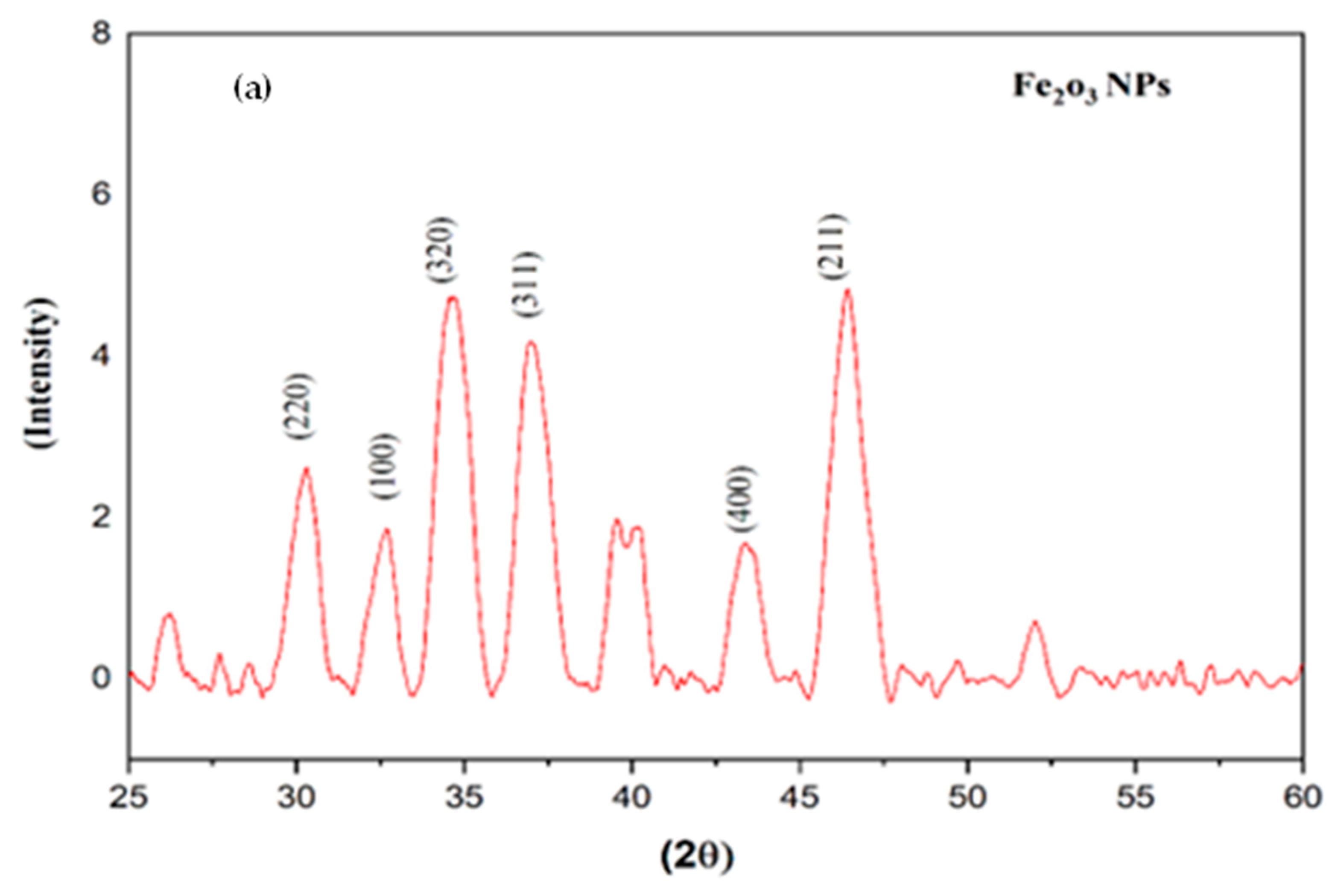
2.1. X-ray Diffraction of Iron and Zinc NPs

The XRD pattern of iron and zinc oxide NPs mycosynthesized from mushroom extract is shown in Figure 1. The diffraction patterns of the synthesized nanoparticles were recorded from 10° to 70°. The diffraction peaks of the synthesized material have a standard structure with several peaks, indicating iron at 2θ = 30.26°, 32.33°, 34.67°, 36.06°, 43.47°, and 46.36°, were obtained. The peaks were consistent with JCPDS card No.76-0958. Full Width at Half Maximum (FWHM) measurements for planes of reflection (220), (100), (320), (311), (400), and (211) were made. Zinc oxide NPs have a hexagonal crystalline structure and were confirmed through the peaks at 2θ values of 32.9°, 34.99°, 58.65°, 67.8° corresponding to the (100), (101), (110), and (200) crystalline planes. The average crystalline size of the iron nanoparticles was 16.8 nm, and the average crystalline size of zinc nanoparticles was 18.53 nm, as determined from the XRD patterns by applying Debye–Scherer’s formula [15].

2.2. FITR Analysis of Iron and Zinc Nanoparticles

The FTIR analysis of the synthesized iron and zinc oxide nanoparticles is shown in Figure 2, which identified the possible biomolecules present in the mushroom extract. The FTIR spectra of iron NPs exhibited a peak at 3390.26 cm−1 allocated to N−H bond stretching of aliphatic primary amine, indicating the synthesis of Iron NPs. Additionally, peaks at 1408.04 cm−1 and 1640.74 cm−1 were attributed to the imine II and amine I bonds, respectively. Peaks at 1595.40 cm−1, 1027.90 cm−1 and 575.39 cm−1 were assigned to N=H, C−N stretching, and C−I stretching, respectively, in the biomolecules [16]. The peak at 3394 cm−1 is attributed to the strong O−H band of the alcohol. The vibrational modes of the strong N−O bond of nitro compounds and the C−H medium band of alkane compounds are represented by the absorption peaks of zinc NPs at 1549.76 cm−1 and 1407 cm−1, respectively. The peak at 669.07 cm−1 is attributed to the C−Br bond [17].

2.3. SEM Analysis of Iron and Zinc NPs

The SEM micrograph analysis predicted that the synthesized iron and zinc nanoparticles would be irregular in shape. It is also observed from SEM images that the particle was agglomerated because of an attractive force present between particles. A SEM micrograph of the synthesized NPs carried out at various magnifications is displayed in Figure 3. SEM plays an important role in studying the size and surface morphology of the synthesized nanoparticles because the biological behavior of the nanoparticles strongly depends on their size and shape [18].

2.4. EDX Analysis of Iron and Zinc NPs

The existence of the element iron and zin oxide in the NPs can be identified by EDX analysis. The relative percentages of iron and oxygen in the mycosynthesized Fe nanoparticles (52.6% of iron and 30.5% oxygen) are similar to the theoretical expected stoichiometric masses. The relative percentage of zinc and oxygen in the mycosynthesized zinc nanoparticles (6.45% of zinc and 37.58% oxygen) are also similar to the expected stoichiometric masses. Three clear signals for Zn and one each for C, O, and P were observed. Other peaks for elements such as Fe, Ca, and S were also observed and are shown in Figure 4 [19].

2.5. Ultraviolet-Visible Spectrum of Iron and Zinc NPs

During the synthesis of iron and zinc NPs, UV-Visible spectrophotometry was carried out to obtain information on the optical properties of the synthesized iron and zinc NPs. The UV-Vis spectra of iron and zinc nanoparticles were taken in the wavelength range of 200–800 nm. Iron nanoparticles mainly show absorption peaks at a wavelength of 287 nm indicating the formation of Iron NPs. The UV-Vis spectra of iron nanoparticles showed various other peaks between 280 and 300 nm, as shown in Figure 5. The VV-Vis spectra of the Zinc NPs show an absorption peak at 360 nm, which was confirmed through spectroscopy. After 24 h of incubation, a zinc NPs peak in the range of 360 to 380 nm was obtained, confirming the formation of Zinc NPs [20].

2.6. Antifungal Activity of Iron and Zinc oxide NPs

For the determination of the antifungal efficacy of iron and zinc nanoparticles, a pathogenic fungus, Aspergillus niger, was taken from the plant pathology lab of the department of plant science Quaid-i-Azam University Islamabad. Aspergillus niger was cultured on PDA at 26 °C in the dark. The PDA media autoclaved with ZnO and Iron nanoparticles at different concentrations of NP solution was poured into Petri dishes (9 cm diameter). The fungi were inoculated after the PDA media solidified. A disc (1.4 cm) of mycelial material taken from the edge of 7-day-old fungal cultures was placed in the center of each Petri dish. The Petri dishes with the inoculums were then incubated at 26 °C. The ability of ZnO and Iron nanoparticle treatment was estimated at the time intervals of 6 days by measuring the diameter of fungal growth. All tests were performed in triplicate and the values were expressed in centimeters [21]. The antifungal test was carried out using the well diffusion method. Various concentrations (0.75 mg/mL, 0.25 mg/mL, 0.1 mg/mL) of iron and zinc NPs exhibited variable fungus growth inhibition. At higher concentrations (0.75 mg/mL), iron nanoparticles inhibited fungal growth by 72%, whereas, at lower concentrations (0.25 mg/mL), they inhibited fungal growth by 60% [22]. The results of the antifungal activity revealed maximum growth inhibition at 0.75 mg/mL concentrations of zinc NPs, which also exhibited the average growth (75%) of pathogenic fungus. Our findings also show that zinc NPs at lower inhibitory concentrations (0.25 mg/mL and 0.1 mg/mL) were able to inhibit the pathogenic fungus by 59% and 27%, respectively. The results show that the nanoparticles have a significant role in disease regulation in an eco-friendly way. The growth inhibition of Aspergillus niger at various concentrations of mycosynthesized Iron and Zinc are shown in Figure 6 [23]. The comparatives antifungal study of various nanoparticles is described in Table 1.

3. Discussion

In this study, aqueous mushroom extract was used as a reducing and capping agent for the synthesis of mycosynthesized iron and zinc NPs. Extensive studies reveal that proteins and carbohydrates constitute the main components within the dried biomass of mushrooms. All these major constituents might have played role in the formation and stabilization of stable elemental iron and zinc at a nanoscale. During the reaction, the formation of iron and zinc NPs was observed by the change in the mixture color (Iron and zinc Solution+ mushroom extract) from light brown to darkish brown. The synthesis was also affirmed with a UV-Vis spectrophotometer that showed a broad absorption peak at a wavelength of 360–380 nm, which indicated a successful synthesis, as shown in Figure 5. It is inferred from a literature review that physicochemical and morphological characteristics of iron and zinc oxide NPs largely depend on the type and species of fungus and the reaction conditions, such as pH, temperature, and synthesis method [30].
The mushroom sample used was collected from Abbottabad kpk and identified through morphological and molecular-based methods. The mycosynthesized iron and zinc nanoparticles were characterized using XRD, FTIR, UV, and SEM, and their antifungal activities were tested against the pathogenic fungus Aspergillus niger. The nanoparticles show good results against Aspergillus niger.
Various characterization techniques were used to study the physicochemical properties of the mycosynthesized iron and zinc nanoparticles, including Fourier transform infrared spectroscopy (FTIR), X-ray diffraction analysis (XRD), UV spectroscopy, and scanning electron microscopy (SEM). The smaller size of synthesized nanoparticles resulted in a large surface area. Furthermore, the XRD diffraction patterns showed that the nanoparticles were highly crystalline. Our XRD results agreed well with the previous literature [31]. Debye–Scherer’s formula was used to calculate the diameter of nanoparticles from XRD spectra. The formula is D = k.λ//ß cos θ, where D is the average crystalline size, k is Debye–Scherer constant (=0.94) λ is the wavelength of the Cukα-radiation (0.154 nm), ß FWHM of the peak, and θ is the Bragg angle. The XRD results revealed that the antimicrobial properties of nanoparticles are highly dependent on their crystal-like nature and the size of the nanoparticles. The crystalline NPs damaged the cell wall of the fungus due to extraordinary antifungal activity [32].
FTIR spectra revealing the functional groups of the mycosynthesized NPs were acquired. FTIR was used to identify the biomolecules present in the mushroom extract, and iron and zinc nanoparticles. The mycosynthesis of iron nanoparticles is carried out by various enzymatic actions, like NADPH-dependent reductase, oxidoreductase, nitrate reductase, and hydrolysis in conjugation with the relevant precursor salt. These biomolecule interactions with the nanoparticles were studied using FTIR. The FTIR spectrum of the mycosynthesized iron NPs exhibited a peak at 3390.26 cm−1 allocated to N-H bond stretching, indicating the synthesis of iron NPs. Additionally, peaks at 1408.04 cm−1 and 1640.74 cm−1 were attributed to the imine II and amine I bonds, respectively [33]. Peaks of 1595.40 cm−1, 1027.90 cm−1, and 575.39 cm−1 were assigned to N=H bonding, C-N stretching, and C-I stretching, respectively. The FTIR spectrum of the mycosynthesized zinc NPs shows various peaks revealing the characteristic functional groups that exist in the zinc NPs [34]. The peak at 3266.22cm−1 was attributed to the strong O-H band of the alcohol. The absorption peaks at 1549.76 cm−1 and 1407 cm−1 were attributed to the vibrational modes N-O strong bond of nitro compounds, and the C-H medium band of alkane compounds, respectively. The C-Br bond is responsible for the peak at 669.07 cm−1 [24]. SEM analysis of the mycosynthesized iron and zinc nanoparticles was used to determine the surface morphology and size of the particles. The boundaries between each nanoparticle are not clearly visible in the lower magnification SEM images, even though the particles are well distributed. Higher magnification photographs show the surface morphology of granules, whereas lower magnification images show monodispersive clusters in the form of assemblies. It is also observed that the particles have agglomerated into clusters, and these agglomerations are seen in the SEM image of the subject particles because of the attractive force that has brought them together [35]. The agglomeration can be removed by the addition of surfactant, but it will lead to a loss of potential characteristics of the nanoparticles. SEM plays an important role in the study of the size and morphology of the nanoparticles because the biological behavior of the nanoparticles strongly depends on their size and shape [36]. UV-Vis spectroscopic analysis of the mycosynthesized iron and zinc oxide nanoparticles was carried out extracellularly using wild mushrooms. UV-Vis spectroscopy is an important technique to establish the formation of nanoparticles [37]. The excitation of an absorption peak at 370 nm confirms the synthesis of mycosynthesized zinc nanoparticles.
The characteristic band of Zinc oxide NPs usually appearing the range 350–380 nm. A continuous increase in the absorbance in the UV spectrum clearly indicates the increased formation of Zinc NPs [38]. The iron nanoparticles mainly show absorption peaks at a wavelength of 287 nm indicating the formation of iron NPs. The UV-Vis spectrum of the iron nanoparticles showed various peaks between 280 and 300 nm and matched well the previous literature [39].

4. Materials and Methods

In the present study, both iron and zinc oxide NPs were synthesized via biosynthesis and green procedures. Iron chloride hexahydrate CAS 10025-77-1 (FeCl3) and dehydrated zinc acetate (C4H10O6Zn) were used as the precursor salts in the synthesis of the iron and zinc oxide nanoparticles. The mushroom Mushroom extract was used as a reducing agent during the synthesis.

4.1. Collection of Mushroom Material

Mushroom samples were collected from Abbottabad kpk during August 2022 and were brought to the mycology lab for NP synthesis.

4.2. Preparation of Zinc and Iron Myconanoparticles

For the synthesis of the iron and zinc oxide nanoparticles, 50 mL of aqueous mushroom extract was taken and slowly mixed into 50 mL of iron and zinc oxide solution. These solutions were kept on a hot plate for 6 h at a temperature of 100 °C and magnetically stirred at 300 rpm. The color of the iron solution changed from light brown to dark brown, while the color of the zinc oxide solution remained unchanged. The pH of the iron solution was 1.6 and that of the zinc solution was 1.8. The solutions were centrifuged at 15,000 rpm for 10 min. After centrifugation, the supernatant was discarded. After washing, the nanoparticles were transferred into Petri dishes and dried on a hot plate at 300 °C. The dry particles were ground into a fine powder and stored at room temperature for further chemical and physical characterization.

4.3. Characterization of Iron and Zinc NPs

The following laboratory methods were used for size identification and morphological characterization of the iron and zinc oxide nanoparticles. The phase composition, surface morphology, crystal structure, and other parameters of the mycosynthesized nanoparticles were investigated using powder X-ray diffraction (XRD, Bruker, D8, Billerica, MA, USA) analysis in the 2θ range from 10° to 70°. An FTIR spectrometer (PECTRUM,65 Manufacturer SKU, Waltham, MA, USA) was used to record the functional groups of the mycosynthesized nanoparticles in the frequency range of 4000–400 cm−1. The UV-Vis spectra (arana BSI sCMOS, Kymera 328i, Oxford, UK) of the mycosynthesized nanoparticles were taken in the special range of 200–800 nm using a spectrophotometer.

4.4. In Vitro Antifungal Activity

The antifungal activity of the mycosynthesized iron and zinc oxide nanoparticles against Aspergillus was performed by the well diffusion method. Different concentrations of iron and zinc oxide nanoparticles (0.25, 0.50, and 0.75 mL) were made to check the antifungal activity, and each concentration was added to PDA media separately. Three replicates of each concentration were made. The isolated fungus from lychee fruit was cut into 2–4 nm inoculum discs and carefully placed in the center of the PDA media dishes. In sterilized conditions, different concentrations of nanoparticle treatment were given to fungi on separate PDA media dishes, and a PDA media dish without nanoparticle treatment served as a positive control. Fungi showed different levels of growth inhibition at different nanoparticle concentrations after being kept in incubators for a week at 26–27 °C. In all Petri dishes, the growth inhibition was estimated by the following formula. Growth inhibition% = (C − T)/C × 100, where C = Average fungal growth and T = Average fungal growth in NPs-treated dish.

5. Conclusions

Wild mushrooms can be used for the synthesis of iron and zinc oxide nanoparticles. The aqueous extract of the wild mushroom, which contains large amounts of carbohydrates, polysaccharides, proteins, and phenolic compounds, was responsible for the formation, and stabilization, and acted as a capping agent, of the particles. The FTIR analysis confirmed the functional groups present in the iron and zinc nanoparticles. The sharp and strong diffraction peaks observed in the XRD patterns confirmed the crystallinity of the mycosynthesized iron and zinc oxide nanoparticles. The mycosynthesized iron nanoparticles were 16.8 nm in size and the zinc oxide nanoparticles were 18.53 nm in size. Both types of nanoparticles were found to be irregular in shape, which was confirmed by SEM analysis. The different functional groups of iron NPs were found to be N-H, O=C=O, C=N, N=H, S=O, and C-I, and of zinc oxide NPs O-H, N-O, C-H, and C-Br, making the synthesized iron and zinc oxide effective antimicrobial agents. The mycosynthesized iron and zinc oxide nanoparticles can both be used as effective antifungal agents.

Author Contributions

Conceptualization, A.K. (Asif Kamal 1) and M.S.; methodology, A.K. (Asif Kamal 1), M.S., M.A. and A.K. (Asif Kamal 2); software, A.K. (Asif Kamal 1) and M.B.; validation, A.K. (Asif Kamal 2) and M.S.; formal analysis, A.K. (Asif Kamal 2) and M.B.; investigation and resources, M.A.; data curation, D.A.A.F. and A.M.A.-M.; writing—original draft preparation, A.K. (Asif Kamal 1) and A.M.A.-M.; writing—review and editing, D.A.A.F. and A.M.A.-M.; visualization, D.H. and A.K. (Asif Kamal 2); supervision and project administration, S.A. All authors have read and agreed to the published version of the manuscript.

Funding

The authors extend their appreciation to the Researchers Supporting Project number (RSP2023R247), King Saud University, Riyadh, Saudi Arabia.

Data Availability Statement

All data and materials support the published claims and comply with field standards.

Acknowledgments

The authors extend their appreciation to the Researchers Supporting Project number(RSP2023R247), King Saud University, Riyadh, Saudi Arabbia.

Conflicts of Interest

The authors declare that they have no conflict of interest.

References

  1. Afsah-Hejri, L.; Hajeb, P.; Ara, P.; Ehsani, R.J. A comprehensive review on food applications of terahertz spectroscopy and imaging. CRFSFS 2019, 18, 1563–1621. [Google Scholar] [CrossRef] [PubMed]
  2. Carmina, E.; Stanczyk, F.Z.; Chang, L.; Miles, R.A.; Lobo, R.A. The ratio of androstenedione: 11β-hydroxyandrostenedione is an important marker of adrenal androgen excess in women. Int. J. Fertil Steril. 1992, 58, 148–152. [Google Scholar] [CrossRef]
  3. Feynman, R.P. An invitation to enter a new field of physics. Int. J. Eng. Sci. 1960, 23, 8. [Google Scholar]
  4. Fischer, M.W.; Money, N.P. Why mushrooms form gills: Efficiency of the lamellate morphology. Fungal. Genet. Biol. 2010, 114, 57–63. [Google Scholar] [CrossRef]
  5. Gianola, D.; de los Campos, G.; Toro, M.A.; Naya, H.; Schön, C.-C.; Sorensen, D.J.G. Do molecular markers inform about pleiotropy? J. Genet. 2015, 201, 23–29. [Google Scholar] [CrossRef]
  6. Imran Din, M.; Rani, A. Recent advances in the synthesis and stabilization of nickel and nickel oxide nanoparticles: A green adeptness. Int. J. Anal. Chem. 2016, 28, 33–48. [Google Scholar] [CrossRef]
  7. Kamal, A.; Saba, M.; Farooq, M. Biocompatible formulations based on mycosynthesized iron oxide nanoparticles: Fabrication, characterization, and biological investigation. J. Basic Microbiol. 2022, 11, 34–41. [Google Scholar] [CrossRef] [PubMed]
  8. Kumar, N.S.; Ganapathy, M.; Sharmila, S.; Shankar, M.; Vimalan, M.; Potheher, I.V. ZnO/Ni(OH)2 core-shell nanoparticles: Synthesis, optical, electrical and photoacoustic property analysis. J. Alloys Compd. 2017, 12, 33–55. [Google Scholar]
  9. Bahadoran, A.; Jabarabadi, M.K.; Mahmood, Z.H.; Bokov, D.; Janani, B.J.; Fakhri, A. Quick and sensitive colorimetric detection of amino acid with functionalized-silver/copper nanoparticles in the presence of cross linker, and bacteria detection by using DNA-template nanoparticles as peroxidase activity. Spectrochim. Acta Part A Mol. Biomol. Spectrosc. 2022, 268, 120636. [Google Scholar] [CrossRef]
  10. Lai, Y.; Fakhri, A.; Janani, B.J. Synergistic activities of silver indium sulfide/nickel molybdenum sulfide nanostructures anchored on clay mineral for light-driven bactericidal performance, and detection of uric acid from gout patient serum. J. Photochem. Photobiol. B Biol. 2022, 234, 112526. [Google Scholar] [CrossRef]
  11. Marchiol, L. Synthesis of metal nanoparticles in living plants. Ital. J. Agron. 2012, 7, e37. [Google Scholar] [CrossRef]
  12. Mueller, G.M.; Schmit, J.P. Fungal biodiversity: What do we know? What can we predict. Anim. Biodivers. Conserv. 2017, 16, 1–5. [Google Scholar] [CrossRef]
  13. Ogwal Engola, A.P. Ecology and nutraceutical value of edible indigenous mushrooms. A case study of Kyebe Subcounty in Rakai District, Uganda. M.R.T. 2018, 15, 184–245. [Google Scholar]
  14. Hu, B.; Cui, Y.; Yang, X.; Xu, X.; Janani, B.J.; Fakhri, A. Fabrication of novel rational Ti-Sn doped Cu-ferrite nanoparticles for robust photocatalysis reaction, magnetic resonance imaging, and chemo-magneto-photo-thermal therapy. Surf. Interfaces 2022, 33, 102226. [Google Scholar] [CrossRef]
  15. Padmavathy, N.; Vijayaraghavan, R. Enhanced bioactivity of ZnO nanoparticles—An antimicrobial study. STAM 2008, 9, 035004. [Google Scholar] [CrossRef]
  16. Patra, J.K.; Baek, K.-H. Green nanobiotechnology: Factors affecting synthesis and characterization techniques. J. Nanomater. 2014, 18, 71–80. [Google Scholar] [CrossRef]
  17. Rajabi, H.; Stamm, K.; Appel, E.; Gorb, S. Micro-morphological adaptations of the wing nodus to flight behaviour in four dragonfly species from the family Libellulidae (Odonata: Anisoptera). Arthropod Struct. Dev. 2018, 47, 442–448. [Google Scholar] [CrossRef]
  18. Sharmila, G.; Fathima, M.F.; Haries, S.; Geetha, S.; Kumar, N.M.; Muthukumaran, C. Green synthesis, characterization and antibacterial efficacy of palladium nanoparticles synthesized using Filicium decipiens leaf extract. J. Mol. Struct. 2017, 1138, 35–40. [Google Scholar] [CrossRef]
  19. Souto, E.B.; Dias-Ferreira, J.; Craveiro, S.A.; Severino, P.; Sanchez-Lopez, E.; Garcia, M.L.; Mahant, S. Therapeutic interventions for countering leishmaniasis and Chagas’s disease: From traditional sources to nanotechnological systems. J. Pathog. 2019, 8, 119. [Google Scholar] [CrossRef]
  20. Khalil, A.T.; Ovais, M.; Ullah, I.; Ali, M.; Shinwari, Z.K.; Maaza, M. Biosynthesis of iron oxide (Fe2O3) nanoparticles via aqueous extracts of Sageretia thea (Osbeck.) and their pharmacognostic properties. Green Chem. Lett. Rev. 2017, 10, 186–201. [Google Scholar] [CrossRef]
  21. He, L.; Liu, Y.; Mustapha, A.; Lin, M. Antifungal activity of zinc oxide nanoparticles against Botrytis cinerea and Penicillium expansum. Microbiol. Res. 2021, 166, 207–215. [Google Scholar] [CrossRef]
  22. Tuskan, G.A.; Difazio, S.; Jansson, S.; Bohlmann, J.; Grigoriev, I.; Hellsten, U.; Putnam, N.; Ralph, S.; Rombauts, S.; Salamov, A.; et al. The genome of black cottonwood, Populus trichocarpa (Torr. & Gray). Science 2006, 313, 1596–1604. [Google Scholar] [PubMed]
  23. Kamal, A.; Haroon, U.; Manghwar, H.; Alamer, K.H.; Alsudays, I.M.; Althobaiti, A.T.; Iqbal, A.; Akbar, M.; Farhana; Anar, M.; et al. Biological Applications of Ball-Milled Synthesized Biochar-Zinc Oxide Nanocomposite Using Zea mays L. Molecules 2022, 27, 5333. [Google Scholar] [CrossRef] [PubMed]
  24. Zubair, M.S.; Munis, M.F.H.; Alsudays, I.M.; Alamer, K.H.; Haroon, U.; Kamal, A.; Ali, M.; Ahmed, J.; Ahmad, Z.; Attia, H. First Report of Fruit Rot of Cherry and Its Control Using Fe2O3 Nanoparticles Synthesized in Calotropis procera. Molecules 2022, 27, 4461. [Google Scholar] [CrossRef] [PubMed]
  25. Saqib, S.; Munis, M.F.H.; Zaman, W.; Ullah, F.; Shah, S.N.; Ayaz, A.; Farooq, M.; Bahadur, S. Synthesis, characterization and use of iron oxide nano particles for antibacterial activity. Microsc. Res. Tech. 2019, 82, 415–420. [Google Scholar] [CrossRef]
  26. Ali, M.; Wang, X.; Haroon, U.; Chaudhary, H.J.; Kamal, A.; Ali, Q.; Saleem, M.H.; Usman, K.; Alatawi, A.; Ali, S.; et al. Antifungal activity of Zinc nitrate derived nano Zno fungicide synthesized from Trachyspermum ammi to control fruit rot disease of grapefruit. Ecotoxicol. Environ. Saf. 2022, 233, 113311. [Google Scholar] [CrossRef]
  27. Kamal, A.; Saba, M.; Ullah, K.; Almutairi, S.M.; AlMunqedhi, B.M.; Ragab abdelGawwad, M. Mycosynthesis, Characterization of Zinc Oxide Nanoparticles, and Its Assessment in Various Biological Activities. Crystals 2023, 13, 171. [Google Scholar] [CrossRef]
  28. Sandhya, J.; Kalaiselvam, S. Biogenic synthesis of magnetic iron oxide nanoparticles using inedible borassus flabellifer seed coat: Characterization, antimicrobial, antioxidant activity and in vitro cytotoxicity analysis. Mater. Res. Express 2020, 7, 015045. [Google Scholar] [CrossRef]
  29. Umar, L.; Hamzah, Y.; Setiadi, R.N. Biosensor signal improvement using current mirror topology for dissolved oxygen measurement. Meas. Sci. Technol. 2019, 30, 065102. [Google Scholar] [CrossRef]
  30. Vanathi, P.; Rajiv, P.; Narendhran, S.; Rajeshwari, S.; Rahman, P.K.; Venckatesh, R. Biosynthesis and characterization of phyto mediated zinc oxide nanoparticles: A green chemistry approach. Mater. Lett. 2014, 134, 13–15. [Google Scholar] [CrossRef]
  31. Sumanth, B.; Lakshmeesha, T.R.; Ansari, M.A.; Alzohairy, M.A.; Udayashankar, A.C.; Shobha, B.; Niranjana, S.R.; Srinivas, C.; Almatroudi, A. Mycogenic synthesis of extracellular zinc oxide nanoparticles from Xylaria acuta and its nanoantibiotic potential. Int. J. Nanomed. 2020, 15, 8519–8536. [Google Scholar] [CrossRef]
  32. Oussou-Azo, A.F.; Nakama, T.; Nakamura, M.; Futagami, T.; Vestergaard, M.C.M. Antifungal potential of nanostructured crystalline copper and its oxide forms. Nanomater 2020, 10, 1003. [Google Scholar] [CrossRef] [PubMed]
  33. Devi, H.S.; Boda, M.A.; Shah, M.A.; Parveen, S.; Wani, A.H. Green synthesis of iron oxide nanoparticles using Platanus orientalis leaf extract for antifungal activity. Green Process. Synth. 2019, 8, 38–45. [Google Scholar] [CrossRef]
  34. Baskar, R.; Dai, J.; Wenlong, N.; Yeo, R.; Yeoh, K.W. Biological response of cancer cells to radiation treatment. Front. Mol. Biosci. 2014, 1, 24. [Google Scholar] [CrossRef] [PubMed]
  35. Devi, L.S.; Joshi, S.R. Ultrastructures of silver nanoparticles biosynthesized using endophytic fungi. J. Microsc. Ultrastruct. 2015, 3, 29–37. [Google Scholar]
  36. Laurent, S.; Forge, D.; Port, M.; Roch, A.; Robic, C.; Vander Elst, L.; Muller, R.N. Magnetic iron oxide nanoparticles: Synthesis, stabilization, vectorization, physicochemical characterizations, and biological applications. Chem. Rev. 2008, 108, 2064–2110. [Google Scholar] [CrossRef]
  37. Katata-Seru, L.; Moremedi, T.; Aremu, O.S.; Bahadur, I. Green synthesis of iron nanoparticles using Moringa oleifera extracts and their applications: Removal of nitrate from water and antibacterial activity against Escherichia coli. J. Mol. Liq. 2018, 256, 296–304. [Google Scholar] [CrossRef]
  38. Anthony, K.J.P.; Murugan, M.; Jeyaraj, M.; Rathinam, N.K.; Sangiliyandi, G. Synthesis of silver nanoparticles using pine mushroom extract: A potential antimicrobial agent against E. coli and B. subtilis. J. Ind. Eng. Chem. 2014, 20, 2325–2331. [Google Scholar] [CrossRef]
  39. Ganesan, R.; Manigandan, S.; Samuel, M.S.; Shanmuganathan, R.; Brindhadevi, K.; Chi, N.T.L.; Duc, P.A.; Pugazhendhi, A. A review on prospective production of biofuel from microalgae. Biotechnol. Rep. 2020, 27, e00509. [Google Scholar] [CrossRef]
Figure 1. XRD analysis of (a) iron and (b) ZnO nanoparticles.
Catalysts 13 00400 g001aCatalysts 13 00400 g001b
Figure 2. FTIR analysis of (a) iron and (b) ZnO nanoparticles.
Catalysts 13 00400 g002aCatalysts 13 00400 g002b
Figure 3. SEM Images of (a) iron and (b) ZnO nanoparticles.
Catalysts 13 00400 g003
Figure 4. EDX analysis of iron and ZnO nanoparticles.
Catalysts 13 00400 g004
Figure 5. UV-Vis spectroscopic analysis of iron (a) and ZnO (b) nanoparticles.
Catalysts 13 00400 g005
Figure 6. Growth inhibition of pathogenic fungi at different concentrations (0.75 mg/mL (A), 0.50 mg/mL (B), 0.25 mg/mL (C) of iron and zinc oxide nanoparticles.
Catalysts 13 00400 g006
Table 1. Comparison of antifungal activity of green synthesized iron and zinc oxide nanoparticles.
NPsSourceAntifungal PotentialReference
MB-ZnOPlants60%Zubair, M.S et al., 2022 [24]
Fe2O3Plants65%Saqib’s et al., 2019 [25]
IONPsMetal62%Ali, M et al., 2020 [26]
ZnOplant60%Kamal, A et al., 2022 [27]
ZnONPsMushroom70%Sandhya, J et al., 2020 [28]
IONPsFungi56%Umar, L et al., 2021 [29]
Disclaimer/Publisher’s Note: The statements, opinions and data contained in all publications are solely those of the individual author(s) and contributor(s) and not of MDPI and/or the editor(s). MDPI and/or the editor(s) disclaim responsibility for any injury to people or property resulting from any ideas, methods, instructions or products referred to in the content.

