Article

Co–HOAT Complexes Change Their Antibacterial and Physicochemical Properties with Morphological Evolution

1
Key Laboratory of Environment Chemistry, Faculty of Chemical and Environmental Science, Qujing Normal University, Qujing 655000, China
2
The Education Ministry Key Laboratory of Resource Chemistry, Shanghai Key Laboratory of Rare Earth Functional Materials, Shanghai Normal University, Shanghai 200234, China
*
Authors to whom correspondence should be addressed.
These authors contributed equally to this work.
Catalysts 2024, 14(3), 173; https://doi.org/10.3390/catal14030173
Submission received: 2 January 2024 / Revised: 13 February 2024 / Accepted: 19 February 2024 / Published: 27 February 2024
(This article belongs to the Special Issue Advances in Photocatalytic Biomaterials)

Abstract

:
Antibacterial coordination compounds have attracted tremendous attention ascribed to their excellent designability. However, how the morphological evolution of these complexes influences their antibacterial and physicochemical properties has never been investigated based on proposed mechanisms. Thus, a series of Co–HOAT coordination compounds synthesized from inorganic to organic cobalt sources were prepared. We propose that with the same HOAT ligand, inorganic Co–HOAT nanosheets possess higher sterilization rates compared with organic Co–HOAT nanoparticles. This is explained by the different steric hindrance of cobalt sources. Relatively small steric hindrance could lead to ample active positions for inorganic cobalt ions to coordinate with both N and O atoms in HOAT. Meanwhile, organic Co2+ ions could only unite with N atoms in HOAT. Furthermore, by theoretical calculation, cobalt ions with adequate coordination sites are beneficial for developing nanosheet morphologies. Meanwhile, the Co–HOAT complexes with a lower density of electron clouds present higher sterilization rates due to the anchoring effect of electrostatic attraction. The proposed mechanism is that Co2+ released from compounds could cause multiple toxic effects to bacteria anchored by Co–HOATs. Finally, Co–HOATs’ behaviors have excellent antimicrobial properties without environmental limitations. In conclusion, the Co–HOATs appear to be a potential antibacterial catalyst in the antimicrobial field.

1. Introduction

Recently, the continuing challenges of microorganisms, such as bacteria, fungi and viruses, still cause a huge threat to public human health and economic development [1]. Thus, it is urgent to explore an effective method to prevent the damage of infectious diseases induced by microorganisms. Many traditional strategies, such as ultraviolet [2], ozone [3], chloridum [4], thermocatalytic [5], electrocatalytic [6], and antibacterial, have been proposed. However, the bottlenecks of limited space, high energy cost, and many harmful bi-products restrict their further study [7,8]. As an emerging method, photocatalysis based on photocatalysts has attracted tremendous attention in various scientific fields [9]. This is due to its advantages of low cost, being environmental friendly, and sustainability [10]. Especially, photocatalytic antimicrobial therapy has emerged as a more effective and promising strategy compared with conventional sterilization [11]. During the antibacterial process, reactive oxygen species (ROS) [12], heavy metal ions [13], or other toxic species [14] for killing bacteria can be achieved when catalysts are stimulated by visible light. At present, the common antibacterial materials are mainly focused on metal oxide (Zn–CdO [15], Cr–TiO2 [16], AgBr/CeO2 [17], Cu-doped TiO2 [18], Cu2O [19], SnO2/HRP [20] and ZnO [21]); sulfide (bio-ZnS/CuS [22], CuS [23], Ag/Ag2S/reduced graphene oxide (rGO) [24] and BaWO4–MoS2 [25]); graphene oxide and its derivates [26]; bismuth-based material (3D-BiOCl@PDA [27], BiOBr [28] and Au/BiOI [29]); carbon nanotubes (CNTs) [30], g-C3N4 [31], and complexes (ZIF-8 [32], Al–MOFs [33], CuO/Cu2O@MIL-125-NH2 [34], MIL-125(Ti)/PVDF [35], Cu–BTC MOFs [36] and ZIF-67/ZnO [12]).
Among these, metal complexes are regarded as good photocatalysts with potential applications in drug delivery [37], environmental purification [38], and antibacterial processes [39]. Ligand–metal complexes have received increasing attention, owing to their unique structures, wide range of biological activities, diversity of substituents, and ease of synthesis [40]. Particularly, most metal complexes possess excellent antibacterial activity compared with free ligands and metals [41]. The high potent activities of the complexes are reduced by coordination and chelation. They lead metal complexes to act as more controlling and potent antimicrobial agents, and then restrain the growth of the microorganisms [42]. Among antibacterial metal complexes, metals of Co(II), Ni(II), Cu(II), and Zn(II) have gained substantial interest. This is ascribed to that fact that these metals are beneficial for accelerating functional inactivation by binding the proteins and DNA of cells [43,44]. Among the reported organic ligands, benzotriazole and its derivatives have attracted huge attention in various applications, such as biological and chemical fields [45]. As one of the key benzotriazole derivatives, 1-hydroxy-7-azabenzotriazole (HOAT) has been widely used as a condensing agent [46], coupling agent [47], and catalyst [48] due to its large ring tension and N–N and C–N bonds [49]. The derivative’s functional structure is beneficial for HOAT to form complexes by construction with Co2+. Recently, the proposed mechanisms of antibacterial activity by metal complexes have mainly focused on disrupting cell membranes by oxidative stress stemming from cationic transition metal complexes [50], displacing DNA-bound ethidium bromide by complexes to inactive cell functions [51], or deoxyribonucleic acid fragmentation and avoiding cell propagation by the strong affinity between metal ions and cell walls [52]. However, how the morphological evolution of complexes influences their antibacterial and physicochemical properties has never been investigated.
Herein, in this work, a series Co–HOATs synthesized by inorganic and organic cobalt sources are successfully synthesized by the one-pot method. It is interesting to observe how the morphology of the Co–HOATs evolves from nanosheets to nanoparticles by TEM and SEM when cobalt sources are changed from inorganic to organic. In addition, Co–HOAT nanosheets have higher sterilization efficiency than Co–HOAT nanoparticles. Aiming to explain this phenomenon, a series of characterizations are conducted. We propose that the different steric hindrance of cobalt sources changes the coordination strategy of cobalt ions and HOAT, according to theoretical calculations. Therefore, inorganic cobalt ions possess sufficient sites to coordinate with both N and O atoms in HOAT. However, organic Co2+ ions can only unite with N atoms in HOAT due to limited coordination sites, which have been partially occupied by organic anions. Furthermore, cobalt ions with adequate coordination sites are beneficial for developing nanosheets’ morphology. Meanwhile, Co–HOAT complexes with a lower density of electron clouds present higher sterilization rates due to the anchoring effect of electrostatic attraction. The proposed mechanism is that Co2+ released from complexes could cause multiple toxic effects to bacteria that is anchored by Co–HOATs. Finally, Co–HOATs have excellent antimicrobial properties without posing environmental limitations. In conclusion, the Co–HOATs appear to be a potential antibacterial catalyst in the antimicrobial field.

2. Experimental Sections

2.1. Materials and Characterization

All reagents were purchased from commercial companies and used without further purification, including 1-hydroxy-7-azabenzotriazole (HOAT) (98%, AR), cobalt (II) perchlorate hexahydrate (Co(ClO4·6H2O), purity: 99.8%), cobalt (II) chloride (CoCl2, 99.7%, AR), cobalt nitrate hexahydrate (Co(NO3)2·6H2O, 99%, AR), cobalt sulfate (CoSO4, purity:99.8%), cobalt acetate ((CH3CO2)2Co, purity: 98%), and cobalt acetylacetonate (C10H14CoO4, 98%), Na3[Co(NO2)6] (99%, AR).
XRD: Powder X-ray diffraction (XRD) measurements were carried out on a Rigaku D/max-3C 2000 single-crystal diffractometer using Cu Kα radiation (λ = 0.15405 nm) (Shanghai, China): around 50 mg catalysts powder were tested.
FTIR: Fourier transform infrared (FT-IR) spectroscopy was performed on NEXUS 470 (Qujing, China): we ground the dried catalyst adequately and added pure potassium bromide (ratio: 1:100) evenly. Finally, the ground samples were placed in a mold and pressed into transparent sheets on a tablet press with a pressure of 5 × 10⁷ Pa for determination.
SEM: Energy dispersive spectroscopy (EDS) analysis was performed with HITACHI S-4800 SEM equipped with an Oxford X-max spectrometer (Shanghai, China): about 1 mg catalysts were prepared to be actualized by using the SEM machine. TEM: The morphology of the catalysts was evaluated by using a JEM-2010 transmission electron microscope (TEM): about 0.5 mg catalysts were dispersed evenly in 50 mL ethyl alcohol to be actualized by TEM machine.
XPS: The surface compositions of samples were measured by using X-ray photoelectron spectroscopy (Shanghai, China, XPS, Versa Probe PHI 5000): about 5 mg catalysts were prepared to be tested.
Raman: Raman spectra was measured by using laser Raman spectroscopy (Qujing, China, SuperLabRam II): about 5 mg catalysts were prepared to be tested.

2.2. Synthesis of Various Co–HOAT Nanoparticles

The Co metal salt, cobalt (II) perchlorate hexahydrate (Co(ClO4·6H2O), (CoCl2/Co(NO3)2·6H2O/CoSO4/(CH3CO2)2Co/C10H14CoO4/Na3[Co(NO2)6], 0.2961 ± 0.0005 g) was weighed into the conical flask, and x mL of methanol was added for dissolution. The appropriate amount of HOAT (n metal salt–n ligands = 1:1) was weighed into a small beaker, and y ml of methanol was added into one beaker for dissolution (x + y = 25 mL). After the metal and ligand were dissolved, respectively, the ligand solution in the small beaker was completely added into the conical flask with the metal sources dissolved. The conical flask was sealed with plastic wrap, bathed in 25 °C water, and the mixed solution was stirred evenly for 3 h; then, removing the plastic wrap on the conical flask and keeping the same conditions (bathed in water with 25 °C and stirred), this continued for another 3 h. When the stirring process was finalized, the conical flask with the solution was put into a 60 °C oven to dry for 12–24 h until the material was completely dry. The material could be used for antimicrobial testing and characterization after it was uniformly ground. The samples obtained by preparations performed by using CoCl2, CoSO4, Na3[Co(NO2)6], Co(NO3)2·6H2O, (Co(ClO4·6H2O), (CH3CO2)2Co, and C10H14CoO4 were denoted as Co–HOAT-1, Co–HOAT-2, Co–HOAT-3, Co–HOAT-4, Co–HOAT-5, Co–HOAT-6 and Co–HOAT-7, respectively.

2.3. The Test of Cobalt Ions in Co–HOAT Nanoparticles

In order to determine the amount of cobalt ions of Co–HOATs released during the antibacterial process, about 0.1 g of material was accurately weighed and added to 10 mL of normal saline to soak for 10 min. Then, the solution was centrifuged and the flame atomic absorption method was used to determine the concentration of cobalt ions; finally, the amount of released cobalt ions per gram of material was calculated.

2.4. Culture and Treatment of Microorganisms

Liquid medium: 2.5 g tryptone, 5.0 g agar powder, 1.250 g yeast extract, and 2.5 g sodium chloride were added into 250 mL water in a Erlenmeyer flask and stirred for 10 min. Next, the pH value of this system was adjusted to 7.2–7.4 by using 1 mol/L sodium hydroxide solution. Finally, the mixture was disinfected in a high-handed sterilization pan to obtain a sterile and liquid medium.
Solid medium: 5.875 g agar powder was added into a 250 mL Erlenmeyer flask and dissolved in 250 mL water by heating to 98 °C. Next, the solution was sealed by using a rubber plug and cooled to room temperature. Finally, the system was disinfected in a high-handed sterilization pan to obtain a sterile and solid medium.
Culture of microorganisms: The antibacterial activity of the samples was investigated by the agar well diffusion method; gram-positive Staphylococcus aureus (ATCC8099) and gram-negative E. coli (ATCC8099) were used in this work. The zone of inhibition test was employed to evaluate the antibacterial activity of various Co–HOATs catalysts. First, gram-positive Staphylococcus aureus or gram-negative E. coli were cultivated in Luria Bertani (LB) media and cultured for 24 h at 37 °C. For the agar well diffusion method, initially, plates containing agar were prepared. Then, the nutrient agar plates were inoculated with 1 mL of bacterial suspension containing around 104 colony forming units (CFU) by using the spread plate method, and wells with 8 mm diameters were created in the plates by using a sterilized stainless-steel cork borer. Next, each nanocatalyst (0.050 g) was inoculated into the wells and placed under visible-light and dark conditions for 10 min, respectively. After that, these dishes were cultivated in the artificial bioclimatic test chamber for 24 h. Finally, these tests were performed twice, and pure metal sources and ligands were used as the control.

2.5. The Videos of Optical Microscope for Anchoring Bacteria

The videos of optical microscope for anchored bacteria by Co–HOATs. About 3 mg catalyst assisted by ultrasound to be uniformly dispersed in 1 mL of bacterial suspension (1.5 × 10⁷ cfu E. coli, OD = 0.1), then sucked a drop of above solution on microscope to obtain the videos under dark condition.

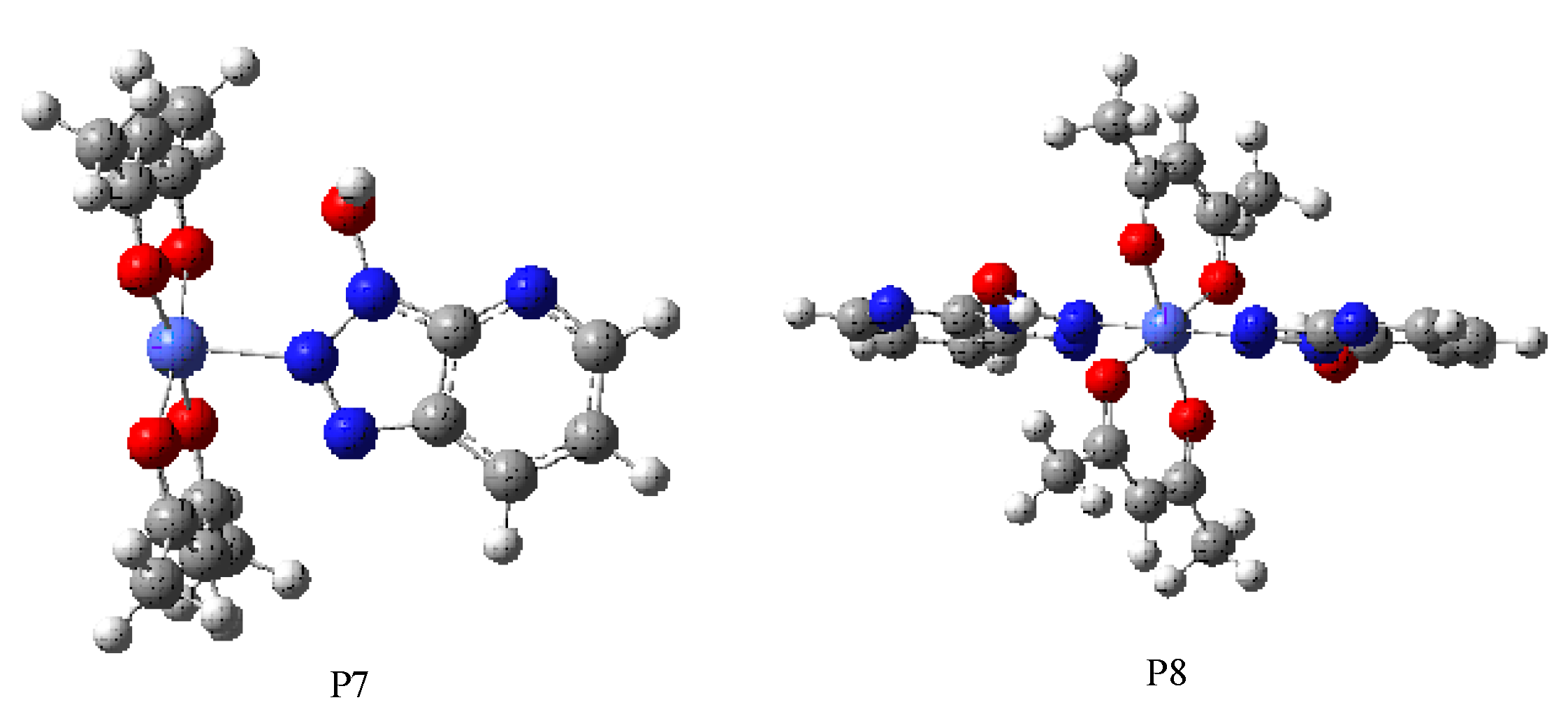
2.6. Molecular Geometries of Calculated Structures

The molecular geometries of the calculated structures were optimized in aqueous solution using the density functional theory (DFT) at the B3LYP level in the 6-311+G basis sets for C, H, O, and N atoms and the LANL2DZ basis sets for the Co atom [53,54]. The solvation of all the optimized structures in water was treated using the polarizable continuum model (PCM) of the self-consistent reaction field (SCRF) theory. All calculations were carried out by using GAUSSIAN 09.

3. Results and Analysis

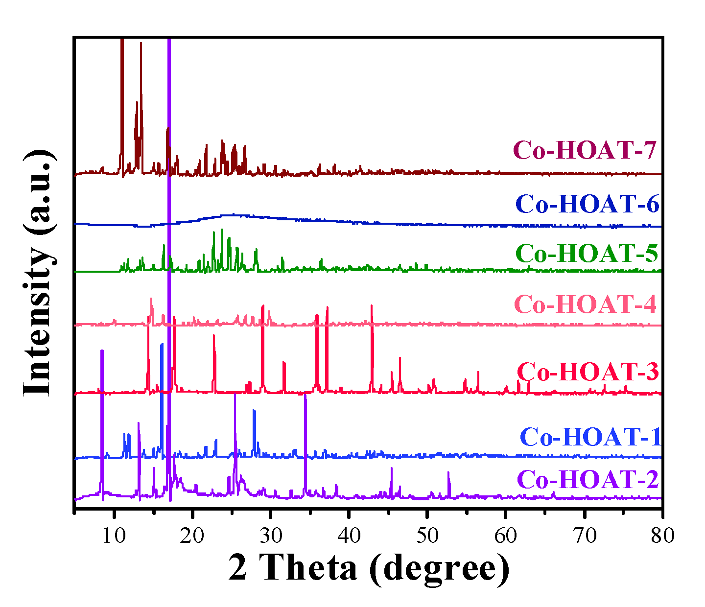
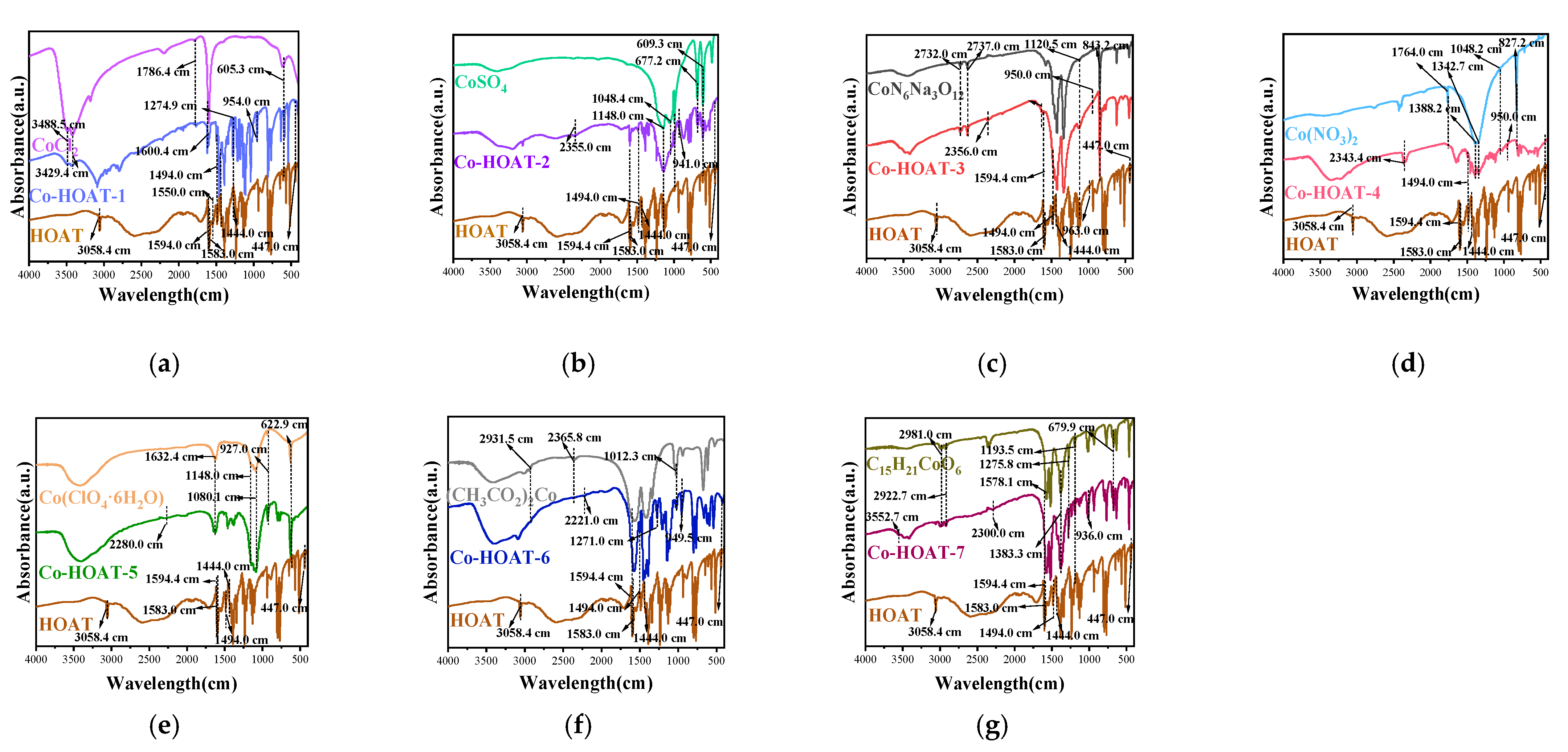
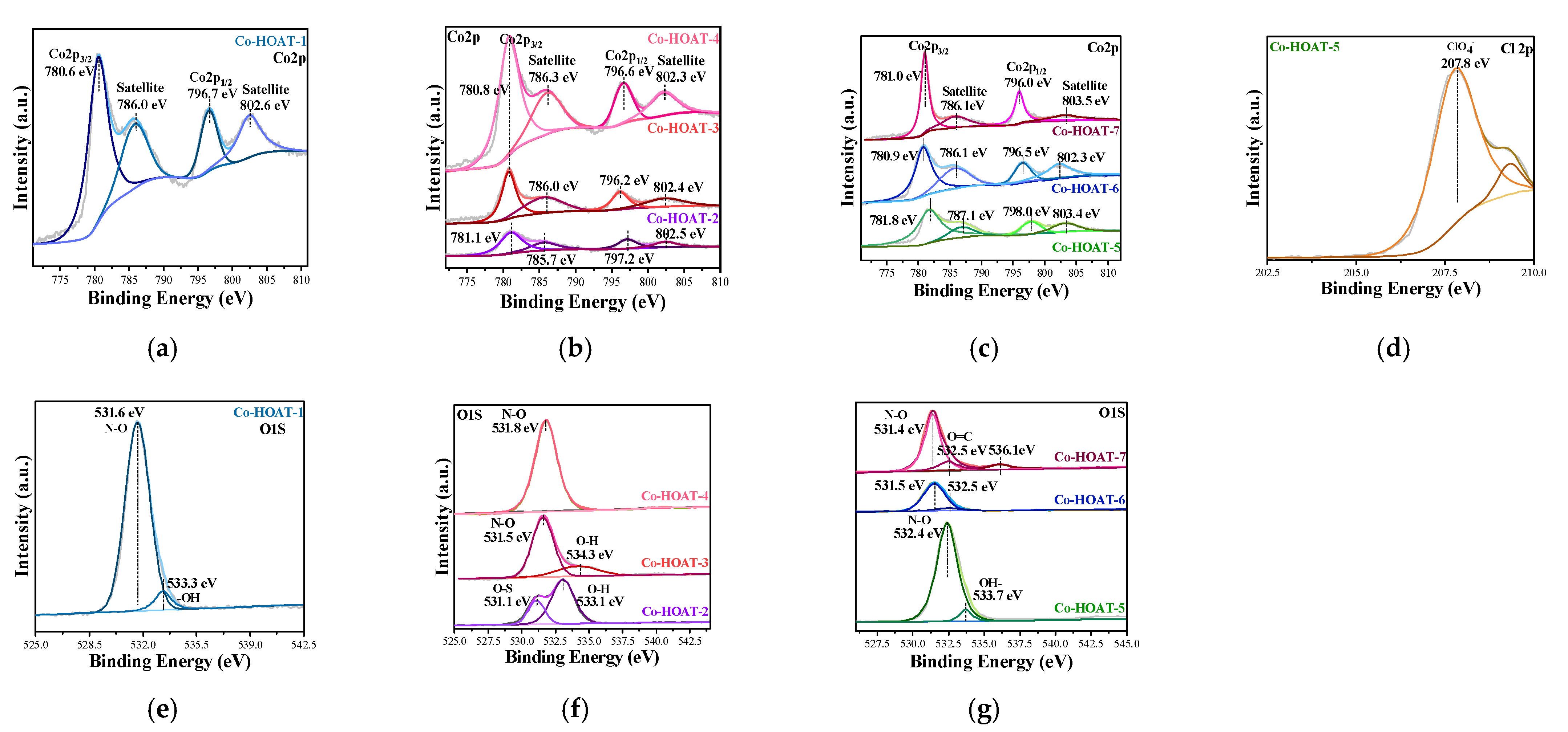
Characterization of Samples

As can be seen in Figure 2, the X-ray diffraction (XRD) patterns of a series of Co–HOATs were collected to evaluate their crystalline phase. It is obvious that all the Co–HOATs demonstrate excellent crystal forms. The inorganic Co–HOATs tend to have a nanosheet morphology, while the organic Co–HOATs grow into small-size nanoparticles. This was verified by using the SEM and TEM images. The morphology of various Co–HOATs can be observed in Figure 1. It is clear that the morphologies of Co–HOAT-1, Co–HOAT-2, Co–HOAT-3, and Co–HOAT-4 present as nanosheets, with flaky distribution and the thickness of these sheets gradually thinning. Meanwhile, Co–HOAT-5 synthesized by (Co(ClO4·6H2O) appeared both as small nanoparticles and with large-size nanosheet morphology. Furthermore, Co–HOATs behave entirely like small nanoparticles with the cobalt source changed to organic salts. It is observed that inorganic cobalt salts are beneficial for Co–HOATs forming large-size nanosheets, while the Co–HOATs grown from organic salts show small nanoparticles. Meanwhile, the EDS mapping spectrum of various Co–HOATs in the Supplementary Information illustrates that the seven Co–HOATs synthesized successfully. This is ascribed to the relevant elements that could be observed, and the fewer N elements in Co–HOAT-7 was mainly due to there being less HOAT that they could combine with C10H14CoO4.
The FTIR spectrum of different Co–HOATs in Figure 3 are shown to further explain difference in the above Co–HOATs by binding situations and chemical bonds. The details of the chemical bonds of the seven Co–HOATs induced by FTIR spectrum are summarized in Supplementary Table S1. It is apparent that the characteristic peaks around 1594, 1494, 1444, 3058.4, and 447 cm−1 are ascribed to HOAT in Co–HOAT-1. In addition, the bonds at 3488.5, 3429.4, 1786.4, and 605.3 cm−1 in Co–HOAT-1 derive from the characteristic peaks of CoCl2. This can also prove that Co–HOAT-1 was successfully synthesized according to FTIR and EDS mapping. Meanwhile, the peaks around at 1594, 1583, 1494, and 1444 cm−1 are attributed to the pyridine ring of HOAT [55]. This can prove that the Co atom is incapable of forming a chemical bond with N in the pyridine ring. This also demonstrates that the Co could not coordinate with -N–OH in HOAT. This is due to the peaks around 1274.9 and 954 cm−1 still being observed, ascribed to the telescopic vibration peak of N–O–H [56] and N–O [57], respectively, in Co–HOAT-1. It can also be seen that the characteristic peaks around 1550.0 cm−1 ascribed to –N=N– have disappeared. Further, the bonds at 1600.4 cm−1 could be observed, which demonstrates the existence of the –R1R2–N– bond in Co–HOAT-1 [58]. It manifests in exhibiting that Co is more willing to bond with the N atoms in position 7 and 8 of HOAT. This is mainly due to large steric hindrance restricting the coordination of cobalt with the nitrogen of the pyridine ring.
The FTIR spectrum of Co–HOAT-2 and Co–HOAT-3 can be seen in Figure 3b,c. The bonds at around 1148 cm−1, 1048.4 cm−1, 609.3 cm−1, and 677.2 cm−1 are attributed to the characteristic peaks of CoSO4. In addition, the characteristic peaks at 2732.0, 2737.0, 1120.5, 2165.5, and 843.2 cm−1 are derived from Na3[Co(NO2)6]. This could prove that Co–HOAT-2 and Co–HOAT-3 are successfully prepared when referring to the general spectrum of HOAT in Figure 3a. The characteristic peaks of N–O–H at 1267 cm−1 disappear in both Co–HOAT-2 and Co–HOAT-3, and the wavelength of the N–O bond (941.0 and 950.0 cm−1) tends to be lower compared with Co–HOAT-1 (954.0 cm−1). In addition, the emerging bonds at 2355.0 cm−1 and 2356.0 cm−1 indicate the –N=N+– bond in Co–HOAT-2 and Co–HOAT-3, respectively [59]. This could further certify that the Co in both CoSO4 and Na3[Co(NO2)6] tends to coordinate with the N and O atoms in HOAT. The FTIR spectrum in Figure 3d shows the characteristic peaks and chemical bonds of Co–HOAT-4. The characteristic peaks at around 1764.0 cm−1, 1342.7 cm−1, 1388.2 cm−1, 1048.2 cm−1, and 827.2 cm−1 belong to Co(NO3)2. They demonstrate that Co–HOAT-4 was synthesized favorably. The coordination method of Co in Co–HOAT-4 was similar to that in Co–HOAT-3. The characteristic peaks of N–O–H at 1267.0 cm−1 disappear, while the N–O peak at 950.0 cm−1 could be observed. Meanwhile, the emerging bonds at 2343.4 cm−1 derive from the –N=N+– bond in Co–HOAT-4. This allow us to conclude that Co is likely to bind with the O and N atom of HOAT at the same time. It shows that the wavelengths of N–O in Co–HOAT-2, Co–HOAT-3, and Co–HOAT-4 present as higher compared with Co–HOAT-1, respectively. This is mainly due to the induced effects of electrons of the Co–O bond when the O atom in N–O coordinates with Co.
The coordination strategies of Co with HOAT in Co–HOAT-5, Co–HOAT-6, and Co–HOAT-7 are similar. As can be seen in Figure 3e–g, the characteristic peaks at 1632.4 cm−1, 1148.0 cm−1, 1080.1 cm−1, and 622.9 cm−1 are ascribed to (Co(ClO4·6H2O). In addition, it is clear that the characteristic peaks at 2931.5 cm−1, 2365.8 cm−1, and 1012.3 cm−1 stemmed from (CH3CO2)2Co. And the characteristic peaks at 1383.3 cm−1, 2981.0 cm−1, 2922.7 cm−1, 1578.1 cm−1, 1275.8 cm−1 1193.5 cm−1, and 679.9 cm−1 are ascribed to C10H14CoO4. This shows Co–HOAT-5, Co–HOAT-6, and Co–HOAT-7 were prepared very well. It can clearly be seen that the emerging –N=N+– bond at 2280 cm−1 appears in Co–HOAT-5 when the Co combines with the N atoms in HOAT [59]. This concludes that Co only could combine with the N atoms of HOAT, which could be further verified by the absence of obvious characteristic peaks of N–O–H around 1276 cm−1 and N–O– at 927.0 cm−1. The assembly methods of Co coordinate with HOAT in Co–HOAT-6 and Co–HOAT-7, and are similar to that in Co–HOAT-5. The appearance of the characteristic peaks of N–O–H (1271.0 and 1275.8 cm−1) and N–O– (949.5 and 936.0 cm−1) in Co–HOAT-6 and Co–HOAT-7, respectively, exhibit that the Co could not combine with -N–OH in HOAT. However, the emerging peaks of –N=N+– (2221.0 cm−1, 2300.0 cm−1) bonds could be observed in Co–HOAT-6 and Co–HOAT-7 when Co combined with the N atoms of HOAT [58]. This shows that the Co only could combine with the N atoms of HOAT in both Co–HOAT-6 and Co–HOAT-7.
In conclusion, the Co in CoCl2 is more willing to bond with the N atoms of HOAT in Co–HOAT-1. Controversially, CoSO4, Na3[Co(NO2)6], and Co(NO3)2·6H2O are likely to bind with the O and N atoms in HOAT at the same time in Co–HOAT-2, Co–HOAT-3, and Co–HOAT-4. Meanwhile, the Co of (Co(ClO4·6H2O), (CH3CO2)2Co, and C10H14CoO4 was only coordinated with the N atoms in HOAT.
In order to further verify the coordination strategies of Co–HOATs, the Raman spectra of as-prepared samples of a series Co–HOATs are illustrated in Figure 4a,b. In addition, the potential coordinated methods of Co with HOAT in seven Co–HOATs were derived from the FTIR and Raman spectra presented in Scheme S1 of the Supplementary Information. They show that there are little peaks at 2987–2994 cm−1 ascribed to the Cl−1 observed in Co–HOAT-1 [60]. Meanwhile, the yellow part in Figure 4b is refer to the characteristic peaks from 524 cm−1 to 554 cm−1 are ascribed to Co–N bond [61]. This demonstrates that the CoCl2 combines with the N atoms in HOAT in Co–HOAT-1. In addition, the characteristic peaks at 446.0 cm−1, 559.0 cm−1, 610.0 cm−1, 980.0 cm−1, and 1141.8 cm−1 of SO42− in Co–HOAT-2 could be observed [62]. Meanwhile, little peaks at 535.2 cm−1 and 492.8 cm−1 belonging to Co–N– and Co–O– bonds [63] exist. Similar to Co–HOAT-2, the peaks at 674.0 cm−1 and 1041.0 cm−1 of the NO2 [64] and NO3 [65] functional groups could also be seen, as well as the Co–N– (532.0 cm−1, 530.5 cm−1) and Co–O– (492.8 cm−1, 492.8 cm−1) bonds in Co–HOAT-3 and Co–HOAT-4 at the same time, respectively. We can conclude that the cobalt from CoSO4, Na3[Co(NO2)6] and Co(NO3)2·6H2O united with HOAT by carrying its anions, respectively. Furthermore, the characteristic peaks of ClO4 (933.5 cm−1) [66] and Co–N– (537.3 cm−1) could be found in Co–HOAT-5. It could also be seen Co–HOAT-6 and Co–HOAT-7 own a D peak at 1346.0 cm−1 and a G peak at 1593.0 cm−1, which are ascribed to the characteristic carbon peaks [67]. This is mainly due to the fact that the entirety of (Co(ClO4·6H2O), (CH3CO2)2Co, and C10H14CoO4 could coordinate with HOAT in Co–HOAT-5, Co–HOAT-6, and Co–HOAT-7, respectively. This lead cobalt to only unite with the N atoms in HOAT due to larger steric hindrance. All the Raman spectra results correspond well with the FTIR spectrum in Figure 3.
The XPS spectra in Figure 5 were actualized to further discuss the micro-component of the Co–HOATs and survey the valence of different elements. Figure 5a exhibits the valence of the Co species from the fitting curves of all the Co–HOATs, respectively. It can be seen that the positions of Co(II) (796.7 eV) of Co 2p1/2, and Co(III) (780.6 eV) of Co 2p3/2 exist in CO–HOAeT-1. The satellite peaks of Co(II) and Co(III) are 802.6 eV and 786.0 eV [68], respectively. Compared with Co–HOAT-1, both the characteristic peaks of Co(III) (781.1 eV) and Co(II) (797.2 eV) in Co–HOAT-2 present more positive shifts in Figure 5b. Meanwhile, their satellite peaks show more negative shifts, respectively. Meanwhile, positive shifts existed in the satellite and characteristic Co(II) peaks of Co–HOAT-3 (802.4, 796.2 eV) and Co–HOAT-4 (802.3, 796.6 eV), respectively. On the contrary, the satellite and characteristic peaks of Co(III) exhibited negative shifts. While the Co(II) with its satellite peaks present more negative binding energy in Co–HOAT-4. This is ascribed to the N–Co–SO4 and N–Co–NO2 bonds being detected in Co–HOAT-2 and Co–HOAT-3 according to the FTIR and Raman spectra, respectively. The cobalt-bound anions caused the Co(III) peaks to be greatly affected. Thus, more electrons transferred from Co to anions, inducing a higher binding energy in the Co(III) peaks in Co–HOAT-2 and Co–HOAT-3 compared with that in Co–HOAT-1 [69], respectively. Meanwhile, the entire [Co(NO2)6]3− ions could coordinate with the O and N atoms in Co–HOAT-3, while the Co2+ ions with less NO3 could unite with HOAT in Co–HOAT-4. The difference in anions (N–O–Co–NO2, N–O–Co) lead to the Co(II) peaks being greatly influenced. Therefore, the binding energy of Co(II) in Co–HOAT-4 is lower than that of Co–HOAT-2 and Co–HOAT-3. The chemical valences of the Co species in Co–HOAT-5, Co–HOAT-6, and Co–HOAT-7 are different in Figure 5c. It is clear that the Co(III) (781.8 eV) peaks were found to be more positive in Co–HOAT-5 than that in inorganic Co–HOATs. The positions of satellite peaks of Co(III) (787.1 eV) and Co(II) in Co–HOAT-5 (803.4 eV) present at greater positivity compared with other Co–HOATs. However, all the characteristic peaks of Co(III) (780.9 eV) and Co(II) (796.5 eV), along with their satellite peaks, in Co–HOAT-6 tend to be lower than that of Co–HOAT-5. At the same time, the positions of Co(III) (781.0 eV), along with its satellite peaks (786.1 eV), and Co(II) (796.0 eV) in Co–HOAT-7 are more negative compared with those of Co–HOAT-5. This is mainly due to the entirety of the organic cobalt salts being able to coordinate with the N atoms in HOAT. This resulted in N–Co–O–Cl bonds in Co–HOAT-5, N–Co–O–C=O in Co–HOAT-6, and N–Co–CH–C=O bonds in Co–HOAT-7 appearing. The anions bound with Co2+ were varied, leading to different chemical cobalt environments, which can be observed in Supplementary Scheme S1.
The XPS spectra of O1s in Figure 5d–f were implemented to further investigate the chemical bonds in Co–HOATs. The N–O peaks at 531.6 eV and O–H at 533.3 eV can be observed [70], which manifested the N–OH chemical bond still existing in Co–HOAT-1. This result is very consistent with the analysis of FTIR for Co–HOAT-1. We can also observe the O–S peaks at 531.1 eV [71] and the O–H bond at 533.1 eV in Co–HOAT-2. It can be verified from the Raman spectra that there is a Co–O band in Co–HOAT-2 due to Co coordinating with the N–O– in HOAT. Then, it can be proposed that the coexistence of O–S and O–H of O1s in Co–HOAT-2 is mainly owed to the emerging SO4-H bond. Different from Co–HOAT-2, Co–HOAT-3 possesses an N–O bond at 531.6 eV and an O–H bond at 534.3 eV. Meanwhile, only the N–O peaks at 531.8 eV in Co–HOAT-4 are observed compared with Co–HOAT-3. This indicated that the entirety of [Co(NO2)6]3− ions in Na3[Co(NO2)6] could coordinate with the O and N atoms in Co–HOAT-3. The emerging O–H bond have be derived from H in the aqueous phase integrating with negative [Co(NO2)6]3− ions. Unlike [Co(NO2)6]3−, it is difficult for H to combine with the Co(NO3)2 molecule due to electrical neutrality. The micro component and valence of different elements in organic Co–HOATs are various. As can be seen in Figure 5c, the O 1s peaks of 532.4 and 533.7 eV are typical for the N–O and OH peaks in Co–HOAT-5, respectively. In addition, the coexistent peaks of ClO4 at 207.8 eV [72] could further prove that (Co(ClO4·6H2O) only coordinated with the N atoms in HOAT. Co–HOAT-6 and Co–HOAT-7 are the opposite, and the obvious peaks of N–O (531.5, 531.4 eV) and C=O (532.5, 532.5 eV) could be perceived [73], respectively. The N–O bonds could be ascribed to the N–O–H of HOAT in both Co–HOAT-6 and Co–HOAT-7. Meanwhile, the C=O peak in Co–HOAT-6 is less distinct than that in Co–HOAT-7. This is mainly due to there being four C=O bonds in C10H14CoO4, while (CH3CO2)2Co only owns two C=O bonds. The appearance of the O–H peaks can further certify the C10H14CoO4 and (CH3CO2)2Co could only combine with N atoms in HOAT in Co–HOAT-6 and Co–HOAT-7, respectively. All of the above results prove that the entirety of organic salts participate in the coordination process with N atoms in organic Co–HOATs, while there are inorganic anions that coordinate with both N and O atoms in Co–HOAT-1, Co–HOAT-2, Co–HOAT-3, and Co–HOAT-4, respectively.
In order to better explain the experimental results, the involved complexes’ structures were calculated. The calculated structures are shown in Figure 6 and Figure 7. The blue, red, grey and whilt pellets represet nitrogen, oxygen, carbon and hydrogen atoms, respectively, in Figure 6 and Figure 7. The relative energies of all species in water are listed in Table 1. The calculated results show that the coordination complexes between Co2+ and 1-hydroxy-7-azabenzotriazole (HOAT) have five different kinds of conformations. It is worth noting that there are three types of di-coordination complexes, and all of them are planar. As can be seen from Table 1, the binding energy of P23 is −6.98 eV, which is the lowest among all complexes. From the above discussion, it can be seen that P23 is the most stable structure, and the result is better in accordance with the experiment. This further proves that the Co2+ ions in Co–HOATs synthesized by CoCl2 are more reluctant to coordinate with the N atoms of HOAT. In order to further illustrate the experimental result, the structure of Co–HOAT-7 synthesized by cobalt acetylacetone is illustrated in Figure 7. It is found that the four active positions of the Co2+ ion are occupied before cobalt acetylacetone combines with HOAT. However, the only two positions of Co ions left could be occupied by the HOAT organic ligand. This demonstrates that small steric hindrance could lead to ample active positions for inorganic cobalt ions to coordinate with both the N and O atoms in HOAT. Meanwhile, organic Co2+ ions could only unite with the N atoms in HOAT. In this case, cobalt ions with adequate coordination sites are beneficial for developing nanosheet morphologies.

4. Antibacterial Assay

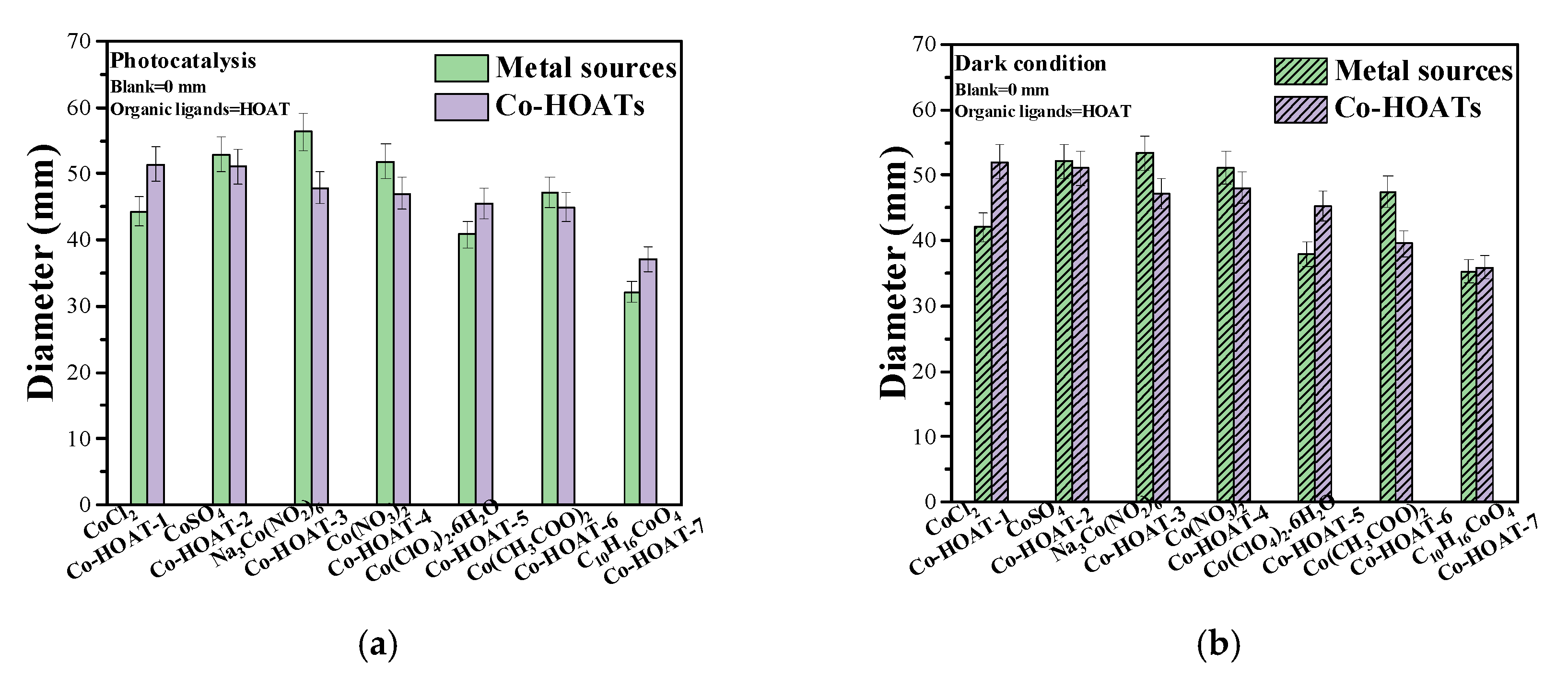
The Zone of Inhibition Test

The zone of inhibition test was executed to investigate the antibacterial performance of the Co–HOATs. Figure 8a,b displays the diameters of inhibition zones formed around Co–HOAT-1, Co–HOAT-2, Co–HOAT-3, Co–HOAT-4, Co–HOAT-5, Co–HOAT-6, and Co–HOAT-7 and their cobalt salts, respectively. On the whole, it is evidenced that there are slight differences in sterilization activity between the dark and visible-light conditions for all the tested samples. This demonstrates that the Co–HOATs in this manuscript could achieve their antimicrobial capacity under no environmental constraints. It can also be observed that Co–HOAT-1 could obviously generate inhibition zone diameters of 52.08 mm and 51.45 mm under the dark and visible-light conditions, respectively. In addition, the diameters in the dark (51.05 mm) and solar light (51.06 mm) are similar to each other in Co–HOAT-2. The antibacterial activity of Co–HOAT-3 exhibits a mild declining trend compared with that of Co–HOAT-2. The inhibition zone diameters are 48.06 mm and 47.03 mm under the dark and photocatalysis conditions, respectively. Moreover, the sterilization rate of Co–HOAT-4 is evaluated by inhibition zone diameter. The 47.2 mm and 47.84 diameters also show that the antibacterial performance of the Co–HOATs were not limited by the environment. Furthermore, the antimicrobial rate maintains its steady downward trend until the value reaches about 45.25 mm and 45.50 mm under the dark and visible-light conditions. The inhibition zone diameter of Co–HOAT-6 (39.6 mm) and Co–HOAT-7 (35.94 mm) exhibit a dramatic decrease under dark condition, respectively. Meanwhile, the diameters present under photocatalysis were 44.92 mm and 37.04 mm, respectively. Although both Co–HAOT and metal sources exhibit excellent sterilization rate, the main mechanism of antibacterial activity for metal sources is complete reliance upon the released cobalt ions and their toxicity. Compared with Co–HOATs, the released Co2+ ions are uncontrollable due to the absence of organic units [13]. Furthermore, the high toxicity of cobalt sources are not beneficial for further antibacterial research.
In the case of using the same organic ligand of HOAT, Co–HOAT-1, Co–HOAT-2, Co–HOAT-3, and Co–HOAT-4 induced by inorganic cobalt salts presented higher sterilization rates compared with Co–HOAT-5, Co–HOAT-6, and Co–HOAT-7. Moreover, the proposed antibacterial mechanism emphasizes that bacteria could be anchored by catalysts via electrostatic interaction [74] or hydrophobic interaction [75]. This is beneficial for shortening the distance between the metal ions and bacteria and increasing the probability of the metal ions damaging the bacteria. Furthermore, this would lead to better sterilization by cracking the cell membrane and active intracellular substance, rather than by killing bacteria randomly [76]. As with the Co–HOATs, firstly, the bacteria were anchored by the Co–HOATs to prolong the sterilization time. Then, the Co2+ ions released from the Co–HOATs could quickly gather on the surface of the microorganisms. This is ascribed to the opposing electrostatic force between bacteria and Co2+ ions when bacteria touches Co2+ ions. It has been shown that the surface of bacteria membrane present electronegative performance [12]. Furthermore, this can be proved by the Co–HOATs with different electron clouds. It is obviously observed that the entirety of Co–HOAT-1 presents lower density electron clouds, which are ascribed to the existence of a single bond (N–N) and fewer electronegative groups (Co–Cl). Meanwhile, the electron clouds of Co–HOAT-2, Co–HOAT-3, and Co–HOAT-4 seem to be similar. This is mainly due to the three samples’ own –N=N+–OH bonds and their similar electronegative groups (Co–O and Co–N). Unlike the above Co–HOATs, the density of electron clouds for Co–HOAT-5, Co–HOAT-6, and Co–HOAT-7 were found to be much higher. This is mainly ascribed to the three Co–HOATs possessing not only –N=N+–OH bonds, but also more negative electron groups (Co–O–Cl=O, Co–O–C=O, and Co–O–C=O–), respectively. This would result in it being more beneficial for Co–HOATs to have less electron clouds to anchor bacteria by electrostatic interaction. Then, the Co2+ ions could penetrate into the bacterial insides through the damaged membranes. This would result in them destroying the charge balance of the bacteria and even cause serious cell collapse, killing bacteria by bacteriolysis. Thus, the more bacteria that are anchored by Co–HOATs, the better the antimicrobial properties are. Videos using an optical microscope are provided to further verify the bacteria-anchoring process in the Supplementary Materials. It can be seen that the Co–HOAT catalysts are present as nanosheets in the bacterial suspension, and a lot of bacteria are swimming around the Co–HOATs. One bacterium is noticed to lightheartedly move towards a piece of Co–HOAT. Several seconds later, it loses its freedom when anchored by the Co–HOATs. Compared with fixed bacteria, other unanchored E. coli could move out of control and go to anywhere they want. This indicated that the Co–HOATs possess the capacity to capture bacteria by potential electrostatic attraction.
To further investigate the main factor of sterilization, the cobalt ratio in each sample is actualized in Supplementary Table S2. It can be seen that the difference in cobalt ratio present among each Co–HOAT is negligible. This demonstrates that the concentration of cobalt ions released during the sterilization process is not the essential factor in antibacterial activity. The main roles of Co2+ ions during the sterilization process is reliant on their ability to change the functions of proteins by interacting with some of the functional groups on the protease, such as sulfhydryl groups (ASH), amino groups (ANH2), and hydroxyl groups (AOH) [77].

5. Conclusions

In conclusion, the Co–HOATs were successfully synthesized and exhibited excellent sterilization activity when probed with S. aureus bacteria during the dark and visible-light systems. The Co–HOAT nanosheets possessed higher sterilization rates than the Co–HOAT nanoparticles. The XRD, SEM, TEM, Raman, XPS, FTIR, and theoretical calculations were characterized to explain this phenomenon. It was found that with the same HOAT ligand, small steric hindrance could lead to there being ample active positions for inorganic cobalt ions to coordinate with both N and O atoms in HOAT. Meanwhile, organic Co2+ ions could only unite with the N atoms in HOAT. Thus, cobalt ions with adequate coordination sites are beneficial for developing nanosheet morphologies. Meanwhile, it is beneficial for inorganic Co–HOATs to anchor bacteria due to them presenting lower densities of electron clouds. This is the main reason that Co–HOATs with a nanosheet morphology show more efficient antibacterial activity than nanoparticles. The proposed mechanism in this manuscript is that Co2+ released from Co–HOATs could cause multiple toxic effects to anchored bacteria via Co–HOATs’ reactive oxygen species and lipid peroxidation. In conclusion, the Co–HOATs appear to be potential antibacterial catalysts in the antimicrobial field.

Supplementary Materials

The following supporting information can be downloaded at: https://www.mdpi.com/article/10.3390/catal14030173/s1, Figure S1: The EDS mapping spectrum of Co–HOAT-1, Co–HOAT-2, Co–HOAT-3, Co–HOAT-4, Co–HOAT-5, Co–HOAT-6 and Co–HOAT-7, respectively, Table S1: The existed potential chemical bonds in seven Co–HOATs derived from FTIR spectra, Table S2: The cobalt ratio in catalysts of Co–HOAT-1, Co–HOAT-2, Co–HOAT-3, Co–HOAT-4, Co–HOAT-5, Co–HOAT-6 and Co–HOAT-7, respectively, Scheme S1: The potential structure of seven Co–HOATs derived from FTIR and Raman spectra.

Author Contributions

Conceptualization, Y.M.; methodology, Y.Z.; software, S.Y.; validation, F.L.; formal analysis, Z.B.; investigation, Y.Z.; resources, S.Y.; data curation, F.L.; writing—original draft preparation, X.X.; writing—review and editing, M.D. and H.L.; visualization, X.X.; supervision, M.D.; project administration, Z.B.; funding acquisition, Y.M. All authors have read and agreed to the published version of the manuscript.

Funding

This work is supported by Technological Innovation Team for Green Catalysis and Energy Materials Yunnan Institutions of Higher Learning, Surface project of Yunnan Province science and technology Department (202101BA070001-050), Yunnan Province University Collaborative Innovation Center (Qujing Green Photovoltaic Industry Collaborative Innovation Center), Technology Talent and Platform Plan Project of Yunnan Provincial Department of Science and Technology (202305AF150088), Special Basic Cooperative Research Programs of Yunnan Provincial Undergraduate Universities (202301BA070001-078), and Scientific Research Fund project of Education Department of Yunnan Province (2023J1035).

Data Availability Statement

All authors confirm that the data supporting the findings of this study are available within the article and its Supplementary Materials.

Conflicts of Interest

The authors declare that they have no known competing financial interests or personal relationships that could have appeared to influence the work reported in this paper.

References

  1. Lin, N.; Verma, D.; Saini, N.; Arbi, R.; Munir, M.; Jovic, M.; Turak, A. Antiviral nanoparticles for sanitizing surfaces: A roadmap to self-sterilizing against COVID-19. Nano Today 2021, 40, 101267. [Google Scholar] [CrossRef] [PubMed]
  2. Wegener, M.; Hansen, M.J.; Driessen, A.A.-O.; Szymanski, W.A.-O.; Feringa, B.A.-O. Photocontrol of Antibacterial Activity: Shifting from UV to Red Light Activation. J. Am. Chem. Soc. 2017, 139, 17979–17986. [Google Scholar] [CrossRef] [PubMed]
  3. Dodd, M.C.; Kohler, H.P.E.; Von Gunten, U. Oxidation of antibacterial compounds by ozone and hydroxyl radical: Elimination of biological activity during aqueous ozonation processes. Environ. Sci. Technol. 2009, 43, 2498–2504. [Google Scholar] [CrossRef] [PubMed]
  4. Wu, P.; Xie, R.; Imlay, K.; Shang, J.K. Visible-light-induced bactericidal activity of titanium dioxide codoped with nitrogen and silver. Environ. Sci. Technol. 2010, 44, 6992–6997. [Google Scholar] [CrossRef] [PubMed]
  5. Li, Z.; Yi, Z.; Li, Z.; Zou, Z. Photocatalytic and Thermocatalytic Conversion of Methane. Solar RRL 2021, 5, 2000596. [Google Scholar] [CrossRef]
  6. Valenzuela, L.; Faraldos, M.; Bahamonde, A.; Rosal, R. Critical review on the use of photocatalysis and photoelectrocatalysis to create antimicrobial surfaces. Curr. Opin. Chem. Eng. 2021, 34, 100762. [Google Scholar] [CrossRef]
  7. Kim, W.; Zhu, W.; Hendricks, G.L.; Van Tyne, D.; Steele, A.D.; Keohane, C.E.; Fricke, N.; Conery, A.L.; Shen, S.; Pan, W.; et al. A new class of synthetic retinoid antibiotics effective against bacterial persisters. Nature 2018, 556, 103–107. [Google Scholar] [CrossRef] [PubMed]
  8. Ding, M.; Shi, F.; Zhu, H.; Yang, Y.; Zhong, J.; Luo, L.; Huo, Y.; Li, H. Photoelectrocatalytic bacterial inactivation of Acinetobacter baumannii on Cu2O/TiO2@Cu mesh photoanodes. Catal. Sci. Technol. 2020, 10, 7378–7385. [Google Scholar] [CrossRef]
  9. Helali, S.; Polo-López, M.I.; Fernández-Ibáñez, P.; Ohtani, B.; Amano, F.; Malato, S.; Guillard, C. Solar photocatalysis: A green technology for E. coli contaminated water disinfection. Effect of concentration and different types of suspended catalyst. J. Photochem. Photobiol. A Chem. 2014, 276, 31–40. [Google Scholar] [CrossRef]
  10. Gong, J.; Li, C.; Wasielewski, M.R. Advances in solar energy conversion. Chem. Soc. Rev. 2019, 48, 1862–1864. [Google Scholar] [CrossRef]
  11. Ran, B.; Ran, L.; Wang, Z.; Liao, J.; Li, D.; Chen, K.; Cai, W.; Hou, J.; Peng, X. Photocatalytic Antimicrobials: Principles, Design Strategies, and Applications. Chem. Rev. 2023, 123, 12371–12430. [Google Scholar] [CrossRef] [PubMed]
  12. Shi, F.; Ding, M.; Tong, H.; Yang, Y.; Zhang, J.; Wang, L.; Li, H.; Huo, Y. Photoelectrocatalytic sterilization on thorn-like ZIF-67/ZnO hybrid photoanodes. J. Environ. Chem. Eng. 2022, 10, 107385. [Google Scholar] [CrossRef]
  13. Liu, J.; Wu, D.; Zhu, N.; Wu, Y.; Li, G. Antibacterial mechanisms and applications of metal-organic frameworks and their derived nanomaterials. Trends Food Sci. Technol. 2021, 109, 413–434. [Google Scholar] [CrossRef]
  14. Jiao, Y.; Niu, L.-N.; Ma, S.; Li, J.; Tay, F.R.; Chen, J.-H. Quaternary ammonium-based biomedical materials: State-of-the-art, toxicological aspects and antimicrobial resistance. Prog. Polym. Sci. 2017, 71, 53–90. [Google Scholar] [CrossRef]
  15. Gupta, V.K.; Fakhri, A.; Tahami, S.; Agarwal, S. Zn doped CdO nanoparticles: Structural, morphological, optical, photocatalytic and anti-bacterial properties. J. Colloid Interface Sci. 2017, 504, 164–170. [Google Scholar] [CrossRef] [PubMed]
  16. Gomez-Polo, C.; Larumbe, S.; Gil, A.; Muñoz, D.; Fernández, L.R.; Barquín, L.F.; García-Prieto, A.; Fdez-Gubieda, M.L.; Muela, A. Improved photocatalytic and antibacterial performance of Cr doped TiO2 nanoparticles. Surf. Interfaces 2021, 22, 100867. [Google Scholar] [CrossRef]
  17. Chen, C.; Wang, D.; Li, Y.; Huang, H.; Ke, Y. ‘Flower-like AgBr/CeO2 Z-scheme heterojunction photocatalyst with enhanced visible light photocatalytic and antibacterial activities. Appl. Surf. Sci. 2021, 565, 150534. [Google Scholar] [CrossRef]
  18. Yuzer, B.; Aydın, M.I.; Con, A.H.; Inan, H.; Can, S.; Selcuk, H.; Kadmi, Y. Photocatalytic, self-cleaning and antibacterial properties of Cu(II) doped TiO2. J. Environ. Manag. 2022, 302, 114023. [Google Scholar] [CrossRef]
  19. Wang, K.; Lv, M.; Si, T.; Tang, X.; Wang, H.; Chen, Y.; Zhou, T. Mechanism analysis of surface structure-regulated Cu2O in photocatalytic antibacterial process. J. Hazard. Mater. 2024, 461, 132479. [Google Scholar] [CrossRef]
  20. Aihemaiti, X.; Wang, X.; Li, Y.; Wang, Y.; Xiao, L.; Ma, Y.; Qi, K.; Zhang, Y.; Liu, J.; Li, J. Enhanced photocatalytic and antibacterial activities of S-scheme SnO2/Red phosphorus photocatalyst under visible light. Chemosphere 2022, 296, 134013. [Google Scholar] [CrossRef]
  21. Qi, K.; Cheng, B.; Yu, J.; Ho, W. Review on the improvement of the photocatalytic and antibacterial activities of ZnO. J. Alloys Compd. 2017, 727, 792–820. [Google Scholar] [CrossRef]
  22. Ma, H.; Wang, K.; Zeng, Q.; Li, P.; Lyu, S.; Li, B.; Luo, X.; Jiang, L.; Cao, M.; Liao, B.; et al. Photocatalytic properties and antibacterial mechanisms of microbial-derived ZnS/CuS nanocomposites. J. Environ. Chem. Eng. 2023, 11, 111425. [Google Scholar] [CrossRef]
  23. Agboola, P.O.; Haider, S.; Shakir, I. Copper sulfide and their hybrids with carbon nanotubes for photocatalysis and antibacterial studies. Ceram. Int. 2022, 48, 10136–10143. [Google Scholar] [CrossRef]
  24. Huo, P.; Liu, C.; Wu, D.; Guan, J.; Li, J.; Wang, H.; Tang, Q.; Li, X.; Yan, Y.; Yuan, S. Fabricated Ag/Ag2S/reduced graphene oxide composite photocatalysts for enhancing visible light photocatalytic and antibacterial activity. J. Ind. Eng. Chem. 2018, 57, 125–133. [Google Scholar] [CrossRef]
  25. Nivetha, P.; Kavitha, B.; Kalanithi, M. Investigation of photocatalytic and antimicrobial activities of BaWO4–MoS2 nanoflowers. J. Sci. Adv. Mater. Dev. 2021, 6, 65–74. [Google Scholar] [CrossRef]
  26. Dou, L.; Yuan, M.; Li, D.; Zou, S.; Tan, S.; Zhao, Z.; Cai, G. Friction spun spandex/rGO/Ag/polyester core-sheath yarn with antibacterial activity for wearable sensors. Surf. Interfaces 2024, 44, 103746. [Google Scholar] [CrossRef]
  27. Chilivery, R.; Zhang, R.; Chen, G.; Yao, D.; Fan, D.; Lu, F.; Song, Y. Facile in situ construction of novel hybrid 3D-BiOCl@PDA heterostructures with vacancy induced charge transfer for efficient visible light driven photocatalysis and antibacterial activity. Colloid. Surface A 2023, 656, 130415. [Google Scholar] [CrossRef]
  28. Sun, J.; Mo, S.; Zhang, Z.; Wen, J.; Guo, D.; Li, Y.; Liu, L. Optimizing the electronic structure of BiOBr via constructing oxygen-rich vacancies for highly efficient NIR light-driven antibacterial activity. Chem. Eng. J. 2022, 450, 137980. [Google Scholar] [CrossRef]
  29. Hsu, C.-L.; Li, Y.-J.; Jian, H.-J.; Harroun, S.G.; Wei, S.-C.; Ravindranath, R.; Lai, J.-Y.; Huang, C.-C.; Chang, H.-T. Green synthesis of catalytic gold/bismuth oxyiodide nanocomposites with oxygen vacancies for treatment of bacterial infections. Nanoscale 2018, 10, 11808–11819. [Google Scholar] [CrossRef]
  30. Kong, X.; Liu, X.; Zheng, Y.; Chu, P.K.; Zhang, Y.; Wu, S. Graphitic carbon nitride-based materials for photocatalytic antibacterial application. Mater. Sci. Eng. R Rep. 2021, 145, 100610. [Google Scholar] [CrossRef]
  31. Guo, J.; Zhou, J.; Sun, Z.; Wang, M.; Zou, X.; Mao, H.; Yan, F. Enhanced photocatalytic and antibacterial activity of acridinium-grafted g-C3N4 with broad-spectrum light absorption for antimicrobial photocatalytic therapy. Acta Biomater. 2022, 146, 370–384. [Google Scholar] [CrossRef] [PubMed]
  32. Zhang, A.; Qiao, Q.; Pei, Z.; Wang, Z.; Guo, J.; Li, Y.; Zhai, G.; Fei, P.; Lu, J.; Jia, H. Polyurethane-Based T-ZIF-8 Nanofibers as Photocatalytic Antibacterial Materials. ACS Appl. Nano Mater. 2023, 6, 22173–22184. [Google Scholar] [CrossRef]
  33. Zhang, J.; Li, P.; Zhang, X.; Ma, X.; Wang, B. Aluminum Metal–Organic Frameworks with Photocatalytic Antibacterial Activity for Autonomous Indoor Humidity Control. ACS Appl. Mater. Interfaces 2020, 12, 46057–46064. [Google Scholar] [CrossRef] [PubMed]
  34. Abdelhameed, R.M.; Darwesh, O.M.; El-Shahat, M. Titanium-based metal-organic framework capsulated with magnetic nanoparticles: Antimicrobial and photocatalytic degradation of pesticides. Microporous Mesoporous Mater. 2023, 354, 112543. [Google Scholar] [CrossRef]
  35. Zhou, S.; Gao, J.; Zhu, J.; Peng, D.; Zhang, Y.; Zhang, Y. Self-cleaning, antibacterial mixed matrix membranes enabled by photocatalyst Ti-MOFs for efficient dye removal. J. Membr. Sci. 2020, 610, 118219. [Google Scholar] [CrossRef]
  36. Kaur, R.; Kaur, A.; Kaur, R.; Singh, S.; Bhatti, M.S.; Umar, A.; Baskoutas, S.; Kansal, S.K. Cu-BTC metal organic framework (MOF) derived Cu-doped TiO2 nanoparticles and their use as visible light active photocatalyst for the decomposition of ofloxacin (OFX) antibiotic and antibacterial activity. Adv. Powder Technol. 2021, 32, 1350–1361. [Google Scholar] [CrossRef]
  37. Savić-Gajić, I.M.; Savić, I.M. Drug design strategies with metal-hydroxyquinoline complexes. Expert Opin. Drug Dis. 2020, 15, 383–390. [Google Scholar] [CrossRef] [PubMed]
  38. Crespi, A.F.; Zomero, P.N.; Pérez, A.L.; Brondino, C.D.; Molina, A.I.; Linck, Y.G.; Monti, G.A.; Fernández, M.A.; Rodríguez-Castellón, E.; Lázaro-Martínez, J.M. Montmorillonite materials with paramagnetic metal complexes: Structural studies and catalytic degradation of emerging pollutants. J. Environ. Chem. Eng. 2023, 11, 111420. [Google Scholar] [CrossRef]
  39. Liu, Y.; Jiang, C.; Peng, L.; Li, Z.; Wang, J.; Liao, X.; Guo, W. Discovery of metal complexes with antibacterial properties in aqueous extracts of Radix scutellariae and a study of the antibacterial properties of the baicalin–manganese complex. Inorg. Chem. Front. 2023, 10, 6506–6518. [Google Scholar] [CrossRef]
  40. Wang, P.; Dong, Y.; Liu, G. Green and cost-effective carboxylic acid Fe complex functionalized cotton fabrics: Sunlight-driven catalytic and antibacterial activities, mechanical and thermal properties. Cellulose 2018, 25, 3663–3678. [Google Scholar] [CrossRef]
  41. Liang, J.; Sun, D.; Yang, Y.; Li, M.; Li, H.; Chen, L. Discovery of metal-based complexes as promising antimicrobial agents. Eur. J. Med. Chem. 2021, 224, 113696. [Google Scholar] [CrossRef] [PubMed]
  42. El-Sawaf, A.K.; El-Essawy, F.; Nassar, A.A.; El-Samanody, E.-S.A. Synthesis, spectral, thermal and antimicrobial studies on cobalt(II), nickel(II), copper(II), zinc(II) and palladium(II) complexes containing thiosemicarbazone ligand. J. Mol. Struct. 2018, 1157, 381–394. [Google Scholar] [CrossRef]
  43. Pettinari, C.; Pettinari, R.; Di Nicola, C.; Tombesi, A.; Scuri, S.; Marchetti, F. Antimicrobial MOFs. Coord. Chem. Rev. 2021, 446, 214121. [Google Scholar] [CrossRef]
  44. Wang, S.; Wang, Y.; Peng, Y.; Yang, X. Exploring the Antibacteria Performance of Multicolor Ag, Au, and Cu Nanoclusters. ACS Appl. Mater. Interfaces 2019, 11, 8461–8469. [Google Scholar] [CrossRef]
  45. DelRe, C.; Jiang, Y.; Kang, P.; Kwon, J.; Hall, A.; Jayapurna, I.; Ruan, Z.; Ma, L.; Zolkin, K.; Li, T.; et al. Near-complete depolymerization of polyesters with nano-dispersed enzymes. Nature 2021, 592, 558–563. [Google Scholar] [CrossRef]
  46. Han, C.; Lee, J.P.; Lobkovsky, E.; Porco, J.A. Catalytic Ester−Amide Exchange Using Group (IV) Metal Alkoxide−Activator Complexes. J. Am. Chem. Soc. 2005, 127, 10039–10044. [Google Scholar] [CrossRef]
  47. Ferrer, F.J.; Carpino, L.A. Synthesis of the Two Isomeric Benzo Derivatives of 1-Hydroxy-7-azabenzotriazole and Preliminary Studies of Their Effectiveness as Coupling Reagents. In Peptides: The Wave of the Future, Proceedings of the Second International and the Seventeenth American Peptide Symposium, San Diego, CA, USA, 9–14 June 2001; Lebl, M., Houghten, R.A., Eds.; Springer: Dordrecht, The Netherlands, 2001; pp. 95–96. [Google Scholar]
  48. Chen, Y.; Liu, Y.W.; Xie, Y.; Zhang, H.H.; Zhang, Z. Preparation and anti-corrosion performance of superhydrophobic silane/graphene oxide composite coating on copper. Surf. Coat. Technol. 2021, 423, 127622. [Google Scholar] [CrossRef]
  49. Stierstorfer, J.; Klapötke, T.M. High Energy Materials. Propellants, Explosives and Pyrotechnics. By Jai Prakash Agrawal. Angew. Chem. Int. Ed. 2010, 49, 6253. [Google Scholar] [CrossRef]
  50. Nandanwar, S.K.; Kim, H.J. Anticancer and Antibacterial Activity of Transition Metal Complexes. ChemistrySelect 2019, 4, 1706–1721. [Google Scholar] [CrossRef]
  51. Utthra, P.P.; Pravin, N.; Raman, N. Scrutinizing the DNA damaging and antimicrobial abilities of triazole appended metal complexes. J. Photochem. Photobiol. B Biol. 2016, 158, 136–144. [Google Scholar] [CrossRef]
  52. Andiappan, K.; Sanmugam, A.; Deivanayagam, E.; Karuppasamy, K.; Kim, H.-S.; Vikraman, D. Detailed investigations of rare earth (Yb, Er and Pr) based inorganic metal-ion complexes for antibacterial and anticancer applications. Inorg. Chem. Commun. 2023, 150, 110510. [Google Scholar] [CrossRef]
  53. Becke, A.D. A new mixing of Hartree–Fock and local density-functional theories. J. Chem. Phys. 1993, 98, 1372–1377. [Google Scholar] [CrossRef]
  54. Lee, C.; Yang, W.; Parr, R.G. Development of the Colle-Salvetti correlation-energy formula into a functional of the electron density. Phys. Rev. B 1988, 37, 785–789. [Google Scholar] [CrossRef] [PubMed]
  55. Al-Saif, F.A.; Al-Humaidi, J.Y.; Binjawhar, D.N.; Refat, M.S. Six new palladium(II) mixed ligand complexes of 2-, 3-, 4-monosubstituted derivative of pyridine ring with caffeine moiety: Synthesis, spectroscopic, morphological structures, thermal, antimicrobial and anticancer properties. J. Mol. Struct. 2020, 1218, 128547. [Google Scholar] [CrossRef]
  56. Nikolić, A.; Jović, B.; Krstić, V.; Tričković, J. N–H...O hydrogen bonding. FT-IR, NIR and 1H NMR study of N-methylpropionamide—Dialkyl ether systems. J. Mol. Struct. 2008, 889, 328–331. [Google Scholar] [CrossRef]
  57. Alt, H.C.; Gomeniuk, Y.V.; Bittersberger, F.; Kempf, A.; Zemke, D. Analysis of electrically active N–O complexes in nitrogen-doped CZ silicon crystals by FTIR spectroscopy. Mater. Sci. Semicond. Process. 2006, 9, 114–116. [Google Scholar] [CrossRef]
  58. Wu, L.; Wang, X. A Concise Handbook of Spectral Interpretation; People’s Medical Publishing House: Beijing, China, 2013; pp. 55–86. [Google Scholar]
  59. Zhang, H.; Peng, Q.; Zhang, Y.; Zhang, R. Organic Spectroscopy; Chemical Industry Press: Beijing, China, 2005; pp. 254–300. [Google Scholar]
  60. Wang, L.; Morita, A.; North, N.M.; Baumler, S.M.; Springfield, E.W.; Allen, H.C. Identification of Ion Pairs in Aqueous NaCl and KCl Solutions in Combination with Raman Spectroscopy, Molecular Dynamics, and Quantum Chemical Calculations. J. Phys. Chem. B 2023, 127, 1618–1627. [Google Scholar] [CrossRef] [PubMed]
  61. Song, J.; Yu, D.; Wu, X.; Xie, D.; Sun, Y.; Vishniakov, P.; Hu, F.; Li, L.; Li, C.; Maximov, M.Y.; et al. Interfacial coupling porous cobalt nitride nanosheets array with N-doped carbon as robust trifunctional electrocatalysts for water splitting and Zn-air battery. Chem. Eng. J. 2022, 437, 135281. [Google Scholar] [CrossRef]
  62. Zangmeister, C.D.; Pemberton, J.E. Raman Spectroscopy and Atomic Force Microscopy of the Reaction of Sulfuric Acid with Sodium Chloride. J. Am. Chem. Soc. 2000, 122, 12289–12296. [Google Scholar] [CrossRef]
  63. Chlebda, D.K.; Jodłowski, P.J.; Jędrzejczyk, R.J.; Łojewska, J. 2D-COS of in situ μ-Raman and in situ IR spectra for structure evolution characterisation of NEP-deposited cobalt oxide catalyst during n-nonane combustion. Spectrochim. Acta A 2017, 186, 44–51. [Google Scholar] [CrossRef]
  64. Liu, J.J.; Siegler, M.A.; Karlin, K.D.; Moënne-Loccoz, P. Direct Resonance Raman Characterization of a Peroxynitrito Copper Complex Generated from O2 and NO and Mechanistic Insights into Metal-Mediated Peroxynitrite Decomposition. Angew. Chem. Int. Ed. 2019, 58, 10936–10940. [Google Scholar] [CrossRef] [PubMed]
  65. Waterland, M.R.; Myers Kelley, A. Far-ultraviolet resonance Raman spectroscopy of nitrate ion in solution. J. Chem. Phys. 2000, 113, 6760–6773. [Google Scholar] [CrossRef]
  66. Zapata, F.; Ortega-Ojeda, F.; García-Ruiz, C.; González-Herráez, M. Selective Monitoring of Oxyanion Mixtures by a Flow System with Raman Detection. Sensors 2018, 18, 2196. [Google Scholar] [CrossRef] [PubMed]
  67. Pan, J.; Lv, M.; Bai, H.; Hou, Q.; Li, M.; Wang, Z. Effects of Metamorphism and Deformation on the Coal Macromolecular Structure by Laser Raman Spectroscopy. Energy Fuels 2017, 31, 1136–1146. [Google Scholar] [CrossRef]
  68. Yuan, X.; Ge, H.; Wang, X.; Dong, C.; Dong, W.; Riaz, M.S.; Xu, Z.; Zhang, J.; Huang, F. Controlled Phase Evolution from Co Nanochains to CoO Nanocubes and Their Application as OER Catalysts. ACS Energy Lett. 2017, 2, 1208–1213. [Google Scholar] [CrossRef]
  69. Wu, Q.; Wang, B.; Fu, Y.; Zhang, Z.; Yan, P.; Liu, T. MOF-derived Co/CoO particles prepared by low temperature reduction for microwave absorption. Chem. Eng. J. 2021, 410, 128378. [Google Scholar] [CrossRef]
  70. Ahmad, Z.; Li, Y.; Yang, J.; Geng, N.; Fan, Y.; Gou, X.; Sun, Q.; Chen, J. A Membrane-Supported Bifunctional Poly(amidoxime-ethyleneimine) Network for Enhanced Uranium Extraction from Seawater and Wastewater. J. Hazard. Mater. 2022, 425, 127995. [Google Scholar] [CrossRef] [PubMed]
  71. Gupta, N.K.; Bae, J.; Kim, K.S. Metal organic framework derived NaCoxOy for room temperature hydrogen sulfide removal. Sci. Rep. 2021, 11, 14740. [Google Scholar] [CrossRef]
  72. Ren, Z.; Xu, X.; Gao, B.; Li, Y.; Kong, J.; Shang, Y.; Song, W.; Zhang, Q. Capture of perchlorate by a surface-modified bio-sorbent and its bio-regeneration properties: Adsorption, computations and biofouling. Chemosphere 2017, 185, 152–161. [Google Scholar] [CrossRef]
  73. Fernandez, V.; Morgan, D.; Bargiela, P.; Fairley, N.; Baltrusaitis, J. Combining PCA and nonlinear fitting of peak models to re-evaluate C 1s XPS spectrum of cellulose. Appl. Surf. Sci. 2023, 614, 156182. [Google Scholar] [CrossRef]
  74. Pang, L.; Dai, C.; Bi, L.; Guo, Z.; Fan, J. Biosafety and Antibacterial Ability of Graphene and Graphene Oxide In Vitro and In Vivo. Nanoscale Res. Lett. 2017, 12, 564. [Google Scholar] [CrossRef]
  75. Xu, X.; Ding, M.; Liu, K.; Lv, F.; Miao, Y.; Liu, Y.; Gong, Y.; Huo, Y.; Li, H. The synthesis and highly effective antibacterial properties of Cu-3, 5-dimethy l-1, 2, 4-triazole metal organic frameworks. Front. Chem. 2023, 11, 1124303. [Google Scholar] [CrossRef]
  76. Vecitis, C.D.; Zodrow, K.R.; Kang, S.; Elimelech, M. Electronic-Structure-Dependent Bacterial Cytotoxicity of Single-Walled Carbon Nanotubes. ACS Nano 2010, 4, 5471–5479. [Google Scholar] [CrossRef]
  77. Miao, Y.; Xu, X.; Liu, K.; Wang, N. Preparation of novel Cu/TiO2 mischcrystal composites and antibacterial activities for Escherichia coli under visible light. Ceram. Int. 2017, 43, 9658–9663. [Google Scholar] [CrossRef]
Figure 1. (a) SEM and (b) TEM images of different powder samples of Co–HOAT-1, 2, 3, 4, 5, 6, and 7, respectively.
Catalysts 14 00173 g001
Figure 2. XRD patterns of different powder samples of Co–HOAT-1, 2, 3, 4, 5, 6, and 7, respectively.
Catalysts 14 00173 g002
Figure 3. FTIR spectra of (ag) Co–HOAT-1, 2, 3, 4, 5, 6, and 7 with their organic ligands of HOAT and metal sources of CoCl2, CoSO4, Na3[Co(NO2)6], Co(NO3)2·6H2O, (Co(ClO4·6H2O), (CH3CO2)2Co, and C10H14CoO4, respectively.
Catalysts 14 00173 g003
Figure 4. Raman spectra of Co–HOAT-1, 2, 3, 4, 5, 6, and 7, respectively, in the spectra from (a) 25 to 3425 cm−1 and (b) 200 to 600 cm−1.
Catalysts 14 00173 g004
Figure 5. XPS of Co 2p in (a) Co–HOAT-1, (b) Co–HOAT-2, Co–HOAT-3, and Co–HOAT-4; (c) Co–HOAT-5, Co–HOAT-6, and Co–HOAT-7; (d) Cl 2p of Co–HOAT-5, O 1s of (e) Co–HOAT-1; (f) Co–HOAT-2, Co–HOAT-3, and Co–HOAT-4; (g) Co–HOAT-5, Co–HOAT-6, and Co–HOAT-7.
Catalysts 14 00173 g005
Figure 6. The optimized geometries of the stationary points at the B3LYP/6-311+G level.
Catalysts 14 00173 g006
Figure 7. The optimized structure of cobalt acetylacetone as substrate.
Catalysts 14 00173 g007
Figure 8. The zone of inhibition test observed around Co–HOAT-1, 2, 3, 4, 5, 6, and 7 with their metal sources, respectively, under (a) photocatalysis and (b) dark conditions.
Catalysts 14 00173 g008
Table 1. Relative energies (ΔE, in kJ/mol) at the B3LYP/6-311+G level for the stationary points in the water phase.
SpeciesECo2+ (Hatree)EHOAT (Hatree)EComplex (Hatree)∆E (Hatree)∆E (eV)
P21−142.6476629−974.439454−1117.5374599−0.2535470−6.90
P22−142.6476629−974.439454−1117.517432−0.2335195−6.35
P23−142.6476629−974.439454−1117.540615−0.2566421−6.98
P3−142.6476629−1461.659181−1604.735925−0.1936112−5.27
P4−142.6476629−1948.878908−2092.063041−0.1430971−3.90
P5−142.6476629−2436.098635−2579.282433−0.041063−1.12
P6−142.6476629−2923.318362−3066.657249−0.095178−2.59
Disclaimer/Publisher’s Note: The statements, opinions and data contained in all publications are solely those of the individual author(s) and contributor(s) and not of MDPI and/or the editor(s). MDPI and/or the editor(s) disclaim responsibility for any injury to people or property resulting from any ideas, methods, instructions or products referred to in the content.

