Review

Engineering Xylose Isomerase for Industrial Applications

College of General Education, Kookmin University, Seoul 02707, Republic of Korea
Catalysts 2024, 14(9), 597; https://doi.org/10.3390/catal14090597
Submission received: 12 August 2024 / Revised: 1 September 2024 / Accepted: 4 September 2024 / Published: 5 September 2024
(This article belongs to the Special Issue New Trends in Industrial Biocatalysis, 2nd Edition)

Abstract

:
Xylose isomerase (XI), also known as glucose isomerase, is an aldose isomerase that converts D-glucose to D-fructose and D-xylose to D-xylulose. This enzyme is widely used in the production of high-fructose corn syrup and bioethanol. Enhancing the efficiency of XI is critical for its use in industrial applications. To improve the enzymatic efficiency of XI in the desired reaction environment, various protein engineering studies have used rational engineering and directed evolution. This review introduces the molecular features and structural studies of XI. Additionally, it provides a structural analysis of the functional characteristics of the engineering sites discovered through biochemical and computational experiments in engineered XI research. This review will offer crucial insights for future XI engineering aimed at enhancing its industrial applications.

Graphical Abstract

1. Introduction

Xylose isomerase (XI: EC 5.3.1.5), also known glucose isomerase (GI), is widely distributed in nature and produced by many microorganisms and plants [1,2,3]. XI catalyzes the reversible isomerization of D-glucose or D-xylose to D-fructose or D-xylulose, respectively [2]. Moreover, GI shows promiscuous activity toward other saccharides, including D-allose, L-arabinose, D-ribose, and L-rhamnulose [4,5,6,7,8].
XI is a functional tetramer and requires metal ions for its isomerization reaction [9,10,11]. XI-mediated catalysis was elucidated through chemical modification, isotope exchange, and X-ray crystallography, revealing a hydride shift mechanism [12,13,14,15]. The isomerase reaction of XI follows three major steps: (i) the ring opening of the substrate, (ii) isomerization with a hydride shift from C2 to C1, and (iii) the ring closure of the product [9]. In the ring-opening step, a conserved histidine residue near site M1 catalyzes proton transfer from O1 to O5 of the substrate. During isomerization, the substrate adopts an open-chain conformation. O2 and O4 of the substrate bind to site M1, and, once bound, O1 and O2 bind to site M2 in the transition state. These interactions, in addition to a conserved lysine residue, catalyze the hydride shift necessary for isomerization.
XI is an important enzyme that is extensively used in various industries, such as food, medicine, and bioethanol production [16,17,18,19]. The isomerizing activity of XI, which converts D-glucose to D-fructose, is the reason for its major application in the food industry [20]. XI is used in the production of high-fructose corn syrup, a key ingredient in numerous food and beverage products, which serves as a sweetener [21,22,23]. The isomerization property of XI, which converts D-xylose to D-xylulose, is important for the bioconversion of xylose to ethanol for the production of bioethanol from hemicellulose [17,24,25,26].
Studies on the development of XI are being conducted to ensure a higher production efficiency and cost savings by meeting specific industrial requirements. This includes identifying XI from new strains and performing XI engineering to obtain more efficient variants [4,27,28,29,30]. Additionally, studies on XI inhibitors are being conducted to enhance the efficiency of its industrial application [31,32,33,34].
Various XIs have been engineered for industrial applications [35,36,37,38,39], but the increase in their activity has not been fully explained at the atomic level. This review describes the amino acid sequences and structures of XIs and introduces notable XI engineering research. In addition, the molecular properties of the XI engineering site were analyzed using the experimental crystal structure and model structure generated by AlphaFold [40]. XI can reversibly isomerize D-glucose or D-xylose into D-fructose or D-xylulose, respectively, and thus can be called XI or GI. In this study, XI has been used when describing its general properties, such as its amino acid sequence and protein structure. However, in the review of engineering studies on this enzyme, the same nomenclature (XI or GI) as reported in the original publications is used. This review provides insights into enzyme engineering to increase XI activity for future applications.

2. Structure

2.1. XI Structures in Protein Data Bank

In total, 192 XI/GI structures from 18 organisms have been deposited in the Protein Data Bank (PDB) (https://www.rcsb.org/, accessed on 9 August 2024) (Table 1). Of these, 120 GI structures were derived from Streptomyces rubiginosus and most are used as models for crystallographic techniques rather than functional studies. More than two crystal structures of XI from S. rubiginosus, Arthrobacter sp. NRRL B3728, Piromyces sp. E2, Actinoplanes missouriensis, and Streptomyces olivochromogenes have been determined (Table 1). These XI structures include different states such as their native form, substrate-binding form, and inhibitor-binding form. Accordingly, these crystal structures are useful for understanding the various conformations of the active site of XI and serve as valuable templates for structure-based protein engineering.
Based on their amino acid sequence lengths, the XI family can be divided into class I (~390 amino acids) and class II (~440 amino acids), with class II containing the longer protein due to an N-terminal extension [10]. GIs from S. rubiginosus, Arthrobacter sp. NRRL B3728, A. missouriensis, S. olivochromogenes, Streptomyces diastaticus, Streptomyces sp. F-1, Streptomyces sp. SK, Bacteroides thetaiotaomicron VPI-5482, Geobacillus stearothermophilus, Pectobacterium atrosepticum SCRI1043, Streptomyces albus, Streptomyces avermitilis, Streptomyces murinus, Thermus caldophilus, and Thermus thermophilus HB8 belong to class I. The sequence alignment of class I XIs revealed variability in the lengths of the N- and C-terminal regions across XIs (Figure 1). Additionally, in AspXI and AmiXI, amino acid insertions were observed in the loop between the α10-helix and β10-strand, as well as in the α14-helix, compared with other class I XIs. In SsfXI, three amino acids near the N-terminus of the α4-helix were deleted relative to other class I XIs. Despite the differences in amino acid sequence length among class I XIs, their catalytic and metal-binding residues are conserved (Figure 1).
XIs from Piromyces sp. E2, B. thetaiotaomicron VPI-5482, G. stearothermophilus, Thermoanaerobacterium thermosulfurigenes, and Thermotoga neapolitana belong to class II (Figure 2). The sequence alignment of class II XIs revealed variability in the lengths of their N- and C-terminal regions. Notably, in GstXI, one amino acid near the N-terminus of the α3-helix is missing compared with other class II XIs. The N-terminal region (~50 amino acids) of class II XIs, which differentiates them from class I XIs, shows partial amino acid conservation and forms a loop in their crystal structure (see below). The catalytic and metal-binding residues in class II XIs are conserved (Figure 2).

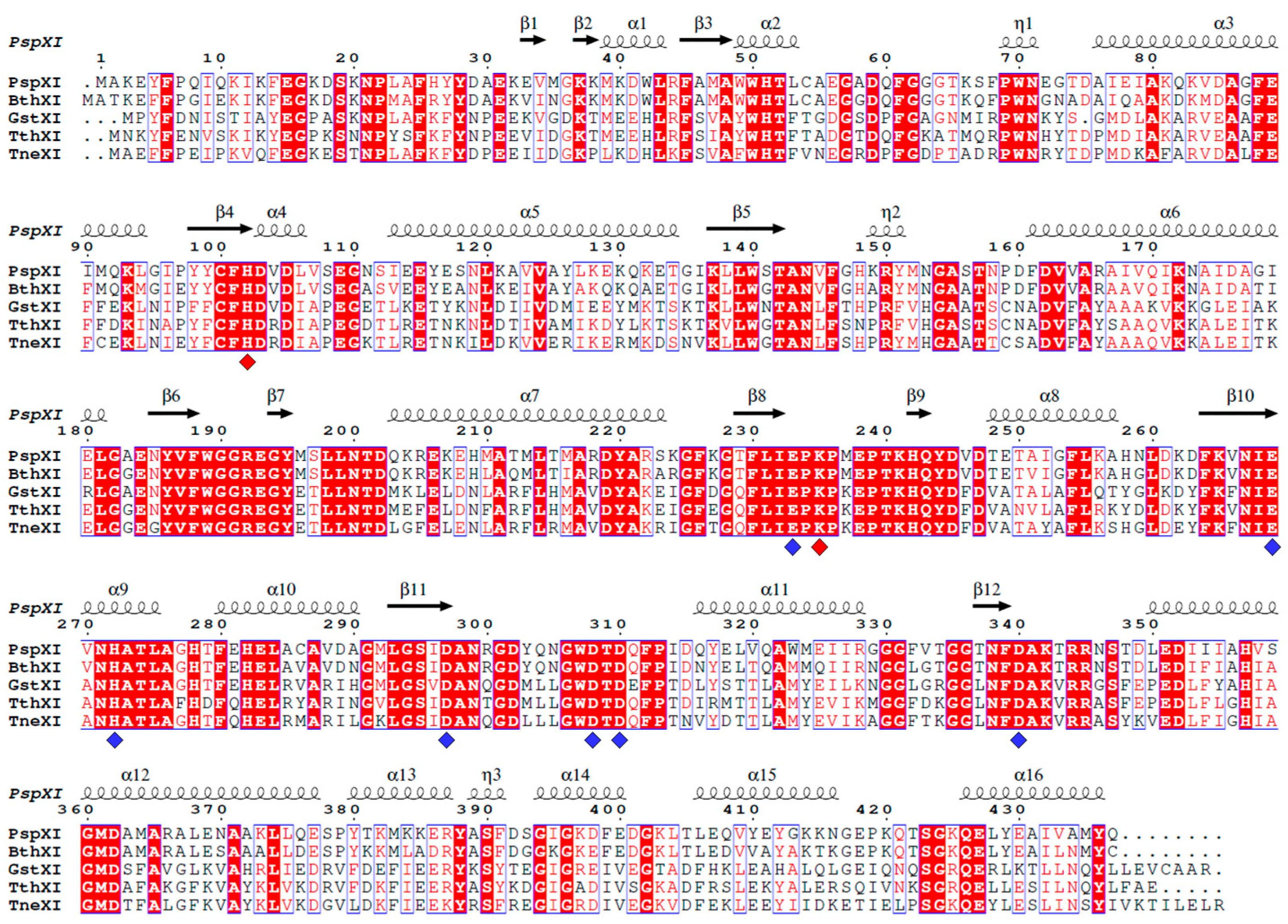
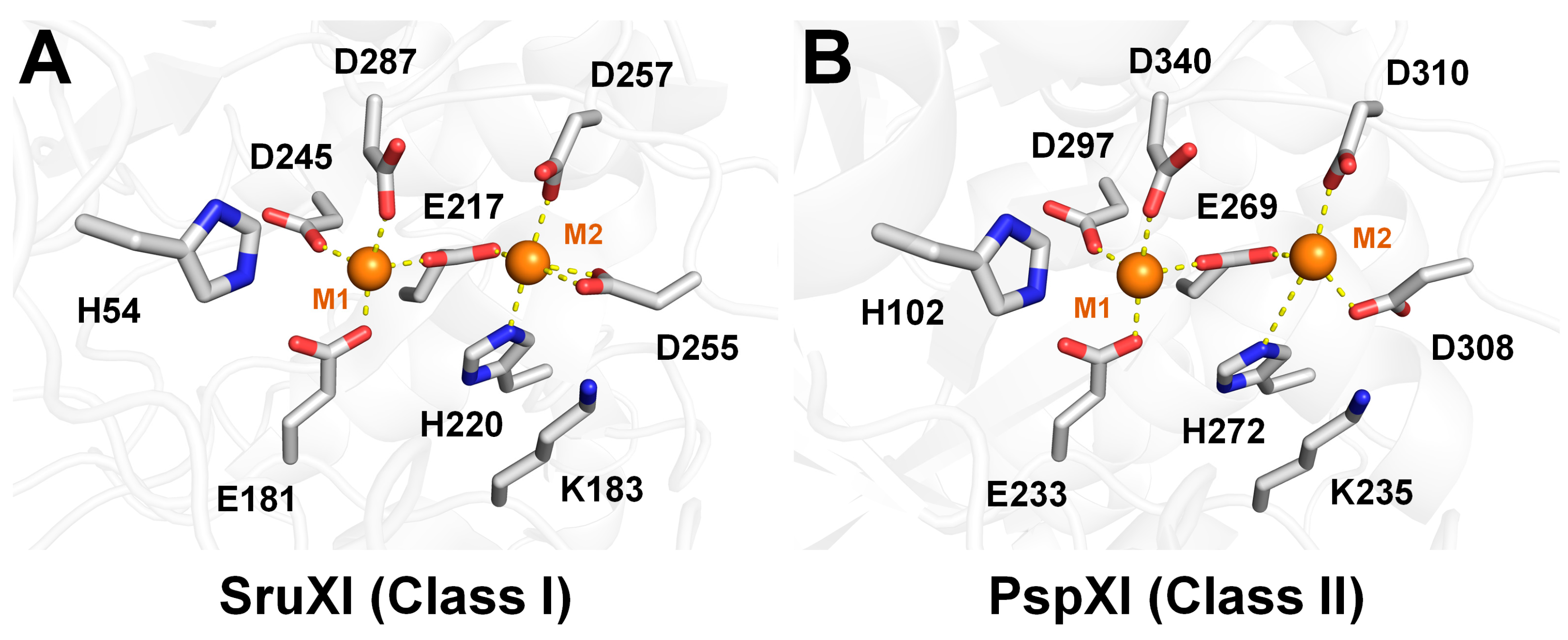
2.2. Structural Analysis of Class I and II XIs

To explain the structural features of XIs, the structures of SruXI and PspXI, representing class I and class II XI, respectively, are described and compared. Both class I and II XIs typically have a TIM-barrel domain at the N-terminal and an α-helical domain at the C-terminal (Figure 3A,B). The N-terminal region of class II PspXI is extended compared with that of class I SruXI (Figure 3B). The TIM-barrel domain of XIs contains an active site with metal-binding residues, whereas the C-terminal domain plays a role in stabilizing tetrameric assembly. The superimposition of SruXI (PDB code: 1XIS) and PspXI (PDB code: 5NH8) reveals distinct conformations between the TIM barrel and α-helical domains, with an r.m.s. deviation of 1.194–1.271 Å (Figure 3C).
Class I and II XIs form a tetrameric assembly with 222 symmetry (Figure 3D,E). Within this assembly, two TIM-barrel domains create substrate-binding channels, whereas the C-terminal domain interacts with the surrounding TIM-barrel domains. The C-terminal domain is crucial for maintaining a tight tetrameric formation, stabilized by interactions such as hydrogen bonds and salt bridges among various amino acids [28].
Tetrameric XI forms four active sites for isomerization. The substrate-binding channel is in the central region of the TIM-barrel domain. The metal ions that bind the active site or enhance the activity of XI can vary depending on the specific XI or strain [10,41,42,43]. Two metal-binding sites, M1 and M2, are at the active site of XI. At M1, a metal ion is coordinated by conserved aspartic or glutamic acid, whereas at M2 the metal ion is coordinated by conserved aspartic acid, glutamic acid, and histidine residues. Additionally, metal ions at both M1 and M2 are coordinated by water molecules. A conserved histidine residue, involved in substrate ring opening, is near M1, whereas a conserved lysine residue, involved in the hydride shift, is near M2.
Studies have shown that class I XIs exhibit optimal activity on glucose substrates in the presence of Mg2+ or a combination of Mg2+ and Co2+ [43,44]. In contrast, class II XIs show optimal activity on glucose substrates in the presence of Co2+ and on xylose substrates with Mn2+ [43,44]. Although the preferred metal ions may vary between class I and class II XIs, the metal-binding configurations at their active sites show a high degree of similarity.
For example, in class I SruXI, the metal ion at M1 is coordinated by E181, D245, D287, and E217, while the metal ion at M2 is coordinated by E217, D255, H220, and D257 (Figure 4A). E217 can interact with both metal ions at M1 and M2. H54, which is involved in ring opening, is located approximately 7.7 Å away from M1, whereas K183, which is involved in the hydride shift, is 4.8 Å away from M2.
In class II PspXI, the metal ion at M1 is coordinated by E233, D297, D340, and E269, whereas the metal ion at M2 is coordinated by E269, D308, H272, and D310 (Figure 4B). E269 interacts with both metal ions at M1 and M2. H102, which is involved in ring opening, is approximately 7.1 Å away from M1, whereas K183, involved in the hydride shift, is 4.6 Å away from M2. These results indicate that the metal-binding residues and catalytic residues of class I and II XIs are sequentially and positionally conserved.

3. Engineering of XI

3.1. GI from Thermoanaerobacterium saccharolyticum

The native GI produced by the thermophilic anaerobic bacterium T. saccharolyticum (TsaGI) was purified and its physicochemical and catalytic properties were characterized [45]. The optimal temperature and pH for TsaGI activity were 80 °C and pH 7.0–7.5, respectively. Xu et al., referencing studies that enhanced enzyme activity at Trp139 of Clostridium thermosulfurogenes [46], performed a site-saturation mutagenesis on the Trp139 of TsaGI [39]. A total of 17 mutants of TsaGI were obtained, excluding two failed constructions, W139P and W139R. Enzyme activity measurements showed that mutants W139F (240% relative activity), W139C (214%), W139S (205%), and W139T (200%) showed >2-fold enzyme activity compared with wild-type TsaGI (TsaGI-WT) (Figure 5A). Additionally, the mutants W139N (196%), W139I (190%), W139K (175%), W139L (122%), W139D (129%), W139Q (139%), W139V (127%), W139Y (118%), and W139G (103%) demonstrated improved enzyme activity compared with TsaGI-WT. In contrast, the mutants W139H (92%), W139A (90%), W139E (87%), and W139M (43%) showed reduced enzyme activity (Figure 5A). The best mutant, W139F, with a 2.4-fold increase in its specific activity at 80 °C, was characterized in more detail [39]. The specific activity of TsaGI-W139F at 80 °C was 2.4-fold higher than that of TsaGI-WT. TsaGI-W139F retained 50% of its initial activity after 20 h at 80 °C, whereas TsaGI-WT’s activity was halved after 12 h. This suggests that TsaGI-W139F has increased thermal stability, making it potentially more effective for industrial applications. Both TsaGI-WT and TsaGI-W139F had an optimal activity temperature of 90 °C and became inactive above this temperature, indicating that W139 is not critical for determining the optimal temperature for isomerization.
A metal effect analysis showed the highest thermal stability was achieved in the presence of 5 mM Mg2+ and 250 μM Co2+. Using these conditions, kinetic experiments on TsaGI-W139F and TsaGI-WT were performed at pH 6.5 and 80 °C. The Km, Vmax, Kcat, Kcat/Kmax, and specific activity of TsaGI-WT were 149.4 mM, 0.0557 μmol/min, 881.1 min−1, 5.90, and 17.62 U/mg, respectively. The Km, Vmax, Kcat, Kcat/Kmax, and specific activity of TsaGI-F139F were 51.3 mM, 0.0843 μmol/min, 2979.1 min−1, 58.06, and 59.58.62 U/mg, respectively. These results indicate that TsaGI-W139F is a successful mutant with high catalytic efficiency, making it suitable for industrial applications [39].
This biochemical experiment clearly indicated that the position of W139 in TsaGI is critical for enhancing enzyme activity. However, the study did not provide insights into how the W139 mutant affects TsaGI activity. To understand the impact of the W139 mutation, a model of the structure of TsaGI was generated and analyzed, because an experimental structure was lacking. The model structure of TsaGI showed that W139 was within the substrate-binding site (Figure 5B). The side chain of W139 was surrounded by hydrophobic residues, such as V186, C99, W49, and L138. W139 was in close proximity to the M1-binding residues E232, D296, and D339, at distances of 4.8, 4.0, and 5.4 Å, respectively. It is approximately 5.2 Å from H101 and ~6.0 Å from the potential M1. The side chain of W139 was oriented toward M1 and played a role in shaping the substrate-binding pocket at M1. Therefore, mutations in W139 may alter the shape of the substrate-binding pocket at M1 and affect the conformation of His101, which is involved in ring opening.
To understand the structural effects of TsaGI mutants, model structures for W139C, W139F, W139I, W139K, W139N, W139S, and W139T were generated. These mutants exhibited >50% increase in enzyme activity compared with TsaGI-WT. All mutants had an increased volume inside the substrate-binding pocket compared to TsaGI-WT. Specifically, W139C, W139I, W139N, W139S, and W139T, which have shorter side chains, created more volume inside the substrate-binding site than TsaGI-WT. The effects on substrate-binding volume and hydrophobicity vary depending on the characteristics of the mutant amino acids. These results suggest that the W139 mutants enhanced TsaGI activity by altering its substrate-binding volume, shape, and hydrophobicity around M1.

3.2. GI from Geobacillus caldoxylosilyticus TK4

Recombinant GI from the thermophilic bacterium G. caldoxylosilyticus TK4 (GcaGI) was obtained and subjected to mutagenesis to enhance its performance at high temperatures and low pHs, with a lower Km value for glucose [36]. Based on the reported mutagenesis studies and sequence alignment, the His99, Val184, and Asp102 of GcaGI were substituted by Qln, Thr, and Asn, respectively.
GcaGI-WT had the highest enzyme activity at pH 7.5, with its activity reducing to <70% at pHs > 8 and pHs < 7.0. The optimal pH values for GcaGI-H99Q, GcaGI-D102N, and GcaGI-V184T were pH 6.0, 6.0, and 6.5, respectively, indicating a shift toward an acidic environment for the optimal pH of the GcaGI mutants. GcaGI-H99Q, GcaGI-D102N, and GcaGI-V184T maintained >80% of their enzyme activity within the pH ranges of 6.0–6.5, 5.5–6.5, and 6.0–7.0, respectively. GcaGI-WT showed highest activity at 80 °C, whereas all GcaGI mutants exhibited their maximum activity at 85 °C. All GcaGI mutants exhibited high activity at >85 °C, whereas GcaGI-WT lost its enzyme activity at this temperature. This indicates that the thermostability of all GcaGI mutants improved compared to that of GcaGI-WT. The results of the kinetic analysis showed that the Km of GcaGI-WT, H99Q, V184T, and D102N were approximately 39, 173, 76, and 125 mM, respectively. The Vmax of GcaGI-WT, H99Q, V184T, and D102N were approximately 0.7, 0.3, 0.3, and 0.2 U/mg, respectively.
The highest enzyme activity of GcaGI-WT, H99Q, V184T, and D102N was observed in the presence of Co2+, Co2+, Mn2+ and Cu2+, respectively. Na+ enhanced the activity of GcaGI-H99Q but inhibited the activity of GcaGI-V184T and GcaGI-D102N. These mutagenesis and biochemical results clarified that the mutant positions of H99, V184, and D102 in GcaGI are critical for changing its optimal pH and temperature and improving its thermostability. The effect of metal ions on the activity of all GcaGI mutants was unique. Based on the literature, H99 and D102 were considered to play important roles in the catalytic site, whereas V184 was related to the substrate-binding pocket [36].
To better understand how the mutant sites of GcaGI affect enzyme activity, the model structures of GcaGI and its mutants were analyzed. Analysis of the model structure of GcaGI showed that H99 and D102 are posited at the entrance of the active site and V184 is near M1 (Figure 6A). The substrate accessibility channel and active site regions of GcaGI-WT had a negatively charged surface (Figure 6B).
In the model structure of GcaGI-H99Q, the electrostatic charge distribution at the mutant site was similar to that of GcaGI-WT, despite the different properties of histidine and glutamine. However, the surface shape at the position of the mutation site in GcaGI-H99Q was slightly changed (Figure 6B). The model structure of GcaGI-D102N showed a positively charged surface at the mutation site, which significantly differed from that in GcaGI-WT (Figure 6B). In the model structure of GcaGI-V184T, the mutant residue was buried in the deeper region of the substrate-binding pocket near M1, and the size of the mutated and original residues was almost identical (Figure 6B). Thus, significant structural changes were not observed between GcaGI-V184T and GcaGI-WT. Taken together, the substrate-binding entrance and the vicinity of the M1-binding site of GcaGI affected the enzyme’s activity, including optimal pH and temperature, thermostability, and kinetic parameters.

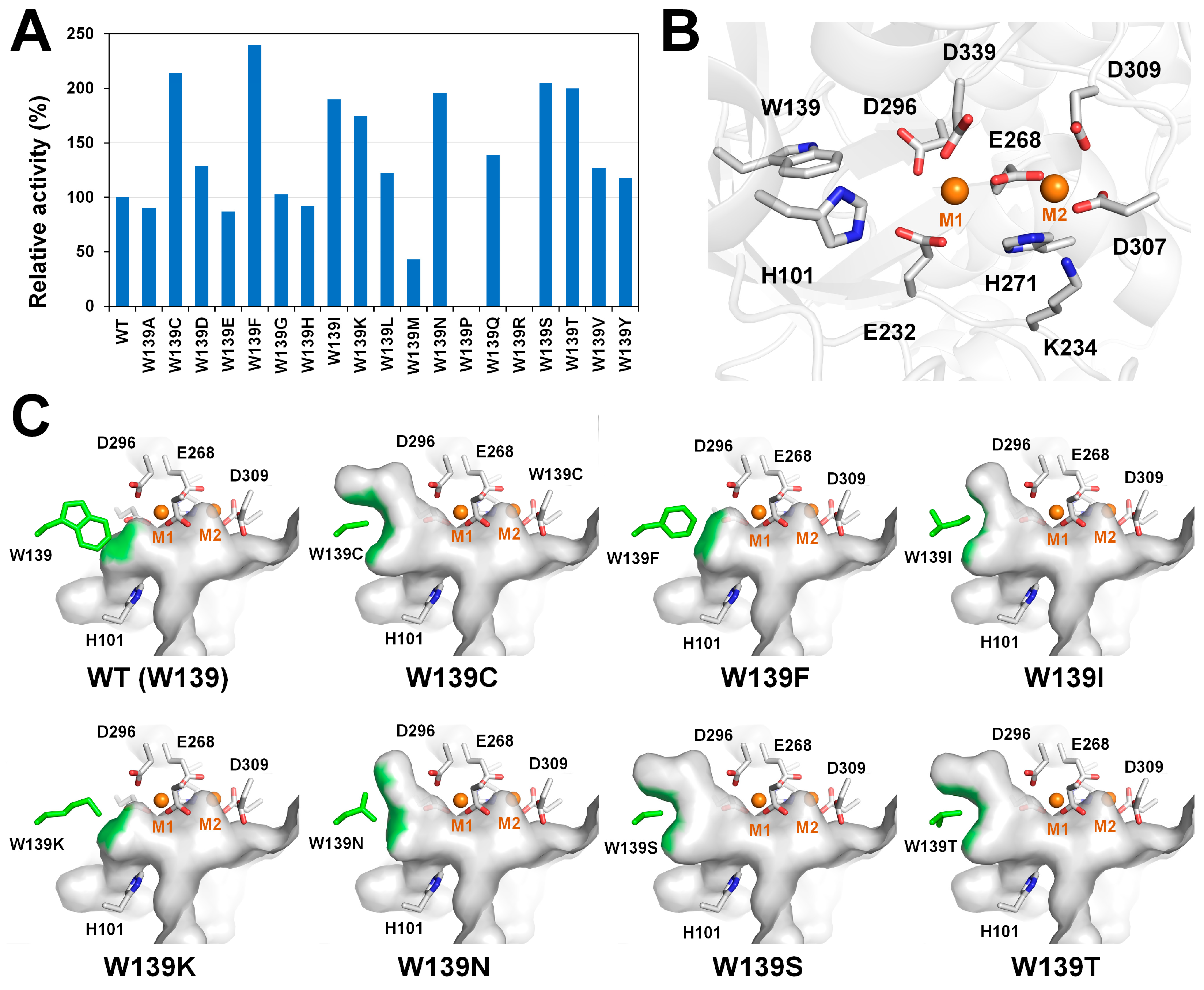
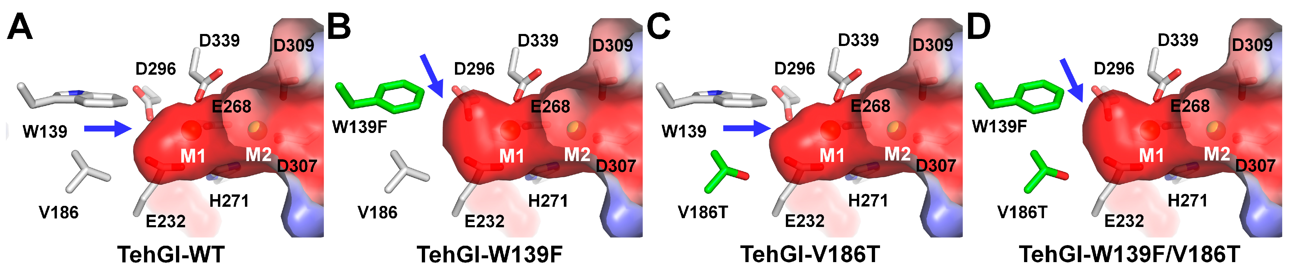
3.3. GI from Thermoanaerobacter ethanolicus

The use of thermostable GI to improve the economics and competitiveness of industrial production is attractive [38]. To obtain thermostable GI, Liu et al. performed a genome-mining approach to screen thermophilic GIs from GenBank, using Thermotoga maritima, with its conserved motif FSVAFWHTF as the template [38]. GI from T. ethanolicus CCSD1 (TehGI, WP_003868244.1) was selected; its recombinant protein exhibited a good soluble expression. The specific activity of TehGI was 39.9 U/mg, and a 53.8% conversion could be obtained using glucose as a substrate. Based on the model structure analysis and the literature, Liu et al. further engineered two sites (W139 and V186) to improve the activity and thermostability of TehGI. TehGI-W139F was generated to enlarge the active site pocket and improve its activity and thermal stability. TehGI-V186T and TehGI-V186S were designed to enhance the binding of hydrophilic D-glucose. The optimal temperature for TehGI-WT and its mutants was 90 °C. The residual activities of TehGI-WT, TehGI-W139F, TehGI-V186T, TehGI-V186S, and W139F/V186T after 24 h at 90 °C were 56%, 65%, 59%, 45%, and 68%, respectively. Kinetic studies showed that the Km (mM)/Vmax (µmol/min/mg)/kcat (s−1) for TehGI-WT, TehGI-W139F, TehGI-V186T, TehGI-V186S, and W139F/V186T was 421.0/27.0/22.6, 322.4/33.3/27.9, 255.4/27.8/23.3, 1361.9/71.8/60.1, and 245.2/29.2/24.5, respectively. TehGI-W139F/V186T showed the highest activity (92.1 U/mg) and thermostability. Additionally, the catalytic efficiency (Kcat/Km) of TehGI-W139F/V186T was 1.86-fold higher than that of TehGI-WT.
This mutagenesis and biochemical study clearly showed that the W139F and V186T mutations of TehGI are critical for improving enzyme activity, but a functional analysis of its structural basis has not been performed. To better understand this mutation site, the structural positions of W139F and V186T in TehGI were analyzed using model structures (Figure 7). In TehGI-WT, W139 and V186 are near M1 at the active site of TehGI (Figure 7A). The side chains of Trp193 and V186 form the substrate-binding pocket at M1 of the active site. In TehGI-W139F and TehGI-W139F/V186T, the volume of the deeper substrate-binding region inside M1 increased due to the reduced volume of the W139F side chain, whereas the side chain volumes of Val186 and V186T were the same (Figure 7B–D). However, V186 may affect the surface charge of the deeper substrate-binding region, changing it from hydrophobic to hydrophilic. These structural results indicate that changes in the deeper substrate-binding region are important for enhancing GI activity.
Jin et al. further engineered TehGI-W139F/V186T (named TehGI-M) using a conservation analysis of its primary structure to further improve its thermostability and catalytic capacity [37]. Based on the sequence alignment of TehGI with other GIs from T. thermosulfurigenes (sequence identity: 86.4%), G. stearothermophilus (72.7%), and T. neapolitana (71.5%), nine sites (38, 130, 137, 218, 229, 278, 299, 316, and 367) of non-strict conservation, which were predicted to be beneficial for improving catalytic capacity, were mutated [37]. TehGI-M-L38M, TehGI-M-V137L, and TehGI-M-L38M/V137L exhibited 1.61-, 1.68-, and 2.0-fold higher activity, respectively, than TehGI-M. The specific activity of TehGI-M, TehGI-M-L38M, TehGI-M-V137L, and TehGI-M-L38M/V137L was approximately 85.1, 136.2, 142.9, and 169.3 U mg−1, respectively. The Km values of TehGI-M, TehGI-M-L38M, TehGI-M-V137L, and TehGI-M-L38M/V137L were approximately 234.2, 199.9, 112.2, and 85.9 mM, respectively. The Vmax values of TehGI-M, TehGI-M-L38M, TehGI-M-V137L, and TehGI-M-L38M/V137L were approximately 29.2, 39.8, 32.5, and 42.7 µmol/min/mg, respectively.
To evaluate their potential applications, the isomerization reactions catalyzed by TehGI-M and TehGI-M-L38M/V137L (named TehGI-M2) were analyzed. The time courses of the bioconversion of D-glucose to high-fructose corn syrup showed that after 1.5 h, the yield of D-fructose by TehGI-M2 and TehGI-M was 60.2% and 51.8%, respectively. At equilibrium, the final yield of D-fructose by TehGI-M2 and TehGI-M was 67.3% at 2.5 h and 53% upon prolonged processing, respectively. This study performed an analysis of the mutation sites L38M and V137L of TehGI-M2 using a model structure generated by Modeller 9.23 [37]. The overall structure of TehGI-M2, which includes the N-terminal TIM-barrel domain and the C-terminal helical domain, displayed a typical GI architecture. However, the explanation of the model structure analysis of TehGI-M2 was incorrect. This study suggested that the interconnected helices of the L38M mutant had increased polarity, leading to tighter tetramer binding, thereby improving thermostability. However, L38M of TehGI was at the bottom of the TIM-barrel fold and not involved in tetrameric assembly. L38M was surrounded by other hydrophobic amino acids and likely contributed to stabilizing the hydrophobic core of the TIM-barrel fold. Moreover, the study docked a glucose molecule to TehGI-M and TehGI-M2. Metal ions, such as Mn2+, Mg2+, or Co2+, which bind two metal-binding sites in the active site of GI, are essential for substrate binding and isomerization [9,10]. Accordingly, this glucose docking study must be performed using the TehGI model structure containing metal ions at its active site. However, this study docked glucose onto the model structure of TehGI without metal ions at its active site [37]. Consequently, in the docking results, glucose did not bind to M1 but interacted with His101, Glu232, and Asp339 of TehGI-M and Glu232, Glu268, and Asp339 of TehGI-M2. As a result, this study contains errors in some aspects of its structural interpretation and does not accurately explain the direct functions of L38M and substrate binding [37].

3.4. XI from Piromyces sp. E2

The enzymatic function of XI, which converts xylose to xylulose, can be utilized for bioethanol production [17,24,25,26]. Saccharomyces cerevisiae is widely used for producing bioethanol through XI-related metabolic pathways [47,48,49]. Bae et al. developed a recombinant strain of S. cerevisiae capable of secreting XI from Piromyces sp. E2 (named PspXI here) that can continuously convert xylose to xylulose in a medium before cell uptake during fermentation [35]. The culture condition for yeast is pH 5.0, whereas the optimal pH for PspXI is pH 7.0, with PspXI exhibiting only 50% enzyme activity at pH 5.0. To enhance the extracellular conversion of xylose to xylulose during yeast fermentation, a directed evolution experiment was performed to obtain a PspXI mutant with higher activity at low pHs. The procedure was as follows: (1) An error-prone PCR was performed, and the resulting products were transformed into S. cerevisiae 2805Δgal80. (2) First screening: UDX medium at pH 4.0. (3) Second screening: YPX medium at pH 4.0 in a 96-well deep-well plate. (4) Third screening: YPX medium at pH 4.0 in a flask to confirm growth and xylose consumption.
Compared to the WT, a strain with 36% higher xylose consumption and 20% improved growth was obtained. In this clone, the Glu56 and Ile252 residues of PspXI were substituted with alanine and methionine, respectively. To investigate how these mutations affected its enzyme activity, PspXI-WT, PspXI-E56A, PspXI-I252M, and PspXI-E56A/I252M were cultured in YPX medium for 72 h. The secreted XI yield between the PspXI-WT and PspXI mutants was not significantly different. The optimal pH values of PspXI-WT, PspXI-E56A, PspXI-I252M, and PspXI-E56A/I252M were 7.0, 6.0, 7.0, and 5.0, respectively. The cell growth (OD600) and xylose consumed (g) by PspXI-WT, PspXI-E56A, PspXI-I252M, and PspXI-E56A/I252M were approximately 15.3/9.0, 18.6/13.4, 16.5/10.8, and 16.3/10.6, respectively. Accordingly, the xylose consumption of the strains expressing PspXI-E56A/I252M increased by >20% compared with PspXI-WT. At pH 5.0, the relative activity of PspXI-WT and PspXI-E56A/I252M was approximately 60% and 120%, respectively, compared with the maximum activity of XI-WT at pH 7.0. When SR8/TFP3 strains containing PspXI-WT and PspXI-E56A/I252M were cultured for 48 h, the xylose consumption (g/L), ethanol production (g/L), and yield (EtOH/Xyl) of the strains containing PspXI-WT and PspXI-E56A/I252M were approximately 67.9/25.0/0.36 and 71.2/26.7/0.37, respectively. This study improved PspXI activity at pH 5.0 using two amino acid mutations. However, the study did not investigate how E56A and I252M enhanced PspXI activity and shifted the optimal pH.
To provide insights into the optimal pH shift of the PspXI used during fermentation with S. cerevisiae in the bioethanol production industry, the mutation sites E56A and I252M are analyzed here using the crystal structure of XI from Piromyces sp. E2 (PDB code: 5NH7). In the crystal structure of PspXI, E56 is in the TIM-barrel domain and positioned near the substrate access surface, changing the shape of the substrate-binding channel’s entrance (Figure 8A). In particular, E56 of PspXI-WT led to the substrate-binding entrance having a negative charged state, whereas E56A showed a neutral charged state, which may affect substrate accessibility and cause an optimal pH shift (Figure 8B). The I252 of PsiXI is in the α-helix region on the opposite side of the substrate-binding site in the TIM-barrel domain and is surrounded by hydrophobic amino acids, such as V266, A287, Ala290, and M292 (Figure 8B). Therefore, I252M changed the hydrophobicity of the hydrophobic core at the bottom of the TIM-barrel domain, although it was unlikely to directly affect pH, which is consistent with the unchanged optimal pH of XI-I252M. Nevertheless, because the optimal pH of the XI-E56A/I252M double mutant shifted to a more acidic range, I252M may indirectly influence pH through its interactions with other amino acids.

4. Discussion

XIs are widely used in various industries, including the food and bioenergy fields. This review introduces the engineered XIs that have increased enzymatic activity through mutagenesis and biochemical experiments. The mutation sites of engineered XIs were analyzed using experimentally determined structures or model structures to understand how these engineered sites affect their catalytic activity. The XI engineering sites reviewed here can be categorized as follows: (i) active sites, (ii) substrate-binding entrance regions, and (iii) regions not directly involved in substrate binding or activation.
In engineering active sites, the interior region of M1 in the substrate-binding site of XI is crucial for enhancing its activity. For example, in class II XIs, the substitution of W139 with phenylalanine in TsaGI and TehGI effectively increased their enzyme activity [38,39]. The model structure analysis of XI showed that substituting W139 with other amino acids can change the shape and charge distribution of the substrate-binding site around M1. Typically, substituting W139 with amino acids that have shorter side chains increased the space within M1. Additionally, across TsaGI mutants, the volumes of the substrate-binding sites for W139C and W139S or W139T and W139V were almost identical due to their similar side chain lengths; however, these mutants exhibited different enzyme activities, indicating that not only the side chain volume but also the charge state of the side chain residue affected enzyme activity. In the saturation mutagenesis of TsaGI, W139F, which showed the highest enzyme activity, was highlighted. However, other mutants, such as W139C, W139F, W139I, W139K, W139N, W139S, and W139T, had activity that was increased by >50% compared with the WT [39], suggesting that these amino acids could be considered in XI engineering studies.
Substituting the valine around M1 with threonine increased XI activity. A model structure analysis showed that V184T in CgaGI and V186T in TehGI did not affect the volume of the active site pocket, and thus did not hinder substrate access [36,37]. Substituting Val with Thr introduced a change in the charge on the surface of the active site pocket, which may enhance catalytic efficiency through additional hydrogen bonding with the substrate [46,50]. Notably, in CgaGI, the V184T mutant affected not only its catalytic activity but also its optimal pH and temperature. Accordingly, this engineering position could be useful for altering optimal enzyme characteristics as well as enzyme activity.
Next, the engineering of the substrate-binding channel or entrance region of XI was important in improving its catalytic activity. The substitutions of H99Q and D102N of GcaGI in the substrate-binding channel and E56A of PsiXI in the substrate-binding entrance significantly improved enzyme activity [35,36]. The substitution of these amino acids could potentially affect the charge state of the protein surface. A model structure analysis showed that GcaGI-D102Q changed the negative charge surface to a positive charge surface, whereas PsiXI-E56A changed from a negative to a neutral charge surface. Notably, PsiXI-E56A influenced the optimal pH of the protein, providing insights for engineering XI in order to shift its pH.
Finally, there were cases where mutation sites not related to substrate binding or approach affected XI activity. Although the PsiXI-I252M mutation is opposite the substrate-binding site and is involved in protein folding, it influenced XI activity and indirectly affected its optimal pH [35]. This indicates that amino acids involved in protein folding can potentially impact XI activity.
This review not only highlights notable engineering cases related to XI engineering but also introduced the general amino acid and structural characteristics of XI. XI structures deposited in the PDB include not only its native forms but also its complexes with substrates, such as glucose or xylose, and inhibitors, such as xylitol (Table 1). The various states of these XI structures revealed different conformations of the active site and the substrate-binding channel of XI. Therefore, these structures provide detailed information about XI’s molecular dynamics, offering valuable insights for understanding its mutation sites and rational designs that improve its enzyme activity. Based on these structures, rational protein engineering can be achieved by substituting amino acids around the active site and substrate-binding channel or entrance. However, if there are no experimentally determined structures available for structure-based protein engineering, AI-based structure prediction programs, such as AlphaFold, can be used to obtain information for understanding a protein’s structure and function [40]. These modeling programs can offer valuable insights into the potential changes in volume or charge state at mutation sites, which can be useful for predicting substrate accessibility.
In the XI engineering studies reviewed, isomerase activity was measured using specific substrates and metal ions. As reported in previous studies, the same XI could exhibit different activities depending on whether glucose or xylose was used as the substrate, and the metal ions that provide optimal activity could vary [43,44]. Additionally, the type of metal ions bound to the active site could influence the optimal conditions and activity of XI [10,41,42,43]. Therefore, future XI engineering efforts should include evaluations of various substrates and metal ions in addition to simple activity comparisons with WT XI.

5. Conclusions

This review introduced significant results from the engineering of XI. Key residues used in XI engineering were analyzed using crystal and model structures. The structural analysis of the engineered XI revealed that targeting the deeper regions of its active site and the entrance of its substrate-binding pocket presents a promising strategy for enhancing its catalytic activity. This review provides useful insights into engineering XI to improve its catalytic activity for industrial applications.

Funding

This work was funded by the National Research Foundation of Korea (NRF) (NRF-2021R1I1A1A01050838).

Data Availability Statement

No new data were created or analyzed in this study.

Conflicts of Interest

The author declares no conflicts of interest.

References

  1. DiCosimo, R.; McAuliffe, J.; Poulose, A.J.; Bohlmann, G. Industrial use of immobilized enzymes. Chem. Soc. Rev. 2013, 42, 6437. [Google Scholar] [CrossRef] [PubMed]
  2. Bhosale, S.H.; Rao, M.B.; Deshpande, V.V. Molecular and industrial aspects of glucose isomerase. Microbiol. Rev. 1996, 60, 280–300. [Google Scholar] [CrossRef] [PubMed]
  3. Nam, K.H. Glucose Isomerase: Functions, Structures, and Applications. Appl. Sci. 2022, 12, 428. [Google Scholar] [CrossRef]
  4. Barreto, M.Q.; Garbelotti, C.V.; de Moura Soares, J.; Grandis, A.; Buckeridge, M.S.; Leone, F.A.; Ward, R.J. Xylose isomerase from Piromyces sp. E2 is a promiscuous enzyme with epimerase activity. Enzym. Microb. Technol. 2023, 166, 110230. [Google Scholar] [CrossRef]
  5. Kasumi, T.; Hayashi, K.; Tsumura, N. Purification and enzymatic properties of glucose isomerase from Streptomyces griseofuscus, S-41. Agric. Biol. Chem. 1981, 45, 619–627. [Google Scholar] [CrossRef]
  6. Kim, Y.-S.; Kim, D.-Y.; Park, C.-S. Production of l-rhamnulose, a rare sugar, from l-rhamnose using commercial immobilized glucose isomerase. Biocatal. Biotransform. 2017, 36, 417–421. [Google Scholar] [CrossRef]
  7. Langan, P.; Sangha, A.K.; Wymore, T.; Parks, J.M.; Yang, Z.K.; Hanson, B.L.; Fisher, Z.; Mason, S.A.; Blakeley, M.P.; Forsyth, V.T.; et al. L-Arabinose Binding, Isomerization, and Epimerization by D-Xylose Isomerase: X-Ray/Neutron Crystallographic and Molecular Simulation Study. Structure 2014, 22, 1287–1300. [Google Scholar] [CrossRef]
  8. Chen, Z.; Chen, J.; Zhang, W.; Zhang, T.; Guang, C.; Mu, W. Recent research on the physiological functions, applications, and biotechnological production of d-allose. Appl. Microbiol. Biotechnol. 2018, 102, 4269–4278. [Google Scholar] [CrossRef]
  9. Kovalevsky, A.Y.; Hanson, L.; Fisher, S.Z.; Mustyakimov, M.; Mason, S.A.; Trevor Forsyth, V.; Blakeley, M.P.; Keen, D.A.; Wagner, T.; Carrell, H.L.; et al. Metal Ion Roles and the Movement of Hydrogen during Reaction Catalyzed by D-Xylose Isomerase: A Joint X-ray and Neutron Diffraction Study. Structure 2010, 18, 688–699. [Google Scholar] [CrossRef]
  10. Lee, M.; Rozeboom, H.J.; de Waal, P.P.; de Jong, R.M.; Dudek, H.M.; Janssen, D.B. Metal Dependence of the Xylose Isomerase from Piromyces sp. E2 Explored by Activity Profiling and Protein Crystallography. Biochemistry 2017, 56, 5991–6005. [Google Scholar] [CrossRef]
  11. Allen, K.N.; Lavie, A.; Glasfeld, A.; Tanada, T.N.; Gerrity, D.P.; Carlson, S.C.; Farber, G.K.; Petsko, G.A.; Ringe, D. Role of the Divalent Metal Ion in Sugar Binding, Ring Opening, and Isomerization by D-Xylose Isomerase: Replacement of a Catalytic Metal by an Amino Acid. Biochemistry 2002, 33, 1488–1494. [Google Scholar] [CrossRef] [PubMed]
  12. Lee, C.Y.; Bagdasarian, M.; Meng, M.H.; Zeikus, J.G. Catalytic mechanism of xylose (glucose) isomerase from Clostridium thermosulfurogenes. Characterization of the structural gene and function of active site histidine. J. Biol. Chem. 1990, 265, 19082–19090. [Google Scholar] [CrossRef]
  13. Collyer, C.A.; Henrick, K.; Blow, D.M. Mechanism for aldose-ketose interconversion by D-xylose isomerase involving ring opening followed by a 1,2-hydride shift. J. Mol. Biol. 1990, 212, 211–235. [Google Scholar] [CrossRef] [PubMed]
  14. Collyer, C.A.; Blow, D.M. Observations of reaction intermediates and the mechanism of aldose-ketose interconversion by D-xylose isomerase. Proc. Natl. Acad. Sci. USA 1990, 87, 1362–1366. [Google Scholar] [CrossRef] [PubMed]
  15. Asboth, B.; Naray-Szabo, G. Mechanism of Action of D-Xylose Isomerase. Curr. Protein Pept. Sci. 2000, 1, 237–254. [Google Scholar] [CrossRef]
  16. van Maris, A.J.A.; Winkler, A.A.; Kuyper, M.; de Laat, W.T.A.M.; van Dijken, J.P.; Pronk, J.T. Development of Efficient Xylose Fermentation in Saccharomyces cerevisiae: Xylose Isomerase as a Key Component. In Biofuels; Advances in Biochemical Engineering/Biotechnology; Springer: Berlin/Heidelberg, Germany, 2007; pp. 179–204. [Google Scholar]
  17. Van Vleet, J.H.; Jeffries, T.W. Yeast metabolic engineering for hemicellulosic ethanol production. Curr. Opin. Biotechnol. 2009, 20, 300–306. [Google Scholar] [CrossRef]
  18. Kwak, S.; Jin, Y.-S. Production of fuels and chemicals from xylose by engineered Saccharomyces cerevisiae: A review and perspective. Microb. Cell Fact. 2017, 16, 82. [Google Scholar] [CrossRef]
  19. Hou, J.; Qiu, C.; Shen, Y.; Li, H.; Bao, X. Engineering of Saccharomyces cerevisiae for the efficient co-utilization of glucose and xylose. FEMS Yeast Res. 2017, 17, fox034. [Google Scholar] [CrossRef]
  20. Hassan, M.E.; Awad, G.E.A.; MohyEldin, M.S.; Haroun, B.M.; El-Diwany, A.I.; Elnashar, M.M. Carbohydrate microcapsules tailored and grafted for covalent immobilization of glucose isomerase for pharmaceutical and food industries. Biotechnol. Lett. 2022, 45, 175–189. [Google Scholar] [CrossRef]
  21. Visuri, K.; Klibanov, A.M. Enzymatic production of high fructose corn syrup (HFCS) containing 55% fructose in aqueous ethanol. Biotechnol. Bioeng. 2004, 30, 917–920. [Google Scholar] [CrossRef]
  22. Mohamed, S.A.; Janee, S.; Saha, S.; Sharmin, S.; Hasan, A.Q.F.; Zohora, U.S.; Moni, R.; Islam, M.Z.; Rahman, M.S. Construction and investigation of multi-enzyme immobilized matrix for the production of HFCS. PLoS ONE 2024, 19, e0292931. [Google Scholar] [CrossRef]
  23. Majore, K.; Ciprovica, I. Bioconversion of Lactose into Glucose–Galactose Syrup by Two-Stage Enzymatic Hydrolysis. Foods 2022, 11, 400. [Google Scholar] [CrossRef] [PubMed]
  24. Qu, C.; Chen, L.; Fu, H.; Wang, J. Engineering Thermoanaerobacterium aotearoense SCUT27 with argR knockout for enhanced ethanol production from lignocellulosic hydrolysates. Bioresour. Technol. 2020, 310, 123435. [Google Scholar] [CrossRef]
  25. Vieira, I.P.V.; Cordeiro, G.T.; Gomes, D.E.B.; Melani, R.D.; Vilela, L.F.; Domont, G.B.; Mesquita, R.D.; Eleutherio, E.C.A.; Neves, B.C. Understanding xylose isomerase from Burkholderia cenocepacia: Insights into structure and functionality for ethanol production. AMB Express 2019, 9, 73. [Google Scholar] [CrossRef]
  26. Kobayashi, Y.; Sahara, T.; Ohgiya, S.; Kamagata, Y.; Fujimori, K.E. Systematic optimization of gene expression of pentose phosphate pathway enhances ethanol production from a glucose/xylose mixed medium in a recombinant Saccharomyces cerevisiae. AMB Express 2018, 8, 139. [Google Scholar] [CrossRef]
  27. Suzuki, T.; Morimoto, K. Characterization of D-xylose isomerase from Shinella zoogloeoides NN6 and its application for producing D-allulose and two D-ketopentoses in a one-pot multi-step transformation. J. Gen. Appl. Microbiol. 2022, 68, 175–183. [Google Scholar] [CrossRef] [PubMed]
  28. Nam, K.H. Structural Analysis of Xylose Isomerase from Streptomyces avermitilis. Crystals 2024, 14, 446. [Google Scholar] [CrossRef]
  29. Silva, P.C.; Ceja-Navarro, J.A.; Azevedo, F.; Karaoz, U.; Brodie, E.L.; Johansson, B. A novel d-xylose isomerase from the gut of the wood feeding beetle Odontotaenius disjunctus efficiently expressed in Saccharomyces cerevisiae. Sci. Rep. 2021, 11, 4766. [Google Scholar] [CrossRef]
  30. Park, S.-H.; Kwon, S.; Lee, C.W.; Kim, C.M.; Jeong, C.S.; Kim, K.-J.; Hong, J.W.; Kim, H.J.; Park, H.H.; Lee, J.H. Crystal Structure and Functional Characterization of a Xylose Isomerase (PbXI) from the Psychrophilic Soil Microorganism, Paenibacillus sp. J. Microbiol. Biotechnol. 2019, 29, 244–255. [Google Scholar] [CrossRef] [PubMed]
  31. Bae, J.E.; Hwang, K.Y.; Nam, K.H. Structural analysis of substrate recognition by glucose isomerase in Mn2+ binding mode at M2 site in S. rubiginosus. Biochem. Biophys. Res. Commun. 2018, 503, 770–775. [Google Scholar] [CrossRef]
  32. Bae, J.E.; Kim, I.J.; Nam, K.H. Crystal structure of glucose isomerase in complex with xylitol inhibitor in one metal binding mode. Biochem. Biophys. Res. Commun. 2017, 493, 666–670. [Google Scholar] [CrossRef]
  33. Nam, K.H. Room-Temperature Structure of Xylitol-Bound Glucose Isomerase by Serial Crystallography: Xylitol Binding in the M1 Site Induces Release of Metal Bound in the M2 Site. Int. J. Mol. Sci. 2021, 22, 3892. [Google Scholar] [CrossRef]
  34. Xu, Y.; Nam, K.H. Xylitol binding to the M1 site of glucose isomerase induces a conformational change in the substrate binding channel. Biochem. Biophys. Res. Commun. 2023, 682, 21–26. [Google Scholar] [CrossRef] [PubMed]
  35. Bae, J.-H.; Kim, M.-J.; Sung, B.H.; Jin, Y.-S.; Sohn, J.-H. Directed evolution and secretory expression of xylose isomerase for improved utilisation of xylose in Saccharomyces cerevisiae. Biotechnol. Biofuels 2021, 14, 223. [Google Scholar] [CrossRef] [PubMed]
  36. Dokuzparmak, C.; Colak, A.; Kolcuoglu, Y.; Akatin, M.Y.; Ertunga, N.S.; Tuncay, F.O. Development of Some Properties of a Thermophilic Recombinant Glucose Isomerase by Mutation. Appl. Biochem. Microbiol. 2020, 56, 164–172. [Google Scholar] [CrossRef]
  37. Jin, L.-Q.; Jin, Y.-T.; Zhang, J.-W.; Liu, Z.-Q.; Zheng, Y.-G. Enhanced catalytic efficiency and thermostability of glucose isomerase from Thermoanaerobacter ethanolicus via site-directed mutagenesis. Enzym. Microb. Technol. 2021, 152, 109931. [Google Scholar] [CrossRef]
  38. Liu, Z.-Q.; Zheng, W.; Huang, J.-F.; Jin, L.-Q.; Jia, D.-X.; Zhou, H.-Y.; Xu, J.-M.; Liao, C.-J.; Cheng, X.-P.; Mao, B.-X.; et al. Improvement and characterization of a hyperthermophilic glucose isomerase from Thermoanaerobacter ethanolicus and its application in production of high fructose corn syrup. J. Ind. Microbiol. Biotechnol. 2015, 42, 1091–1103. [Google Scholar] [CrossRef] [PubMed]
  39. Xu, H.; Shen, D.; Wu, X.-Q.; Liu, Z.-W.; Yang, Q.-H. Characterization of a mutant glucose isomerase from Thermoanaerobacterium saccharolyticum. J. Ind. Microbiol. Biotechnol. 2014, 41, 1581–1589. [Google Scholar] [CrossRef] [PubMed]
  40. Jumper, J.; Evans, R.; Pritzel, A.; Green, T.; Figurnov, M.; Ronneberger, O.; Tunyasuvunakool, K.; Bates, R.; Žídek, A.; Potapenko, A.; et al. Highly accurate protein structure prediction with AlphaFold. Nature 2021, 596, 583–589. [Google Scholar] [CrossRef]
  41. Callens, M.; Tomme, P.; Kersters-Hilderson, H.; Cornelis, R.; Vangrysperre, W.; De Bruyne, C.K. Metal ion binding to d-xylose isomerase from Streptomyces violaceoruber. Biochem. J. 1988, 250, 285–290. [Google Scholar] [CrossRef]
  42. Callens, M.; Kerstershilderson, H.; Vangrysperre, W.; Debruyne, C.K. D-Xylose Isomerase from Streptomyces-Violaceoruber—Structural and Catalytic Roles of Bivalent-Metal Ions. Enzym. Microb. Technol. 1988, 10, 695–700. [Google Scholar] [CrossRef]
  43. Epting, K.L.; Vieille, C.; Zeikus, J.G.; Kelly, R.M. Influence of divalent cations on the structural thermostability and thermal inactivation kinetics of class II xylose isomerases. FEBS J. 2005, 272, 1454–1464. [Google Scholar] [CrossRef] [PubMed]
  44. Van Bastelaere, P.; Vangrysperre, W.; Kersters-Hilderson, H. Kinetic studies of Mg2+-, Co2+- and Mn2+-activated D-xylose isomerases. Biochem. J. 1991, 278, 285–292. [Google Scholar] [CrossRef]
  45. Lee, C.Y.; Zeikus, J.G. Purification and characterization of thermostable glucose isomerase from Clostridium thermosulfurogenes and Thermoanaerobacter strain B6A. Biochem. J. 1991, 273, 565–571. [Google Scholar] [CrossRef]
  46. Meng, M.; Lee, C.; Bagdasarian, M.; Zeikus, J.G. Switching substrate preference of thermophilic xylose isomerase from D-xylose to D-glucose by redesigning the substrate binding pocket. Proc. Natl. Acad. Sci. USA 1991, 88, 4015–4019. [Google Scholar] [CrossRef] [PubMed]
  47. Qiu, Y.; Wu, M.; Bao, H.; Liu, W.; Shen, Y. Engineering of Saccharomyces cerevisiae for co-fermentation of glucose and xylose: Current state and perspectives. Eng. Microbiol. 2023, 3, 100084. [Google Scholar] [CrossRef]
  48. Coimbra, L.; Malan, K.; Fagúndez, A.; Guigou, M.; Lareo, C.; Fernández, B.; Pratto, M.; Batista, S. Fermentation of D-xylose to Ethanol by Saccharomyces cerevisiae CAT-1 Recombinant Strains. Bioenerg. Res. 2022, 16, 1001–1012. [Google Scholar] [CrossRef]
  49. Li, F.; Bai, W.; Zhang, Y.; Zhang, Z.; Zhang, D.; Shen, N.; Yuan, J.; Zhao, G.; Wang, X. Construction of an economical Xylose-utilizing Saccharomyces cerevisiae and its ethanol fermentation. FEMS Yeast Res. 2024, 24, foae001. [Google Scholar] [CrossRef]
  50. Sriprapundh, D.; Vieille, C.; Zeikus, J.G. Molecular determinants of xylose isomerase thermal stability and activity: Analysis of thermozymes by site-directed mutagenesis. Protein Eng. Des. Sel. 2000, 13, 259–265. [Google Scholar] [CrossRef]
Figure 1. Structure-based sequence alignment of class I XIs from Streptomyces rubiginosus (SruXI, UniProt code: P24300), Arthrobacter sp. (strain NRRL B3728) (ArtXI, P12070), Actinoplanes missouriensis (AmiXI, P12851), Streptomyces olivochromogenes (SolXI, P15587), Streptomyces diastaticus (SdiXI, P50910), Streptomyces sp. F-1 (SsFXI, A0A1K2FZ20), Streptomyces sp. SK (SsSXI, Q9ZAI3), Streptomyces albus G (SalXI, P24299), Streptomyces avermitilis (SavXI, A0A4D4M698), Streptomyces murinus (SmuXI, P37031), Thermus caldophilus (TcaXI, P56681), and Thermus thermophilus (TthXI2, P26997). Their metal-binding site and catalytic residues are indicated by blue and red diamonds, respectively. Their secondary structure was obtained from the crystal structure of SruXI (PDB code: 1XIS).
Catalysts 14 00597 g001
Figure 2. Structure-based sequence alignment of class I XIs from Piromyces sp. (strain E2) (PspXI, Q9P8C9), Bacteroides thetaiotaomicron (BthXI, Q8A9M2), Geobacillus stearothermophilus (GstXI, P54273), Thermoanaerobacterium thermosulfurigenes (TthXI, P19148), and Thermotoga neapolitana (TneXI, P45687). Their metal-binding site and catalytic residues are indicated by blue and red diamonds, respectively. Their secondary structure was derived from the crystal structure of PspXI (PDB code: 5NH8).
Catalysts 14 00597 g002
Figure 3. The crystal structures of XI from Streptomyces rubiginosus (SruXI, class I) and Piromyces sp. E2 (PspXI, class II). (A) The monomer structures of SruXI (PDB code: 1XIS) and (B) PspXI (PDB code: 5NH8) include the N-terminal TIM-barrel domain and the C-terminal α-helical domain. Class II PspXI features an extended N-terminal region compared to class I SruXI. (C) The superimposition of SruXI and PspXI reveals distinct conformations between the N- and C-terminal domains. The tetrameric formations of (D) class I SruXI and (E) class II PspXI.
Catalysts 14 00597 g003
Figure 4. Active site of class I and II XIs. (A) Class I XI from Streptomyces rubiginosus (PDB code: 1XIS). (B) Class II XI from Piromyces sp. E2 (5NH8). The crystal structures of SruXI and PspXI contain Mn2+ and Ca2+ at their metal-binding sites, respectively.
Catalysts 14 00597 g004
Figure 5. Engineering of W139 in XI from Thermoanaerobacterium saccharolyticum. (A) The site-saturation mutagenesis of W139 in TsaGI and the measurement of the relative activity of TsaGI-WT and its mutants. (B) The active site of the modeled TsaGI structure. The positions of the metal ions were obtained from the crystal structure of SruXI (PDB code: 7DFJ). (C) Surface structure analysis of the engineered TsaGIs for WT, W139C, W139F, W139I, W139K, W139N, W139S, and W139T. Mutation sites are indicated by green sticks.
Catalysts 14 00597 g005
Figure 6. Analysis of the engineering site in the GcaGI model structure. (A) Cartoon representation of GcaGI-WT, GcaGI-H99Q, GcaGI-D102N, and GcaGI-V184T. Mutant sites are indicated by green sticks. The positions of the metal ions were obtained from the crystal structure of SruXI (PDB code: 7DFJ). (B) The electrostatic surfaces of GcaGI-WT, GcaGI-H99Q, GcaGI-D102N, and GcaGI-V184T, with mutant sites highlighted by yellow dotted circles.
Catalysts 14 00597 g006
Figure 7. Analysis of the engineering sites of TehGI using model structures. The electrostatic surface of the active site pocket is displayed, including the mutation sites of (A) TehGI-WT, (B) TehGI-W139F, (C) TehGI-V186T, and (D) TehGI-W139F/V186T. The positions of the metal ions were obtained from the crystal structure of SruXI (PDB code: 7DFJ). Mutations are indicated by green sticks. The change in the volume of the substrate-binding pocket near M1 of TehGI due to mutagenesis is indicated by a blue arrow.
Catalysts 14 00597 g007
Figure 8. Structural analysis of PspXI (PDB code: 5NH7) and PspXI mutants. (A) Cartoon representation of the substrate-binding entrance of PspXI-WT and PspXI-E56A. E56 and E56A on the TIM-barrel domain are separated by the active site pocket. The mutation site and active site are indicated by blue dotted circles. (B) The electrostatic surfaces of PspXI-WT and PspXI-E56A. The mutation site and active site are indicated by yellow dotted circles. (C) Cartoon representation of PspXI-WT and PspXI-I252M. I252 and I252M are in the hydrophobic core and involved in stabilizing XI folding.
Catalysts 14 00597 g008
Table 1. XI structures deposited in Protein Data Bank.
Source Organism Accession No.PDB Code (Ligand)
Type I
Streptomyces
rubiginosus
P243001GW9 (β-L-xylopyranose, Ca2+), 1MNZ (Mg2+), 1O1H (Ca2+, Mg2+), 1OAD (Mn2+, Mg2+), 1XIB (Mn2+), 1XIC (Mn2+, D-xylose), 1XID (Mn2+, ascorbic acid), 1XIE (Mn2+, 1,5-anhydro-D-glucitol), 1XIF (Mn2+, α-D-glucopyranose), 1XIG (Mn2+, xylitol), 1XIH (Mn2+, sorbitol), 1XII (Mn2+, D-xyluose), 1XIJ (Mn2+, threonate ion), 1XIS (Mn2+), 2G4J (Ca2+, Mg2+, Cl), 2GLK (Mn2+, glycerol), 2GUB (no ligand), 2GVE (Co2+), 2XIS (Mg2+, xylitol), 3CWH (Mg2+, D-xylulose), 3GNX (Mn2+, xylitol), 3KBJ (no ligand), 3KBM (Cd2+, α-D-glucopyranose), 3KBN (Ni2+, D-glucose), 3KBS (Cd2+), 3KBV (Ni2+), 3KBW (Ni2+, Mg2+), 3KCJ (no ligand), 3KCL (Cd2+, α-D-glucopyranose), 3KCO (Ni2+, D-glucose), 3N4A (Mn2+, Cl), 3QYS (Ni2+), 3QZA (2H+), 3U3H (Mg2+, (2R)-propane-1,1,2,3-tetrol, formic acid, (4R)-2-methylpentane-2,4-diol), 3XIS (Mg2+, α-D-xylopyranose, D-xylose), 4A8I (Co2+, 1,2-ethanediol), 4A8L (Co2+, 1,2-ethanediol), 4A8N (Co2+, glycerol), 4A8R (Co2+, glycerol), 4DUO (Mg2+, xylitol), 4DVO (Ni2+, sorbitol), 4E3V (Mn2+, Mg2+, proline, sulfate), 4J4K (Zn2+, (4S)-2-methyl-2,4-pentanediol, acetate ion), 4LNC (Mn2+, Mg2+, α-D-glucopyranose), 4QDP (Cd2+, β-L-arabinopyranose),4QDW (Ni2+, L-arabinose), 4QE1 (Cd2+, α-L-ribulofuranose), 4QE4 (Ni2+, β-L-ribulofuranose), 4QE5 (Mg2+, α-L-ribulofuranose), 4QEE (Ni2+, α-L-ribulofuranose), 4QEH (Mg2+, β-L-ribofuranose), 4US6 (Mg2+, Ca2+, Na+, glycerol), 4W4Q (Ca2+), 4XIS (Mn2+, D-xylose, α-D-xylopyranose), 4ZB0 (Mn2+, α-D-glucopyranose, β-D-fructofuranose), 4ZB2 (Mn2+, α-D-glucopyranose, β-D-fructofuranose), 4ZB5 (Mn2+, α-D-glucopyranose), 4ZBC (Mn2+, α-D-glucopyranose, β-D-fructofuranose), 5AVH (no ligand), 5AVN (Mn2+, Ca2+, sulfate ion), 5I7G (no ligand), 5VR0 (Mn2+, Cl, Ca2+, (4R)-2-methylpentane-2,4-diol), 5Y4I (Mg2+, acetate, glycerol), 5Y4J (Mg2+, xylitol), 5ZYC (Mn2+, acetate ion, 1,2-ethanediol), 5ZYD (Mg2+, acetate ion), 5ZYE (Mn2+, α-D-glucopyranose), 6IRK (Mg2+), 6KCA (Mg2+), 6KCC (Mg2+), 6KD2 (Mg2+), 6LL2 (Mg2+), 6OQZ (Mn2+, Mg2+, (4S)-2-methyl-2,4-pentanediol), 6QNC (Mg2+, Co2+, α-D-glucopyranose), 6QND (Mg2+, Co2+, D-glucose), 6QNH (Mg2+, Co2+), 6QNI (Mg2+, Co2+, α-D-glucopyranose), 6QNJ (Mg2+, Co2+, α D-glucopyranose), 6QRR (Mg2+, Mn2+, Na+, 1,2-ethanediol, 2-propanol), 6QRS (Mg2+, Mn2+, Na+, 1,2-ethanediol, 2-propanol), 6QRT (Mg2+, Mn2+, Na+, 1,2-ethanediol, 2-propanol), 6QRU (Mg2+, Mn2+, Na+, 1,2-ethanediol, 2-propanol), 6QRV (Mg2+, Mn2+, Na+, 1,2-ethanediol, 2-propanol), 6QRW (Mg2+, Mn2+, Na+, 1,2-ethanediol, 2-propanol), 6QRX (Mg2+, Mn2+, Na+, 1,2-ethanediol, 2-propanol), 6QRY (Mg2+, Mn2+, Na+, 1,2-ethanediol, 2-propanol), 6QUF (Mn2+, glycerol, sulfate ion), 6QUK (Mn2+, glycerol, sulfate ion), 6RND (Mg2+, α-D-glucopyranose), 6RNF (Mg2+, α-D-glucopyranose), 6VRS (Mn2+), 6YBO (Mg2+, Na+), 6YBR (Mg2+, Na+), 7BJZ (Mn2+), 7BVL (Mg2+), 7BVN (Mg2+), 7CJO (Mg2+, 1,2-ethanediol), 7CJP (1,2-ethanediol), 7CK0 (Mg2+), 7CVK (Mg2+), 7CVM (Mg2+), 7DFJ (Mg2+), 7DFK (Mg2+, xylitol), 7DMM (Mn2+, Ca2+, glycerol), 7E03 (Mg2+), 7NJG (Co2+), 8AW8 (Mg2+, Mn2+, glycerol), 8AW9 (Mg2+, Mn2+, glycerol), 8AWB (Mg2+, Mn2+, glycerol), 8AWC (Mg2+, Mn2+, glycerol), 8AWD (Mg2+, Mn2+, glycerol, di(hydroxyethyl)ether), 8AWE (Mg2+, Mn2+, glycerol), 8AWF (Mg2+, Mn2+, glycerol), 8AWS (Mg2+, Mn2+, β-D-glucopyranose), 8AWU (Mg2+, Mn2+, α-D-glucopyranose), 8AWV (Mg2+, Mn2+, α-D-glucopyranose), 8AWX (Mg2+, Mn2+, β-D-glucopyranose), 8AWY (Mg2+, meso-2,3-butanediol), 8XIA (Mn2+, D-xylose), 9XIA (Mn2+, 3-deoxy-3-methyl-β-D-fructofuranose)
Arthrobacter sp.
NRRL B3728
P120701DID (Mn2+, 2,5-dideoxy-2,5-imino-D-glucitol), 1DIE (Mg2+, 1-deoxynojirimycin), 1XLA (no ligand), 1XLB (Mg2+, xylitol), 1XLD (Mn2+, xylitol), 1XLE (Mn2+), 1XLF (Mn2+, D-gluconic acid), 1XLG (Mg2+, Al3+, xylitol), 1XLH (Al3+), 1XLI (Mn2+, 5-thio-α-D-glucopyranose), 1XLJ (Mn2+, xylitol), 1XLK (Mn2+), 1XLL (Zn2+), 1XLM (Al3+, xylitol), 4XIA (Mg2+, sorbitol), 5XIA (Mg2+, xylitol)
Actinoplanes
missouriensis
P128511BHW (no ligand), 1XIM (Co2+, xylitol), 1XIN (Mg2+, xylitol), 2XIM (Mg2+, xylitol), 2XIN (Co2+, xylitol), 3XIM (Co2+, xylitol), 3XIN (no ligand), 4XIM (Co2+), 5XIM (Mg2+, sorbitol), 5XIN (Mg2+, D-xylose), 6XIM (Mg2+, D-xylose), 7XIM (no ligand), 8XIM (Mg2+, D-xylose), 9XIM (Mn2+, D-xylose)
Streptomyces
olivochromogenes
P155871MUW (Mn2+, Mg2+, HO), 1S5M (Mn2+, Na+, α-D-glucopyranose), 1S5N (Mn2+, Na+, HO, xylitol), 1XYA (Mg2+, HO), 1XYB (Mg2+, D-glucose), 1XYC (Mg2+, 3-O-methylfructose [linear form]), 1XYL (Mg2+, HO), 1XYM (Mg2+, HO, D-glucose), 2GYI (Mg2+, 2,3,4,N-tetrahydroxy-butyrimidic acid)
Streptomyces
diastaticus
P509101CLK (Mg2+, Co2+), 1QT1 (Co2+)
Streptomyces sp. F-1 A0A1K2FZ206N98 (Mg2+, sulfate ion), 6N99 (Mg2+, sulfate ion, (4S)-2-methyl-2,4-pentanediol)
Streptomyces sp. SK Q9ZAI34HHL (Co2+, Mg2+,1,2-ethanediol), 4HHM (Mg2+, Co2+)
Bacteroides
thetaiotaomicron VPI-5482
Q8A9M24XKM (Mn2+)
Geobacillus
stearothermophilus
P542731A0D (Mn2+)
Streptomyces albusP242996XIA (no ligand)
Streptomyces
avermitilis
A0A4D4M6988YUD (Mg2+)
Streptomyces murinusP370311DXI (Mg2+)
Thermus caldophilusP566811BXC (no ligand)
Thermus thermophilus HB8 P269971BXB (no ligand)
Type II
Piromyces sp. E2Q9P8C95NH4 (Mg2+, sulfate ion, glycerol), 5NH5 (Ca2+, Fe2+, Mg2+, sulfate ion, glycerol), 5NH6 (Mg2+, xylitol, sulfate ion), 5NH7 (Mg2+,D-xylose, β-D-xylopyranose, α-D-xylopyranose, sulfate ion), 5NH8 (Ca2+, D-xylose, β-D-xylopyranose, sulfate ion), 5NH9 (Mn2+, D-xylose, β-D-xylopyranose, sulfate ion), 5NHA (Mn2+, sorbitol, sulfate ion), 5NHB (Fe2+, sulfate ion), 5NHC (Co2+, D-xylulose, 4-hydroxyproline), 5NHD (Ni2+, D-xylose, β-D-xylopyranose, α-D-xylopyranose, sulfate ion), 5NHE (Cd2+, D-xylose, β-D-xylopyranose, α-D-xylopyranose, sulfate ion), 5NHM (glycerol, sulfate ion, acetic acid), 5YN3 (Mn2+, glycerol), 6T8E (Ca2+, D-xylose, β-D-xylopyranose, α-D-xylopyranose, sulfate ion), 6T8F (Ca2+, D-xylose, β-D-xylopyranose, α-D-xylopyranose, sulfate ion)
Bacteroides
thetaiotaomicron VPI-5482
Q8A9M24XKM (Mn2+)
Geobacillus
stearothermophilus
P542731A0D (Mn2+)
Thermoanaerobacterium thermosulfurigenesP191481A0C (Co2+)
Thermotoga neapolitanaP456871A0E (Co2+)
Disclaimer/Publisher’s Note: The statements, opinions and data contained in all publications are solely those of the individual author(s) and contributor(s) and not of MDPI and/or the editor(s). MDPI and/or the editor(s) disclaim responsibility for any injury to people or property resulting from any ideas, methods, instructions or products referred to in the content.

