Review

Pseudokinases: From Allosteric Regulation of Catalytic Domains and the Formation of Macromolecular Assemblies to Emerging Drug Targets

by
Andrada Tomoni
1,
Jonathan Lees
1,
Andrés G. Santana
2,
Victor M. Bolanos-Garcia
1,* and
Agatha Bastida
2,*
1
Department of Biological and Medical Sciences, Faculty of Health and Life Sciences, Oxford Brookes University, Oxford OX3 OBP, UK
2
Departamento de Química Bio-orgánica, IQOG, c/Juan de la Cierva 3, E-28006 Madrid, Spain
*
Authors to whom correspondence should be addressed.
Catalysts 2019, 9(9), 778; https://doi.org/10.3390/catal9090778
Submission received: 7 August 2019 / Revised: 10 September 2019 / Accepted: 13 September 2019 / Published: 19 September 2019
(This article belongs to the Special Issue Biocatalysis: Chemical Biosynthesis)

Abstract

:
Pseudokinases are a member of the kinase superfamily that lack one or more of the canonical residues required for catalysis. Protein pseudokinases are widely distributed across species and are present in proteins that perform a great diversity of roles in the cell. They represent approximately 10% to 40% of the kinome of a multicellular organism. In the human, the pseudokinase subfamily consists of approximately 60 unique proteins. Despite their lack of one or more of the amino acid residues typically required for the productive interaction with ATP and metal ions, which is essential for the phosphorylation of specific substrates, pseudokinases are important functional molecules that can act as dynamic scaffolds, competitors, or modulators of protein–protein interactions. Indeed, pseudokinase misfunctions occur in diverse diseases and represent a new therapeutic window for the development of innovative therapeutic approaches. In this contribution, we describe the structural features of pseudokinases that are used as the basis of their classification; analyse the interactome space of human pseudokinases and discuss their potential as suitable drug targets for the treatment of various diseases, including metabolic, neurological, autoimmune, and cell proliferation disorders.

1. Introduction

Pseudokinases represent approximately 10% to 40% of the kinome of a multicellular organism [1,2,3,4]. Systematic analysis of the human genome has revealed that approximately one-tenth of all protein kinases are predicted to be catalytically inactive and therefore signalling occurs through other mechanisms. Pseudokinases are not restricted to multicellular organisms [5,6] and form part of the bigger pseudoenzyme superfamily [7,8,9].
So far, the picture that is emerging is that pseudokinases, like their catalytically active counterparts, play pivotal roles in cellular signalling systems as mediators of protein interactions, often involving allosteric regulation of interaction partners, including bona fide protein kinases; the control of subcellular localisation and trafficking; and the assembly of larger macromolecular complexes. Moreover, pseudokinases are often dysregulated in a variety of diseases ranging from developmental, metabolic and neurological disorders to cancer and autoimmune diseases [10,11,12,13,14,15]. Although important insights into pseudokinase function remain to be established, the fact that several clinically approved kinase inhibitors pharmacologically regulate the noncatalytic functions of active kinases, suggests that similar properties may be exploited in pseudokinases associated with human malignancies [16,17,18,19,20].
Independent crystal structures of pseudokinases have revealed that even though they do not retain canonical motifs found in bona fide catalytically active kinases, they are able to adopt local conformational transitions that resemble those present in the former class. Features emerge the possibility of designing new small molecules that could be of therapeutic value by selectively inducing non-functional conformations in pseudokinases. At the same time, the catalytic deficiencies of pseudokinases make these proteins a powerful toolbox for the study of mechanisms of allosteric regulation [21,22,23].
In this contribution, we show the structural features that are used to classify pseudokinases and discuss the interactome space of human pseudokinases and the promise they represent for the treatment of human diseases, including cancer.

2. Types of Pseudokinases Proteins

Pseudokinases have the same overall kinase domain fold. Namely, an N-terminal lobe, composed of five β-sheets, that is connected to the C-terminal lobe via a flexible hinge region. The catalytic site is located at the lobes interface. However, pseudokinases are proteins that lack one or more key conserved catalytic residues present in active kinases [5,7] (Figure 1).
This includes the lysine residue known to position ATP during phosphoryl transfer, which forms the canonical β3-lys/αC-Glu salt-bridge interaction within the VAIK (Val-Ala-Ile-Lys) motif; the aspartic acid in the catalytic loop, known as the catalytic residue within the HRD (His-Arg-Asp) motif; and the aspartic acid in the activation loop, which binds the divalent cations within the DFG (Asp-Phe-Gly) motif (Figure 2). The N-lobe is highly conserved across the kinome [24,25,26] whereas the C-lobe presents an open surface that facilitates protein/protein interactions [27]. Pseudokinases can adopt conformations that recapitulate features of either the “on” or “off” state of catalytically active protein kinases, and in many cases, these conformations are critical for their physiological roles.
The VAIK motif is located in the β3 strand of the N-lobe, which positions the catalytic lysine to coordinate the α and β phosphates of ATP. The HRD-XX-X motif is required for catalysis. This motif is mapped onto the catalytic loop. The aspartate residue in this motif serves as the catalytic base during ATP hydrolysis, and its substitution results in a catalytically inactive kinase. Several pseudokinases carry HRD mutations or are missing this motif entirely, including HER3, integrin-linked protein kinase (ILK) and Mixed Lineage Kinase Domain-Like (MLKL). Other sequence alterations in pseudokinases include substitutions in the glycine-rich loop located in the N-lobe. The glycine-rich loop usually conforms to the consensus sequence GXGXXG in active kinases. The absence of side chains in the glycine residues allows for close contact of the glycine-rich loop with the adenosine ring of ATP, which enables nucleotide binding and proper positioning of ATP for catalysis. In pseudokinases, the glycines are often replaced by larger amino acids, frequently negatively charged, that interfere with ATP binding.
Conformational changes within the activation loop are sometimes accompanied by the motions of the N-lobe located DFG motif. In the active conformation, the aspartate points into the active site and coordinates a Mg2+ ion that interacts with the β-phosphate and γ-phosphate of the ATP molecule. In the inactive conformational state, the aspartate rotates ~180° away from the active site. Many pseudokinases lack this motif entirely while others carry mutations in their DFG motifs.
Pseudokinases can be classified on the basis of whether or not they can retain the capacity to interact with nucleotides and/or divalent cations [28,29]. The key structural feature of such classification is discussed as follows:
(a) pseudokinases do not bind nucleotides or cations. The pseudokinases Trib1 (pdb: 5cek, 5cem, 6dc0), VRK3 (pdb: 2jll), ROR2 (pdb: 3zzw, 4gt4), Pragmin (pdb: 5ve6, 6ewx) and MviN (pdb: 3otv) proteins belong to this group. The crystal structures of these proteins present highly distorted nucleotide binding sites that indicate the absence of a well-defined catalytic pocket. Nevertheless, the pseudo-active site could potentially be able to accommodate non-conventional ligands [30]. In the human, three Tribbles (TRIB) homologs have been identified: Trib1, Trib2 and Trib3 [31,32,33]. These pseudokinases play important functions in lipoprotein metabolism, immune response and cellular differentiation and proliferation and are organised as an N-terminal PEST region, a pseudokinase domain with an atypical DFG metal-binding motif and a C-terminal binding region.
(b) Pseudokinases bind nucleotides in the absence of cations. This group includes the pseudokinases Stradα (pdb: 2wtk, 3gni), MLKL (pdb: 4btf, 4m67, 4m68, 4m69, 4mwi, 5knj, 5ko1), FAM20A (pdb: 5wrr, 5wrs, 5yh2, 5yh3), CASK (pdb: 3c0g, 3c0H, 3c0I, 3mrf, 3mfs, 3tac), Ulk4 and Trib2/3 [34,35,36]. With the exception of FAM20A, this class of pseudokinases lacks the aspartate residue found in the DFG motifs of active kinases (Figure 2). For example, the X-ray crystal structure of the human MLKL pseudokinase reported by Murphy and co-workers (Figure 3D) was revealed as monomeric protein that lacks two of the three catalytic residues [36]. Nevertheless, native MLKL is able to bind ATP, ADP, GTP, and AMP-PNP in vitro in the absence of divalent ions with an affinity for ATP. The addition of divalent ions drastically decreases the affinity for nucleotide substrates [36]. Furthermore, MLKL illustrates the versatility of a pseudokinase domain, which acts as a molecular switch, as a suppressor of the 4HB executioner domain [37], and as a protein interaction domain to recruit downstream effectors, such as the Cdc37-Hsp90 [38].
In the noncatalytic pseudokinase Stradα, also known as Liver Kinase B1 (LKB1), the pseudo-active site lacks the three catalytic residues (K residue in the VAIK motif, D residue in the HRD motif and a D residue in the DFG motif). Nevertheless, Stradα still binds ATP in an orientation similar to that observed in bona fide protein kinases such as Cdk2 (Figure 3A), including the coordination of the nucleotide by a lysine/tyrosine amino acid residue [34] (Figure 3B). In stark contrast, other pseudokinases such as vaccinia-related kinase 3 (VRK3) appear to have lost the ability to bind ATP [30].
(c) pseudokinases bind cations but not nucleotides. Only the proteins ROP2 (pdb: 2w1z, 3dzo) and PEAK1 (pdb: 6bhc) have been described to bind divalent cations but not nucleotides [39,40]. The crystal structures of both proteins revealed a highly occluded binding site with the DFG and YRDLKPEN motifs clearly visible and the cation binding residues are located in a region of low structural complexity.
(d) pseudokinases bind nucleotides and cations. This group includes the proteins ADCK3 (pdb: 4ped); BSK8 (pdb:4l92, 4l93, 4l94); HER3 (pdb:3kex, 3lmg, 4otw, 4riw, 4rix, 4riy); HSER, ILK (pdb: 3kmu, 3kmw, 3rep, 6mib); IRAK2, KSR1, KSR2 (pdb: 2y4l, 5kkr); PAN3 (pdb: 4bwk, 4bwx, 4bwp, 4cyl, 4cyj, 4czy, 4xr7); RNase L (pdb: 4oav, 4oau, 4o1p, 4o1o); ROP5B (pdb: 3q5z, 3q60, 4lv5); STKLD1, Tyk2jh2 (pdb: 3zon, 4oli, 4wov, 5tkd); and Jak1-2 (pdb: 4l00, 4l01, 4fvp, 4fvq, 4fvr, 5l4n, 5usz, 5uto, and 5ut1-6). Most of these proteins have an intact DFG motif and present kinase activity slightly lower than that of active kinases.
The pseudokinase HER3, a member of the epidermal EGFR family of receptor tyrosine kinases, lacks the canonical catalytic Asp residue although its able to bind tightly to ATP. Ligand-induced heterodimerisation of HER3 with EGFR and HER2 modulates activation of the PI3K/AKT signalling pathway [41,42].
The JAK kinases family (JAK1, JAK2, Jak3 and TYK2) contains a FERM domain, a SH2-like domain and a pseudokinase (JH2) domain adjacent to a C-terminal tyrosine-kinase domain (JH1) (Figure 4) [43]. JAK2 is able to autophosphorylate the residues Ser523 and Tyr570 [44] and residue substitutions in the nucleotide binding site prevent autophosphorylation of both residues.
In addition to JAK2, the JAK family of nonreceptor tyrosine kinases consists of JAK1, JAK3 and TYK2 [45], which share seven regions of sequence termed Janus homology (JH) domains. JH1 consists of a conventional tyrosine kinase domain that becomes activated upon stimulation of type I/II cytokine receptors, and mediates a variety of biological processes including hematopoiesis and immune response regulation [46]. The JH2 domain lacks the catalytic Asp residue of the HRD motif but still appears to regulate Jak2 signalling through ATP binding and/or weak catalytic activity [44]. The tyrosine kinase domain of all JAKs is suppressed on co-expression with the pseudokinase domain, either independently or in tandem. Mutations causing activation of the tyrosine kinase domain occur in haematological malignancies [47], whereas loss-of-function mutations have been identified in immune deficiencies [48]. Despite these advances, the precise molecular mechanism by which the JAK pseudokinase domains suppress the activity of the tyrosine kinase domain remains to be established. In addition to Janus kinases, one pseudokinase that regulates other classes of enzymes is vaccinia-related kinase 3 (VRK3), which is known to allosterically regulate the Erk phosphatase DUSP3/VHR [49].
The pseudokinases KSR1 (RAS1) and KSR2 (RAS2) act as scaffolding proteins that organise the assembly of a Raf–MEK–ERK complex, which drives oncogenesis [50,51,52]. KSR1 and 2 are also important regulators of central metabolic pathways and the immune system [53,54]. KSR2 disease-associated mutations disrupt KSR2-mediated signalling via the Raf–MEK–ERK pathway and have been implicated in obesity, insulin resistance and impaired cellular fuel oxidation [55]. While IRAK1 and IRAK4 are bona fide protein kinases, IRAK2 and IRAK3 are classified as pseudokinases. IRAK2 is a positive regulator of NF-κB pro-inflammatory signalling by promoting TRAF6 polyubiquitylation [56,57] and mutations within the pseudokinase domain can either enhance or suppress the magnitude of this signalling response. IRAK3 binds to and antagonises other IRAK family members, acting as a switch to initiate suppression of the inflammatory response implicating NF-κB signalling [56,58,59].

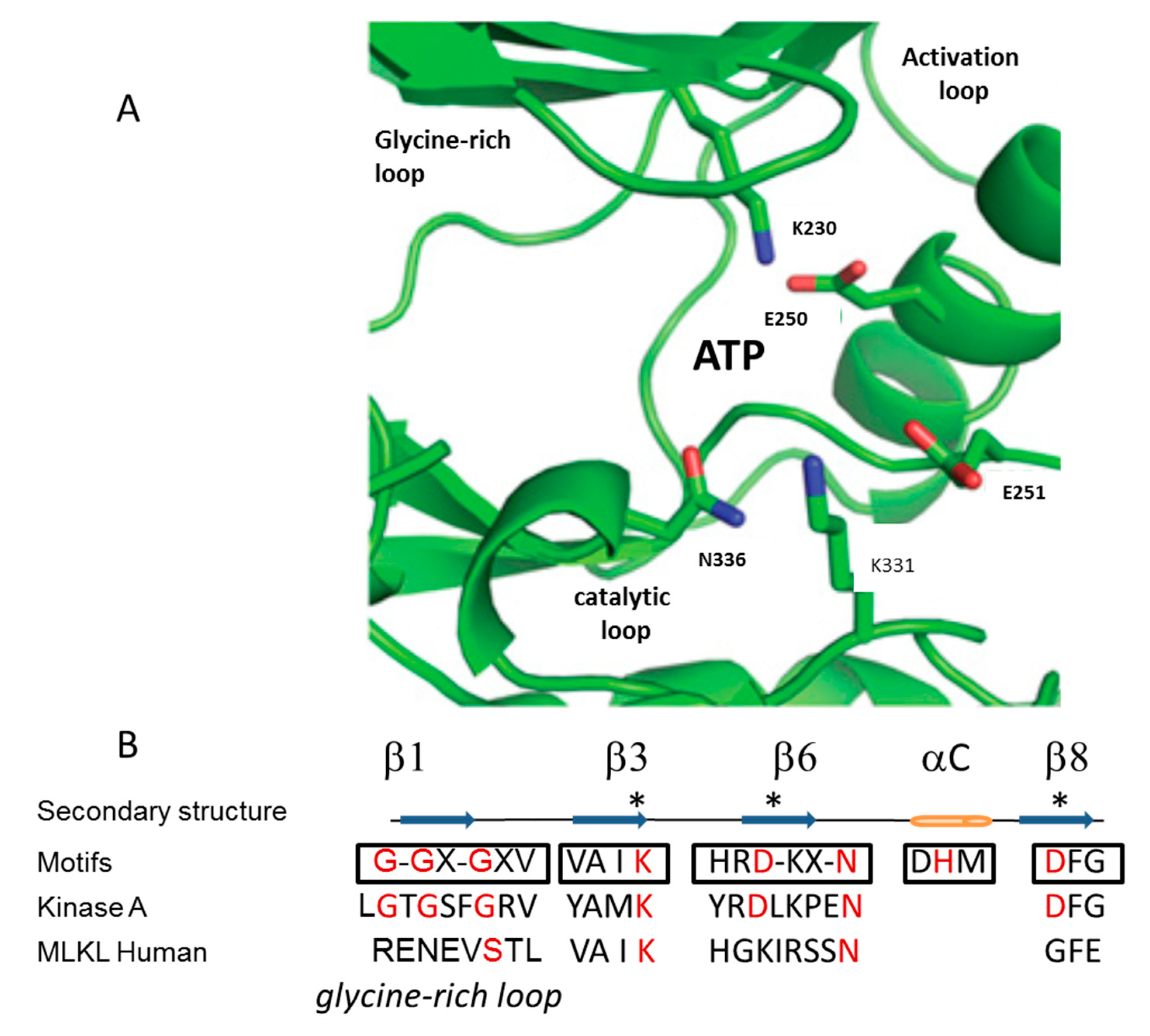
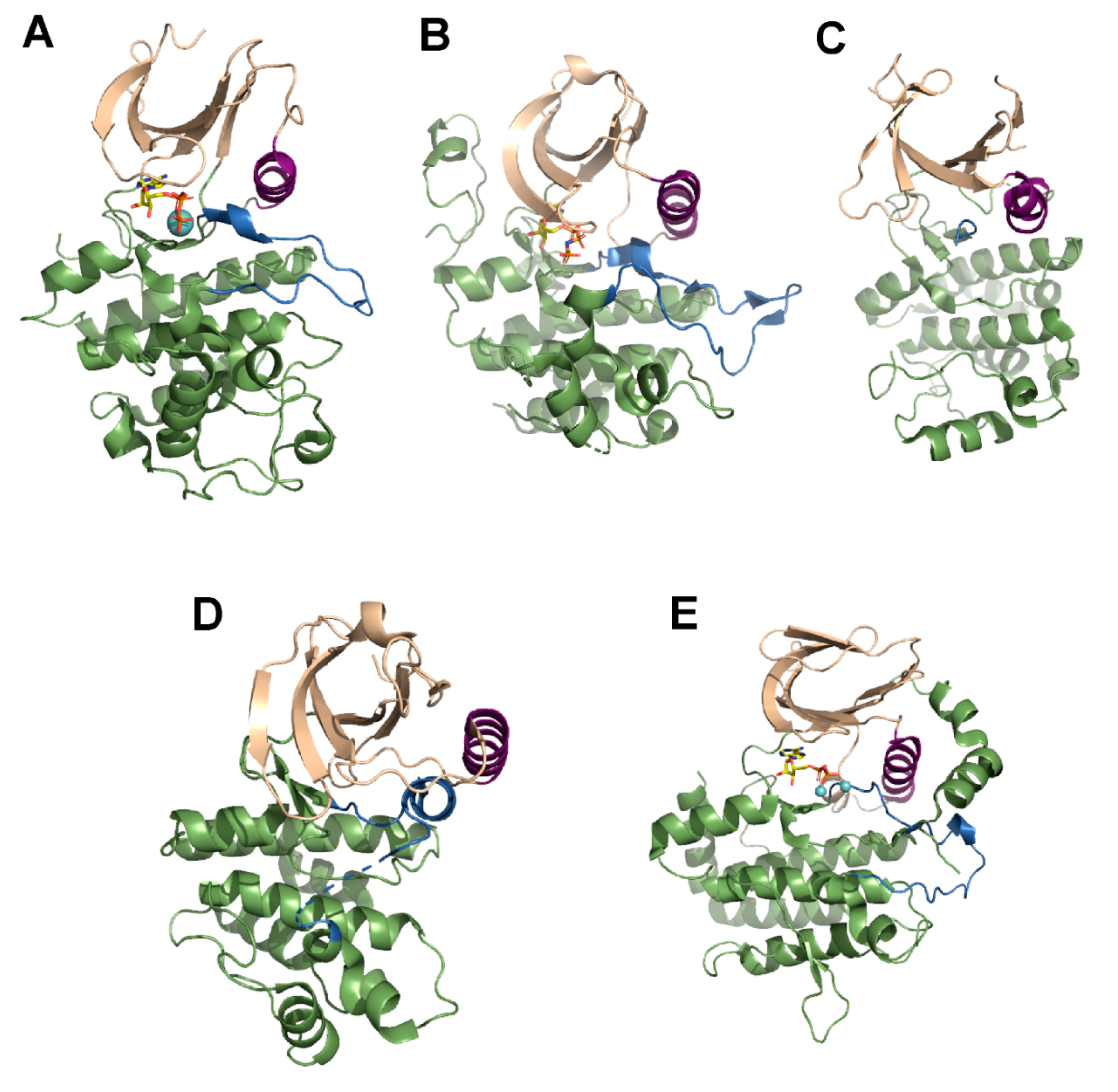
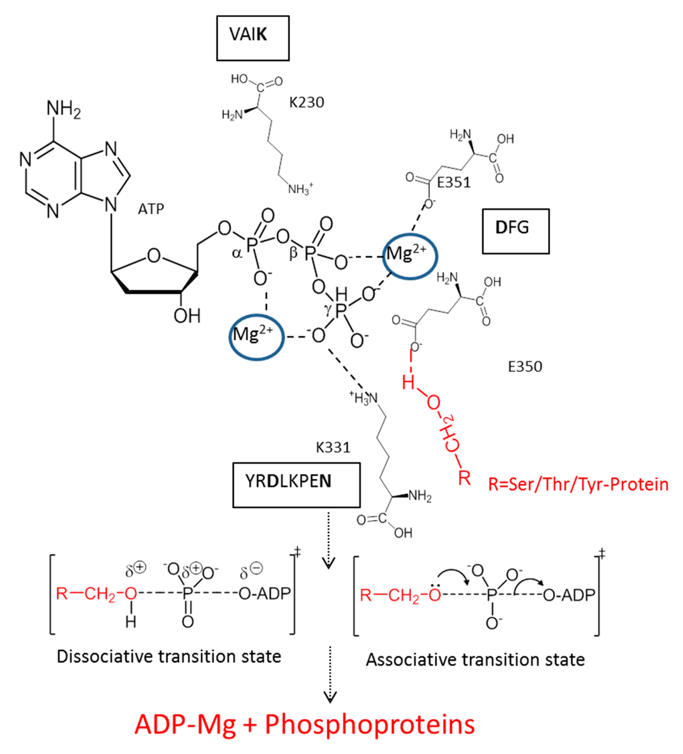
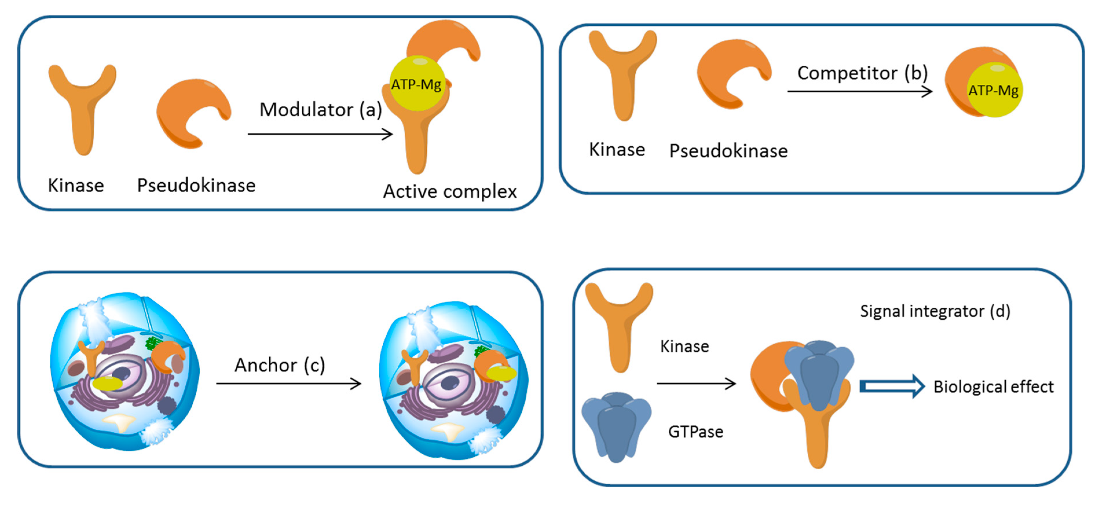
3. Mechanism of Action

The catalytic mechanism of bona fide protein kinases is highly conserved across the tree of life and involves the following steps: (a) nucleotide binding to the active site of the protein, (b) the transfer of the γ-phosphate of the nucleotide to a hydroxyl group of the Ser, Thr, or Tyr residues of the protein and (d) the release of ADP from the active site of the phosphorylated protein (Figure 5).
In contrast, pseudokinases can employ different modes of action: (a) modulating the activity of kinases by serving as dimerisation partners, thereby inhibiting or accelerating kinase activity perhaps through an allosteric transition, (b) competition for substrates of protein kinases, (c) spatial anchor to trap a substrate into particular subcellular location and (d) as a signalling hub that mediates interactions with components of different signalling pathways, or with multiple components of a linear signalling cascade [11] (Figure 6).

4. Pseudokinases and Subcellular Localisation

4.1. Transmembrane Pseudokinases

Nearly 25% of the pseudokinases reported to date contain a functional transmembrane domain. Of these, the homodimeric receptor guanylyl cyclases (RGCs) constitute the largest subgroup, in which the pseudokinase domain occupies the cytoplasmic segment located between the transmembrane domain and a functional guanylyl cyclase domain [60]. The pseudokinase domains associate with each other following ligand binding, which in turn activate the guanylyl cyclase domains probably through an allosteric mechanism [61].

4.2. Nuclear Pseudokinases

The pseudokinase nuclear receptor-binding protein 1 (NRBP1) functions as adaptor and contains a putative Src homology 2 and nuclear receptor-binding domains, as well as sequences that drive protein nuclear import and export [62]. Also in the nucleus resides the Tribbles pseudokinase (TRIB) protein family, whose functions have been described in Section 2. At least in mitosis, the pseudokinase Budding uninhibited by benzimidazoles related 1 (BubR1), functions as a pseudokinase that plays important roles in chromosome segregation but also has been reported to play important functions in DNA repair, ciliogenesis and neuron differentiation [63]. BubR1 together with Budding uninhibited by benzimidazoles 3 (Bub3), Mitotic arrest deficient 2 (Mad2) and Cdc20 assemble to form the Mitotic Checkpoint Complex (MCC), an assembly that regulates the E3 ubiquitin ligase activity of the Anaphase Promoting Complex/Cyclosome (APC/C) [64,65]. BubR1 also functions as a mechano sensor between kinetochores and microtubules [66,67]. Five main regions can be identified in the BubR1 polypeptide chain: (i) two units of the KEN box motif located in the N-terminal region, and one putative destruction box (D-box) motif located in the C-terminal region; (ii) a N-terminal fragment that is organised as a triple-tandem arrangement of the tetratricopeptide repeat (TPR) motif that contributes to the kinetochore localisation of BubR1; (iii) an intermediate, non-conserved region of low structural complexity that is required for the binding to Bub3; (iv) a region harbouring a Cdc20 binding site; and (v) a C-terminal region that contains a serine/threonine kinase domain.
In the human, BubR1 has been reported to function as a pseudokinase [68]. However, at least in the fruit fly the BubR1 kinase domain is catalytically active and phosphorylates the kinetochore protein CENP-E [69]. The phosphorylation of this protein by BubR1 is required for spindle microtubules transition from lateral association to end-on capture. The fact that BubR1 seems to act as a pseudokinase in some species and as a catalytically proficient enzyme in others exemplifies the current challenges in the identification and characterisation of pseudokinases. Other important nuclear pseudokinases are the transformation/transcription domain-associated protein (TRRAP), which functions as a scaffold platform for the assembly of the histone acetyltransferase complex [70,71,72,73,74] and Trib1, which induces nuclear retention of COP1, a highly conserved ubiquitin ligase that regulates diverse cellular processes in plants and metazoans [75].

4.3. Cytoplasmic Pseudokinases

Other pseudokinases are involved in intracellular signalling hubs predominantly in the cytoplasm such as Titin (TTN), a protein firstly characterised as a member of muscle fibre sarcomeres. The N-terminal kinase-like domain of TTN interacts with the zinc finger protein Nbr1, and mutations within the pseudokinase domain that interrupt this interaction are linked to hereditary muscle disease [76]. The TTN pseudokinase domain also recruits the E3 ligases Murf1 and Murf2 [77], and is also proposed to act as a mechanosensor, similar to BubR1 [78]. Another cytoplasmic pseudokinase of particular interest is SCY1-like (SCYLs), which has been implicated in the regulation of intracellular trafficking [79].

5. Pseudokinases and Disease

The use of the bioinformatics resource ProKino (Protein Kinase Ontology Browser) [80] allowed us to map 55 of the human pseudokinases to genes from the open targets project revealing the most enriched disease association to be neurodegenerative disease (p-value = 2e − 9). The most strongly associated gene with this was TBCK. Given the tractability of pseudokinases as drug targets, TBCK appears to be a good drug target that, to the best of our knowledge, remains to be actively pursued. Further, bioinformatics analysis of the 55 human pseudokinases mentioned above indicates that the pathway with the most PKs in the open targets resource is that mediated by Interleukins (p-value = 0.000078).
Although many pseudokinases show a concentration of mutations in the C-terminal subdomain such as Trib1 (Figure 7), other kinases have enrichments at positions other than the kinase domain. One dramatic case is WNK Lysine Deficient Protein Kinase 2 (WNK2) with 38 mutations mapped onto position 1838. The WNK family of Ser/Thr pseudokinases comprises the proteins WNK1 to WNK4 [81] and lacks the conserved β3 lysine assumed to be indispensable for nucleotide binding and stabilisation of the active kinase conformation [82]. Despite this, WNK can regulate diverse intracellular substrates in a phosphorylation-dependent fashion in a process that involves a novel mechanism of catalysis. Namely, the terminal residue in the glycine-rich loop (often a Gly in kinases) is conserved as a Lys residue in WNKs and this residue provides the compensatory charge that is required for productive ATP binding [81]. Neuronal WNK isoforms are associated with hereditary neuropathy and glioma [82]. WNKs are also associated with the control of blood pressure through regulation of SPAK [83,84]. In brief, examples of pseudokinase mutations that lead to impairment of function can be mapped onto the kinase and other adjacent domains and across higher organisms. In the human, such mutations occur in genes encoding for proteins with important roles in metabolic and cell signalling pathways and are associated with metabolic, neurological, autoimmune, and cell proliferation disorders.

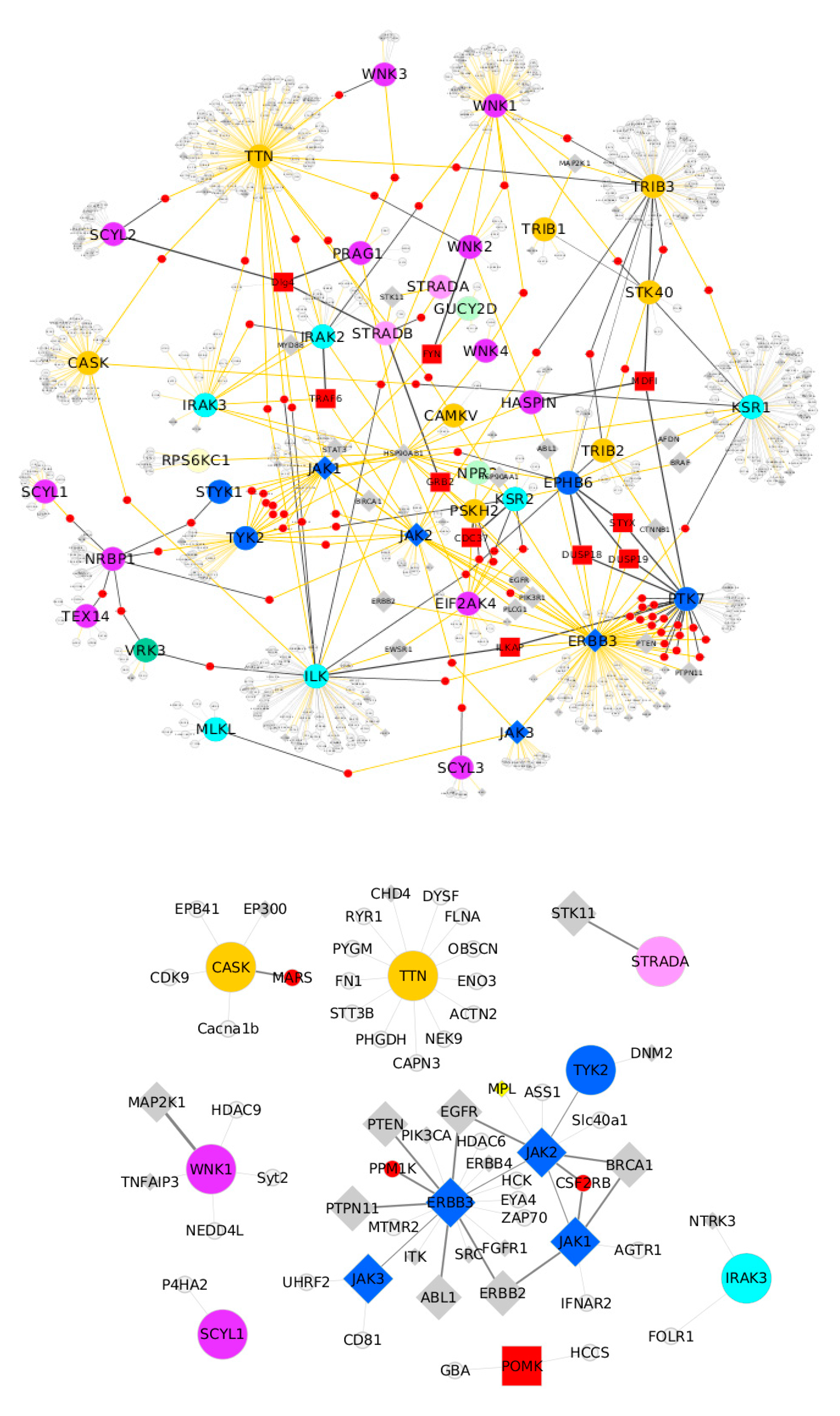
6. Pseudokinases Protein–Protein Interaction Analysis

Interaction analyses indicate that pseudokinases have great potential for drug innovations. Taking the full network from IntAct EBI in the spoke model, it can be seen that pseudokinases form one main connected component. Classifying genes strongly implicated in cancer and other diseases facilitated the identification of a large number of links between pseudokinases and their interacting partners (show as orange links in Figure 8). In the network, colours of nodes represent pseudokinases from the same family except for the red nodes, which indicates proteins that link two or more different pseudokinases. Colouring the nodes in this way reveals that some pseudokinases from the same family share common interaction partners. This interactor sharing is particularly the case for the TK family members where both TYK2 and JAK1 and ERBB3 and PTK7 share a large number of interactors, suggesting co-regulation between these pseudokinases. It is possible to zoom in on the network and to select only those interactions where both genes are disease-associated and either already confirmed as druggable or with strong potential for druggability as designated by the HPA (Human Protein Atlas, 31 July 2019). Gold edges are indicative of potential for druggable interaction therapeutic intervention because one or both of the partners is a known disease gene.
To understand better which regions of the pseudokinases were responsible for physical interactions, we extracted interaction features from Intact restricted to feature lengths ≤20. Results in 9 of the 17 pseudokinases showed interaction features that overlap with the kinase domain in its interactors, indicating that interactions through the kinase domain are often important. Conversely, non-PK regions look equally important because only 4 of the 17 kinases always interacted through the pseudokinase domain.
Pseudokinases with residual or total lack of catalytic activity can still carry out important roles in cell signalling involving pseudo-active sites, allosteric transitions driven by nucleotide binding, and/or direct modulation of binding surfaces topology. Because some promiscuous inhibitors that target the ATP-binding site of bona fide protein kinases can bind the equivalent site in pseudokinases [36], the features of the kinase inhibitors can inform the development of new small size drugs to target pseudokinases. The computational protein–protein interaction analyses described in this contribution together with recent approaches in drug discovery, such as the targeting of specific protein–protein interfaces with small size compounds [85,86,87], further support the view that pseudokinases offer great potential for drug innovations.

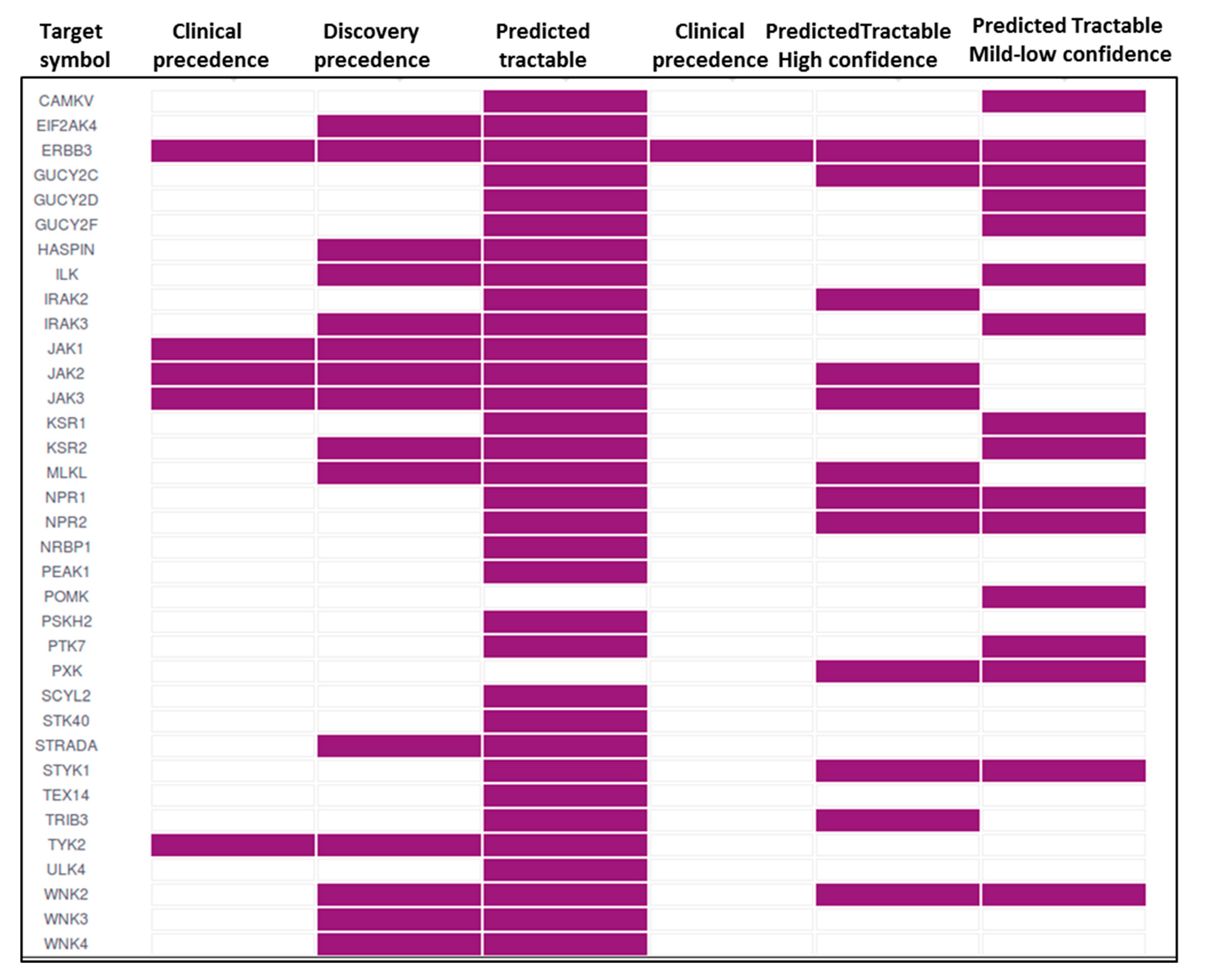
7. Pseudokinases as Drug Targets

Because the vast repertoires of functions exerted by pseudokinases represent a new window to develop innovative strategies for therapeutic intervention to treat human diseases that constitute important yet unmet clinical needs [13,14,15,88,89,90], we mapped pseudokinases to opentargets to visualise their ability to be targeted by small size drugs or antibodies. We carried out an analysis of their druggability in open targets, which showed that a number of pseudokinases can be potential targets of different drug molecule types (e.g., antibody/small molecule) with the Janus kinases representing the most actively pursued targets (Table 1). Our bioinformatics analyses (Figure 9) strongly suggest that pseudokinases have great potential for drug development and revealed that the top diseases targeted are rheumatoid arthritis (five drugs) followed by neoplasm (four drugs). These findings are in good agreement with recent reports by others, which showed that the unique conformations of Tribbles pseudokinases make these proteins good drug targets to treat human acute megakaryocytic leukemia caused by a gain-of-function mutation [91]; that inhibitors of WNK such as WNK463 have potential use in the clinic as antihypertensive drugs [92]; and that a new generation of TYK2 pseudokinase ligands currently in clinical trials may be effective for the treatment of autoimmune diseases [93,94]. An exciting new development in drug discovery is the use of artificial intelligence (AI) to speed up the process and reduce costs by facilitating the rapid identification of drug-like compounds. A recent example of the promise of AI to accelerate the development of new drugs is the identification of potent inhibitors of the Discoidin domain receptor 1 (DDR1) tyrosine kinase using machine learning techniques [95]. It can be anticipated that a similar approach will be used to identify pseudokinase inhibitors, including drugs that interfere with specific protein–protein interactions.

8. Conclusions

Pseudokinases are a subset of the protein kinase superfamily that present inactivating mutations in critical catalytic motifs but signal primarily through noncatalytic mechanisms. Systematic analysis of the human genome has revealed that circa one-tenth of all protein kinases correspond to pseudokinases. These proteins play central roles in the cell and the loss of their regulation can lead to a variety of diseases. Our succinct analysis of the pseudokinase interactome revealed that TYK2, JAK1, ERBB3, and PTK7 pseudokinases share a large number of interactors, suggesting that they are subjected to co-regulation. Several clinically approved kinase inhibitors have been shown to influence the noncatalytic functions of active kinases, providing expectation that similar properties in pseudokinases could be pharmacologically regulated. Recent advances in drug discovery assisted by the use of artificial intelligence approaches may pave the way for the rapid identification of potent pseudokinase inhibitors. Taken these advances together, it can be anticipated that in the coming years pseudokinases are likely to become mainstream drug targets for the treatment of diverse malignancies.

Author Contributions

Conceptualization: V.M.B.-G., J.L. and A.B.; methodology: V.M.B.-G., J.L. and A.B.; software: J.L.; validation: V.M.B.-G., J.L. and A.B.; formal analysis: V.M.B.-G., J.L. and A.B.; investigation: A.T., V.M.B.-G., J.L., A.G.S and A.B.; resources: V.M.B.-G., J.L. and A.B.; writing—original draft preparation: A.T., V.M.B.-G., J.L., A.G.S and A.B.; writing—review and editing: V.M.B.-G., J.L. and A.B.; supervision: V.M.B.-G., J.L. and A.B.; project administration: V.M.B.-G., J.L. and A.B.; funding acquisition: V.M.B.-G. and A.B.

Funding

This research received no external funding.

Conflicts of Interest

The authors declare no conflict of interest.

References

  1. Manning, G.; Whyte, D.B.; Martinez, R.; Hunter, T.; Sudarsanam, S. The protein kinase complement of the human genome. Science 2002, 298, 1912–1934. [Google Scholar] [CrossRef] [PubMed]
  2. Caenepeel, S.; Charydczak, G.; Sudarsanam, S.; Hunter, T.; Manning, G. The mouse kinome: Discovery and comparative genomics of all mouse protein kinases. Proc. Natl. Acad. Sci. USA 2004, 101, 11707–11712. [Google Scholar] [CrossRef] [PubMed] [Green Version]
  3. Plowman, G.D.; Sudarsanam, S.; Bingham, J.; Whyte, D.; Hunter, T. The protein kinases of Caenorhabditis elegans: A model for signal transduction in multicellular organisms. Proc. Natl. Acad. Sci. USA 1999, 96, 13603–13610. [Google Scholar] [CrossRef] [PubMed]
  4. Giamas, G.; Man, Y.L.; Hirner, H.; Bischof, J.; Kramer, K.; Khan, K.; Ahmed, S.S.; Stebbing, J.; Knippschild, U. Kinases as targets in the treatment of solid tumors. Cell. Signal. 2010, 22, 984–1002. [Google Scholar] [CrossRef] [PubMed]
  5. Kwon, A.; Scott, S.; Taujale, R.; Yeung, W.; Kochut, K.J.; Eyers, P.A.; Kannan, N. Tracing the origin and evolution of pseudokinases across the tree of life. Sci. Signal. 2019, 12, eaav3810. [Google Scholar] [CrossRef] [PubMed]
  6. Manning, G.; Reiner, D.S.; Lauwaet, T.; Dacre, M.; Smith, A.; Zhai, Y.; Svard, S.; Gillin, F.D. The minimal kinome of Giardia lamblia illuminates early kinase evolution and unique parasite biology. Genome Biol. 2011, 12, R66. [Google Scholar] [CrossRef] [PubMed]
  7. Ribeiro, A.J.M.; Das, S.; Dawson, N.; Zaru, R.; Orchard, S.; Thornton, J.M.; Orengo, C.; Zeqiraj, E.; Murphy, J.M.; Eyers, P.A. Emerging concepts in pseudoenzyme classification, evolution, and signaling. Sci. Signal. 2019, 12, eaat9797. [Google Scholar] [CrossRef] [PubMed]
  8. Sharir-Ivry, A.; Xia, Y. Using Pseudoenzymes to Probe Evolutionary Design Principles of Enzymes. Evol. Bioinform. 2019, 15, 1176934319855937. [Google Scholar] [CrossRef]
  9. Jeffery, C.J. The demise of catalysis, but new functions arise: Pseudoenzymes as the phoenixes of the protein world. Biochem. Soc. Trans. 2019, 47, 371–379. [Google Scholar] [CrossRef]
  10. Bailey, F.P.; Byrne, D.P.; McSkimming, D.; Kannan, N.; Eyers, P.A. Going for broke: Targeting the human cancer pseudokinome. Biochem. J. 2015, 465, 195–211. [Google Scholar] [CrossRef]
  11. Reiterer, V.; Eyers, P.A.; Farhan, H. Day of the dead: Pseudokinases and pseudophosphatases in physiology and disease. Trends Cell Biol. 2014, 24, 489–505. [Google Scholar] [CrossRef] [PubMed]
  12. Zhang, H.; Photiou, A.; Grothey, A.; Stebbing, J.; Giamas, G. The role of pseudokinases in cancer. Cell. Signal. 2012, 24, 1173–1184. [Google Scholar] [CrossRef] [PubMed]
  13. Boudeau, J.; Miranda-Saavedra, D.; Barton, G.J.; Alessi, D.R. Emerging roles of pseudokinases. Trends Cell Biol. 2006, 16, 443–452. [Google Scholar] [CrossRef] [PubMed]
  14. Eyers, P.; Murphy, J.M. Dawn of the dead: Protein pseudokinases signal new adventures in cell biology. Biochem. Soc. Trans. 2013, 41, 969–974. [Google Scholar] [CrossRef] [PubMed]
  15. Kung, J.E.; Jura, N. Prospects for pharmacological targeting of pseudokinases. Drug Dev. Nat. Rev. Drug Discov. 2019, 18, 501–526. [Google Scholar] [CrossRef] [PubMed]
  16. Fabbro, D.; Cowan-Jacob, S.W.; Moebitz, H. Ten things you should know about protein kinases: IUPHAR Review 14. Br. J. Pharmacol. 2015, 172, 2675–2700. [Google Scholar] [CrossRef] [PubMed] [Green Version]
  17. Salazar, M.; Lorente, M.; Orea-Soufi, A.; Dávila, D.; Erazo, T.; Lizcano, J.; Carracedo, A.; Kiss-Toth, E.; Velasco, G. Oncosuppressive functions of tribbles pseudokinase 3. Biochem. Soc. Trans. 2015, 43, 1122–1126. [Google Scholar] [CrossRef]
  18. Wu, P.; Nielsen, T.E.; Clausen, M.H. FDA-approved small-molecule kinase inhibitors. Trends Pharmacol. Sci. 2015, 36, 422–439. [Google Scholar] [CrossRef] [Green Version]
  19. Knight, Z.A.; Shokat, K.M. Features of Selective Kinase Inhibitors. Chem. Biol. 2005, 12, 621–637. [Google Scholar] [CrossRef] [Green Version]
  20. Davis, M.I.; Hunt, J.P.; Herrgard, S.; Ciceri, P.; Wodicka, L.M.; Pallares, G.; Hocker, M.; Treiber, D.K.; Zarrinkar, P.P. Comprehensive analysis of kinase inhibitor selectivity. Nat. Biotechnol. 2011, 29, 1046–1051. [Google Scholar] [CrossRef]
  21. Zhang, X.; Gureasko, J.; Shen, K.; Cole, P.A.; Kuriyan, J. An allosteric mechanism for activation of the kinase domain of epidermal growth factor receptor. Cell 2006, 125, 1137–1149. [Google Scholar] [CrossRef] [PubMed]
  22. Littlefield, P.; Liu, L.; Mysore, V.; Shan, Y.; Shaw, D.E.; Jura, N. Structural analysis of the EGFR/HER3 heterodimer reveals the molecular basis for activating HER3 mutations. Sci. Signal. 2014, 7, ra114. [Google Scholar] [CrossRef] [PubMed]
  23. Rajakulendran, T.; Sicheri, F. Allosteric protein kinase regulation by pseudokinases: Insights from STRAD. Sci. Signal. 2010, 3, pe8. [Google Scholar] [CrossRef] [PubMed]
  24. Vulpetti, A.; Bosotti, R. Sequence and structural analysis of kinase ATP pocket residues. Farmaco 2004, 59, 759–765. [Google Scholar] [CrossRef] [PubMed]
  25. Huse, M.; Kuriyan, J. The conformational plasticity of protein kinases. Cell 2002, 109, 275–282. [Google Scholar] [CrossRef]
  26. Taylor, S.S.; Kornev, A.P. Protein kinases: Evolution of dynamic regulatory proteins. Trends Biochem. Sci. 2011, 36, 65–77. [Google Scholar] [CrossRef] [PubMed]
  27. Bose, R.; Holbert, M.A.; Pickin, K.A.; Cole, P.A. Protein tyrosine kinase-substrate interactions. Curr. Opin. Struct. Biol. 2006, 16, 668–675. [Google Scholar] [CrossRef]
  28. Murphy, J.M.; Zhang, Q.; Young, S.N.; Reese, M.L.; Bailey, F.P.; Eyers, P.A.; Ungureanu, D.; Hammaren, H.; Silvennoinen, O.; Varghese, L.N.; et al. A robust methodology to subclassify pseudokinases based on their nucleotide-binding properties. Biochem. J. 2013, 457, 323–334. [Google Scholar] [CrossRef]
  29. Jacobsen, A.V.; Murphy, J.M. The secret life of kinases: Insights into non-catalytic signalling functions from pseudokinases. Biochem. Soc. Trans. 2017, 15, 665–681. [Google Scholar] [CrossRef]
  30. Scheeff, E.D.; Eswaran, J.; Bunkoczi, G.; Knapp, S.; Manning, G. Structure of the pseudokinase VRK3 reveals a degraded catalytic site, a highly conserved kinase fold, and a putative regulatory binding site. Structure 2009, 17, 128–138. [Google Scholar] [CrossRef]
  31. Bailey, F.P.; Byrne, D.P.; Oruganty, K.; Eyers, C.E.; Novotny, C.J.; Shokat, K.M.; Kannan, N.; Eyers, P.A. The Tribbles 2 (TRB2) pseudokinase binds to ATP and autophosphorylates in a metal independent manner. Biochem. J. 2015, 467, 47–62. [Google Scholar] [CrossRef] [PubMed]
  32. Foulkes, D.M.; Byrne, D.P.; Bailey, F.P.; Eyers, P.A. Tribbles pseudokinases: Novel targets for chemical biology and drug discovery? Biochem. Soc. Trans. 2015, 43, 1095–1103. [Google Scholar] [CrossRef] [PubMed]
  33. Eyers, P.A.; Keeshan, K.; Kannan, N. Tribbles in the 21st Century: The evolving roles of tribbles pseudokinases in biology and disease. Trends Cell Biol. 2017, 27, 284–298. [Google Scholar] [CrossRef] [PubMed]
  34. Zeqiraj, E.; Filippi, B.M.; Goldie, S.; Navratilova, I.; Boudeau, J.; Deak, M.; Alessi, D.R.; van Aalten, D.M.F. ATP and MO25α regulate the conformational state of the STRADα pseudokinase and activation of the LKB1 tumour suppressor. PLoS Biol. 2009, 7, 1000126. [Google Scholar] [CrossRef] [PubMed]
  35. Cui, J.; Zhu, Q.; Zhang, H.; Cianfrocco, M.A.; Leschziner, A.E.; Dixon, J.E.; Xiao, J. Structure of Fam20A reveals a pseudokinase featuring unique disulfide pattern and inverted ATP-binding. eLife 2017, 6, 1–16. [Google Scholar] [CrossRef] [PubMed]
  36. Murphy, J.M.; Lucet, I.S.; Hildebrand, J.M.; Tanzer, M.C.; Young, S.N.; Sharma, P.; Lessene, G.; Warren, S.A.; Babon, J.J.; Silke, J.; et al. Insights into the evolution of divergent nucleotide-binding mechanisms among pseudokinases revealed by crystal structures of human and mouse MLKL. Biochem. J. 2014, 457, 369–377. [Google Scholar] [CrossRef]
  37. Petrie, E.J.; Sandow, J.J.; Jacobsen, A.V.; Smith, B.J.; Griffin, M.D.W.; Lucet, I.S.; Dai, W.; Young, S.N.; Tanzer, M.C.; Wardak, A.; et al. Conformational switching of the pseudokinase domain promotes human MLKL tetramerization and cell death by necroptosis. Nat. Commun. 2018, 9, 2422. [Google Scholar] [CrossRef]
  38. Jacobsen, A.V.; Lowes, K.N.; Tanzer, M.Z.; Lucet, I.S.; Hildebrand, J.M.; Petrie, E.J.; van Delft, M.F.; Liu, Z.; Conos, S.A.; Zhang, J.-G.; et al. HSP90 activity is required for MLKL oligomerisation and membrane translocation and the induction of necroptotic cell death. Cell Death Dis. 2016, 7, e2051. [Google Scholar] [CrossRef]
  39. Ha, B.H.; Boggon, T.J. The crystal structure of pseudokinase PEAK1 (Sugen Kinase 269) reveals an unusual catalytic cleft and a novel mode of kinase fold dimerization. J. Biol. Chem. 2017, 293, 1642–1650. [Google Scholar] [CrossRef]
  40. Labesse, G.; Gelin, M.; Bessin, Y.; Lebrun, M.; Papoin, J.; Cerdan, R.; Arold, S.T.; Dubremetz, J.F. ROP2 from Toxoplasma gondii: A virulence factor with a protein- kinase foldand no enzymatic activity. Structure 2009, 17, 139–146. [Google Scholar] [CrossRef]
  41. Yarden, Y.; Sliwkowski, M.X. Untangling the ErbB signaling network. Nat. Rev. Mol. Cell. Biol. 2001, 2, 127–137. [Google Scholar] [CrossRef] [PubMed]
  42. Zhang, N.; Chang, Y.; Rios, A.; An, Z. HER3/ErbB3, an emerging cancer therapeutic target. Acta Biochim. Biophys. Sin. 2016, 48, 39–48. [Google Scholar] [CrossRef] [PubMed]
  43. Toms, A.V.; Deshpande, A.; McNally, R.; Jeong, Y.; Rogers, J.M.; Kim, C.U.; Gruner, S.M.; Ficarro, S.B.; Marto, J.A.; Sattler, M.; et al. Structure of a pseudokinase domain switch that controls oncogenic activation of Jak kinases. Nat. Struct. Mol. Biol. 2014, 20, 1221–1223. [Google Scholar] [CrossRef] [PubMed]
  44. Ungureanu, D.; Wu, J.; Pekkala, T.; Niranjan, Y.; Young, C.; Jensen, O.N.; Xu, C.F.; Neubert, T.A.; Skoda, R.C.; Hubbard, S.R.; et al. The pseudokinase domain of JAK2 is a dual-specificity protein kinase that negatively regulates cytokine signaling. Nat. Struct. Mol. Biol. 2011, 14, 971–976. [Google Scholar] [CrossRef] [PubMed]
  45. Laurence, A.; Pesu, M.; Silvennoinen, O.; O’shea, J. JAK kinases in health and disease: An update. Open Rheumatol. J. 2012, 6, 232–244. [Google Scholar] [CrossRef]
  46. Haan, C.; Behrmann, I.; Haan, S. Perspectives for the use of structural information and chemical genetics to develop inhibitors of Janus kinases. J. Cell. Mol. Med. 2010, 14, 504–527. [Google Scholar] [CrossRef] [PubMed]
  47. Hammarén, H.M.; Ungureanu, D.; Grisouard, J.; Skoda, R.C.; Hubbard, S.R.; Silvennoinena, O. ATP binding to the pseudokinase domain of JAK2 is critical for pathogenic activation. Proc. Natl. Acad. Sci. USA 2015, 112, 4642–4647. [Google Scholar] [CrossRef] [Green Version]
  48. Notarangelo, L.D.; Mella, P.; Jones, A.; de Saint Basile, G.; Savoldi, G.; Cranston, T.; Vihinen, M.; Schumacher, R.F. Mutations in severe combined immune deficiency (SCID) due to JAK3 deficiency. Hum. Mutat. 2001, 18, 255–263. [Google Scholar] [CrossRef]
  49. Amand, M.; Erpicum, C.; Bajou, K.; Cerignoli, F.; Blacher, S.; Martin, M.; Dequiedt, F.; Drion, P.; Singh, P.; Zurashvili, T.; et al. DUSP3/VHR is a pro-angiogenic atypical dual-specificity phosphatase. Mol. Cancer 2014, 13, 108–126. [Google Scholar] [CrossRef]
  50. Yoon, S.; Seger, R. The extracellular signal-regulated kinase: Multiple substrates regulate diverse cellular functions. Growth Factors 2006, 24, 21–44. [Google Scholar] [CrossRef]
  51. Rauch, J.; Volinsky, N.; Romano, D.; Kolch, W. The secret life of kinases: Functions beyond catalysis. Cell Commun. Signal. 2011, 9, 23. [Google Scholar] [CrossRef] [PubMed]
  52. Kolch, W. Coordinating ERK/MAPK signalling through scaffolds and inhibitors. Nat. Rev. Mol. Cell. Biol. 2005, 6, 827–837. [Google Scholar] [CrossRef] [PubMed]
  53. Costanzo-Garvey, D.L.; Pfluger, P.T.; Dougherty, M.K.; Stock, J.L.; Boehm, M.; Chaika, O.; Fernandez, M.R.; Fisher, K.; Kortum, R.L.; Hong, E.G.; et al. KSR2 is an essential regulator of AMP kinase, energy expenditure, and insulin sensitivity. Cell Metab. 2009, 10, 366–378. [Google Scholar] [CrossRef] [PubMed]
  54. Revelli, J.P.; Smith, D.; Allen, J.; Jeter-Jones, S.; Shadoan, M.K.; Desai, U.; Schneider, M.; van Sligtenhorst, I.; Kirkpatrick, L.; Platt, K.A.; et al. Profound obesity secondary to hyperphagia in mice lacking kinase suppressor of ras 2. Obesity 2011, 19, 1010–1018. [Google Scholar] [CrossRef]
  55. Pearce, L.R.; Atanassova, N.; Banton, M.C.; Bottomley, B.; van der Klaauw, A.A.; Revelli, J.P.; Hendricks, A.; Keogh, J.M.; Henning, E.; Doree, D.; et al. KSR2 mutations are associated with obesity, insulin resistance, and impaired cellular fuel oxidation. Cell 2013, 7, 765–777. [Google Scholar] [CrossRef] [PubMed]
  56. Rhyasen, G.W.; Starczynowski, D.T. IRAK signalling in cancer. Br. J. Cancer 2015, 112, 232–237. [Google Scholar] [CrossRef] [PubMed]
  57. Lin, S.C.; Lo, Y.C.; Wu, H. Helical assembly in the Myd88-IRAK4-IRAK2 complex in TLR/IL-1R signalling. Nature 2010, 465, 885–890. [Google Scholar] [CrossRef] [PubMed]
  58. Ge, Z.W.; Wang, B.C.; Hu, J.L.; Sun, J.J.; Wang, S.; Chen, X.J.; Meng, S.P.; Liu, L.; Cheng, Z.Y. IRAK3 gene silencing prevents cardiac rupture and ventricular remodeling through negative regulation of the NF-κB signaling pathway in a mouse model of acute myocardial infarction. J. Cell Physiol. 2019, 234, 11722–11733. [Google Scholar] [CrossRef] [PubMed]
  59. Kobayashi, K.; Hernandez, L.D.; Galan, J.E.; Janeway, C.A., Jr.; Medzhitov, R.; Flavell, R.A. IRAK-M is a negative regulator of toll-like receptor signaling. Cell 2002, 110, 191–202. [Google Scholar] [CrossRef]
  60. Kuhn, M. Molecular physiology of membrane guanylyl cyclase receptors. Physiol. Rev. 2016, 96, 751–804. [Google Scholar] [CrossRef]
  61. Biswas, K.H.; Shenoy, A.R.; Dutta, A.; Visweswariah, S.S. The evolution of guanylyl cyclases as multidomain proteins: Conserved features of kinase-cyclase domain fusions. J. Mol. Evol. 2009, 68, 587–602. [Google Scholar] [CrossRef] [PubMed]
  62. Kerr, J.S.; Wilson, C.H. Nuclear receptor-binding protein 1: A novel tumour suppressor and pseudokinase. Biochem. Soc. Trans. 2013, 41, 1055–1060. [Google Scholar] [CrossRef] [PubMed]
  63. Bolanos-Garcia, V.M.; Blundell, T.L. BUB1 and BUBR1: Multifaceted kinases of the cell cycle. Trends Biochem. Sci. 2001, 36, 141–150. [Google Scholar] [CrossRef] [PubMed]
  64. Hein, J.B.; Nilsson, J. Stable MCC binding to the APC/C is required for a functional spindle assembly checkpoint. EMBO Rep. 2014, 15, 264–272. [Google Scholar] [CrossRef] [PubMed]
  65. Alfieri, C.; Chang, L.; Zhang, Z.; Yang, J.; Maslen, S.; Skehel, M.; Barford, D. Molecular basis of APC/C regulation by the spindle assembly checkpoint. Nature 2016, 536, 431–436. [Google Scholar] [CrossRef] [PubMed] [Green Version]
  66. Huang, H.; Hittle, J.; Zappacosta, F.; Annan, R.S.; Hershko, A.; Yen, T.J. Phosphorylation sites in BubR1 that regulate kinetochore attachment, tension, and mitotic exit. J. Cell Biol. 2008, 183, 667–680. [Google Scholar] [CrossRef] [Green Version]
  67. Chan, G.K.T.; Jablonski, S.A.; Sudakin, V.; Hittle, J.C.; Yen, T.J. Human BUBR1 is a mitotic checkpoint kinase that monitors CENP-E functions at kinetochores and binds the cyclosome/APC. J. Cell Biol. 1999, 146, 941–954. [Google Scholar] [CrossRef]
  68. Suijkerbuijk, S.J.; van Dam, T.J.; Karagöz, G.E.; von Castelmur, E.; Hubner, N.C.; Duarte, A.M.; Vleugel, M.; Perrakis, A.; Rüdiger, S.G.; Snel, B.; et al. The vertebrate mitotic checkpoint protein BUBR1 is an unusual pseudokinase. Dev. Cell. 2012, 22, 1321–1329. [Google Scholar] [CrossRef]
  69. Huang, Y.; Lin, L.; Liu, X.; Ye, S.; Yao, P.Y.; Wang, W.; Yang, F.; Gao, X.; Li, J.; Zhang, Y.; et al. BubR1 phosphorylates CENP-E as a switch enabling the transition from lateral association to end-on capture of spindle microtubules. Cell Res. 2019, 29, 562–578. [Google Scholar] [CrossRef]
  70. Li, H.; Cuenin, C.; Murr, R.; Wang, Z.-Q.; Herceg, Z. HAT cofactor Trrap regulates the mitotic checkpoint by modulation of Mad1 and Mad2 expression. EMBO J. 2004, 23, 4824–4834. [Google Scholar] [CrossRef] [Green Version]
  71. McMahon, S.B.; Wood, M.A.; Cole, M.D. The essential cofactor TRRAP recruits the histone acetyltransferase hGCN5 to c-Myc. Mol. Cell. Biol. 2000, 20, 556–562. [Google Scholar] [CrossRef] [PubMed]
  72. Murr, R.; Loizou, J.I.; Yang, Y.-G.; Cuenin, C.; Li, H.; Wang, Z.-Q.; Herceq, Z. Histone acetylation by Trrap-Tip60 modulates loading of repair proteins and repair of DNA double-strand breaks. Nat. Cell Biol. 2006, 8, 91–99. [Google Scholar] [CrossRef] [PubMed]
  73. Ichim, G.; Mola, M.; Finkbeiner, M.; Cros, M.-P.; Herceg, Z.; Hernandez-Vargas, H. The histone acetyltransferase component TRRAP is targeted for destruction during the cell cycle. Oncogene 2014, 33, 181–192. [Google Scholar] [CrossRef] [PubMed]
  74. Tapias, A.; Zhou, Z.-W.; Shi, Y.; Chong, Z.; Wang, P.; Groth, M.; Platzer, M.; Huttner, W.; Herceg, Z.; Yang, Y.G.; et al. Trrap-dependent histone acetylation specifically regulates cell-cycle gene transcription to control neural progenitor fate decisions. Cell Stem Cell 2014, 14, 632–643. [Google Scholar] [CrossRef] [PubMed]
  75. Kung, J.E.; Jura, N. The pseudokinase TRIB1 toggles an intramolecular switch to regulate COP1 nuclear export. EMBO J. 2019, 38, e99708. [Google Scholar] [CrossRef] [PubMed]
  76. Lange, S.; Xiang, F.; Yakovenko, A.; Vihola, A.; Hackman, P.; Rostkova, E. The kinase domain of Titin controls muscle gene expression and protein turnover. Science 2005, 308, 1599–1603. [Google Scholar] [CrossRef]
  77. Bogomolovas, J.; Gasch, A.; Simkovic, F.; Rigden, D.J.; Labeit, S.; Mayans, O. Titin kinase is an inactive pseudokinase scaffold that supports MuRF1 recruitment to the sarcomeric M-line. Open Biol. 2014, 4, 140041. [Google Scholar] [CrossRef] [Green Version]
  78. Puchner, E.M.; Alexandrovich, A.; Kho, A.L.; Hensen, U.; Schafer, L.V.; Brandmeier, B. Mechanoenzymatics of titin kinase. Proc. Natl. Acad. Sci. USA 2008, 105, 13385–13390. [Google Scholar] [CrossRef] [Green Version]
  79. Pelletier, S. SCYL pseudokinases in neuronal function and survival. Neural Regen. Res. 2016, 11, 42–44. [Google Scholar] [CrossRef]
  80. A Partnership to Transform Drug Discovery through the Systematic Identification and Prioritisation of Targets. Available online: www.opentargets.org/ (accessed on 22 August 2019).
  81. Tang, B.L. (WNK)ing at death: With-no-lysine (Wnk) kinases in neuropathies and neuronal survival. Brain Res. Bull. 2016, 125, 92–98. [Google Scholar] [CrossRef]
  82. Xu, B.-E.; English, J.M.; Wilsbacher, J.L.; Stippec, S.; Goldsmith, E.J.; Cobb, M.H. WNK1, a novel mammalian serine/threonine protein kinase lacking the catalytic lysine in subdomain II. J. Biol. Chem. 2000, 275, 16795–16801. [Google Scholar] [CrossRef] [PubMed]
  83. Wu, A.; Wolley, M.; Stowasser, M. The interplay of renal potassium and sodium handling in blood pressure regulation: Critical role of the WNK-SPAK-NCC pathway. J. Hum. Hypertens. 2019, 33, 508–523. [Google Scholar] [CrossRef] [PubMed]
  84. Shekarabi, M.; Zhang, J.; Khanna, A.R.; Ellison, D.H.; Delpire, E.; Kahle, K.T. WNK Kinase Signaling in Ion Homeostasis and Human Disease. Cell Metab. 2017, 25, 285–299. [Google Scholar] [CrossRef] [PubMed] [Green Version]
  85. Pommier, Y.; Kiselev, E.; Marchand, C. Interfacial inhibitors. Bioorg. Med. Chem. Lett. 2015, 25, 3961–3965. [Google Scholar] [CrossRef] [PubMed]
  86. Jin, L.; Wang, W.; Fang, G. Targeting protein-protein interaction by small molecules. Annu. Rev. Pharmacol. Toxicol. 2014, 54, 435–456. [Google Scholar] [CrossRef] [PubMed]
  87. Jubb, H.; Blundell, T.L.; Ascher, D.B. Flexibility and small pockets at protein-protein interfaces: New insights into druggability. Prog. Biophys. Mol. Biol. 2015, 119, 2–9. [Google Scholar] [CrossRef] [PubMed]
  88. Karvonen, H.; Perttilä, R.; Niininen, W.; Barker, H.; Ungureanu, D. Targeting Wnt signaling pseudokinases in hematological cancers. Eur. J. Haematol. 2018, 101, 457–465. [Google Scholar] [CrossRef] [PubMed]
  89. Byrne, D.P.; Foulkes, D.M.; Eyers, P.A. Pseudokinases: Update on their functions and evaluation as new drug targets. Future Med. Chem. 2017, 9, 245–265. [Google Scholar] [CrossRef]
  90. Cowan-Jacob, S.W.; Jahnke, W.; Knapp, S. Novel approaches for targeting kinases: Allosteric inhibition, allosteric activation and pseudokinases. Future Med. Chem. 2014, 6, 541–561. [Google Scholar] [CrossRef]
  91. Yokoyama, T.; Toki, T.; Aoki, Y.; Kanezaki, R.; Park, M.J.; Kanno, Y.; Takahara, T.; Yamazaki, Y.; Ito, E.; Hayashi, Y.; et al. Identification of TRIB1 R107L gain-of-function mutation in human acute megakaryocytic leukemia. Blood 2012, 119, 2608–2611. [Google Scholar] [CrossRef]
  92. Yamada, K.; Park, H.M.; Rigel, D.F.; DiPetrillo, K.; Whalen, E.J.; Anisowicz, A.; Beil, M.; Berstler, J.; Brocklehurst, C.E.; Burdick, D.A.; et al. Small-molecule WNK inhibition regulates cardiovascular and renal function. Nat. Chem. Biol. 2016, 12, 896–898. [Google Scholar] [CrossRef] [PubMed]
  93. Burke, J.R.; Cheng, L.; Gillooly, K.M.; Strnad, J.; Zupa-Fernandez, A.; Catlett, I.M.; Zhang, Y.; Heimrich, E.M.; McIntyre, K.W.; Cunningham, M.D.; et al. Autoimmune pathways in mice and humans are blocked by pharmacological stabilization of the TYK2 pseudokinase domain. Sci. Transl. Med. 2019, 11, eaaw1736. [Google Scholar] [CrossRef] [PubMed]
  94. Moslin, R.; Zhang, Y.; Wrobleski, S.T.; Lin, S.; Mertzman, M.; Spergel, S.; Tokarski, J.S.; Strnad, J.; Gillooly, K.; McIntyre, K.W.; et al. Identification of N-Methyl Nicotinamide and N-Methyl Pyridazine-3-Carboxamide Pseudokinase Domain Ligands as Highly Selective Allosteric Inhibitors of Tyrosine Kinase 2 (TYK2). J. Med. Chem. 2019. [Google Scholar] [CrossRef] [PubMed]
  95. Zhavoronkov, A.; Ivanenkov, Y.A.; Aliper, A.; Veselov, M.S.; Aladinskiy, V.A.; Aladinskaya, A.V.; Terentiev, V.A.; Polykovskiy, D.A.; Kuznetsov, M.D.; Asadulaev, A.; et al. Deep learning enables rapid identification of potent DDR1 kinase inhibitors. Nat. Biotechnol. 2019, 37, 1038–1040. [Google Scholar] [CrossRef] [PubMed]
Figure 1. General structure of a kinase protein domain with key structural motifs.
Catalysts 09 00778 g001
Figure 2. (A) the pseudoactive site of human complex ATP-MLKL pseudokinase structure (PDB:5knj), (B) secondary structure and amino acid sequence alignment of the catalytic motif of protein kinase A and the MLKL pseudokinase domain. (*) Residues known to mediate catalysis.
Catalysts 09 00778 g002
Figure 3. Comparison of the 3D structure of (A) Cdk2 kinase (PDB ID 1QMZ); and representatives pseudokinases (B) Stradα, (PDB ID 2WTK); (C) Trib1 (PDB ID 5CEM); and (D) MLKL (PDB ID 4BTF). In the human, BubR1 seems to function as a pseudokinase. However, the recently reported crystal structure of BubR1 from the fruit fly (Panel E, PDB ID 6JKM) revealed a bona fide protein kinase domain. In each structure, highlighted in colour is the N-terminal lobe (salmon); the alpha-helix (purple) and the activation loop (marine). In the Cdk2 (A) and BubR1 (E) structures, magnesium ions are shown in sphere representation.
Catalysts 09 00778 g003
Figure 4. Domain organisation of JAKs pseudokinases.
Catalysts 09 00778 g004
Figure 5. General catalytic mechanism of catalytically active protein kinases. The relative position of the conserved motifs VAIK, YRDLKPEN and DFG is indicated.
Catalysts 09 00778 g005
Figure 6. Modes of action of pseudokinases; (a) dimerisation (b) competitor (c) anchor and (d) signal integrator.
Catalysts 09 00778 g006
Figure 7. Trib1 is an example of a pseudokinase that contains subregions recurrently mutated. Analysis based on the cosmic 3D Mutation bioinformatics resource.
Catalysts 09 00778 g007
Figure 8. Above, the human pseudokinase interactome. The colours represent family classes. Protein kinase linking nodes are shown in red whereas diamonds indicate HPA genes that are strongly implicated in cancer. Gold edges indicate a potentially therapeutic interaction to target where one of the genes is a cancer or disease gene. Below, a highly druggable sub-network where all nodes are disease-associated genes and have an FDA (Food and Drug Administration) drug or a high potential for druggability as defined by the Human Protein Atlas (HPA) datasets.
Catalysts 09 00778 g008
Figure 9. Pseudokinases mapped to opentargets tractability for both small size drug compounds and antibodies.
Catalysts 09 00778 g009
Table 1. Existing status for pseudokinases in clinical trials as reported in the open targets database.
TargetMax PhaseMolecula TypeDrugs
JAK2Phase IIISmall molecule5
ERBB3Phase IIAntibody4
JAK3Phase IISmall molecule4
JAK1Phase IISmall molecule3
JAK2Phase ISmall molecule3
JAK1Phase IIISmall molecule3
JAK2Phase IISmall molecule3
JAK2Phase IVSmall molecule2
ERBB3Phase IVSmall molecule2
GUCY2CPhase IVprotein2
ERBB3Phase IAntibody2
EPHB6Phase IVSmall molecule1
JAK1Phase IVSmall molecule1
ERBB3Phase IIISmall molecule1
ERBB3Phase IIIAntibody1
ERBB3Phase IISmall molecule1
NPR1Phase IVprotein1

