Article

Simple Preparation of Ceramic-Like Materials Based on 1D-Agx(x=0, 5, 10, 20, 40 mM)/TiO2 Nanostructures and Their Photocatalysis Performance

1
Department of Physics, College of Science, King Faisal University, P.O. Box 400, Al-Ahsa 31982, Saudi Arabia
2
Physics Department, Faculty of Science, Assiut University, Assiut 71516, Egypt
*
Authors to whom correspondence should be addressed.
Crystals 2020, 10(11), 1024; https://doi.org/10.3390/cryst10111024
Submission received: 5 October 2020 / Revised: 26 October 2020 / Accepted: 6 November 2020 / Published: 10 November 2020

Abstract

:
Vertical Agx/TiO2 nanorods were successfully grown by a simple oxidation method of a Ti-Ag coating. The samples were grown in the phase of ceramic-like materials, which can be reusable for many cycles for photocatalysis applications. These ceramic-like Agx/TiO2 nanostructures were prepared by the spin-coating of silver nitrate onto Ti sheets. The presence of silver on the surface of the Ti sheet during the oxidation process helped in the growth of one-dimensional nanostructures. The physical properties of the fabricated ceramic-like nanostructures were studied by varying the concentration of silver on the Ti-sheet before the oxidation. One-dimensional nanostructures with an average size varying within the range of 200–500 nm were grown. The presence of silver made the nanostructure vertically directed. The nanorods were dense at the low and medium concentrations of 5, 10, and 20 mM of silver in contrary to high silver concentrations, where the nanorods were very sparse at 40 mM. Structural analysis showed the anatase and rutile structure of pure TiO2 with distinguishing diffraction lines A(101) and R(110); however, Agx/TiO2 showed a dominant orientation of A(101), confirming the 1D growth. Raman spectra confirmed the presence of TiO2 via the observation of its corresponding phonon modes. The photocatalysis properties of the fabricated ceramic-like nanostructures were performed on methylene blue (MB) as a known target dye. The low- and medium-silver-concentration samples showed a high photocatalytic activity compared to the pure and high-silver-concentration samples.

Graphical Abstract

1. Introduction

Since the last decade, metal-oxide semiconductors have attracted the interests of researchers to study them extensively due to their electronic and optical properties such as their tunable bandgap, controllable morphology, large surface area, high stability, and reusability. These properties make them excellent candidates for different applications related to photocatalysis [1,2,3]. Photocatalytic activity is a crucial mechanism for pollutant materials degradation. That is why this process has received great attention as an environmentally friendly, low cost, and sustainable treatment technology. The photocatalytic activity of metal oxides can originate from two sources: (i) Generation of OH radicals by the oxidation of OH anions, and (ii) generation of radicals by reduction of O2 radicals by the reduction of O2. The radicals and anions can react with pollutants to degrade or otherwise transform them into less toxic byproducts [4].
Among the metal oxides used in pollutant degradation, TiO2 is still the most attractive one because it is environmentally safe, biocompatible, present in large quantities, highly chemical stable, and is a low-cost metal oxide photocatalyst with the ability to efficiently degrade a spectrum of contaminants [5]. Fujishima and Honda were the first to use the photocatalytic activity via titanium dioxide (TiO2) [6]. Since the discovery of its photocatalytic activity, TiO2 materials have gained a rising interest in materials research due to their various applications such as pollutant degradation [7,8,9]. It has a wide bandgap of 3.2 eV, low electron transfer rate, and fast recombination effect [10,11,12]. TiO2 nanostructures have distinctive physical and chemical properties that vary with the size, morphology, phase structure, crystallinity, surface area, and the synthesis process of the nanoparticles where all have a great impact on the photocatalytic activity [13,14]. Different TiO2 nanoparticle (NP) shape configurations have been reported in recent years such as nanotubes [15], nanobelts [16], nanowires [17,18], and nanosheets [19]. However, one-dimensional nanomaterials have attracted much attention due to their unique properties. For example, TiO2 nanorods have been studied extensively due to their unique properties related to the easy charge transfer collection along the axial direction [20].
Metal-doped TiO2 nanostructures have attracted enormous attention in improving the photocatalytic performance of environmental and energy applications [21,22]. The use of noble metals such as gold, silver, and Co with TiO2 nanostructures has been studied extensively and has shown an effect on the bandgap and, consequently, the enhancement in photocatalytic reactions [23,24,25]. Among the noble metals, silver nanoparticles (Ag NPs) are an active catalytic element and have been applied to various catalysts due to their unique physical, chemical, electronic, and optical properties [26,27,28]. These particles can reduce the recombination rate of the photogenerated electron–hole pairs and enhance the photocatalytic activity in visible light [24,29].
Reusable photocatalysts are of great interest to pollutant degradation applications where a higher cycle utilization performance is required. Different studies have been recently reported on the synthesis of reusable photocatalysts, for example, porous nickel-doped titanium dioxide nanoparticles, rGO/TiO2 composites, and TiO2/GO/CuFe2O4 nanocomposites have shown stable and superior photocatalytic performance [30,31,32].
In this work, we present a reusable ceramic-like Agx/TiO2 material that contains one-dimensional nanostructures prepared by a simple method for photocatalysis application. The structure consists of growing nanorods of Agx/TiO2 that result from silver nitrate coated onto the surface of a Ti sheet of 0.5 mm in thickness. The Agx/TiO2 phase is prepared based on the oxidation of the Ti sheet in ambient air at a temperature of 800 °C. The structural and optical properties of the fabricated ceramic-like materials are studied well by using scanning electron microscopy (SEM), X-ray diffraction (XRD), Raman, and UV-Vis. The photocatalysis properties were studied based on the degradation of well-known methylene blue dye to evaluate the Agx/TiO2 degradation efficiency.

2. Experimental Details

For preparing Agx/TiO2, a titanium (Ti) sheet 500 µm in thickness and 99.99% in purity was used. The sheet was cut into small pieces of 2.25 cm2 in area, and they were ultrasonically cleaned well. The solution of silver nitrate (AgNO3) was prepared in advance with different concentrations of 5, 10, 20, and 40 mM. In addition, 1.0 mL of each AgNO3 solution was wisely dropped onto one side of the Ti sheet. The samples were then placed in an alumina boat, which was placed in a furnace in ambient air. The furnace was heated from room temperature up to 800 °C in 120 min and kept at this temperature for 4.0 h. Afterward, the furnace was left to cool to the room temperature, after which the samples were collected. Five samples were prepared based on the variation in silver nitrate, namely, Agx/TiO2, where x refers to the concentration of silver nitrate of x = 0, 5, 10, 20, and 40 mM.
The Agx/TiO2 surface morphology was characterized by field emission scanning electron microscopy (FE-SEM), Model JEOL JMS-7000 (Tokyo, Japan) operating at 15 kV. The crystal structure of prepared samples was investigated by an X-ray diffractometer (Philips Type PW 1710, Almelo, The Netherlands) with CuKα radiation. XRD patterns were recorded in a range of 10–90° with a scanning rate of 2°/min. The optical properties of Agx/TiO2 were evaluated at room tempe rature by measuring the reflectance of the ceramic in the range of 200–800 nm. The Hitachi UV-3600 Plus spectrophotometer (Reinach, Switzerland) with an integrating sphere attachment allows the measurement of the absorbance spectra for the solid. Raman spectra were conducted using a confocal Raman microscope (LabRAM HR800, Horiba Scientific, Villeneuve-D’Ascq, France) connected to a multichannel charge-coupled detector (CCD). A blue laser (He-Cd) with a wavelength of 442 nm and 20 mW output power was used as a source of excitation. We measured the Raman spectra at ambient temperature in a backscattering configuration with a spectral resolution of 0.8 cm−1. Finally, for photocatalysis measurements, a solution of methylene blue (MB) of 1 × 10⁻⁶ M was prepared in advance. Hitachi UV-3600 spectrophotometers with a scanning rate of 4 nm/s were used to record the UV-Vis absorbance spectra of MB. A Pyrex reactor was irradiated by a Xenon lamp in the UV-visible wavelength range of 320–1100 nm (Solar Oriel Sol2A) with a maximum wavelength emission in the range of 500–600 nm. The deterioration of MB in water was determined by measuring the variation in the intensity of the main absorbance peak observed at a wavelength of 664 nm.

3. Results and Discussions

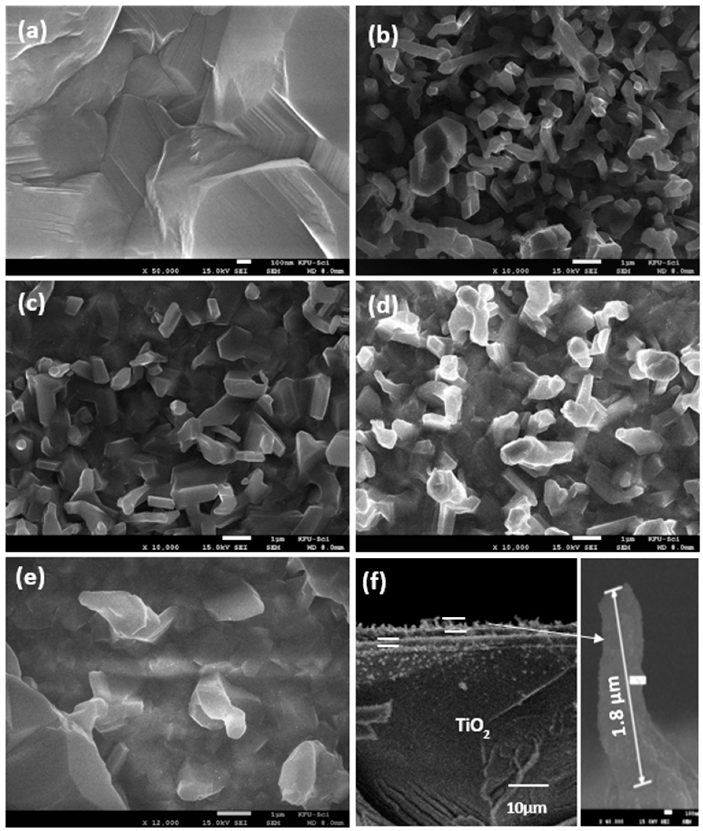
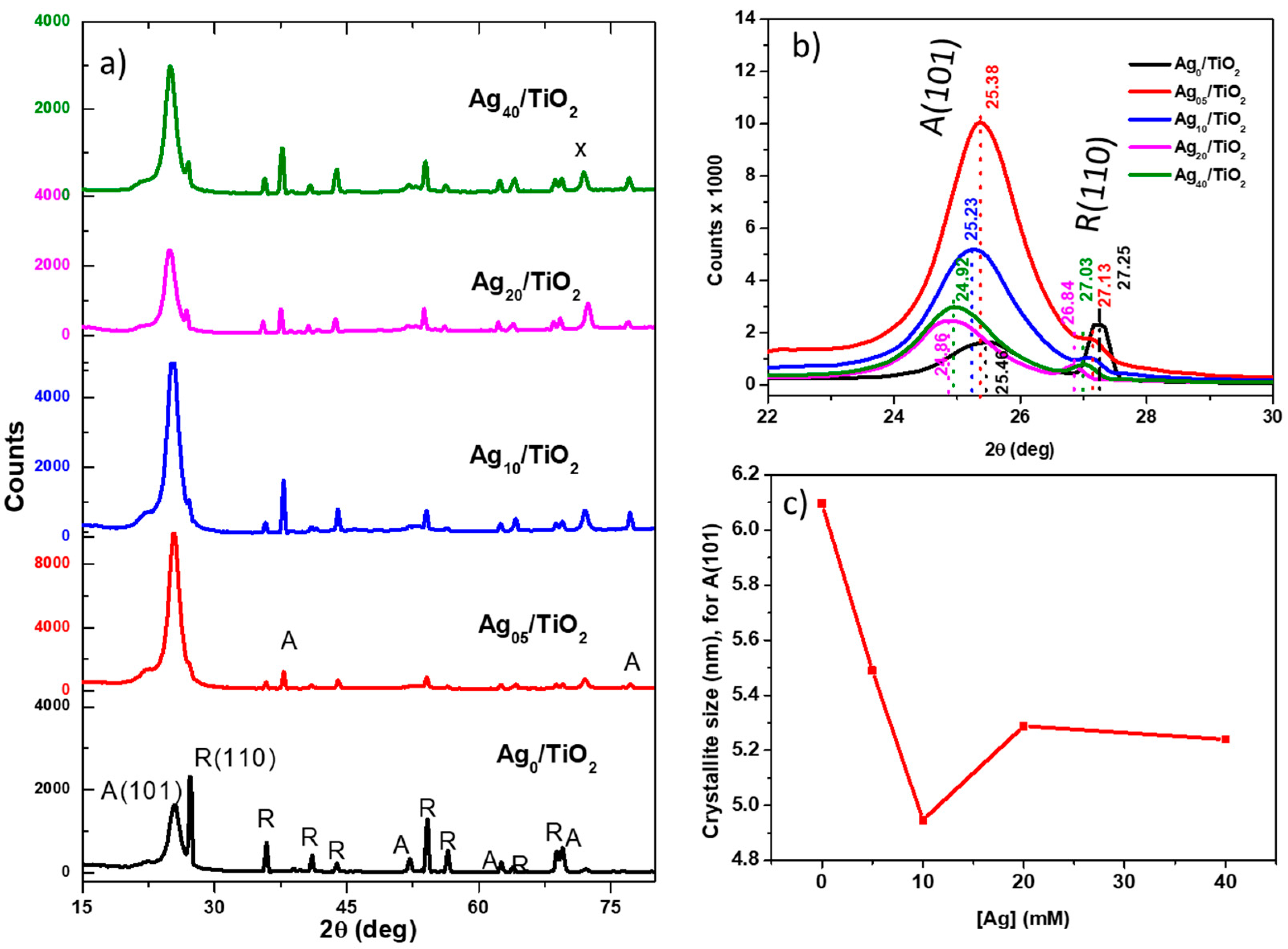
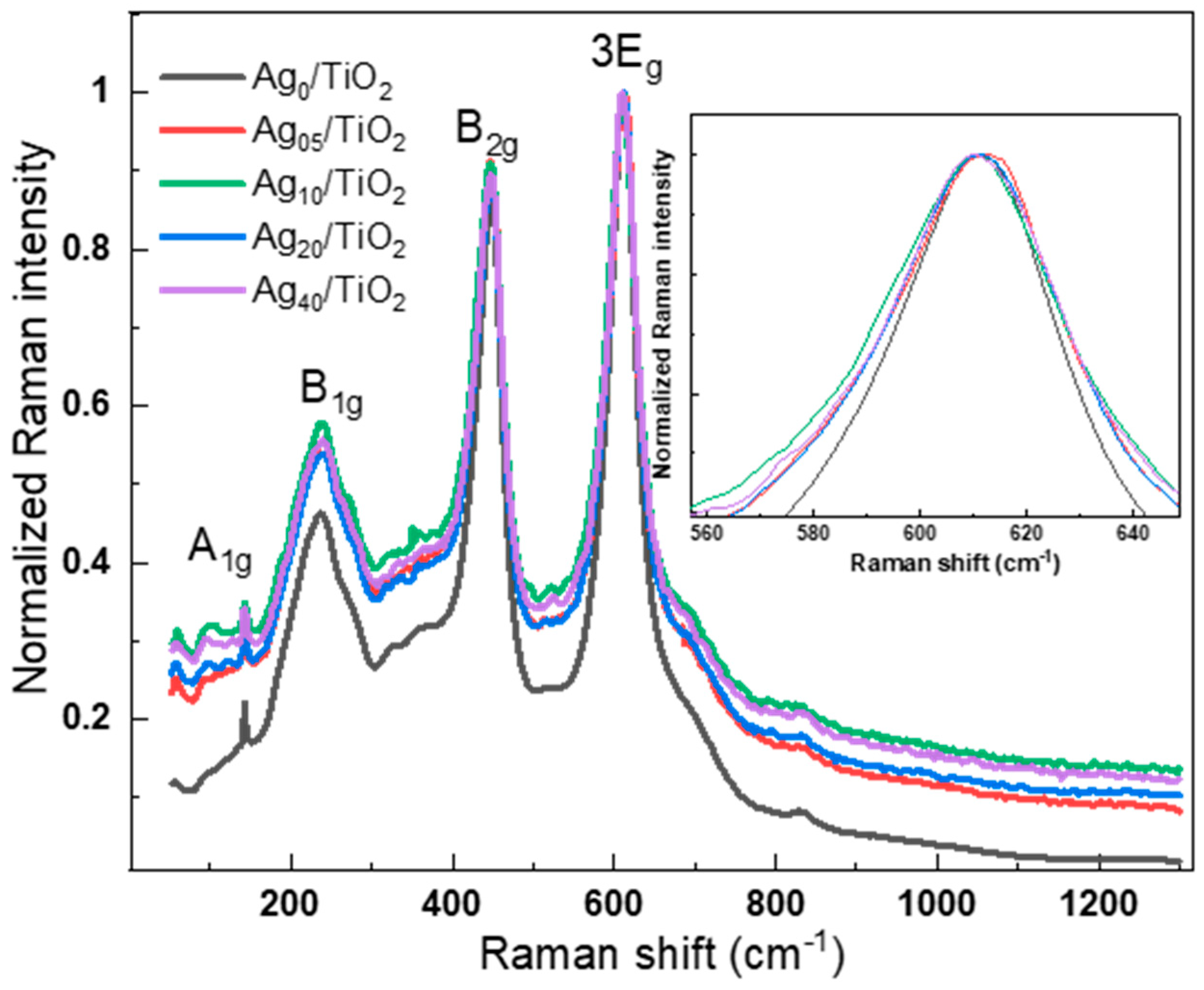
3.1. Structural Characteristics

Field emission scanning electron microscopy (FESEM) measurement was performed on different ceramic-like Agx/TiO2 nanostructures. Figure 1a–e show the topography from the pure TiO2 and Agx/TiO2 surface, confirming the formation of vertical nanorods. Figure 1a shows the appearance of formed grains with small size; however, most of the area contains predominantly larger-size grains without any growth for 1D structures. By adding silver nanoparticles onto the surface, 1D nanostructures with an average size of 200–500 nm became predominant with the shape of nanorods, in addition to the pores between these observed rods, as shown in Figure 1b. By increasing the concentration of silver to 10 mM, the density of nanorods decreased, maintaining a normal direction on the surface of the sheet. By adding more silver, the density of rods decreased and became larger in size compared to the low silver concentration. These nanorods were very sparse for 40 mM of silver, as shown in Figure 1e. The nanorods were about 2 µm in length and 200–500 nm in diameter. It seems that the growth of nanostructures was limited to 2 µm deep beneath the surface, where the sheet was 100 µm thick.
XRD patterns of the as-prepared and Agx/TiO2 nanostructures are shown in Figure 2a. It is confirmed that Agx/TiO2 has been formed due to the oxidation of the Ti sheet in the ambient air. X-ray data analysis indicated that the TiO2 pure phase included the anatase and rutile structures with distinguishing diffraction lines A(101) and R(110) corresponding to JCPDS-21-1272 and JCPDS-01-1292 cards, respectively. The investigated pure TiO2 nanostructure showed more than the preferred orientation related to both phases. On the contrary, Agx/TiO2 samples showed a dominant orientation of A(101), although a low intensity of the diffraction lines for the rutile phase was observed. The preferred orientation of Agx/TiO2 growth confirms the observation of the 1D nanostructure observed in the XRD spectra. It seems that Ag deposited on the surface of Ti enhanced the growth of 1D nanostructures. A non-identified peak located at 72° could be assigned to some impurities existing in the Ti sheets. This peak is observed in all the Agx/TiO2, as shown in Figure S1 (Supplementary Material). Figure 2b shows a shift to the lower angle for (101) and R(110) peaks for Agx/TiO2 upon the formation of nanorods on the TiO2 surface. This shift disappeared in case of the pure and high silver concentration of 40 mM, which did not show nanorods formation. This may be ascribed to the formation of nanorods on the surface. By the well-known Scherrer equation, the crystallite size of the fabricated Agx/TiO2 was calculated and is shown in Figure 2c. The crystallite size of the pure phase was about 6.1 nm, which decreased to 4.9 nm for Ag10/TiO2, and then increased up to ~5.3 nm for Ag20/TiO2 and Ag40/TiO2 samples. The change in the crystallite size was about 20% compared to that of the pure TiO2, which might influence the physical properties of the fabricated composites.
The observed Raman lines belong to both anatase and rutile phases [33]. Figure 3 clearly shows the four phonon modes of TiO2 that are A1g, B1g, B2g, and 3Eg located respectively at 143, 236, 447, and 610 cm−1 [34]. Besides, the mentioned phonon modes of the rutile phase were present in all the samples of Agx/TiO2. On the other side, the inset in Figure 3 shows a shift in the 610 cm−1 modes that occurred during the increase in silver concentration. It could be that the peak located at 143 cm−1 belongs to the E1g mode attributed to the anatase phase. The reason for the predominance of rutile modes could be the crystal orientation of the anatase phase. The nanorods were especially more oriented toward the c axis; hence, the Raman modes were not very active. The second reason could be that the anatase phase region was beneath the nanorods enclosing the bulk region. The latter can be supported by the detection of the anatase phase by XRD analysis.
The Raman peak of pure TiO2 was located at 610.2 cm−1. The peak had a slight blueshift and broadened with the decrease in the intensity of the doped samples. The blue shift and broadness could be related to the increase in vacancies on the surface of the photocatalysts, as previously shown by Parker and Siegel [35,36,37]. However, the formation of the 1D nanostructure due to silver catalysts in the host structure led to a distortion in the lattice that compensated the nonstoichiometric effect, and consequently, the Eg mode redshifted. The latter was observed clearly for Ag10/TiO2; however, for Ag40/TiO2, the Eg mode went back to the pure TiO2 when the nanorods almost disappeared. The increase in the silver concentration affected the amplitude due to the increase in silver optical absorption, as shown in Figure 4. The full-width at half-maximum of the Raman intensity (FWHM) increased when the concentration of silver increased to 10 mM, then decreased for higher silver concentrations. This FWHM behavior reflects the development in the shape and density of 1D nanostructures observed in FESEM images. This FWHM variation also correlates with the FWHM features extracted from the anatase phase A(101) observed by XRD.

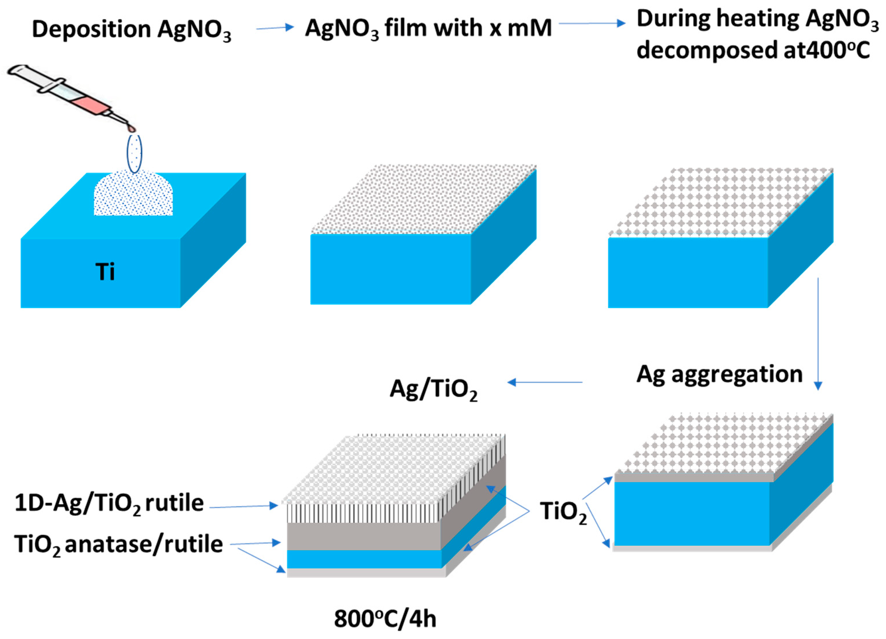
3.2. Growth Mechanism of Ag/TiO2 Nanorods

In Figure 5, we propose a growth mechanism of the 1D nanostructure prepared by Agx/TiO2. After the step of coating AgNO3 on the surface, the sheet was placed on the furnace. It is known that AgNO3 molecules decomposed at ~400 °C, forming Ag aggregations on the surface of the sheet to large Ag droplets at high temperatures of 800 °C. These Ag droplets worked as a catalyst for the growth of TiO2 nanorods at this temperature. The silver droplets moved upward due to this high temperature, pulling/accompanying the oxidized titanium atoms with them. Thus, the Ag/TiO2 1D nanostructure was formed. From the SEM images, we observed that the growth of nanorods was only ~2 µm deep in the sheet compared to 100 µm of the sheet thickness, as shown in Figure 1f. Thus, the oxidation process was expected to gradually extend into the sheet beneath the nanorods. That is why we expect the rutile phase to form almost in the region of the nanorods, while the anatase phase almost formed in the bulk region beneath the rods, as shown in Figure 5. This confirms the appearance of anatase in the XRD greater than that in Raman, where X-rays penetrate deeper inside the material; however, Raman is a surface characteristic. This elucidates why the rutile phase dominated in the Raman spectra for Agx/TiO2 rather than XRD. The results also confirm that the oxidation was not only for the 1D nanostructure region but gradually extended into the bulk. The extension of the oxide region into the bulk made the rods planted or have roots in the bottom part, adequately coherent with the rest of the sheet, forming ceramics-like materials. However, this explanation of this mechanism cannot be proven without performing any elemental analysis techniques in the nanorods.

3.3. Optical Properties

The study of optical properties for the fabricated Agx/TiO2 is important. From these optical properties, we are concerned with the energy band structure and electronic process in the material. We investigated how the light interacts with the material in the wide range of wavelengths of the incident photons. For determining the visual idea for the optical properties of Agx/TiO2 nanorods, we used the measured data of the absorbance (A) for the solid, as shown in Figure 6. It can be seen that the absorbance spectra was low for all samples. The reason is ascribed to the nature of the Ti sheet and the existence of Ag on the surface of TiO2. The spectra of pure TiO2 showed a band peak at 3.22 eV, which was observed for the absorbance reflectance spectra. However, two-band peaks were observed for Agx/TiO2 at 3.22 and 3.84 eV. This blue shift of 3.84 eV could be related to the quantum confinement effect that could be due to the small crystallites or the anisotropy of the effective masses [38,39]. It is seen for the SEM image that both the 1D and bulk surfaces were exposed to the photon beam. However, there was a redshift in the bandgap peak when the Ag increased from 5 to 10 mM rather than 20 and 40 mM. The top region of 1D refers to the rutile phase and the bottom region refers to the anatase phase. Thus, there are two bands ranging between 1.5 and 3.2 eV when we draw a straight line intersect with the x-axis at zero absorbance. Thus, these two bands allowed the Agx/TiO2 samples to work at the visible and UV-regions of the incident light for the photocatalysis application.

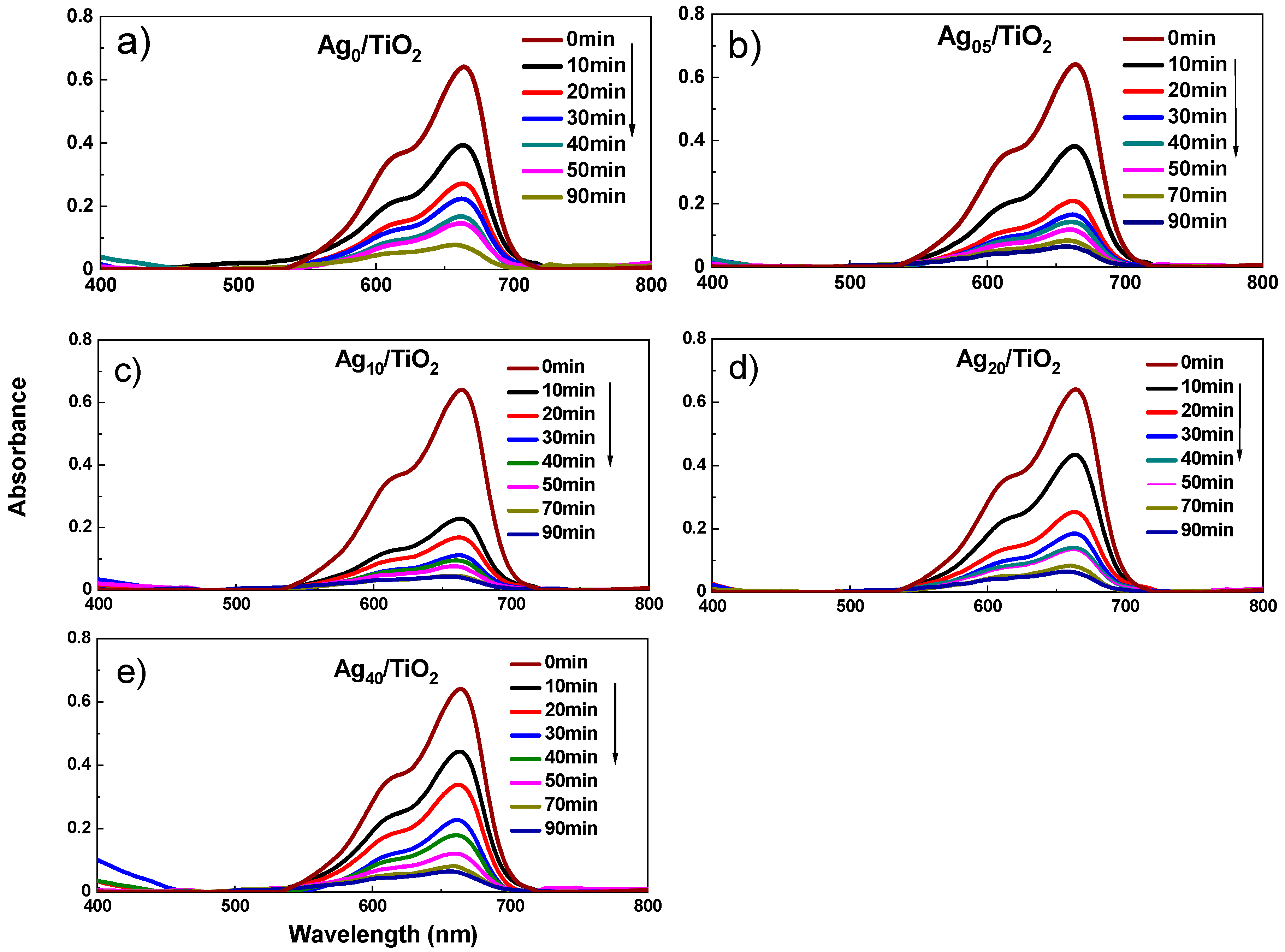
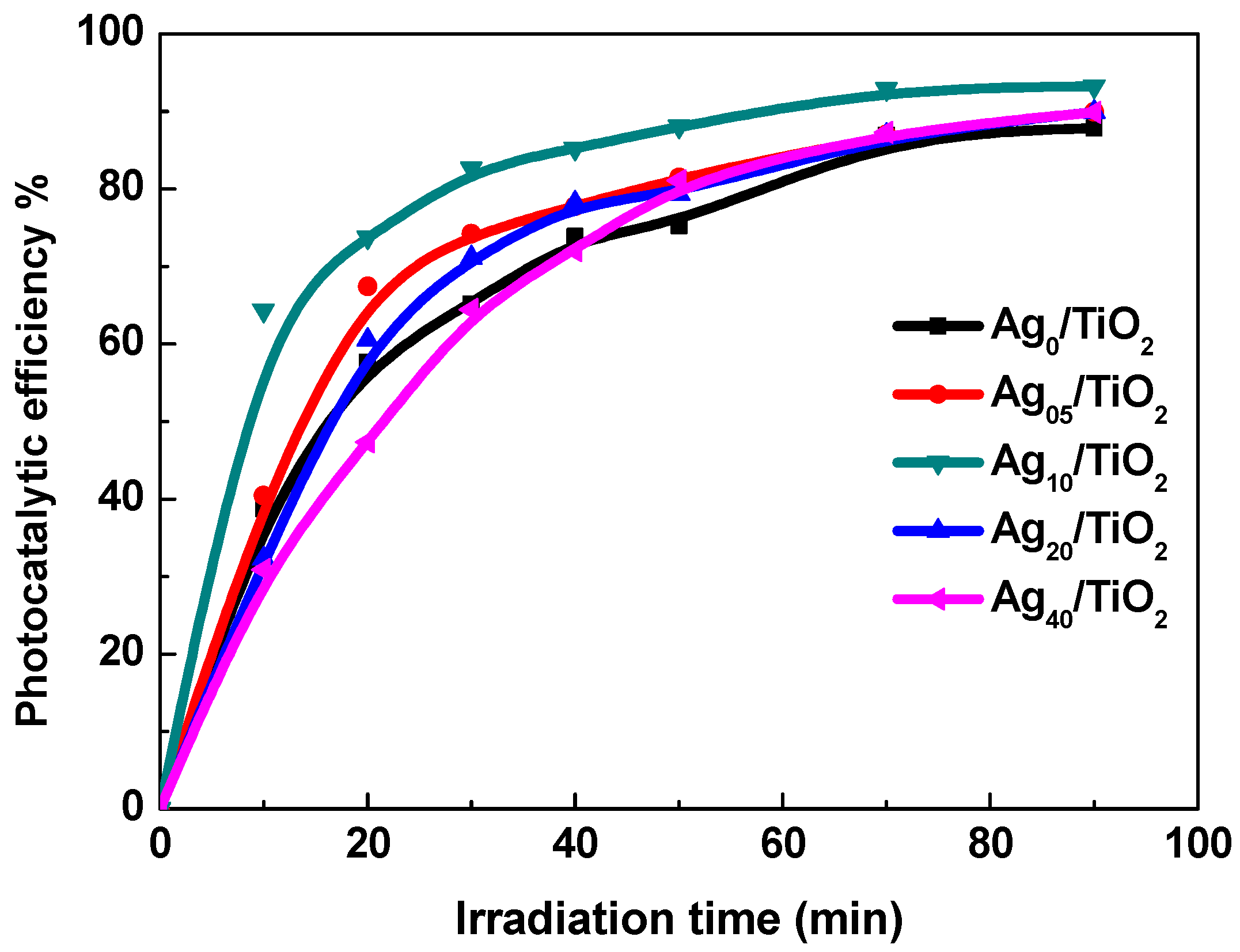
3.4. Photocatalysis Properties

For the photocatalysis study, the absorbance measurement was carried out by using a spectrophotometer UV-1800 series Shimadzu (Reinach, Switzerland) in the wavelength interval of 200–1100 nm. We used methylene blue (MB) as a known target dye for the photocatalytic degradation. A concentration of 10−6 M of MB was prepared and a piece of Agx/TiO2 of 0.5 cm2 in the area was immersed in the aqueous solution. Before starting the measurement, blank experiments were performed to assure that no change in the color of MB could result from the light, and the MB solution was also stirred before each measurement. In addition, we tested it in the dark in order to assure that the removal was due to the photodegradation process and not the surface adsorption. We must mention that we used these samples on different occasions to assure their reproducibility. The absorbance was measured from the sample after a successive irradiation of a duration of 10 min. A decrease in the absorbance was observed in all the samples, as shown in Figure 7. However, the reduction in the absorbance for the Ag10/TiO2 sample was significantly fast compared to the other samples.
To estimate the photocatalytic performance of the fabricated samples, the photocatalytic efficiency (η) of MB has been evaluated by using Equation (1) for monitoring the changes in the main absorbance peak observed at a wavelength of 664 nm:
η % = C t C 0 C 0 × 100
where C0 is the initial dye concentration and Ct the residual dye concentration after the irradiation time (t). Figure 8 shows the photocatalytic efficiency toward the MB for Agx/TiO2 samples.
Significant enhancement in the photocatalytic efficiency for the sample doped with silver, especially the Ag5/TiO2, Ag10/TiO2, and Ag20/TiO2, was observed; however, the sample of Ag40/TiO2 showed an efficiency identical to that of Ag0/TiO2. The highest efficiency was recorded for the Ag10/TiO2 sample. The enhancement in the efficiency of Agx/TiO2 (0 < x < 40) can be explained by the formation of 1D nanorods besides the effect of the presence of Ag content. The 1D nanostructure was not formed for the Ag40/TiO2 sample, although the silver amount was much higher on the surface than the other samples. This was obviously due to the higher excess of silver concentration that stopped the growth of the nanostructure and also the optical loss due to the absorption of silver in the visible wavelength range on the surface of the oxide. Figure 9 shows the efficiency toward MB degradation against the Agx/TiO2 samples after irradiation for 10, 40, and 90 min. Samples with low silver concentration exhibited good performance, and a higher performance was recorded for the Ag10/TiO2 sample, especially at the beginning of the light exposure. However, the Ag40/TiO2 sample exhibited a performance slightly lower than that of the pure TiO2. The performance of the samples with low silver concentrations was high when the concentration of MB was high, showing quick degradation of MB on their surface.
The kinetics reactions of photocatalytic degradation of MB in the presence UV-Vis light irradiation is considered to obey a pseudo-first-order kinetic reaction [40,41]. The integrated rate law for a first-order reaction can be written using the logarithmic relation, as described in Equation (2) [42]:
ln ( c t c 0 ) = k t
where k is the pseudo-first-order rate constant (min−1) and t (min) is the time point where the concentration Ct was measured. The logarithmic relationship MB against the irradiation time obtains a straight line, as shown in Figure 10, indicating a semi-pseudo-first-order reaction. The correlation factor (R2) is less than 1.0 for the fitted lines due to the division of some points. Accordingly, the value of k was estimated from the slope and plotted in the inset figure. The value of k was about 2.6 × 10⁻² min−1 for the Ag0/TiO2, which increased gradually up to 5.0 × 10⁻² min−1 for Ag10/TiO2. As shown from the calculated k, the sample Ag10/TiO2 exhibited a higher reaction rate compared to the others. Thus, the maximum degradation rate was recorded for the nanorods structure while the minimum degradation rate was recorded for the rough TiO2 surface. With the increase in the Ag content on the surface up to 40 mM, the reaction rate constant decreased to 3.1 × 10⁻² min−1; however, it was still higher than the pure phase of TiO2.

4. Conclusions

One-dimensional Agx/TiO2 nanostructures were synthesized successfully by a simple method. The samples were ceramic-like nanostructures. The physical properties of the fabricated ceramic-like nanostructures showed one-dimensional nanostructures with an average size varying within the range of 200–500 nm being grown. The presence of silver made the nanostructure vertically directed with a high density of nanorods at median concentrations of 5, 10, and 20 mM of silver, and a low density at high silver concentrations of 40 mM. The anatase and rutile structure were confirmed for pure TiO2 with distinguishing diffraction lines A(101) and R(110); however, the anatase phase was dominant for Agx/TiO2 with (101) planes, confirming the 1D growth. This dominance can be due to the change in the temperature of the anatase-to-rutile phase transition between Ag0/TiO2 and Agx/TiO2. The degradation of methylene blue based on the fabricated ceramic-like nanostructures showed the photocatalysis activity of these nanostructures, where the median silver concentration samples showed a high photocatalytic activity compared to the pure and high-silver-concentration samples. The kinetics reactions of photocatalytic reaction almost obeyed a pseudo-first-order kinetic reaction. Finally, the most important characteristic of these samples is that they remain hard and can be reusable for further cycles.

Supplementary Materials

The following are available online at https://www.mdpi.com/2073-4352/10/11/1024/s1, Figure S1: XRD peak observed in the range of 2-theta = 71–74°.

Author Contributions

Conceptualization, N.A.S., C.A. and N.M.S.; data curation, N.A.S., C.A., A.A. and N.M.S.; formal analysis, C.A. and N.M.S.; funding acquisition, N.A.S., C.A., A.A.; project administration, N.A.S. and C.A.; resources, A.A.; software, C.A. and N.M.S.; supervision, N.A.S., C.A.; writing—original draft, C.A.; writing—review and editing, N.A.S., C.A. and N.M.S. All authors have read and agreed to the published version of the manuscript.

Funding

This research was funded by the Deanship of Scientific Research at King Faisal University (Saudi Arabia), grant number 187002 and the APC was funded by the same grant number 187002.

Acknowledgments

The authors acknowledge the Deanship of Scientific Research at King Faisal University for the financial support under Ra’ed Track (Grant No. 187002).

Conflicts of Interest

The authors declare no conflict of interest.

References

  1. Byrne, C.; Subramanian, G.; Pillai, S.C. Recent advances in photocatalysis for environmental applications. J. Environ. Chem. Eng. 2018, 6, 3531–3555. [Google Scholar] [CrossRef]
  2. Mills, A.; Le Hunte, S. An overview of semiconductor photocatalysis. J. Photochem. Photobiol. A Chem. 1997, 108, 1–35. [Google Scholar] [CrossRef]
  3. Khan, M.M.; Adil, S.F.; Al-Mayouf, A. Metal oxides as photocatalysts. J. Saudi Chem. Soc. 2015, 19, 462–464. [Google Scholar] [CrossRef] [Green Version]
  4. Hoffmann, M.R.; Martin, S.T.; Choi, W.; Bahnemann, D.W. Environmental Applications of Semiconductor Photocatalysis. Chem. Rev. 1995, 95, 69–96. [Google Scholar] [CrossRef]
  5. Ni, M.; Leung, M.K.H.; Leung, D.Y.C.; Sumathy, K. A review and recent developments in photocatalytic water-splitting using TiO2 for hydrogen production. Renew. Sustain. Energy Rev. 2007, 11, 401–425. [Google Scholar] [CrossRef]
  6. Fujishima, A.; Honda, K. Electrochemical Photolysis of Water at a Semiconductor Electrode. Nature 1972, 238, 37–38. [Google Scholar] [CrossRef]
  7. Pelaez, M.; Nolan, N.T.; Pillai, S.C.; Seery, M.K.; Falaras, P.; Kontos, A.G.; Dunlop, P.S.M.; Hamilton, J.W.J.; Byrne, J.A.; O’Shea, K.; et al. A review on the visible light active titanium dioxide photocatalysts for environmental applications. Appl. Catal. B Environ. 2012, 125, 331–349. [Google Scholar] [CrossRef] [Green Version]
  8. Daghrir, R.; Drogui, P.; Robert, D. Modified TiO2 For Environmental Photocatalytic Applications: A Review. Ind. Eng. Chem. Res. 2013, 52, 3581–3599. [Google Scholar] [CrossRef]
  9. Mahlambi, M.M.; Ngila, C.J.; Mamba, B.B. Recent Developments in Environmental Photocatalytic Degradation of Organic Pollutants: The Case of Titanium Dioxide Nanoparticles—A Review. J. Nanomater. 2015, 2015, 1–29. [Google Scholar] [CrossRef] [Green Version]
  10. Wang, M.; Hu, Y.; Han, J.; Guo, R.; Xiong, H.; Yin, Y. TiO2/NiO hybrid shells: P–n junction photocatalysts with enhanced activity under visible light. J. Mater. Chem. A 2015, 3, 20727–20735. [Google Scholar] [CrossRef]
  11. An, H.R.; Park, S.Y.; Huh, J.Y.; Kim, H.; Lee, Y.C.; Lee, Y.B.; Hong, Y.C.; Lee, H.U. Nanoporous hydrogenated TiO2 photocatalysts generated by underwater discharge plasma treatment for solar photocatalytic applications. Appl. Catal. B Environ. 2017, 211, 126–136. [Google Scholar] [CrossRef]
  12. Shao, J.; Sheng, W.; Wang, M.; Li, S.; Chen, J.; Zhang, Y.; Cao, S. In situ synthesis of carbon-doped TiO2 single-crystal nanorods with a remarkably photocatalytic efficiency. Appl. Catal. B Environ. 2017. [Google Scholar] [CrossRef]
  13. Hochbaum, A.I.; Chen, R.; Delgado, R.D.; Liang, W.; Garnett, E.C.; Najarian, M.; Majumdar, A.; Yang, P. Enhanced thermoelectric performance of rough silicon nanowires. Nature 2008, 451, 163–167. [Google Scholar] [CrossRef]
  14. Park, J.-Y.; Choi, K.-I.; Lee, J.-H.; Hwang, C.-H.; Choi, D.-Y.; Lee, J.-W. Fabrication and characterization of metal-doped TiO2 nanofibers for photocatalytic reactions. Mater. Lett. 2013, 97, 64–66. [Google Scholar] [CrossRef]
  15. Albu, S.P.; Ghicov, A.; Macak, J.M.; Hahn, R.; Schmuki, P. Self-Organized, Free-Standing TiO2 Nanotube Membrane for Flow-through Photocatalytic Applications. Nano Lett. 2007, 7, 1286–1289. [Google Scholar] [CrossRef] [PubMed]
  16. Wu, N.; Wang, J.; Tafen, D.N.; Wang, H.; Zheng, J.-G.; Lewis, J.P.; Liu, X.; Leonard, S.S.; Manivannan, A. Shape-Enhanced Photocatalytic Activity of Single-Crystalline Anatase TiO2 (101) Nanobelts. J. Am. Chem. Soc. 2010, 132, 6679–6685. [Google Scholar] [CrossRef] [Green Version]
  17. Li, H.; Lu, W.; Tian, J.; Luo, Y.; Asiri, A.M.; Al-Youbi, A.O.; Sun, X. Synthesis and Study of Plasmon-Induced Carrier Behavior at Ag/TiO2 Nanowires. Chem.-A Eur. J. 2012, 18, 8508–8514. [Google Scholar] [CrossRef]
  18. Wu, H.B.; Hng, H.H.; Lou, X.W.D. Direct Synthesis of Anatase TiO2 Nanowires with Enhanced Photocatalytic Activity. Adv. Mater. 2012, 24, 2567–2571. [Google Scholar] [CrossRef]
  19. Liu, G.; Yang, H.G.; Wang, X.; Cheng, L.; Pan, J.; Lu, G.Q.; Cheng, H.-M. Visible Light Responsive Nitrogen Doped Anatase TiO2 Sheets with Dominant {001} Facets Derived from TiN. J. Am. Chem. Soc. 2009, 131, 12868–12869. [Google Scholar] [CrossRef]
  20. Ge, M.; Cao, C.; Huang, J.; Li, S.; Chen, Z.; Zhang, K.-Q.; Al-Deyab, S.S.; Lai, Y. A review of one-dimensional TiO2 nanostructured materials for environmental and energy applications. J. Mater. Chem. A 2016, 4, 6772–6801. [Google Scholar] [CrossRef]
  21. Plodinec, M.; Grčić, I.; Willinger, M.G.; Hammud, A.; Huang, X.; Panžić, I.; Gajović, A. Black TiO2 nanotube arrays decorated with Ag nanoparticles for enhanced visible-light photocatalytic oxidation of salicylic acid. J. Alloys Compd. 2019, 776, 883–896. [Google Scholar] [CrossRef]
  22. Qi, H.-P.; Wang, H.-L.; Zhao, D.-Y.; Jiang, W.-F. Preparation and photocatalytic activity of Ag-modified GO-TiO2 mesocrystals under visible light irradiation. Appl. Surf. Sci. 2019, 480, 105–114. [Google Scholar] [CrossRef]
  23. Khan, S.B.; Hou, M.; Shuang, S.; Zhang, Z. Morphological influence of TiO2 nanostructures (nanozigzag, nanohelics and nanorod) on photocatalytic degradation of organic dyes. Appl. Surf. Sci. 2017, 400, 184–193. [Google Scholar] [CrossRef]
  24. Fan, X.; Wan, J.; Liu, E.; Sun, L.; Hu, Y.; Li, H.; Hu, X.; Fan, J. High-efficiency photoelectrocatalytic hydrogen generation enabled by Ag deposited and Ce doped TiO2 nanotube arrays. Ceram. Int. 2015, 41, 5107–5116. [Google Scholar] [CrossRef]
  25. Meng, L.; Huang, T.; Wang, X.; Chen, S.; Yang, Z.; Ren, B. Gold-coated AFM tips for tip-enhanced Raman spectroscopy: Theoretical calculation and experimental demonstration. Opt. Express 2015, 23, 13804–13813. [Google Scholar] [CrossRef]
  26. Zhang, H.; Liang, C.; Liu, J.; Tian, Z.; Wang, G.; Cai, W. Defect-Mediated Formation of Ag Cluster-Doped TiO2 Nanoparticles for Efficient Photodegradation of Pentachlorophenol. Langmuir 2012, 28, 3938–3944. [Google Scholar] [CrossRef]
  27. Subrahmanyam, A.; KP, B.; Rajesh, P.; Kumar, K.J.; MR, K. Surface modification of sol gel TiO2 surface with sputtered metallic silver for Sun light photocatalytic activity: Initial studies. Sol. Energy Mater. Sol. Cells 2012, 101, 241–248. [Google Scholar] [CrossRef]
  28. Li, Y.; Ma, M.; Chen, W.; Li, L.; Zen, M. Preparation of Ag-doped TiO2 nanoparticles by a miniemulsion method and their photoactivity in visible light illuminations. Mater. Chem. Phys. 2011, 129, 501–505. [Google Scholar] [CrossRef]
  29. Huang, Y.-C.; Chang, S.-Y.; Jehng, J.-M. Photocatalytic H2 Generation Efficiencies of TiO2 Nanotube-based Heterostructures Grafted with ZnO Nanorods, Ag Nanoparticles, or Pd Nanodendrites. J. Phys. Chem. C 2017, 121, 19063–19068. [Google Scholar] [CrossRef]
  30. Guan, B.; Yu, J.; Guo, S.; Yu, S.; Han, S. Porous nickel doped titanium dioxide nanoparticles with improved visible light photocatalytic activity. Nanoscale Adv. 2020, 2, 1352–1357. [Google Scholar] [CrossRef] [Green Version]
  31. Corredor, J.; Rivero, M.J.; Ortiz, I. New insights in the performance and reuse of rGO/TiO2 composites for the photocatalytic hydrogen production. Int. J. Hydrogen Energy 2020. [Google Scholar] [CrossRef]
  32. Ismael, A.M.; El-Shazly, A.N.; Gaber, S.E.; Rashad, M.M.; Kamel, A.H.; Hassan, S.S.M. Novel TiO2/GO/CuFe2O4 nanocomposite: A magnetic, reusable and visible-light-driven photocatalyst for efficient photocatalytic removal of chlorinated pesticides from wastewater. RSC Adv. 2020, 10, 34806–34814. [Google Scholar] [CrossRef]
  33. Lottici, P.P.; Bersani, D.; Braghini, M.; Montenero, A. Raman scattering characterization of gel-derived titania glass. J. Mater. Sci. 1993, 28, 177–183. [Google Scholar] [CrossRef]
  34. Mathew, S.; Ganguly, P.; Rhatigan, S.; Kumaravel, V.; Byrne, C.; Hinder, S.; Bartlett, J.; Nolan, M.; Pillai, S. Cu-Doped TiO2: Visible Light Assisted Photocatalytic Antimicrobial Activity. Appl. Sci. 2018, 8, 2067. [Google Scholar] [CrossRef] [Green Version]
  35. Parker, J.C.; Siegel, R.W. Calibration of the Raman spectrum to the oxygen stoichiometry of nanophase TiO2. Appl. Phys. Lett. 1990, 57, 943–945. [Google Scholar] [CrossRef]
  36. Parker, J.C.; Siegel, R.W. Raman microprobe study of nanophase TiO2 and oxidation-induced spectral changes. J. Mater. Res. 1990, 5, 1246–1252. [Google Scholar] [CrossRef]
  37. Melendres, C.A.; Narayanasamy, A.; Maroni, V.A.; Siegel, R.W. Raman spectroscopy of nanophase TiO2. J. Mater. Res. 1989, 4, 1246–1250. [Google Scholar] [CrossRef]
  38. Satoh, N.; Nakashima, T.; Kamikura, K.; Yamamoto, K. Quantum size effect in TiO2 nanoparticles prepared by finely controlled metal assembly on dendrimer templates. Nat. Nanotechnol. 2008, 3, 106–111. [Google Scholar] [CrossRef]
  39. Peng, H.; Li, J. Quantum Confinement and Electronic Properties of Rutile TiO2 Nanowires. J. Phys. Chem. C 2008, 112, 20241–20245. [Google Scholar] [CrossRef]
  40. Türkyılmaz, Ş.Ş.; Güy, N.; Özacar, M. Photocatalytic efficiencies of Ni, Mn, Fe and Ag doped ZnO nanostructures synthesized by hydrothermal method: The synergistic/antagonistic effect between ZnO and metals. J. Photochem. Photobiol. A Chem. 2017, 341, 39–50. [Google Scholar] [CrossRef]
  41. Skinner, A.W.; DiBernardo, A.M.; Masud, A.M.; Aich, N.; Pinto, A.H. Factorial design of experiments for optimization of photocatalytic degradation of tartrazine by zinc oxide (ZnO) nanorods with different aspect ratios. J. Environ. Chem. Eng. 2020, 8, 104235. [Google Scholar] [CrossRef]
  42. de Mendonça, V.R.; Ribeiro, C. Influence of TiO2 morphological parameters in dye photodegradation: A comparative study in peroxo-based synthesis. Appl. Catal. B Environ. 2011, 105, 298–305. [Google Scholar] [CrossRef]
Figure 1. Scanning electron microscopy measurement of: (a) Ag0/TiO2, (b) Ag5/TiO2, (c) Ag10/TiO2, (d) Ag20/TiO2, (e) Ag40/TiO2, and (f) the cross-sectional image for the prepared sample of Ag10/TiO2.
Crystals 10 01024 g001
Figure 2. (a) XRD patterns of Agx/TiO2 samples, (b) expanded 2-theta range (22–30°) for A(101) peak, and (c) the calculated crystallite size from A(101) corresponding to anatase phase.
Crystals 10 01024 g002
Figure 3. Raman spectrum of Agx/TiO2 samples. A quantitative analysis was carried out by fitting the spectra with a Lorentzian function.
Crystals 10 01024 g003
Figure 4. Extracted parameters (Raman peak position, FWHM, amplitude) from the fitting of the phonon mode located at 611 cm−1 by using Lorentzian function, adj. R-squared 0.996.
Crystals 10 01024 g004
Figure 5. Schematic illustration for the preparation steps and growth mechanism of Ag/TiO2 nanorods.
Crystals 10 01024 g005
Figure 6. The absorbance spectra versus incident photon energy for Agx/TiO2 nanostructures.
Crystals 10 01024 g006
Figure 7. The degradation of methylene blue due to the irradiation in the presence of Agx/TiO2 nanostructures; (a) Ag0/TiO2, (b) Ag5/TiO2, (c) Ag10/TiO2, (d) Ag20/TiO2, (e) Ag40/TiO2.
Crystals 10 01024 g007
Figure 8. Photocatalytic efficiency toward methylene blue (MB) versus time of Agx/TiO2, where x = 0, 5, 10, 20, and 40.
Crystals 10 01024 g008
Figure 9. The photocatalytic efficiency toward MB degradation against the Agx/TiO2 samples after irradiation for 10, 40, and 90 min.
Crystals 10 01024 g009
Figure 10. Semi-logarithmic graph of MB concentration versus irradiation time in the presence of Agx/TiO2. The inset shows the first-order reaction rate constants (k) versus the Ag concentration.
Crystals 10 01024 g010
Publisher’s Note: MDPI stays neutral with regard to jurisdictional claims in published maps and institutional affiliations.

