Article

Synthesis of 1-[(Aryl)(3-amino-5-oxopyrazolidin-4-ylidene) methyl]-2-oxo-1,2-dihydroquinoline-3-carboxylic Acid Derivatives and Their Breast Anticancer Activity

by
Ahmed Gaber
1,2,
Walaa F. Alsanie
2,3,
Majid Alhomrani
2,3,
Abdulhakeem S. Alamri
2,3,
Ibrahim M. El-Deen
4 and
Moamen S. Refat
5,*
1
Department of Biology, College of Science, Taif University, P.O. Box 11099, Taif 21944, Saudi Arabia
2
Center of Biomedical Sciences Research, Taif University, P.O. Box 11099, Taif 21944, Saudi Arabia
3
Department of Clinical Laboratories Sciences, The Faculty of Applied Medical Sciences, Taif University, P.O. Box 11099, Taif 21944, Saudi Arabia
4
Department of Chemistry, Faculty of Science, Port Said University, Port Said 42511, Egypt
5
Department of Chemistry, College of Science, Taif University, P.O. Box 11099, Taif 21944, Saudi Arabia
*
Author to whom correspondence should be addressed.
Crystals 2021, 11(5), 571; https://doi.org/10.3390/cryst11050571
Submission received: 31 March 2021 / Revised: 8 May 2021 / Accepted: 18 May 2021 / Published: 20 May 2021
(This article belongs to the Special Issue Research about Vital Organic Chelates and Metal Ion Complexes)

Abstract

:
This research aimed to produce new 1-[(aryl)(3-amino-5-oxopyrazolidin-4-ylidene) methyl]-2-oxo-1,2-dihydroquinoline-3-carboxylic acid derivatives and check their anticancer effect against the breast cancer MCF-7 cell line. The 2-oxo-1,2-dihydroquinoline-3-carboxylic acid (4) compound was obtained by hydrolyzing ethyl 2-oxo-1,2-dihydroquinoline-3-carboxylate (2) with thiourea and anhydrous potassium carbonate ethanol, which was then treated with ethyl 3-substituted 2-cyanoacrylates (6) in the presence of triethylamine in diethyl formamide to give 1-[2-(ethoxy)carbonyl-2-cyano-1-arylvinyl]-2-oxo-1,2-dihydroquinoline-3-carboxylic (7a,d). Cyclization of compound 7 with hydrazine hydrate ethanol inferred the association of 1-[(aryl)(3 amino-5-oxopyrazolidin-4-ylidene)methyl-2-oxo-1,2-dihydroquinol-3-carboxylates (8a,d). Spectroscopic and micro-analytical techniques such as IR, NMR, and elemental analysis were used to validate the structure of the synthesized organic compounds. The anticancer effects of the synthesized compounds 7ad and 8ad were tested by using the MTT assay on the MCF-7 cell line. When compared to the reference compound Dox, the compounds 7b,c and 8ac demonstrated strong anticancer activity against the MCF-7 cell line. The anticancer effects of the synthesized compounds 7ad and 8ad were tested against the MCF-7 cell line, using MTT assay. The compounds 7b,c and 8ac showed significant anticancer activity compared to the reference compound Dox against the MCF-7 cell line.

1. Introduction

Quinoline, a heterocyclic nitrogen compound, has been list by the Food and Drug Administration as a chemotherapy compound [1]. Quinolines are a common pharmacological scaffold that can be found in a wide range of synthetic and natural bioactive compounds [2]. The chemistry of quinolines has been extensively studied over the last century, with various and fascinating biological activities, such as antibacterial, antifungal, anti-inflammatory, anti-malaria, and anticancer properties [3,4,5,6,7,8]. Quinoline derivatives are effective against cancer cells in the breast, prostate, gastrointestinal, colon, and liver [9,10,11,12]. Similar compounds, such as camptothecin and its analogs (irinotecan and topotecan) [13,14], as well as bosutinib [15,16,17], have been used in clinical trials.
The anticancer hybrid drug approach is a cutting-edge synthetic strategy that involves either combining or blending the hepatotoxic moieties of several drugs into a novel molecular structure [18,19]. Better anticancer agents resulted from combining these active pharmacophores in a new molecular architecture [18,19,20,21,22]. Pyrazoloquinolines correspondingly are used for many applications, including antiviral activity against the herpes simplex virus [23], caspase activators, anticancer activity [24,25], and apoptosis inducers [25]. Several quinoline–pyrazole hybrids with potent anti-proliferative activity against human liver, breast, and colon cancer cell lines were developed by Pirol et al. [26]. As a result, finding new medications to treat diseases and infections without causing serious adverse effects in patients is critical.
Consequently, quinoline derivatives are among the most effective molecules, with several studies stating that they have a broad variety of biological activities while being safe for patients [27,28]. Therefore, using a taster of multiple functional groups on the quinoline scaffold to create a new anticancer drug is a good idea. As a result of these findings, it was deemed valuable to synthesize some new 2-oxo-1,2-dihydroquinoline-3-carboxylic acids with pyrazolinone moieties at the N-position to obtain a safe and effective new anticancer drug compound.

2. Materials and Methods

2.1. Chemistry

Melting points (m.p.) of the synthesized compounds were measured in an electrothermal digital melting-point device. IR and NMR spectra were verified on a Shimadzu 470 c as KBr pellets and Bruker 400 DRX-Avance spectrometer in DMSO-d6 as a solvent, respectively. The elemental analysis was achieved on a Perkin-Elmer 2400 series II CHN elemental analyzer. Chemicals that were used in synthesis were purchased from Aldrich, Merck, or Fluka Chemical Companies.

2.1.1. Synthesis of the Starting Materials (2)

Ethyl 2-oxo-1,2-dihydroquinoline-3-carboxylate (2) was obtained by fusion of the mixture 2-aminobenzaldehyde (1) (0.01 mol) and diethyl malonate (0.01 mol) in the presence of piperidine (2 mL) on a hot plate, for 2–3 min; ethanol (30 mL) was added to the reaction mixture and heated under reflux for 2 h. The reaction mixture was cooled, poured into water, and neutralized with dilute hydrochloric acid (2%). The resulting product was filtered off, washed with water, dried, and, finally, the product was crystallized from ethanol to give 2 as colorless crystals, yield 76%, m.p. 155 °C. IR (KBr) υmax =1738, 1686 (C=O), 1603, 1586 (C=C), 1118, 1063 (C–O) cm−1.
1H-NMR (DMSO-d6) δ: 1.32 (t, J = 6.32, 3H, CH3), 4.22 (q, J = 6.32, 2H, OCH3), 7.21–7.82 (m, 4H, Ar-H), 8.86 (s, 1H, H-4 of quinoline ring) ppm.
Anal. Calcd. For C12H11NO3 (217): C, 66.35; H, 5.10; N,6.45. Found: C, 66.16; H, 4.83; N, 6.24.

2.1.2. Synthesis of 2-Oxo-1,2-dihydroquinoline-3-carboxylic acid (4)

Ester 2 (0.01 mol) was added to a solution of thiourea (0.02 mol) and anhydrous potassium carbonate (0.03 mol) in 50 mL ethanol. The reaction mixture was heated under reflux for 4 h, then cooled, poured into water, and neutralized with a few drops from acetic acid. The solid formed was filtered off, washed with water, dried, and crystallized from dimethylformamide to give 4 as colorless crystals, yield 63%, m.p. 270 °C. IR (KBr) υmax = 3368 (NH), 3481–3175 (br–OH), 1716, 1685 (C=O), 1605, 1563 (C=C), 1161, 1009 (C–O) cm−1.
1H-NMR (DMSO-d6) δ: 7.42–7.98 (m, 5H, Ar-H and NH), 8.09 (br. s, 1H, OH), 8.87 (s, 1H, H-4 of quinoline ring).
13C-NMR (DMSO-d6) δ: 163.00, 160.80 (C=O), 154.51 (C–N), 148.28, 134.58, 130.74, 125.56, 119.78, 118.93, 116.60 (carbons of quinoline ring) ppm.
Anal. Calcd. For C10H7NO3 (189): C, 63.44; H, 3.73; N, 7.40. Found: C, 63.26; H, 3.55; N, 7.17.

2.1.3. General Procedures for the Preparation of Ethyl 3-aryl-2-cyanoccrylamates (6ad)

A mixture of ethyl cyanoacetates (0.01 mol), appropriate aromatic aldehydes (namely, benzaldehyde, 4-N,N-(dimethyl)amino benzaldehyde, 0.01 mol), and triethyl amine (0.03 mol) in ethanol (50 mL) was refluxed for 2 h. After cooling, the solution was poured into water and neutralized with dilute acetic acid (2%). The solid formed was washed with water, dried, and recrystallized from a suitable solvent to give the compounds 6ad.
Ethyl 3-phenyl-2-cyanoacrylate (6a) as pale yellow crystals, yield 65%, m.p. 52°C. IR (KBr) υmax = 2232 (CN), 1742 (C=O), 1605, 1582 (C=C), 1038 (C–O) cm−1.
1H-NMR (DMSO-d6) δ:1.3 (t, J = 8.01, 3H, CH3), 7.42–7.75 (m, 5H, Ar-H), 8.32 (s, 1H, H-olefinic) ppm.
Ethyl 3-(4-methoxy) phenyl-2-cyanoacrylate (6b) as yellow crystals, yield 71%, m.p. 71 °C. IR (KBr) υmax = 2238 (CN), 1738 (C=O), 1607, 1586 (C=C), 1078, 1032 (C–O) cm−1.
1H-NMR (DMSO-d6) δ:1.31 (t, J = 8.01, 3H, CH3), 3.88 (s, 3H, OCH3), 4.31 (q, J = 8.01,2H, OCH2), 7.15 (d, J = 10.30, 2H, Ar-H), 8.09 (d, 2H, J = 10.30, 2H, Ar-H), 8.30 (s, 1H, H-olefinic) ppm.
Ethyl 3-(4-hydroxy-3-methoxy) phenyl-2-cyanoacrylate (6c) as yellow crystals, yield 73%, m.p. 115 °C. IR (KBr) υmax = 3300 (br. s, OH), 2232 (CN), 1731 (C=O), 1605, 1587 (C=C), 1174, 1092, 1019 (C–O) cm−1.
1H-NMR (DMSO-d6) δ:1.30 (t, J = 8.01, 3H, CH3), 3.83 (s, 3H, OCH3), 4.30 (q, 2H, J = 8.01,2H, OCH2), 6.97 (d, J = 10.3, 1H, Ar-H), 7.64 (d, J = 10.30, 1H, Ar-H), 7.78 (s, 1H, Ar-H), 8.25 (s, 1H, H-olefinic), 10.57 (s, 1H, OH) ppm.
Ethyl 3-(4-N,N-dimethyl amino)phenyl-2-cyanoacrylate (6d) as orange crystals, yield 76%, m.p. 126 °C. IR (KBr) υmax = 2225 (CN), 1736 (C=O), 1607, 1583 (C=C), 1067 (C–O) cm−1.
1H-NMR (DMSO-d6) δ:1.29 (t, J = 8.01, 3H, CH3), 3.10 (s, 6H, NCCH3), 4.27 (q, J = 8.01, 2H, OCH2), 6.85 (d, J = 10.32, 2H, Ar-H), 7.97 (d, J = 10.32, 2H, Ar-H), 8.13 (s, 1H, H-olefinic) ppm.

2.1.4. General Procedures for Synthesis of 1-[2-Cyano-2-(ethoxy) carbonyl-1-arylvinyl]-2-oxo-1,2-dihydroquinoline-3-carboxylic acid (7ad)

Ethyl 3-aryl-2-cyanoacrylates (6ad), 0.01 mol) was dissolved in the mixture of 30 mL dimethylformamide and 3 mL triethylamine around the bottom flask, then added with 2-oxo-1,2-dihydroquinoline-3-carboxylic acid (4) (0.01 mol). The reaction mixture was heated under reflux for 4 h. It was then cooled, poured into water, and neutralized with dilute hydrochloric acid (2%). The solid formed was separated by filtration and purified by recrystallization, using ethanol solvent, to give compounds 7ad.
1-[2-(ethoxy)carbonyl-2-cyano-1-phenylvinyl]-2-oxo-1,2-dihydroquinoline-3-carboxylic acid (7a) as pale yellow crystals, yield 71%, m.p. 226 °C. IR (KBr) υmax = 3468 (br. OH), 2225 (CN), 1735–1715 (C=O), 1607, 1583 (C=C), 1121, 1080 (C–O) cm−1.
1H-NMR (DMSO-d6) δ:1.32 (t, J = 8.01, 3H, CH3), 4.33 (q, J = 8.01, 2H, OCH2), 7.43–8.10 (m, 9H, Ar-H), 8.42 (s, 1H, OH), 8.89 (s, 1H, H-4 of quinoline ring) ppm.
13C-NMR (DMSO-d6) δ: 162.99 (C-11), 162.28 (C-21), 160.82 (C-2), 155.61 (C-12), 154.51 (C-9), 148.31 (C-4), 134.58 (C-5), 133.92 (C-16), 131.83 (C-13), 131.29 (C-15, 17), 130.75 (C-10), 129.83 (C-14, 18), 125.56 (C-6), 119.75 (C-7), 118.93 (C-8), 116.61 (C-3), 116.11 (C-19), 103.07 (C-20), 62.88 (C-23), 14.47 (C-24) ppm.
Anal. Calcd. For C22H16N2O5 (388): C, 68.04; H, 4.15; N, 7.21. Found: C, 67.87; H, 4.02; N, 7.07.
1-[2-(ethoxy)carbonyl-2-cyano-1-(4-methoxy)phenylvinyl]-2-oxo-1,2-dihydroquinoline-3-carboxylic acid (7b) as pale yellow crystals, yield 73%, m.p. 245 °C. IR (KBr) υmax = 3485 (br. OH), 2224 (CN), 1736–1715 (C=O), 1607, 1583 (C=C), 1125, 1086, 1017 (C–O) cm−1.
1H-NMR (DMSO-d6) δ:1.30 (t, J = 8.01, 3H, CH3), 3.89 (s, 3H, OCH3), 4.30 (q, J = 8.01, 2H, OCH2), 7.04–8.10 (m, 8H, Ar-H), 8.31 (br. s, 1H, OH), 8.90 (s, 1H, H-4 of quinoline ring) ppm.
13C-NMR (DMSO-d6) δ:164.03 (C-11), 163.90 (C-21), 162.84 (C-2), 162.71 (C-16), 154.92 (C-12), 154.74 (C-9), 148.28, 148.09 (C-4), 134.55, 134.43 (C-5), 133.80 (C-14, 18), 130.72, 130.53 (C-10), 128.77, 128.65 (C-6), 125.53, 125.35 (C-7), 124.42, 124.30 (C-13), 119.53 (C-8), 118.73 (C-3), 116.58 (C-19), 115.44, 115.31 (C-15, 17), 99.02, 98.83 (C-20), 62.54, 62.36 (C-230, 56.23, 56.11(OCH3), 14.49, 14.30 (C-24) ppm.
Anal. Calcd. For C23H18N2O6 (418): C, 66.03; H, 4.34; N, 6.70. Found: C, 65.75; H, 4.11; N, 6.49.
1-[2-(ethoxy)carbonyl-2-cyano-1-(4-hydroxy-3-methoxy)phenylvinyl]-2-oxo-1,2-dihydroquinoline-3-carboxylic acid (7c) as yellow crystals, yield 69%, m.p. 233 °C. IR (KBr) υmax = 3865–2980 (br. OH), 2227 (CN), 1736–1714 (C=O), 1608, 1540 (C=C), 1135, 1187, 1081 (C–O) cm−1.
1H-NMR (DMSO-d6) δ:1.30 (t, J = 8.01, 3H, CH3), 3.83 (s, 3H, OCH3), 4.29 (q, J = 8.01, 2H, OCH2), 6.95–8.10 (m, 7H, Ar-H), 8.24 (br. s, 1H, OH), 8.88 (s, 1H, H-4 of quinoline ring), 10.59 (br. s., 1H, OH) ppm.
13C-NMR (DMSO-d6) δ: 163.11 (C-11), 162.99 (C-21), 160.82 (C-2), 157.17 (C-16), 154.93 (C-17), 153.17 (C-9), 148.30 (C-4), 148.26 (C-12), 134.57 (C-5), 130.74 (C-10), 127.61 (C-14), 125.55 (C-6), 123.30 (C-13), 119.73 (C-7), 118.93 (C-8), 117.13 (C-18), 116.60 (C-3), 116.48 (C-19), 114.55 (C-15), 97.45 (C-20), 62.43 (C-23), 56.03 (OCH3), 14.55 (C-24) ppm.
Anal. Calcd. For C23H18N2O7 (434): C, 63.59; H, 4.18; N, 6.45. Found: C, 63.33; H, 3.98; N, 6.27.
1-[2-(ethoxy)carbonyl-2-cyano-1-(4-N,N-dimethylamine)phenylvinyl]-2-oxo-1,2-dihydroquinoline-3-carboxylic acid (7d) as orange crystals, yield 72%, m.p. 238 °C. IR (KBr) Vmax = 3385 (br. OH), 2223 (CN), 1734–1716 (C=O), 1608, 1581 (C=C), 1093, 1023 (C–O) cm−1.
1H-NMR (DMSO-d6) δ:1.28 (t, J = 8.01, 3H, CH3), 3.09 (s, 6H, N(CH3)2), 4.26 (q, J = 8.01, 2H, OCH2), 6.82–7.99 (m, 8H, Ar-H), 8.10 (s, 1H, OH), 8.88 (s, 1H, H-4 of quinoline ring) ppm.
13C-NMR (DMSO-d6) δ: 163.96 (C-11), 162.98 (C-21), 160.81 (C-2), 154.65 (C-9), 154.51 (C-12), 154.19 (C-16), 148.30 (C-4), 134.57 (C-5), 134.28 (C-14, 18), 130.74 (C-10), 125.55 (C-6), 119.73 (C-7), 118.75 (C-8), 118.06 (C-1), 116.60 (C-3), 112.18 (C-15, 17), 92.44 (C-20), 61.92 (C-23), 40.59 (N(OCH3)2, 14.63 (C-24) ppm.
Anal. Calcd. For C24H21N3O5 (431): C, 66.81; H, 4.91; N, 9.74. Found: C, 66.63; H, 4.58; N, 9.58.

2.1.5. General Procedures for Preparation of 1-[(Aryl)(3-amino-5-oxopyrazolidine-4-ylidene)methyl]-2-oxo-1,2-dihydroquinoline-3-carboxylic acid (8ad)

A mixture of compound 7 (0.01 mol) and hydrazine hydrate (0.02 mol) in ethanol (50 mL) was heated under reflux for 2 h, then cooled, poured into ice-water, and neutralized with a few drops of acetic acid. The precipitate formed was filtered, washed with water, and dried. Finally, the product was recrystallized from a suitable solvent to give 8.
1-((3-Amino-5-oxo-1H-pyrazol-4(5H)-ylidene)(phenyl)methyl)-2-oxo-1,2-dihydroquinoline-3-carboxylic acid (8a) as pale yellow crystals, yield 61%, m.p. 255 °C. IR (KBr) Vmax = 3380–2993 (br. OH), 3228, 3225, 3171 (NH2, NH), 1725–1690 (C=O), 1625 (C=N), 1605, 1582 (C=C), 1021 (C–O) cm−1.
1H-NMR (DMSO-d6) δ: 6.97 (s, 2H, NH2), 7.35–7.91 (m, 9H, Ar-H), 8.74 (s, 1H, H-4 quinoline ring), 11.15 (s, 1H, NH), 11.33 (s, 1H, OH carboxylic acid).
13C-NMR (DMSO-d6) δ:163.70 (C-23), 163.27 (C-11), 162.89 (C-2), 162.04 (C-12), 159.12 (C-20), 134.26 (C-9), 134.03 (C-4), 133.58 (C-13), 132.11 (C-16), 131.70 (C-5), 131.29 (C-6), 130.79 (C-7), 129.47, (C-15), 128.96, 128.86 (C-14, 18), 120.09 (C-8), 118.67 (C-19), 117.01 (C-3) ppm.
Anal. Calcd. For C20H14N4O4 (374): C, 64.17; H, 3.74; N, 14.97. Found: C, 64.02; H, 3.55; N, 14.68.
1-((3-Amino-5-oxo-1H-pyrazol-4(5H)-ylidene)(4-methoxyphenyl)methyl)-2-oxo-1,2-dihydroquinoline-3-carboxylic acid (8b) as pale yellow, yield 68%, m.p. 260 °C. IR (KBr) υmax = 3401–2970 (br. OH), 3325, 3221, 3183 (NH2 and NH), 1724–1693 (C=O), 1616 (C=N), 1605, 1563 (C=C), 1117, 1086, 1039 (C–O) cm−1.
1H-NMR (DMSO-d6) δ: 3.85 (s, 3H, OCH3) 6.98–7.81 (m, 10H, Ar-H and NH2), 8.65, 8.75 (s, 1H, H-4 of quinoline ring), 11.14 (s, 1H, NH), 11.42 (S, 1H, OH carboxylic acid) ppm.
13C-NMR (DMSO-d6) δ: 163.26 (C-23), 163.02 (C-11), 162.54 (C-2), 162.45 (C-12), 159.12 (C-20), 133.72 (C-9), 133.27 (C-4), 131.77 (C-5), 131.30 (C-10), 130.81, 130.46 (C-14, 18), 127.05 (C-6), 126.56 (C-7), 120.09, (C-19), 118.68 (C-8), 117.01, (C-3), 114.99, 114.88 (C-15, 17), 55.918 (C-24) ppm.
Anal. Calcd. For C21H16N4O5 (404): C, 62.38; H, 3.96; N, 13.86. Found: C, 62.11; H, 3.72; N, 13.63.
1-[(3-Amino-5-oxo-1,5-dihydro-4H-pyrazol-4-ylidene)(4-hydroxy-3-methoxyphenyl)methyl]-2-oxo-1,2-dihydroquinoline-3-carboxylic acid (8c) as yellow, yield 63%, m.p. 269 °C. IR (KBr) υmax = 3421–2960 (br. OH), 3361, 3230, 3181 (NH2 and NH), 1727–1690 (C=O), 1623 (C=N), 1607, 1591 (C=C), 1119, 1098, 1036 (C–O) cm−1.
1H-NMR (DMSO-d6) δ: 3.85 (s, 3H, OCH3) 6.97–7.72 (m, 9H, Ar-H and NH2), 8.75 (s, 1H, H-4 of quinoline ring), 11.16 (s, 1H, NH), 11.36 (s, 1H, OH carboxylic acid) ppm.
13C-NMR (DMSO-d6) δ:163.28 (C-23), 162.89, (C-11), 161.14 (C-2), 159.12 (C-23), 150.93 (C-20), 148.48 (C-16), 148.43 (C-17), 133.73 (C-9), 133.16 (C-4), 131.82 (C-5), 131.30 (C-10), 125.97 (C-6), 125.34 (C-7), 124.01 (C-8), 120.09 (C-14), 118.73 (C-3), 118.67 (C-19), 117.00 (C-13), 116.02 (C-18), 110.63 (C-15), 55.95 (C-24) ppm.
Anal. Calcd. For C21H16N4O6 (420): C, 60.00; H, 3.81; N, 13.33. Found: C, 59.83; H, 3.63; N, 13.11.
1-{(3-Amino-5-oxo-1,5-dihydro-4H-pyrazol-4-ylidene)[4-(dimethylamino)phenyl]methyl}-2-oxo-1,2-dihydroquinoline-3-carboxylic acid (8d) as pale orange, yield 71%, m.p. 261 °C. IR (KBr) υmax = 3391–2915 (br. OH), 3361, 3222, 3161 (NH2 and NH), 1720–1691(C=O), 1623 (C=N), 1603, 1585 (C=C), 1118, 1063 (C–O) cm−1.
1H-NMR (DMSO-d6) δ: 3.02 (s, 6H, N(CH3)2) 6.76–7.72 (m, 10H, Ar-H and NH2), 8.63 (s, 1H, H-4 of quinoline ring), 11.16 (br. s, 1H, NH), 11.61 (s, 1H, OH carboxylic acid),) ppm.
13C-NMR (DMSO-d6) δ:163.27 (C-23), 163.03 (C-11), 161.84 (C-2), 159.12 (C-23), 152.96 (C-20), 152.43 (C-16), 133.73 (C-9), 132.80 (C-4), 131.83 (C-5), 131.29 (C-10), 130.65 (C-14), 129.97 (C-7), 122.00 (C-6), 120.91 (C-8), 120.09 (C-13), 118.83, (C-19), 117.01, (C-3), 112.13, (C-15), 40.59 (C-24) ppm.
Anal. Calcd. For C22H19N5O4 (417): C, 63.31; H, 4.56; N, 16.78. Found: C, 63.13; H, 4.29; N, 16.56.

2.2. Biological Assay

2.2.1. In vitro Anticancer Effect of the Compounds 7ad and 8ad Against MCF-7 Cell Line

A cytotoxicity test was conducted, using the MTT technique, to investigate the effect of the synthesized compounds 7ad and 8ad as anticancer medications [29]. Cells were started at the concentration 104 cells/well and distributed in a 96-plate and allowed to bind to the plate for 24 h before adding the synthesized compounds. Six wells were set for each dose. The 96-plates were incubated at 37 °C with 5% CO2. MTT was added to each well at a final concentration of 0.5 mg/mL after two days, and the plates were incubated at 37 °C for an additional four hours in the presence of 5% CO2. An ELISA reader was used to calculate color intensity. To create a survival curve for the MCF-7 cancer cell line after the particular concentration of the synthesized compounds, a link between the survivor curve and drug consumption was drawn. Data are calculated as the mean ± of three different experiments.

2.2.2. Cell-Cycle Analysis

To investigate the effect of the synthesized 7c compound on the cell cycle of MCF-7, cells at the concentration of 2 × 10⁵/well were incubated for two days with compound No. 7 at its IC50 value (1.73 μM). After being exposed to this procedure, cells were washed twice with ice-cold phosphate saline (PBS) buffer, centrifuged, and fixed in ice-cold 70% ethanol at 4 °C for 30 min. Next, cells were washed in 1xPBS solution for 30 min at 37 °C and then collected by centrifugation at low speed (2000 rpm) for 5 min. Finally, cells were stained with a propidium iodide solution at a final concentration of 1 μg/mL. The samples were gently mixed and left at room temperature for 20 min in the dark. A BD FACSCanto flow cytometer was used to look at the DNA material (BD Biosciences Systems, San Jose, CA, USA). FACSDiva software (BD Biosciences Systems, San Jose, CA, USA) was used to interpret the data (BD Biosciences Systems).

2.3. Statistical Analysis

Statistical comparisons were accomplished by a one-way ANOVA with the Duncan test, using IBM SPSS version 26 (IBM, Armonk, NY, USA). A probability level of 0.05 or lower was considered statistically significant; ** p < 0.01.

3. Results and Discussion

3.1. Chemistry

To synthesize the 3-amino pyrazolinones target carrying 2-oxo-1,2-dihydroquinoline-3-carboxylic acid, the reaction sequence is shown in Scheme 1, Scheme 2 and Scheme 3. Reaction steps include condensation of 2-aminobenzaldehyde (1) with diethyl malonate in presence of a base catalyst for giving ethyl 2-oxo-1,2-dihydroquinoline-3-carboxylate (2). Treatment of compound (2) with thiourea in ethanol in the presence of anhydrous potassium carbonate under reflux was expected to result in the administration of 2-mercapto-4-hydroxy-5,6-dihydroquinolino[4,3-b]pyrimidine-5-wan. (3) However, only 2-oxo-1,2-dihydroquinoline-3-carboxylic acid (4) was produced (Scheme 1).
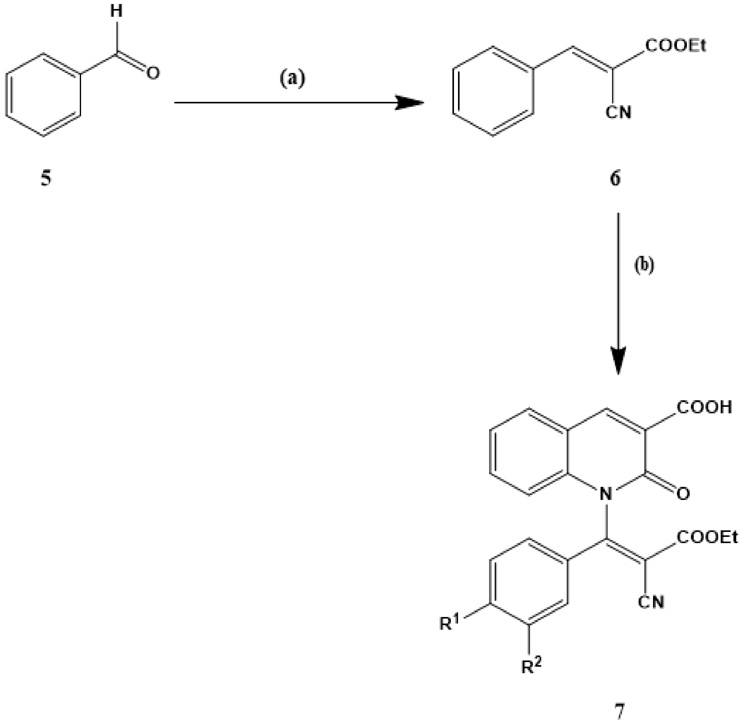
The NMR spectrum of compound 4 showed multiple signals at δ = 7.43–8.01 ppm, with four protons of the aromatic ring and one proton of the NH group. In addition, the 1H-NMR spectrum data for compound 4 gave conclusive evidence of two single signals at 8.89 and 8.10 ppm, indicating the proton of the quinolinone ring at position 4 and the hydroxyl function (OH) position of the carboxyl group. The 13C-NMR spectrum of compound 4 showed four signals at 163.00, 160.82, 154.51, and 148.30 ppm assigned to carbon for two carbonyl groups (C=O), C–N, and C-4 from the quinolinone ring. In addition, the 13C-NMR spectrum of compound 4 showed six-carbon signals in the 134.59–116.61 ppm region due to the carbon residue in the quinolinone ring. Condensation of a variety of aromatic aldehydes (5ad) with ethyl cyanoacetate into ethanol in the presence of a base catalyst resulted in the formation of ethyl 3-aryl-2-cyanoacrylate (6ad). The compound 1-(2-(ethoxy)carbonyl-2-cyano-1-arylvinyl)-2-oxo-1,2-dihydroquinoline-3-carboxylic (7ad) was prepared by reaction of quinoline-3-carboxylic acid (4) with ethyl 3-aryl-2-cyanoacrylates (i.e., ethyl 3-phenyl-2-cyanoacrylate, ethyl 3-(4-methoxy) phenyl-2-cyanoacrylate, ethyl 3-(4-hydroxy-3-methoxy) phenyl-2-cyanoacrylate, and ethyl 3-(4-N,N-dimethylamino) phenyl-2-cyanoacrylate) in the presence of triethylamine as a primary catalyst in dimethyl formation under the reflux (Scheme 2).
The structure of the compounds obtained was determined (7ad) by spectral data. In the infrared spectra, large expansion bonds belonging to OH, C=O for ester, amide, and acid groups, C=C, and CN were observed. 1H-NMR spectra of 7ad compounds showed two sharp signals at δ = 1.27–1.34 ppm as triple signal and δ = 4.25–4.35 ppm as quadruple signal due to protons of the ethoxy groups (OCH2CH3) (Supplementary Materials Figures S1–S8).
Regarding compounds 7ad, two protons appeared as singlet signal at δ 8.88–8.89 ppm refer to the H-4 of quinolinone ring and hydroxyl function (OH) for the carboxylic acid groups. The entire aromatic proton peaks in the 1H-NMR spectra were also recorded at estimated regions (Supplementary Materials Figures S1–S8). The 13C-NMR spectra of compounds 7ad displayed three-carbon signals at δ 164.81–162.99 ppm, 162.99–162.28 ppm, and δ 162.51–160.81 ppm due to carbons of three carbonyl function (C=O) of ester, amide, and acid (Supplementary Materials Figures S1–S8). In addition, the carbons of cyano groups (CN) for the compounds 7ad in the 13C-NMR spectra were observed with the expected chemical shift in the region of δ 103.07–92.44 ppm (Supplementary Materials Figures S1–S8). At the final step, the compounds 7ad were reacted with hydrazine hydrate in ethanol under reflux to give 1-[(aryl)(3-amino-5-oxopyrazolidine-4-ylidene)methyl]-2-oxo-1,2-dihydroquinoline-3-carboxylic acid (8ad, Scheme 3).
IR, 1H-NMR, 13C-NMR, and elemental analyses have described all the new compounds 8ad. The IR spectra of compounds 8ad revealed the absence of carbonyl function of ester and cyano groups; the appearance of new stretching bonds at 3385 and 3228 indicated the presence of amino (NH2) and NH groups. The 1H-NMR spectra of compounds 8ad showed the appearance of new signals at δ = 6.76–7.99 and 11.19–11.16 ppm for characteristic amino (NH2) and amino NH groups, with the absence of any signals for the ethoxy protons at δ 1.32 and 4.32 ppm (Supplementary Materials Figures S9–S16). Moreover, the 1H-NMR spectra of compounds 8ad displayed the presence of the characteristic singlet signals in the region at δ 11.33–11.68 ppm and refer to the protons of hydroxy (OH) groups (Supplementary Materials Figures S9–S16), which confirmed the structure of compound 8 in enol form (Scheme 3). The protons of the aromatic rings and quinolinone were observed within the expected chemical transformation regions and showed the expected integrative values. The 13C-NMR spectra of compounds 8ad (Supplementary Materials Figures S9–S16) showed the absence of any carbon signals for the ethoxy and cyano groups that were present in compounds 7ad (Supplementary Materials Figures S1–S8).

3.2. In Vitro Cytotoxic Activity Against MCF-7 Cell Line

To examine the anticancer activity of the prepared quinolone derivatives, the effect of the synthesized compounds on the viability of the MCF-7 cell line was measured by using colorimetric MTT assay after 48 h of incubation. The percentage of cell viability of quinolone derivatives and reference compound are presented in Table 1. The reference compound in our assay is Doxorubicin (Dox). Interestingly, among the quinolone derivatives compound, 1-[2-(ethoxy)carbonyl-2-cyano-1-(4-hydroxy-3-methoxy)phenylvinyl]-2-oxo-1,2-dihydroquinoline-3-carboxylic acid (compound 7c) was found to be superior to the other quinolone derivatives in terms of anticancer activity and showed significant anti-proliferative activity compared to the reference compound Dox with IC50 value of 1.73 ± 0.27 μg/mL.

3.3. Cell-Cycle Analysis

Due to the significance of the cell cycle in the process of tumor cell proliferation, MCF-7 cell growth inhibition, as a result of cell-cycle arrest, was evaluated by using DNA flow cytometric assay. In this assay, MCF-7 cells were dealt with compound 7c at a concentration of 1.73 μM value for 48 h. It is evident from Figure 1 that compound 7c induced cell arrest on 36.04% of cells at the G2/M phase, in comparison with the untreated cells, which had 9.88%. Moreover, compound 7c revealed G0 phase arrest marked by the appearance of a peak at the G0 phase of the cell-cycle distribution profile, which indicates MCF-7 cell apoptosis (Figure 1).
These data suggest that compound 7c causes perturbations during the cell-cycle progression, especially at the G2/M stage. Cell cycle and apoptosis play remarkable roles in the regulatory mechanisms of the development and growth of the cell. The impacts of numerous substances that are utilized as anticancer drugs were discovered to be through the capturing of the cell cycle during the stages G0/G1, S, and G2/M, which exhilarated and initiated apoptosis [30,31,32,33]. Consequently, we presume that the impact of compound 7c on the MCF-7 cell line was through the instabilities influences of the compound 7c in cell-cycle progression particularly at the G2/M stage.

4. Conclusions

In conclusion, we have developed a rapid and efficient synthetic route for the synthesis of 1-[(aryl)(3-amino-5-oxopyrazolidin-4-ylidene) methyl]-2-oxo-1,2-dihydroquinoline-3-carboxylic acid derivatives. The present synthetic pathway has the advantages of operational simplicity, moderate reaction conditions, and good to high yield of bioactive products. Our method is simple, as no extraordinary apparatus, reagents, or chemicals for workup are required, and the compound formed is filtered and purified just by simple crystallization. This synthesis is also beneficial in terms of economy, as well as avoiding any hazardous chemicals. The effect of the synthesized compounds on the viability of MCF-7 cell line was measured by using colorimetric MTT assay after 48 h of incubation. The compounds 7b, 7c, 8b, and 8c showed significant anticancer activity compared to the reference compound Dox against the MCF-7 cell line.

Supplementary Materials

The following are available online at https://www.mdpi.com/article/10.3390/cryst11050571/s1. Figure S1: 1H-NMR spectrum of compound 7a. Figure S2: 13C-NMR spectrum of compound 7a. Figure S3: 1H-NMR spectrum of compound 7b. Figure S4: 13C-NMR spectrum of compound 7b. Figure S5: 1H-NMR spectrum of compound 7c. Figure S6: 13C-NMR spectrum of compound 7c. Figure S7: 1H-NMR spectrum of compound 7d. Figure S8: 13C-NMR spectrum of compound 7d. Figure S9: 1H-NMR spectrum of compound 8a. Figure S10: 13C-NMR spectrum of compound 8a. Figure S11: 1H-NMR spectrum of compound 8b. Figure S12: 13C-NMR spectrum of compound 8b. Figure S13: 1H-NMR spectrum of compound 8c. Figure S14: 13C-NMR spectrum of compound 8c. Figure S15: 1H-NMR spectrum of compound 8d. Figure S16: 13C-NMR spectrum of compound 8d.

Author Contributions

Methodology, A.G., I.M.E.-D., and M.S.R.; software, I.M.E.-D., and M.S.R.; validation, A.G., W.F.A., M.A., and A.S.A.; formal analysis, A.G., M.A., A.S.A., I.M.E.-D., and M.S.R.; data curation, W.F.A., M.A., A.S.A., and I.M.E.-D.; writing—original draft preparation, I.M.E.-D., and M.S.R.; writing—review and editing, A.G., and M.S.R.; supervision, W.F.A., and M.S.R.; project administration, A.G.; funding acquisition, A.G., and W.F.A. All authors have read and agreed to the published version of the manuscript.

Funding

This research was funded by Taif University Researchers Supporting Project number (TURSP-2020/39), Taif University, Taif, Saudi Arabia.

Institutional Review Board Statement

Not applicable.

Informed Consent Statement

Not applicable.

Data Availability Statement

The data presented in this study are available within the article and in the Supplementary Materials.

Acknowledgments

The authors are appreciative the support of Taif University Researchers Supporting Project number (TURSP-2020/39), Taif University, Taif, Saudi Arabia.

Conflicts of Interest

The authors declare no conflict of interest.

References

  1. Vitaku, E.; Smith, D.T.; Njardason, J.T. Analysis of the Structure Diversity, Substitution Patterns, and Frequency of nitrogen heterocycles among U.S. FDA Approved pharmaceuticals. J. Med. Chem. 2014, 57, 10257. [Google Scholar] [CrossRef] [PubMed]
  2. Lautz, T.B.; Jie, C.; Clark, S.; Naiditch, J.A.; Jafari, N.; Qiu, Y.Y.; Zheng, X.; Chu, F.; Madonna, M.B. The effect of vorinostat on the development of resistance to doxorubicin in neuroblastoma. PLoS ONE 2012, 7, e40816. [Google Scholar] [CrossRef] [PubMed] [Green Version]
  3. Lee, J.H.; Choy, M.L.; Marks, P.A. Mechanisms of Resistance to Histone Deacetylase Inhibitors. In Advances in Cancer Research; Steven, G., Ed.; Academic Press: San Diego, CA, USA, 2012; Volume 116, pp. 39–86. [Google Scholar]
  4. Richon, V.M. Cancer biology: Mechanism of antitumour action of vorinostat (suberoylanilide hydroxamic acid), a novel histone deacetylase inhibitor. Br. J. Cancer 2006, 95, 2–6. [Google Scholar] [CrossRef]
  5. Basu, H.S.; Mahlum, A.; Mehraein-Ghomi, F.; Kegel, S.J.; Guo, S.; Peters, N.R.; Wilding, G. Pre-treatment with anti-oxidants sensitizes oxidatively stressed human cancer cells to growth inhibitory effect of suberoylanilide hydroxamic acid (SAHA). Cancer Chemother. Pharmacol. 2011, 67, 705–715. [Google Scholar] [CrossRef] [PubMed] [Green Version]
  6. Mann, B.S.; Johnson, J.R.; He, K.; Sridhara, R.; Abraham, S.; Booth, B.P.; Verbois, L.; Morse, D.E.; Jee, J.M.; Pope, S.; et al. Vorinostat for treatment of cutaneous manifestations of advanced primary cutaneous T-cell lymphoma. Clin. Cancer Res. 2007, 13, 2318–2322. [Google Scholar] [CrossRef] [Green Version]
  7. Kelly, W.K.; O1Connor, O.A.; Krug, L.M.; Chiao, J.H.; Heaney, M.; Curley, T.; MacGregore-Cortelli, B.; Tong, W.; Secrist, J.P.; Schwartz, L.; et al. Phase I study of an oral histone deacetylase inhibitor, suberoylanilide hydroxamic acid, in patients with advanced cancer. J. Clin. Oncol. 2005, 23, 3923–3931. [Google Scholar] [CrossRef]
  8. Rundall, B.K.; Denlinger, C.E.; Jones, D.R. Combined histone deacetylase and NF-kB inhibition sensitizes non-small cell lung cancer to cell death. Surgery 2005, 138, 360–367. [Google Scholar] [CrossRef] [PubMed]
  9. Panda, P.; Chakroborty, S. Navigating the Synthesis of Quinoline Hybrid Molecules as Promising Anticancer Agents. ChemistrySelect 2020, 5, 10187–10199. [Google Scholar] [CrossRef]
  10. Jain, S.; Chandra, V.; Jain, P.K.; Pathak, K.; Pathak, D.; Vaidya, A. Comprehensive review on current developments of quinoline-based anticancer agents. Arab. J. Chem. 2019, 12, 4920–4946. [Google Scholar] [CrossRef] [Green Version]
  11. Matveeva, M.D.; Purgatorio, R.; Voskressensky, L.G.; Altomare, C.D. Pyrrolo[2,1-a]isoquinoline scaffold in drug discovery: Advances in synthesis and medicinal chemistry. Future Med. Chem. 2019, 11, 2735–2755. [Google Scholar] [CrossRef]
  12. Cancer Today. Available online: http://gco.iarc.fr/today/home (accessed on 19 May 2019).
  13. Chen, Y.L.; Huang, C.J.; Huang, Z.Y.; Tseng, C.H.; Chang, F.S.; Yang, S.H.; Lin, S.R.; Tzeng, C.C. Synthesis and antiproliferative evaluation of certain 4-anilino-8-methoxy-2-phenylquinoline and 4-anilino-8-hydroxy-2-phenylquinoline derivatives. Bioorg. Med. Chem. 2006, 14, 3098–3105. [Google Scholar] [CrossRef] [PubMed]
  14. Chen, Y.L.; Zhao, Y.L.; Lu, C.M.; Tzeng, C.C.; Wang, J.P. Synthesis, cytotoxicity, and anti-inflammatory evaluation of 2-(furan-2-yl)-4-(phenoxy)quinoline derivatives. Bioorg. Med. Chem. 2006, 14, 4373–4378. [Google Scholar] [CrossRef]
  15. Feng, Y.; Lau, E.; Scortegagna, M.; Ruller, C.; De, S.K.; Barile, E.; Krajewski, S.; Aza-Blanc, P.; Williams, R.; Pinkerton, A.B.; et al. Inhibition of melanoma development in the Nras(Q61K)::Ink4a−/− mouse model by the small molecule BI-69A11. Pigm. Cell Melanoma Res. 2013, 26, 136–142. [Google Scholar] [CrossRef] [Green Version]
  16. Gholap, A.R.; Toti, K.S.; Shirazi, F.; Kumari, R.; Bhat, M.K.; Deshpande, M.V.; Srinivasan, K.V. Synthesis and evaluation of antifungal properties of a series of the novel 2-amino-5-oxo-4-phenyl-5,6,7,8-tetrahydroquinoline-3-carbonitrile and its analogues. Bioorg. Med. Chem. 2007, 15, 6705–6715. [Google Scholar] [CrossRef]
  17. Fakhfakh, M.A.; Fournet, A.; Prina, E.; Mouscadet, J.F.; Franck, X.; Hocquemiler, R.; Figadere, B. Synthesis and biological evaluation of substituted quinolines: Potential treatment of protozoal and retroviral co-infection. Bioorg. Med. Chem. 2003, 11, 5013. [Google Scholar] [CrossRef]
  18. Srivastava, B.K.R.; Joharapurkar, A.; Raval, S.; Patel, J.Z.; Soni, R.; Raval, P.; Gite, A.; Goswami, A.; Sadhwani, N.; Gandhi, N.; et al. Biarylpyrazole inverse Agonists at the Cannabinoid CBI Receptor: Importance of the C-3 Carboxamide oxygen/Lysine3.28(192) interaction. J. Med. Chem. 2007, 50, 5951. [Google Scholar]
  19. Kim, M.; Park, S.B. An improved synthesis of pyrimidine- and pyrazole-based acyclo-C-nucleosides as carbohydrides. Tetrahed. Lett. 2008, 49, 5080. [Google Scholar]
  20. Prekupec, S.; Makuc, D.; Plavec, J.; Suman, L.; Kral, M.J.; Pavelic, K.; Balzarin, J.I.; Clercq, E.D.; Mintas, M.; Malic, S. Novel C-6 fluorinated acyclic side chain pyrimidine derivatives: Synthesis, 1H and 13C NMR conformation studies, and antiviral and cytostatic evaluations. J. Med. Chem. 2007, 50, 3037. [Google Scholar] [CrossRef] [PubMed]
  21. Agarwal, A.; Srivastava, k.; Puri, S.K.; Chauhan, P.M.S. Synthesis of 2,4,6-trisubstituted pyrimidinesas antimalarial agents. Bioorg. Med. Chem. Lett. 2005, 13, 4645. [Google Scholar] [CrossRef] [PubMed]
  22. Singh, S.K.; Vobbalareddy, S.; Shivaramakrishna, S.; Krishnamaraju, A.; Rajjak, S.A.; Gasturi, S.R.; Akhilaband, Y.V.; Raoa, K. Methanesulfonamide group at position-4 of the C-5-phenyl ring of 1,5-diarylpyrazole affords a potent class of cyclooxygenase-2 (COX-2) inhibitors. Bioorg. Med. Chem. Lett. 2004, 14, 1683. [Google Scholar] [CrossRef] [PubMed]
  23. Dias, L.R.S.; Salvador, R.R.S. Pyrazole carbohydrazide derivatives of pharmaceutical interest. Pharmaceuticals 2012, 5, 317. [Google Scholar] [CrossRef] [Green Version]
  24. Koca, I.; Ozgur, A.; Coskun, K.A.; Tutar, A. Synthesis and anticancer activity of acylthioureas bearing pyrazole moiety. Bioorg. Med. Chem. 2013, 21, 3859. [Google Scholar] [CrossRef]
  25. Selim, M.; Zahran, M.; Belal, A.; Samir, M.; Shedid, S.; Mehany, A.; Elhagali, G.; Ammar, Y. Hybridized Quinoline Derivatives as Anticancer Agents: Designe, Synthesis, biological Evaluation and Molecular Docking. Anti Cancer Agents Med. Chem. 2019, 19, 439. [Google Scholar] [CrossRef]
  26. Pirol, S.C.; Caliskan, B.; Sahin, I.D.; Banoglu, E. Synthesis and preliminary mechanistic evaluation of 5-(p-tolyl)-1-(quinolin-2-yl)pyrazole-3-carboxylic acid amides with potent antiproliferative activity on human cancer cell lines. Eur. J. Med. Chem. 2014, 87C, 140–149. [Google Scholar] [CrossRef]
  27. Bekhit, A.A.; El-Sayed, O.A.; Aboul-Enein, H.Y.; Siddiqui, Y.M.; Al-Ahdal, M.N. Synthesis of aldehyde-sugar derivatives of pyrazoloquinoline as inhibitors of herpes simplex virus type 1 replication. J. Enzyme. Inhib. Med. Chem. 2004, 19, 33. [Google Scholar] [CrossRef] [PubMed] [Green Version]
  28. Zhang, H.Z.; Claassen, G.; Grogran-Grundy, C.; Tseng, B.; Drewe, J.; Cai, S.X. Discovery and structure-activity relationship of N-phenyl-1H-pyrazolo[3,4-b]quinoline-4-amines as a new series of potent apoptosis inducers. Bioorg. Med. Chem. 2008, 16, 222. [Google Scholar] [CrossRef] [PubMed]
  29. Kwan, Y.P.; Saito, T.; Ibrahim, D.; Al-Hassan, F.M.; Ein Oon, C.; Chen, Y.; Jothy, S.L.; Kanwar, J.R.; Sasidharan, S. Evaluation of the cytotoxicity, cell-cycle arrest, and apoptotic induction by Euphorbia hirta in MCF-7 breast cancer cells. Pharm. Biol. 2016, 54, 1223. [Google Scholar] [PubMed] [Green Version]
  30. Zeydi, M.M.; Kalantarian, S.J.; Kazeminejad, Z. Overview on developed synthesis procedures of coumarin heterocycles. J. Iran. Chem. Soc. 2020, 17, 3031–3094. [Google Scholar] [CrossRef]
  31. Torres, K.; Horwitz, S.B. Mechanisms of taxol-induced cell death are concentration dependent. Cancer Res. 1998, 58, 3620–3626. [Google Scholar] [PubMed]
  32. Murray, A.W. Recycling the cell cycle: Cyclins revisited. Cell 2004, 116, 221–234. [Google Scholar] [CrossRef] [Green Version]
  33. Zaki, I.; Abdelhameid, M.K.; El-Deen, I.M.; Abdel Wahab, A.H.A.; Ashmawy, A.M.; Mohamed, K.O. Design, synthesis and screening of 1,2,4-triazinone derivatives as potential antitumor agents with apoptosis inducing activity on MCF-7 breast cancer cell line. Eur. J. Med. Chem. 2018, 56, 563. [Google Scholar] [CrossRef] [PubMed]
Scheme 1. Synthesis of 2-oxo-1,2-dihydroquinoline-3-carboxylic acid (4). Reagents and conditions: (a) diethyl malonate, piperidine/fusion; (b) thiourea, potassium carbonate, ethanol/reflux.
Crystals 11 00571 sch001
Scheme 2. Synthesis of 1-[2-cyano-2-ethoxycarbonyl-1-arylvinyl]-2-oxo-1, 2-dihydroquinoline-3-carboxylic acid derivatives (7ad). Reagents and conditions: (a) ethyl cyanoacetate, Et3N, EtOH reflux; (b) 2-oxo-1,2-dihydroquinoline-3-carboxylic acid, Et3N DMF/reflux. (7a, R1 = R2 = H; 7b, R1 = OCH3, R2 = H; 7c, R1 = OH, R2 = OCH3; 7d, R1 = N(CH3)2, R2 = H).
Crystals 11 00571 sch002
Scheme 3. Synthesis of 2-oxo-1,2-dihydroquinoline-3-carboxylic acid (8ad) bearing pyrazole ring. Reagents and conditions: (a) hydrazine hydrate, EtOH/reflux (a, R1 = R2 = H; b, R1 = OCH3, R2 = H; c, R1 = OH, R2 = OCH3; d, R1 = N(CH3)2, R2 = H).
Crystals 11 00571 sch003
Figure 1. The effect of compound 7c at the concentration of 1.73 μM on the MCF-7 cell-cycle phases. (A) Representative flow cytometry data of the cell-cycle phases. (B) Percentage of the total cell populations at each cell-cycle phase. ** p < 0.01 according to Tukey test.
Crystals 11 00571 g001
Table 1. In vitro anticancer activity of quinolinone derivatives 7ad and 8ad against MCF-7 cell line.
CompoundIC50 Values (μM)/MCF-7
7a>50
7b3.87 ± 0.33 c
7c1.73 ± 0.27 a
7d>50
8a17.32 ± 0.44 e
8b5.67 ± 0.21 d
8c4.03 ± 0.60 c
8d>50
Dox2.82 ± 0.07 b
a–e Each value is the mean of three experiments ± SEM. Different superscript letters designate significant differences (p < 0.05), using Duncan’s multiple range test.
Publisher’s Note: MDPI stays neutral with regard to jurisdictional claims in published maps and institutional affiliations.