Article

The Effect of Crystal Seeds on Calcium Carbonate Ion Pair Formation in Aqueous Solution: A ReaxFF Molecular Dynamics Study

1
State Key Laboratory of High-Efficiency Flexible Coal Power Generation and Carbon Capture Utilization and Storage, Xi’an TPRI Water Management and Environmental Protection Co., Ltd., Xi’an 710054, China
2
Huaneng Zhengning Power Plant, Qingyang 745306, China
3
Huaneng Gansu Energy Development Co., Ltd., Lanzhou 730070, China
4
School of Materials Science and Engineering, Xi’an University of Science and Technology, Xi’an 710054, China
*
Authors to whom correspondence should be addressed.
Crystals 2022, 12(11), 1547; https://doi.org/10.3390/cryst12111547
Submission received: 25 September 2022 / Revised: 21 October 2022 / Accepted: 25 October 2022 / Published: 29 October 2022

Abstract

:
The effect of crystal seeds on calcium carbonate (CaCO3) cluster formation in aqueous solution is of interest in the fields of geochemistry, inorganic chemistry, atmospheric science, biomedicine, biomineralization, and tissue engineering. Due to an instantaneous and microscopic process, it is still experimentally challenging to directly capture the CaCO3 pre-nucleation. This study employed reactive force field (ReaxFF) molecular dynamics simulations to explore the variation among CaCO3 ion pairs in an aqueous solution with or without crystal seeds. The results show that the addition of crystal seeds can improve CaCO3 ion pair formation. We found that the surface of the calcite phase, compared with the metastable vaterite phase, prefers to attach the ion pairs from solution via proton transfer. This work sheds light on the effect of different crystal seeds on CaCO3 ion pair formation as a precursor of pre-nucleation clusters.

1. Introduction

As an insoluble mineral, calcium carbonate (CaCO3) is commonly found in nature and the household [1,2,3]. Crustaceans and marine organisms are mainly comprised of CaCO3 [4,5,6]. Moreover, CaCO3 is the most common fouling substance forming on the surface of the heat exchanger and transfer in engineering [7,8]. Thus, the precipitation/crystallization of CaCO3 has significant implications for optimizing the functionality of tissue biomineralization and economic outcomes.
The nucleation of CaCO3 has been investigated extensively to understand calcium carbonate crystal formation. In theory, the classical nucleation theory (CNT) [9] was proposed to illustrate CaCO3 nucleation in aqueous solutions [10]. A single-step activated process in CNT conflicts with the pre-nucleation process in the nucleation of calcium carbonate [11]. The dynamical nucleation theory (DNT) combined with the liquid drop model [12,13] was later developed to predict the CaCO3 nucleation rate [14]. Nonclassical pathways for CaCO3 nucleation refer to the pre-nucleation cluster (PNC) aggregation and the transformation of amorphous calcium carbonate (ACC), usually operating under a wide array of biologically and environmentally relevant conditions [15,16]. Various ions, polymers, functional groups, etc., were used to control the nucleation and growth of calcium carbonate crystals [17,18,19,20]. Although CaCO3 with various desired microstructures and polymorphs can be obtained, many aspects of the CaCO3 nucleation and crystallization phenomenon remain unclear [21].
In order to understand the mechanism of CaCO3 precipitation from solution, various investigations were performed for the pre-nucleation stage of CaCO3 from a modeling perspective. The monomer structure of CaCO3 hydrate (five water molecules and monodentate coordination mode) was obtained by using Car–Parrinello molecular dynamics (CPMD) simulations [22,23]. The role of the known hydrated CaCO3 polymorphs in forming pre-nucleation clusters in both aqueous and CO2-rich environments was investigated by ab-initio molecular dynamics (AIMD) simulations [24]. Classical molecular dynamics (MD) simulations able to generate nanoseconds to microseconds of simulation of large systems have been extensively used to study calcium carbonate precipitation from solution. In addition, the structure, dynamics, and energetics of hydrated CaCO3 clusters have been analyzed using MD simulations [25,26]. However, the ion pairing and multiple ion binding in calcium carbonate solution are difficult to capture by MD simulations with an empirical force field. Proton transfer is a very important process of CaCO3 precipitation/crystallization. Reactive force fields (ReaxFF) combined with Van der Waals, electrostatic interactions, and covalent terms can address reactivity within the system and are attractive for use in the investigation of CaCO3 nucleation and growth [27].
In this study, ReaxFF MD simulations were employed to study the pre-nucleation of CaCO3 in an aqueous solution. Different anhydrous crystalline polymorphs of CaCO3 were chosen as the template for ion pair formation. The coordination, hydration structure, calcium ions mobility, and concentration distribution are analyzed to reveal the formation and evolution of the CaCO3 ion pair. This work provides evidence of the formation of the CaCO3 ion pair induced by the existence of CaCO3 polymorphs.

2. Simulation Models and Methods

2.1. Models

There are three polymorphs (Figures S1 and S2) (see supplementary materials) of crystalline CaCO3: calcite [28], aragonite [29], and vaterite [30]. The metastable vaterite, in particular, has multiple structures [30]. In this study, three relatively stable vaterite structures (vaterite_p3221, vaterite_c2, and centered_vaterite_c1_1) were chosen as the calcium carbonate seeds (Figure S2). The surface models of all CaCO3 polymorphs were built based on the law of the lowest surface energy and the close-packed plane (Figure 1). It is worth noting that calcium-terminated surfaces for calcite and aragonite were observed owing to their thermodynamically stable forms, while the structures of the most unstable polymorph, vaterite, showed unsaturated oxygen atoms on their surfaces.
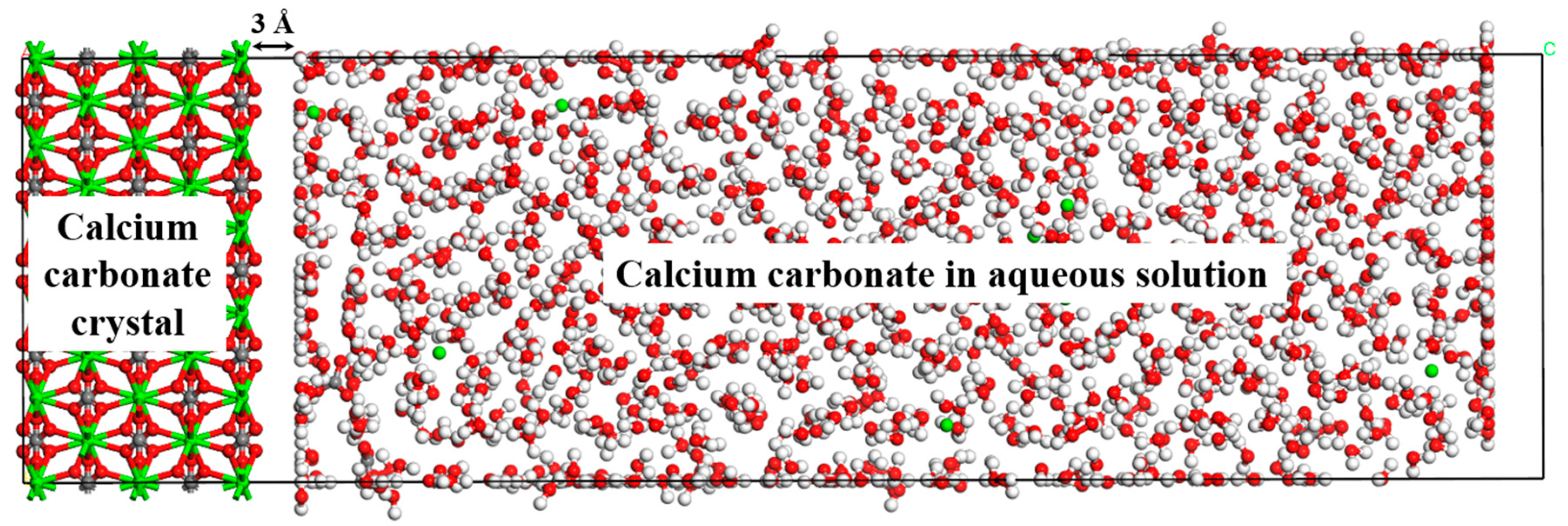
Supersaturation denotes the solution state above the threshold concentration for crystallizing. Supersaturated solution models were built to study the formation of the pre-nucleation CaCO3 cluster in the aqueous solution. High supersaturations (0.5, 1, 2, and 4 mol/L CaCO3 concentration) were chosen in the current calculation owing to the calcium carbonate growth time scales accessible to molecular dynamic simulation. Previously, experimental results have shown that amorphous calcium carbonate forms exceptionally rapidly with increasing calcium carbonate concentrations [31]. Typically, high supersaturations of CaCO3 have been used for the MD simulation of calcium carbonate growth [26,27]. In our study, the initial homogeneous solution systems were created at a very low density by Packmol software [32]. The solution systems were then minimized and deformed to match the desired density (1 g/cm3) and shape of the CaCO3 surfaces, as shown in Figure 1. The computational model consisted of two phases: the CaCO3 crystals as seeds and the CaCO3 solution for growth (Figure 2).

2.2. Methods

Molecular dynamics (MD) simulations were performed using LAMMPS software [33,34]. ReaxFF was employed to simulate the formation of CaCO3 clusters involving the variation of chemical bonds. Aiming at metal carbonates, Duin et al. [27] developed ReaxFF parameters to study their chemical dynamics in aqueous solutions. In this study, we performed a series of canonical ensemble (NVT) ReaxFF MD simulations for CaCO3 cluster formation on the surface of different crystal seeds. The simulated system was equilibrated for 1 ns using a time step of 0.25 fs at a temperature of 360 K (Nosé-Hoover thermostat) [35,36]. According to experimental results [21], vaterite and calcite are generally formed at ambient temperature, while aragonite becomes the main phase above 348 K. A further temperature increase, 360 K or more instead of ambient temperature, can result in less time for cluster formation in MD simulations [25,26]. After the thermal equilibration step, the production step with 5 ns NVT run was performed for the structure and dynamic analysis.

3. Results and Discussion

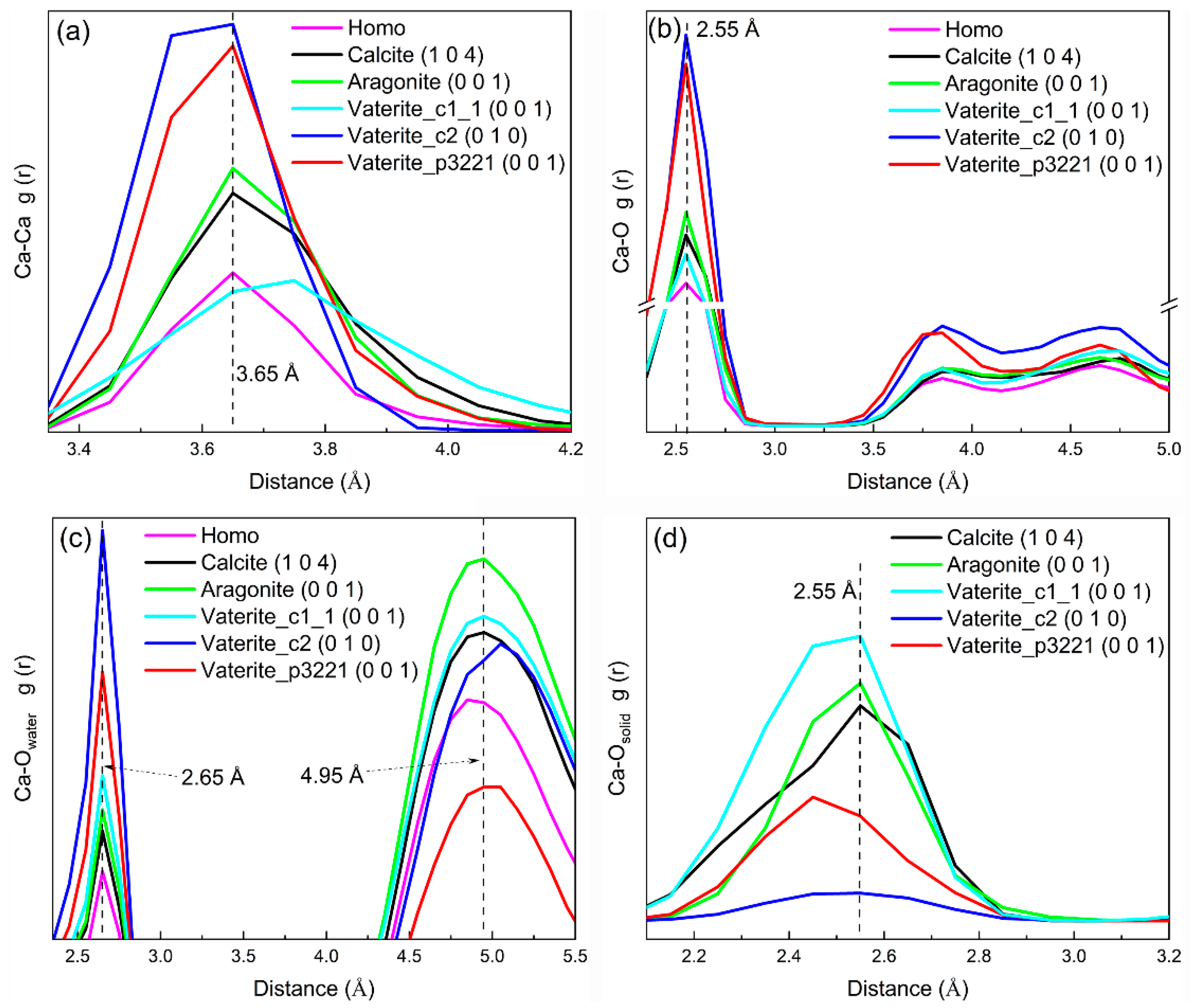
Figure 3 and Figure S3a–d show the radial distribution functions (RDFs) for Ca-Ca, Ca-O (in HCO3), Ca-Owater (in H2O), and Ca-Osolid (in crystalline CaCO3), respectively. The RDFs were generated by averaging the results of the last 2 ns MD simulations in the production stage. Here, the semi-equilibrium structures were presented by RDFs instead of transient structures. Comparing the RDFs of crystalline CaCO3 polymorphs (Figure S4) with the CaCO3 in solution reveals that calcium carbonate in solution has the characteristic of short-range order and long-range disorder as an amorphous structure (Figure 3). We then calculated RDFs for different CaCO3 concentrations (Figure S5). The 2 mol/L concentration, which is the lower-limit concentration for showing relatively obvious RDF characteristics, was chosen in the current study. Since the first peak of RDFs is the strongest one, it can be concluded that small CaCO3 clusters were formed in both homogeneous and heterogeneous systems in this study. The presence of obvious peaks of Ca-Ca RDFs in homogeneous solution at 3.65, 4.95, and 6.85 Å was compared with the two peaks at ~4.0 and ~6.0 Å from the results generated by semi-empirical force field MD simulations [26]. The position of the first Ca-O RDF peak is located at 2.55 Å (Figure 3b) and is slightly larger than the experimental value (2.41 Å) for stabilized amorphous calcium carbonate [37]. For other Ca-O pairs, their first RDF peaks are found at 2.55 Å (Ca-Owater) and 2.65 Å (Ca-Osolid). Considering a higher temperature (360 K) than the experimental condition (ambient temperature), a slight expansion of Ca-O pairs is reasonable.
The intensity of the first peak of Ca-O and C-O in a homogeneous solution is relatively small, indicating a weak tendency for the combination of Ca2+ and CO32− (Figure 3a,b and Figure S6). On the contrary, crystal seeds can strengthen the formation of CaCO3 in solution (stronger peaks in Figure 3). In particular, both the vaterite_c2 and vaterite_p3221 phases show a positive effect in the formation of neutral ion pairs (relatively strong intensity of all peaks in Figure 3b and Figure S5).
A similar increasing trend can be found in the RDFs of the Ca-Owater pair for hydration number (Figure 3c). Reported results showed that the Ca and Owater bonding weakens when the Ca-O (HCO3) RDF peaks are strong [26]. Our study demonstrates that the existence of vaterite_c2 and vaterite_p3221 phases is beneficial for combining calcium and oxygen in solution (Figure 3c,d).
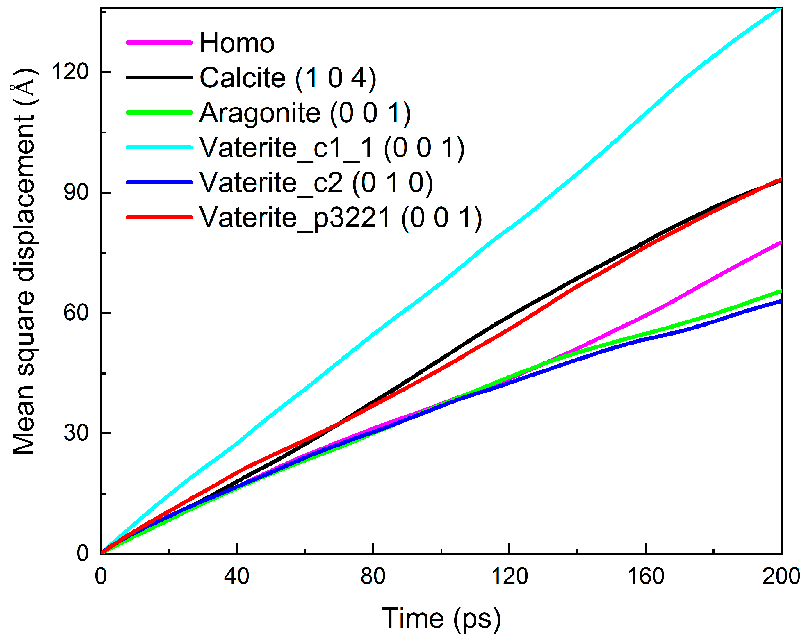
The slopes of the mean square displacement (Figure 4 and Figure S7) are the tracer diffusion coefficients, denoting the mobility of calcium ions in different systems. The effect of different concentrations of CaCO3 on its diffusion has been illustrated in Figure S7. The results showed that a higher density of CaCO3 (4 mol/L) could result in largely weakened diffusion. Therefore, 2 mol/L concentration was chosen for further analysis. Moreover, adding vaterire_c1_1, calcite, or vaterite_p3221 phase could enhance the diffusion of calcium ions (Figure 4), whereas the aragonite and vaterite_c2 phases barely influenced the diffusion of calcium ions. The low mobility of calcium ions in the solution–vaterire_c2 system can be attributed to more binding pairs based on the previous RDF analysis (Figure 3).
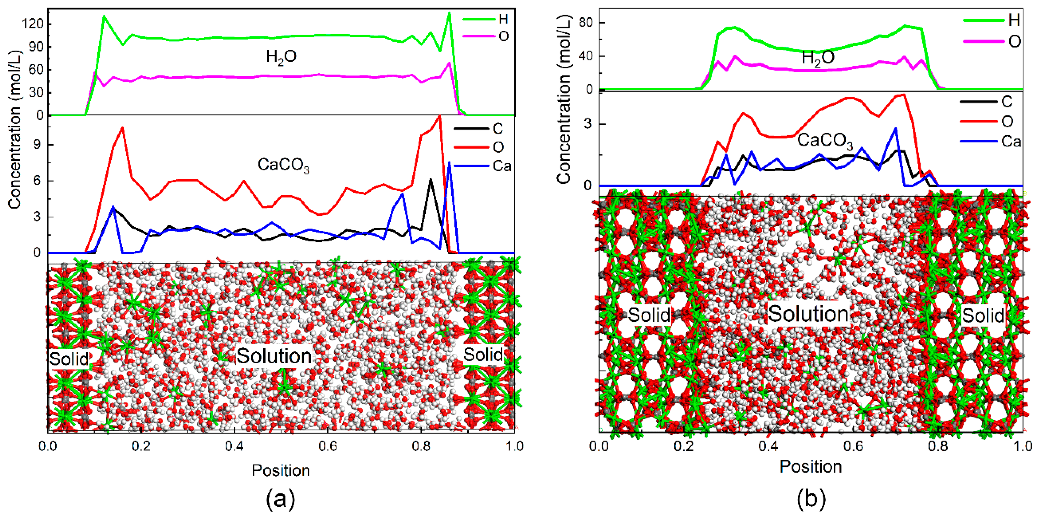
The effect of crystal seeds on calcium carbonate ion pair formation in the aqueous solution was further verified by the concentration profiles of water and calcium carbonate along the direction normal to the solution–solid interface (Figure 5). The solution phase is equally packed along the left and right surface of crystal seeds, generating two interface areas via solution–solid surface atomic interaction. In addition, a zone can be identified within the interface where both the solution and solid atoms overlap. This overlap zone exhibits monotonical decay of the solution density to zero upon approaching the solid surface. The highest concentration of the three types of atoms (Ca, C, and O) that constitute CaCO3 can be found in the solution–calcite interface zone (Figure 5a). The concentration of CaCO3 is high in the solution area when vaterite is used as crystal seeds (Figure 4b). This trend is consistent with the RDFs results shown in Figure 3.
Furthermore, a high concentration of water in the solution–solid interface zone can be observed (top in Figure 5), especially in the solution–calcite system. The water layer at the interface is reported to hinder calcium ions from moving toward the surface of the solid phase [25]. However, in this study, a large amount of CaCO3 can be observed inside the water layer at the interface (Figure 5a).
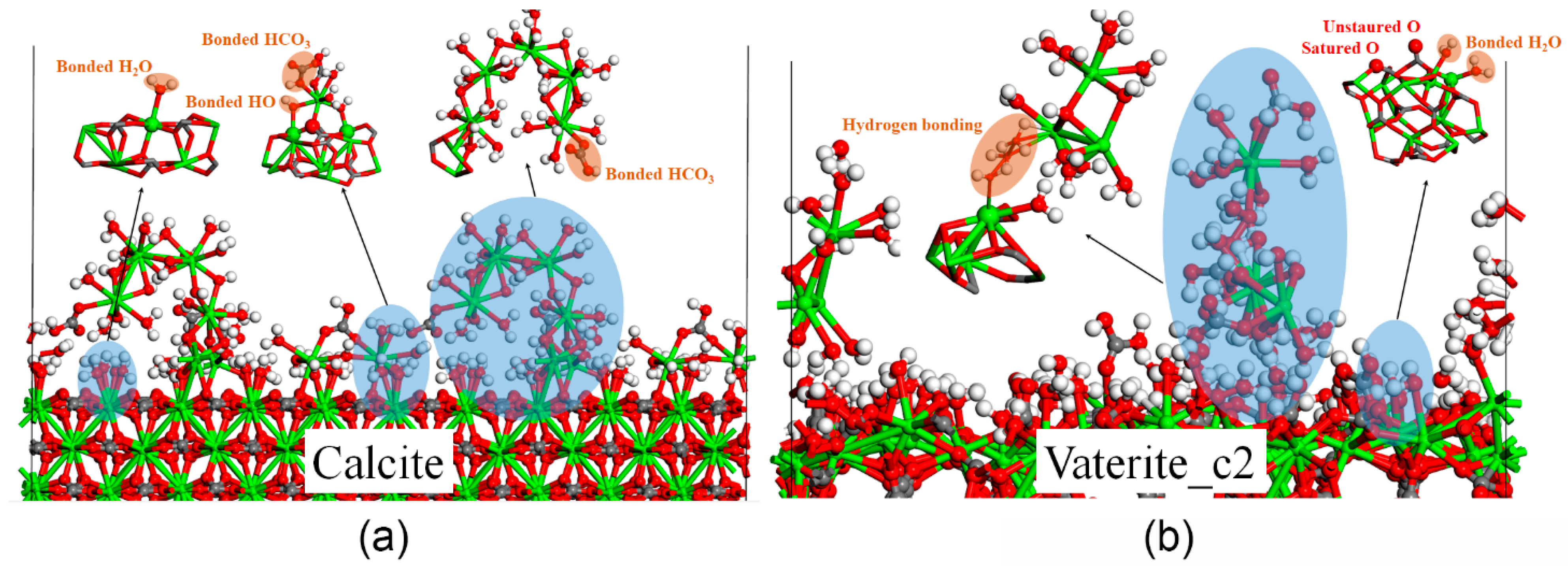
In order to understand the coexistence of a large number of water and CaCO3 molecules, the atomic structures at the solution–solid interface were analyzed and delineated in Figure 6. As shown in Figure 6, water molecules from the solution prefer to bond with the terminated calcium atoms regardless of the type of CaCO3 polymorphs. In the case of calcite, all terminal calcium atoms with a surface density of 0.05 Å−2 are completely saturated by water molecules. The surface density of the top calcium atoms for vaterite (~0.03 Å−2) is smaller than that for the calcite phase. Therefore, noticeable water layers can be observed at the solution–calcite interface (Figure 5).
Once calcium ions approach the surface of calcite crystal, the adsorbed water molecules release one proton and become electron negative, attracting the positive Ca2+ (Figure 6a). More water molecules then surround the Ca2+ to form its first hydration sphere. This hydration sphere around Ca2+, instead of CO32-, has also been reported by other researchers [25,38]. Further growth repeats the process of water dissociation and bonding to new Ca2+.
The connection between the ion pairs and vaterite surface can be attributed to hydrogen bonding among water molecules (Figure 6b). The indirect connection resulted in ion pairs being mainly observed in solution and not on the surface of the vaterite crystal.

4. Conclusions

In this study, the effect of calcium carbonate polymorphs on calcium carbonate ion pair formation in an aqueous solution was investigated by employing ReaxFF molecular dynamic simulations. The results demonstrated that adding crystal seeds, especially vaterite_c2, can improve the combination of calcium carbonate ion pairs. The mobility of Ca2+ in solution can be attributed to the bonding number and the local concentration. A high concentration of water and CaCO3 coexisted at the solution–calcite interface. Atomic structure analysis indicated that the terminal Ca atoms on the crystal surface can be saturated by water molecules from the solution. Moreover, the formation of the hydration sphere of Ca2+ was initially preferred in solution. Later, the CO32- ions bond to Ca2+ by substituting the water in the hydration sphere. The surface water molecules on the calcite crystal release one proton to bond with the hydration sphere of Ca2+. The surface of the metastable vaterite_c2 phase was also found to be connected to the hydration sphere of Ca2+ in solution by hydrogen bonding among water molecules. Thus, the formation of the calcium carbonate ion pair depends on crystal seeds.

Supplementary Materials

The following supporting information can be downloaded at: https://www.mdpi.com/article/10.3390/cryst12111547/s1, Figure S1: Unit cells of CaCO3 polymorphs (a) Calcite and (b) Aragonite; Figure S2: Unit cells of the multiple structures of vaterite CaCO3; Figure S3: Radial distribution functions (RDF) (a) Ca-Ca, (b) Ca-O (in HCO3), (c) Ca-Owater (in H2O), and (d) Ca-Osolid (in crystalline CaCO3) in a 2 mol/L CaCO3 solution at 360 K; Figure S4: Radial distribution functions for crystalline CaCO3 polymorphs; Figure S5: Radial distribution functions for different concentration of CaCO3 on the surface of calcite; Figure S6: Radial distribution functions for C-O pair of CaCO3 in solution; Figure S7: Mean square displacement of different concentration of CaCO3 on the surface of calcite.

Author Contributions

Conceptualization, Z.W. and B.Z.; methodology, Y.Y. and B.Z.; software, Z.W. and B.Z.; validation, Q.J., D.H. and B.Z.; formal analysis, D.H. and J.L.; investigation, Z.W. and Y.Y.; resources, Q.J. and Y.S.; data curation, J.W. (Jing Wang), Y.L., W.X., S.W. and J.W. (Jinlei Wang); writing—original draft preparation, Z.W.; writing—review and editing, B.Z.; project administration, Z.W.; funding acquisition, Z.W. All authors have read and agreed to the published version of the manuscript.

Funding

This research was funded by the Science and Technology Project of China Huaneng Group Co., Ltd. (HNKJ21-H44).

Data Availability Statement

Data is contained within the article. The data presented in this study can be seen in the content above.

Conflicts of Interest

The authors declare no conflict of interest.

References

  1. Jimoh, O.A.; Ariffin, K.S.; Hussin, H.B.; Temitope, A.E. Synthesis of Precipitated Calcium Carbonate: A Review. Carbonates Evaporites 2018, 33, 331–346. [Google Scholar] [CrossRef]
  2. Morse, J.W.; Arvidson, R.S.; Lüttge, A. Calcium Carbonate Formation and Dissolution. Chem. Rev. 2007, 107, 342–381. [Google Scholar] [CrossRef] [PubMed]
  3. Sulpis, O.; Jeansson, E.; Dinauer, A.; Lauvset, S.K.; Middelburg, J.J. Calcium Carbonate Dissolution Patterns in the Ocean. Nat. Geosci. 2021, 14, 423–428. [Google Scholar] [CrossRef]
  4. Meldrum, F.C. Calcium Carbonate in Biomineralisation and Biomimetic Chemistry. Int. Mater. Rev. 2003, 48, 187–224. [Google Scholar] [CrossRef]
  5. Woldetsadik, A.D.; Sharma, S.K.; Khapli, S.; Jagannathan, R.; Magzoub, M. Hierarchically Porous Calcium Carbonate Scaffolds for Bone Tissue Engineering. ACS Biomater. Sci. Eng. 2017, 3, 2457–2469. [Google Scholar] [CrossRef] [PubMed]
  6. Xu, X.; Han, J.T.; Cho, K. Formation of Amorphous Calcium Carbonate Thin Films and Their Role in Biomineralization. Chem. Mater. 2004, 16, 1740–1746. [Google Scholar] [CrossRef]
  7. Andritsos, N.; Karabelas, A.J. Calcium Carbonate Scaling in a Plate Heat Exchanger in the Presence of Particles. Int. J. Heat Mass Transfer 2003, 46, 4613–4627. [Google Scholar] [CrossRef]
  8. Liang, Y.; Xu, Y.; Jia, M.; Wang, J. Experimental Study on the Influence of an Alternating Magnetic Field on the CaCO3 Fouling of a Heat Transfer Surface. Int. J. Heat Mass Transfer 2022, 183, 122156. [Google Scholar] [CrossRef]
  9. Erdemir, D.; Lee, A.Y.; Myerson, A.S. Nucleation of Crystals from Solution: Classical and Two-Step Models. Acc. Chem. Res. 2009, 42, 621–629. [Google Scholar] [CrossRef]
  10. Henzler, K.; Fetisov, E.O.; Galib, M.; Baer, M.D.; Legg, B.A.; Borca, C.; Xto, J.M.; Pin, S.; Fulton, J.L.; Schenter, G.K.; et al. Supersaturated Calcium Carbonate Solutions Are Classical. Sci. Adv. 2018, 4, eaao6283. [Google Scholar] [CrossRef]
  11. Wallace, A.F.; Hedges, L.O.; Fernandez-Martinez, A.; Raiteri, P.; Gale, J.D.; Waychunas, G.A.; Whitelam, S.; Banfield, J.F.; De Yoreo, J.J. Microscopic Evidence for Liquid-Liquid Separation in Supersaturated CaCO3 Solutions. Science 2013, 341, 885–889. [Google Scholar] [CrossRef] [PubMed] [Green Version]
  12. Schenter, G.K.; Kathmann, S.M.; Garrett, B.C. Dynamical Nucleation Theory: A New Molecular Approach to Vapor-Liquid Nucleation. Phys. Rev. Lett. 1999, 82, 3484–3487. [Google Scholar] [CrossRef]
  13. Reguera, D.; Reiss, H. Extended Modified Liquid Drop−Dynamical Nucleation Theory (Emld−Dnt) Approach to Nucleation:  A New Theory. J. Phys. Chem. B 2004, 108, 19831–19842. [Google Scholar] [CrossRef]
  14. Dou, X.; Huang, H.; Han, Y. The Role of Diffusion in the Nucleation of Calcium Carbonate. Chin. J. Chem. Eng. 2022, 43, 275–281. [Google Scholar] [CrossRef]
  15. Gebauer, D.; Völkel, A.; Cölfen, H. Stable Prenucleation Calcium Carbonate Clusters. Science 2008, 322, 1819–1822. [Google Scholar] [CrossRef] [Green Version]
  16. Avaro, J.T.; Wolf, S.L.P.; Hauser, K.; Gebauer, D. Stable Prenucleation Calcium Carbonate Clusters Define Liquid–Liquid Phase Separation. Angew. Chem. Int. Ed. 2020, 59, 6155–6159. [Google Scholar] [CrossRef]
  17. Deng, H.; Shen, X.-C.; Wang, X.-M.; Du, C. Calcium Carbonate Crystallization Controlled by Functional Groups: A Mini-Review. Front. Mater. Sci. 2013, 7, 62–68. [Google Scholar] [CrossRef]
  18. Levi, Y.; Albeck, S.; Brack, A.; Weiner, S.; Addadi, L. Control over Aragonite Crystal Nucleation and Growth: An in Vitro Study of Biomineralization. Chem. Eur. J. 1998, 4, 389–396. [Google Scholar] [CrossRef]
  19. Mergelsberg, S.T.; Riechers, S.L.; Graham, T.R.; Prange, M.P.; Kerisit, S.N. Effect of Cd on the Nucleation and Transformation of Amorphous Calcium Carbonate. Cryst. Growth Des. 2021, 21, 3384–3393. [Google Scholar] [CrossRef]
  20. Mahadevan, G.; Ruifan, Q.; Hian Jane, Y.H.; Valiyaveettil, S. Effect of Polymer Nano- and Microparticles on Calcium Carbonate Crystallization. ACS Omega 2021, 6, 20522–20529. [Google Scholar] [CrossRef]
  21. Ma, M.; Wang, Y.; Cao, X.; Lu, W.; Guo, Y. Temperature and Supersaturation as Key Parameters Controlling the Spontaneous Precipitation of Calcium Carbonate with Distinct Physicochemical Properties from Pure Aqueous Solutions. Cryst. Growth Des. 2019, 19, 6972–6988. [Google Scholar] [CrossRef]
  22. Tommaso, D.D.; de Leeuw, N.H. The Onset of Calcium Carbonate Nucleation: A Density Functional Theory Molecular Dynamics and Hybrid Microsolvation/Continuum Study. J. Phys. Chem. B 2008, 112, 6965–6975. [Google Scholar] [CrossRef] [PubMed]
  23. Di Tommaso, D.; Ruiz-Agudo, E.; de Leeuw, N.H.; Putnis, A.; Putnis, C.V. Modelling the Effects of Salt Solutions on the Hydration of Calcium Ions. Phys. Chem. Chem. Phys. 2014, 16, 7772–7785. [Google Scholar] [CrossRef] [Green Version]
  24. Chaka, A.M. Ab Initio Thermodynamics of Hydrated Calcium Carbonates and Calcium Analogues of Magnesium Carbonates: Implications for Carbonate Crystallization Pathways. ACS Earth Space Chem. 2018, 2, 210–224. [Google Scholar] [CrossRef]
  25. Tribello, G.A.; Bruneval, F.; Liew, C.; Parrinello, M. A Molecular Dynamics Study of the Early Stages of Calcium Carbonate Growth. J. Phys. Chem. B 2009, 113, 11680–11687. [Google Scholar] [CrossRef] [PubMed]
  26. Wang, B.-B.; Xiao, Y.; Xu, Z.-M. Variation in Properties of Pre-Nucleation Calcium Carbonate Clusters Induced by Aggregation: A Molecular Dynamics Study. Crystals 2021, 11, 102. [Google Scholar] [CrossRef]
  27. Dasgupta, N.; Chen, C.; van Duin, A.C.T. Development and Application of Reaxff Methodology for Understanding the Chemical Dynamics of Metal Carbonates in Aqueous Solutions. Phys. Chem. Chem. Phys. 2022, 24, 3322–3337. [Google Scholar] [CrossRef] [PubMed]
  28. Onawole, A.T.; Hussein, I.A.; Carchini, G.; Sakhaee-Pour, A.; Berdiyorov, G.R. Effect of Surface Morphology on Methane Interaction with Calcite: A Dft Study. RSC Adv. 2020, 10, 16669–16674. [Google Scholar] [CrossRef]
  29. De Villiers, J.P.R. Crystal Structures of Aragonite, Strontianite, and Witherite. Am. Mineral. 1971, 56, 758–767. [Google Scholar]
  30. Demichelis, R.; Raiteri, P.; Gale, J.D.; Dovesi, R. The Multiple Structures of Vaterite. Cryst. Growth Des. 2013, 13, 2247–2251. [Google Scholar] [CrossRef]
  31. Wolf, S.E.; Leiterer, J.; Kappl, M.; Emmerling, F.; Tremel, W. Early Homogenous Amorphous Precursor Stages of Calcium Carbonate and Subsequent Crystal Growth in Levitated Droplets. J. Am. Chem. Soc. 2008, 130, 12342–12347. [Google Scholar] [CrossRef] [PubMed]
  32. Martínez, L.; Andrade, R.; Birgin, E.G.; Martínez, J.M. Packmol: A Package for Building Initial Configurations for Molecular Dynamics Simulations. J. Comput. Chem. 2009, 30, 2157–2164. [Google Scholar] [CrossRef] [PubMed]
  33. Plimpton, S. Fast Parallel Algorithms for Short-Range Molecular Dynamics. J. Comput. Phys. 1995, 117, 1–19. [Google Scholar] [CrossRef] [Green Version]
  34. LAMMPS Molecular Dynamics Simulator. Available online: http://lammps.sandia.gov (accessed on 7 August 2019).
  35. Hoover, W.G. Canonical Dynamics Equilibrium Phase Space Distributions. Phys. Rev. A 1985, 31, 1695–1697. [Google Scholar] [CrossRef] [PubMed] [Green Version]
  36. Nosé, S. A Unified Formulation of the Constant Temperature Molecular-Dynamics Methods. J. Chem. Phys. 1984, 81, 511–519. [Google Scholar] [CrossRef] [Green Version]
  37. Michel, F.M.; MacDonald, J.; Feng, J.; Phillips, B.L.; Ehm, L.; Tarabrella, C.; Parise, J.B.; Reeder, R.J. Structural Characteristics of Synthetic Amorphous Calcium Carbonate. Chem. Mater. 2008, 20, 4720–4728. [Google Scholar] [CrossRef]
  38. Iskrenova-Tchoukova, E.; Kalinichev, A.G.; Kirkpatrick, R.J. Metal Cation Complexation with Natural Organic Matter in Aqueous Solutions: Molecular Dynamics Simulations and Potentials of Mean Force. Langmuir 2010, 26, 15909–15919. [Google Scholar] [CrossRef] [PubMed]
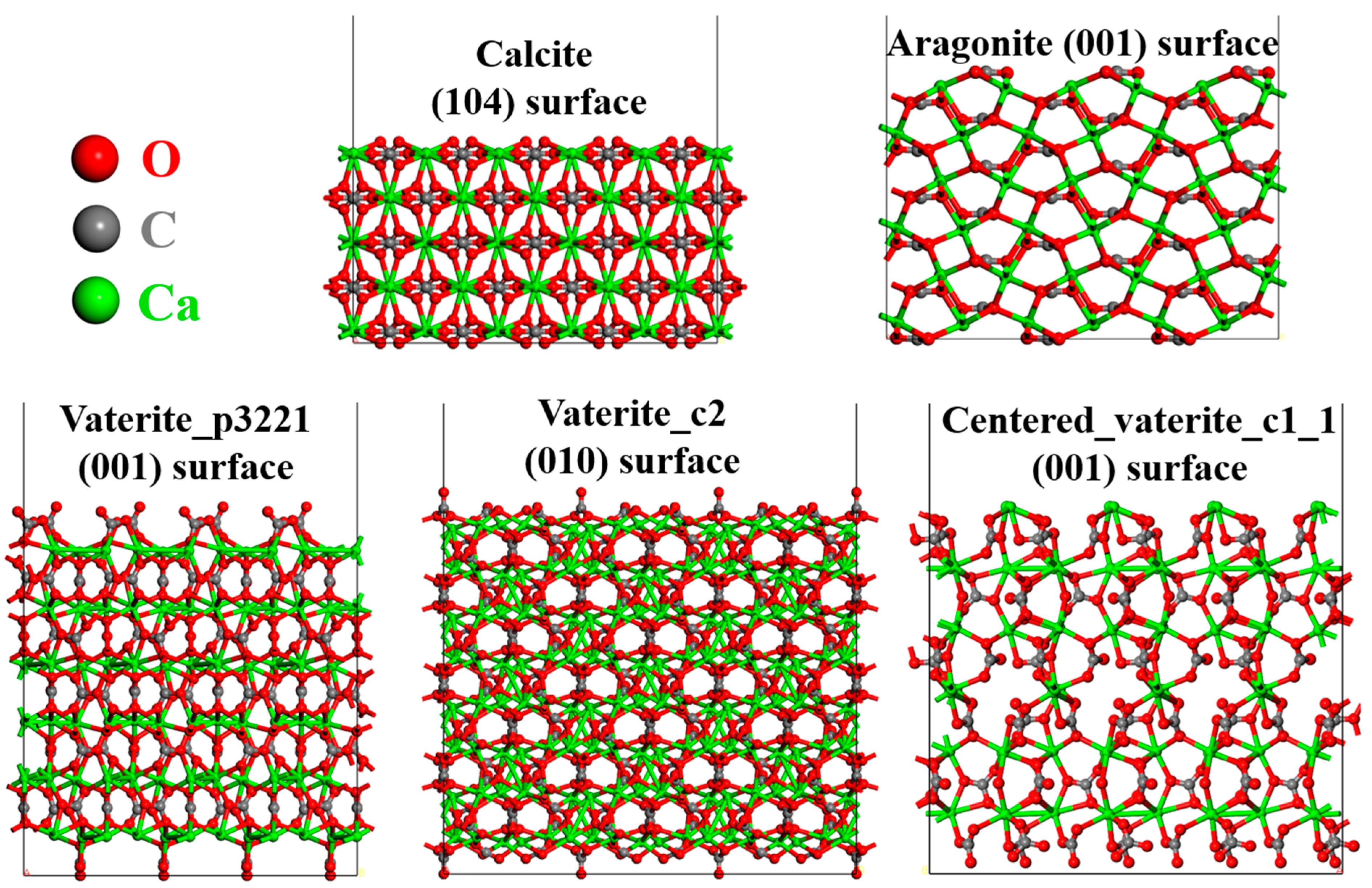
Figure 1. Surface models of different crystal structures of calcium carbonate.
Crystals 12 01547 g001
Figure 2. Computational model consisting of CaCO3 crystals and CaCO3 aqueous solution. The initial width of the crystal–solution interface was 3 Å.
Crystals 12 01547 g002
Figure 3. Radial distribution functions (RDFs): (a) Ca-Ca, (b) Ca-O (in HCO3), (c) Ca-Owater (in H2O), and (d) Ca-Osolid (in crystalline CaCO3) in a 2 mol/L CaCO3 solution at 360 K.
Crystals 12 01547 g003
Figure 4. Mean square displacement of calcium ions in different models.
Crystals 12 01547 g004
Figure 5. Profile of water and calcium carbonate in the direction perpendicular to (a) calcite and (b) vaterite crystal.
Crystals 12 01547 g005
Figure 6. Cluster formation on the surface of (a) calcite and (b) vaterite crystal. The light-blue shaded areas are the selected parts for clear visualization. In addition, the light-orange shaded areas are for those specified groups or interactions (hydrogen bonding here).
Crystals 12 01547 g006
Publisher’s Note: MDPI stays neutral with regard to jurisdictional claims in published maps and institutional affiliations.