
Preparation of Hydroxyapatite-Titanium Dioxide Composite from Eggshell by Hydrothermal Method: Characterization and Antibacterial Activity

 ,
,  ,
, 
Abstract
:1. Introduction
2. Materials and Methods
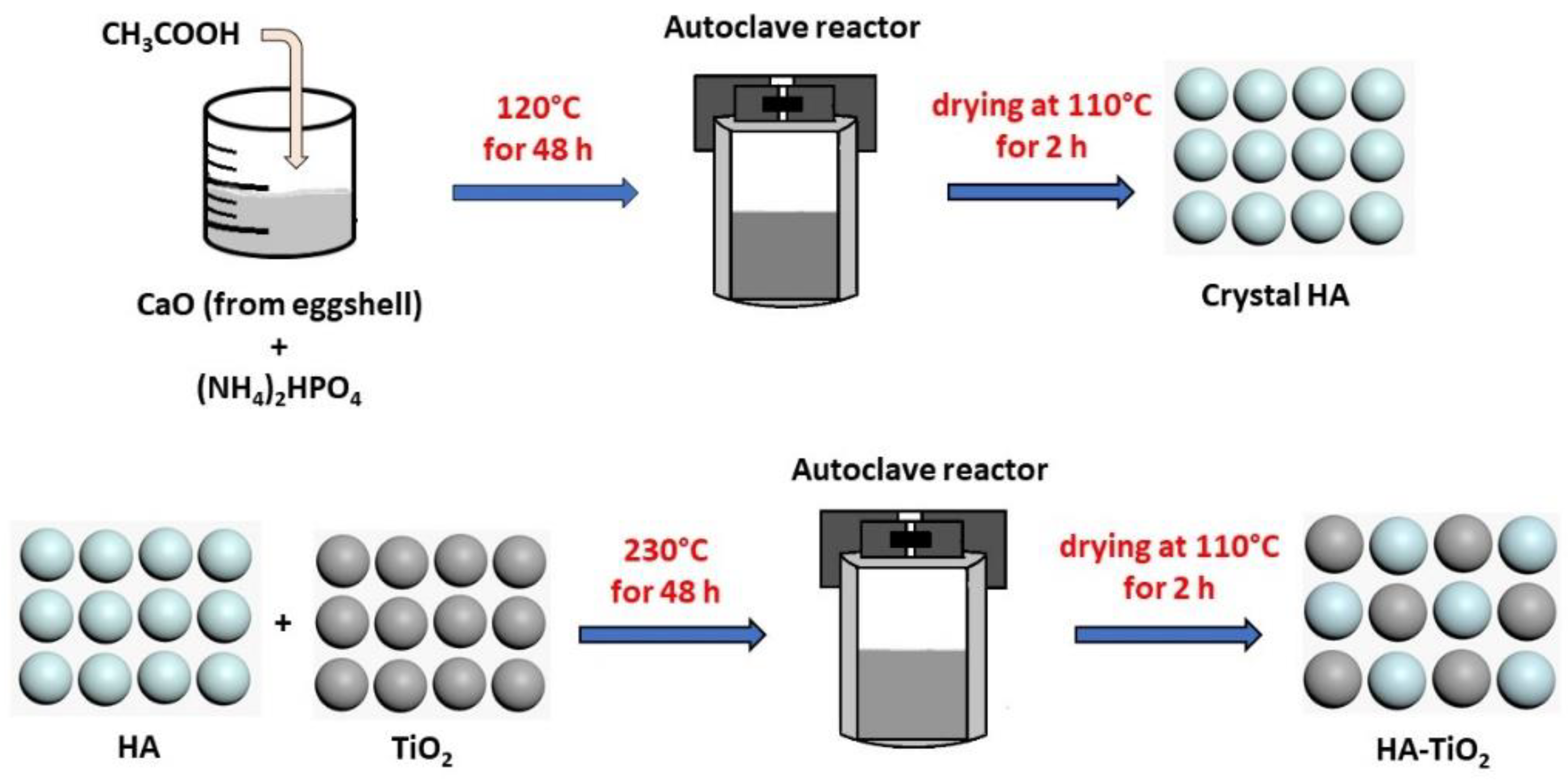
2.1. Synthesis of HA-TiO2 Composites by Hydrothermal Method
2.2. Composite Characterization
2.3. Determination of Antibacterial Activity
2.3.1. Disc Diffusion Method
2.3.2. Optical Density Method
3. Results and Discussions
3.1. XRD Characterization
3.2. FTIR Analysis
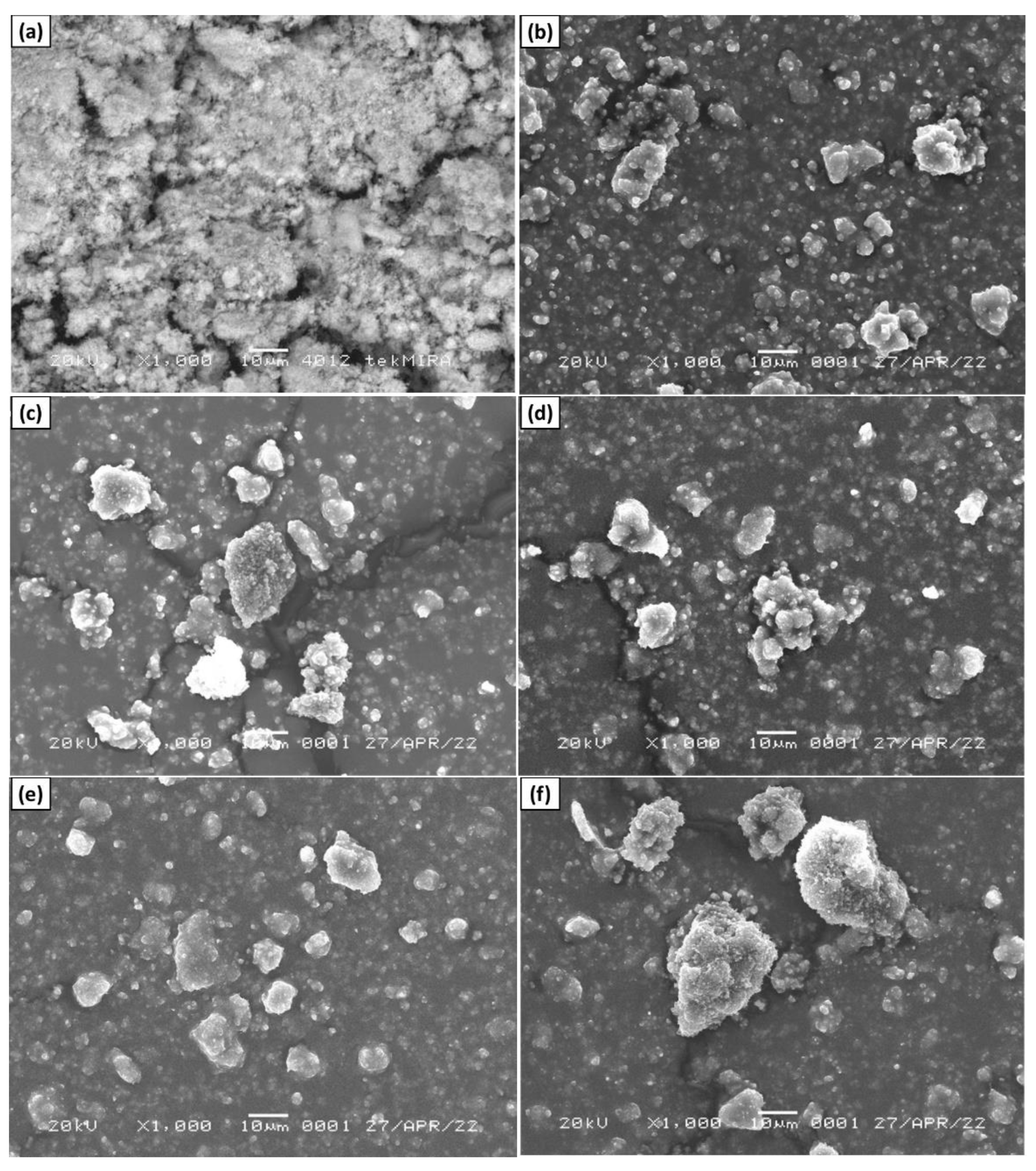
3.3. SEM Analysis
3.4. Antibacterial Activity
4. Conclusions
Supplementary Materials
Author Contributions
Funding
Institutional Review Board Statement
Informed Consent Statement
Data Availability Statement
Acknowledgments
Conflicts of Interest
References
- Uskoković, V. Ion-doped hydroxyapatite: An impasse or the road to follow? Ceram. Int. 2020, 46, 11443. [Google Scholar] [CrossRef]
- Agbeboh, N.I.; Oladele, I.O.; Daramola, O.O.; Adediran, A.A.; Olasukanmi, O.O.; Tanimola, M.O. Environmentally sustainable processes for the synthesis of hydroxyapatite. Heliyon 2020, 6, e03765. [Google Scholar] [CrossRef] [PubMed]
- Mozartha, M. Hidroksiapatit dan aplikasinya di bidang kedokteran gigi. Cakradonya Dent. J. 2015, 7, 835. [Google Scholar]
- Pajor, K.; Pajchel, L.; Kolmas, J. Hydroxyapatite and fluorapatite in conservative dentistry and oral implantology—A review. Materials 2019, 12, 2683. [Google Scholar] [CrossRef] [PubMed] [Green Version]
- Hamzah, S.; Yatim, N.I.; Alias, M.; Ali, A.; Rasit, N.; Abuhabib, A. Extraction of hydroxyapatite from fish scales and its integration with rice husk for ammonia removal in aquaculture wastewater. Indones. J. Chem. 2019, 19, 1019. [Google Scholar] [CrossRef] [Green Version]
- Li, T.T.; Ling, L.; Lin, M.C.; Peng, H.K.; Ren, H.T.; Lou, C.W.; Lin, J.H. Recent advances in multifunctional hydroxyapatite coating by electrochemical deposition. J. Mater. Sci. 2020, 55, 6352. [Google Scholar] [CrossRef]
- Panda, N.N.; Pramanik, K.; Sukla, L.B. Extraction and characterization of biocompatible hydroxyapatite from fresh water fish scales for tissue engineering scaffold. Bioprocess Biosyst. Eng. 2014, 37, 433. [Google Scholar] [CrossRef]
- Hikmah, N.; Nugroho, J.J.; Natsir, N.; Rovani, C.A.; Mooduto, L. Enamel remineralization after extracoronal bleaching using nano-hydroxyapatite (nHA) from synthesis results of blood clam (anadara granosa) shells. J. Dentomaxillofac. Sci. 2019, 4, 28. [Google Scholar] [CrossRef] [Green Version]
- Cacciotti, I. Cationic and anionic substitutions in hydroxyapatite. In Handbook of Bioceramics and Biocomposites; Springer: Cham, Switzerland, 2016; pp. 145–211. [Google Scholar]
- Cacciotti. Multisubstituted hydroxyapatite powders and coatings: The influenoopinge codoping on the hydroxyapatite performances. Int. J. Appl. Ceram. 2019, 16, 186–1884. [Google Scholar]
- Pu’ad, N.M.; Koshy, P.; Abdullah, H.Z.; Idris, M.I.; Lee, T.C. Syntheses of hydroxyapatite from natural sources. Heliyon 2019, 5, e01588. [Google Scholar]
- Gergely, G.; Wéber, F.; Lukács, I.; Tóth, A.L.; Horváth, Z.E.; Mihály, J.; Balázsi, C. Preparation and characterization of hydroxyapatite from eggshell. Ceram. Int. 2010, 36, 803. [Google Scholar] [CrossRef]
- Noviyanti, A.R.; Rahayu, I.; Fauzia, R.P. The effect of Mg concentration to mechanical strength of hydroxyapatite derived from eggshell. Arab. J. Chem. 2021, 14, 103032. [Google Scholar] [CrossRef]
- Noviyanti, A.R.; Akbar, N.; Deawati, Y.; Ernawati, E.E.; Malik, Y.T.; Fauzia, R.P. A novel hydrothermal synthesis of nanohydroxyapatite from eggshell-calcium-oxide precursors. Heliyon 2020, 6, e03655. [Google Scholar] [CrossRef]
- Sirait, M.; Sinulingga, K.; Siregar, N.; Damanik, Y.F. Synthesis and characterization of hydroxyapatite from broiler eggshell. AIP Conf. Proc. 2020, 2221, 110030. [Google Scholar]
- Rivera, E.M.; Araiza, M.; Brostow, W.; Castano, V.M.; Dıaz-Estrada, J.R.; Hernández, R.; Rodrıguez, J.R. Synthesis of hydroxyapatite from eggshells. Mat. Lett. 1999, 41, 128. [Google Scholar] [CrossRef]
- Monmaturapoj, N.; Thepsuwan, W.; Mai-Ngam, K.; Ngernpimai, S.; Klinsukhon, W.; Prahsarn, C. Preparation and properties of hydroxyapatite/titania composite for microbial filtration application. Adv. Appl. Ceram. 2014, 113, 267. [Google Scholar] [CrossRef]
- Monmaturapoj, N.; Sri-On, A.; Klinsukhon, W.; Boonnak, K.; Prahsarn, C. Antiviral activity of multifunctional composite based on TiO2-modified hydroxyapatite. Mater. Sci. Eng. C 2018, 92, 96. [Google Scholar] [CrossRef]
- Nosaka, Y.; Matsushita, M.; Nishino, J.; Nosaka, A.Y. Nitrogen-doped titanium dioxide photocatalysts for visible response prepared by using organic compounds. Sci. Technol. Adv. Mater. 2005, 6, 143. [Google Scholar] [CrossRef]
- Niimi, M.; Masuda, T.; Kaihatsu, K.; Kato, N.; Nakamura, S.; Nakaya, T.; Arai, F. Virus purification and enrichment by hydroxyapatite chromatography on a chip. Sens. Actuators B Chem. 2014, 201, 185. [Google Scholar] [CrossRef]
- Bystrova, A.; Dekhtyar, Y.D.; Popov, A.; Coutinho, J.; Bystrov, V. Modified hydroxyapatite structure and properties: Modeling and synchrotron data analysis of modified hydroxyapatite structure. Ferroelectrics 2015, 475, 135–147. [Google Scholar] [CrossRef]
- Hübner, W.; Blume, A.; Pushnjakova, R.; Dekhtyar, Y.; Hein, H.J. The influence of X-ray radiation on the mineral/organic matrix interaction of bone tissue: An FT-IR microscopic investigation. Int. J. Artif. Organs 2005, 28, 66–73. [Google Scholar] [CrossRef] [PubMed]
- Nikpour, M.R.; Rabiee, S.M.; Jahanshahi, M. Synthesis and characterization of hydroxyapatite/chitosan nanocomposite materials for medical engineering applications. Compos. B. Eng. 2012, 43, 1881. [Google Scholar] [CrossRef]
- Silva-Holguín, P.N.; Reyes-López, S.Y. Synthesis of hydroxyapatite-Ag composite as antimicrobial agent. Dose-Response 2020, 18, 1. [Google Scholar] [CrossRef] [PubMed]
- Ghosh, R.; Swart, O.; Westgate, S.; Miller, B.L.; Yates, M.Z. Antibacterial copper–hydroxyapatite composite coatings via electrochemical synthesis. Langmuir 2019, 35, 5957–5966. [Google Scholar] [CrossRef]
- Irshad, M.A.; Nawaz, R.; ur Rehman, M.Z.; Adrees, M.; Rizwan, M.; Ali, S.; Ahmad, S.; Tasleem, S. Synthesis, characterization and advanced sustainable applications of titanium dioxide nanoparticles: A review. Ecotoxicol. Environ. Saf. 2021, 212, 111978. [Google Scholar] [CrossRef]
- Anandgaonker, P.; Kulkarni, G.; Gaikwad, S.; Rajbhoj, A. Synthesis of TiO2 nanoparticles by electrochemical method and their antibacterial application. Arab. J. Chem. 2019, 12, 1815. [Google Scholar] [CrossRef] [Green Version]
- Luthfiah, A.; Permana, M.D.; Deawati, Y.; Firdaus, M.L.; Rahayu, I.; Eddy, D.R. Photocatalysis of nanocomposite titania–natural silica as antibacterial against Staphylococcus aureus and Pseudomonas aeruginosa. RSC Adv. 2021, 11, 38528. [Google Scholar] [CrossRef]
- Sidane, D.; Rammal, H.; Beljebbar, A.; Gangloff, S.C.; Chicot, D.; Velard, F.; Khireddine, H.; Montagne, A.; Kerdjoudj, H. Biocompatibility of sol-gel hydroxyapatite-titania composite and bilayer coatings. Mater. Sci. Eng. C 2017, 7, 650. [Google Scholar] [CrossRef] [Green Version]
- Pu’ad, N.M.; Haq, R.A.; Noh, H.M.; Abdullah, H.Z.; Idris, M.I.; Lee, T.C. Synthesis method of hydroxyapatite: A review. Mater. Today Proc. 2020, 29, 233. [Google Scholar]
- Teraoka, K.; Nonami, T.; Yokogawa, Y.; Taoda, H.; Kameyama, T. Preparation of TiO2-coated hydroxyapatite single crystals. J. Mater. Res. 2000, 15, 1243. [Google Scholar] [CrossRef]
- Ortiz, G.M.H.; Parra, R.; Fuchs, V.; Fanovich, M.A. TiO2-HA composites obtained by combination of sol–gel synthesis and a supercritical CO2 drying process. J. Solgel. Sci. Technol. 2022, 101, 205–214. [Google Scholar] [CrossRef]
- Poorraeisi, M.; Afshar, A. Synthesizing and comparing HA–TiO2 and HA–ZrO2 nanocomposite coatings on 316 stainless steel. SN Appl. Sci. 2019, 1, 155. [Google Scholar] [CrossRef] [Green Version]
- Wang, X.; Li, B.; Zhou, L.; Ma, J.; Zhang, X.; Li, H.; Liang, C.; Liu, S.; Wang, H. Influence of surface structures on biocompatibility of TiO2/HA coatings prepared by MAO. Mater. Chem. Phys. 2018, 215, 339. [Google Scholar] [CrossRef]
- Vemulapalli, A.K.; Penmetsa, R.M.R.; Nallu, R.; Siriyala, R. HAp/TiO2 nanocomposites: Influence of TiO2 on microstructure and mechanical properties. J. Compos. Mater. 2020, 54, 765. [Google Scholar] [CrossRef]
- He, G.; Hu, J.; Wei, S.C.; Li, J.H.; Liang, X.H.; Luo, E. Surface modification of titanium by nano-TiO2/HA bioceramic coating. Appl. Surf. Sci. 2008, 255, 442. [Google Scholar] [CrossRef]
- Nathanael, A.J.; Lee, J.H.; Mangalaraj, D.; Hong, S.I.; Rhee, Y.H. Multifunctional properties of hydroxyapatite/titania bio-nano-composites: Bioactivity and antimicrobial studies. Powder Technol. 2012, 228, 410. [Google Scholar] [CrossRef]
- Monmaturapoj, N.; Thepsuwan, W.; Wanakitti, S.; Mongkolkachit, C.; Mai-ngam, K.; Ngernpimai, S.; Klinsukhon, W.; Prahsarn, C. Honeycomb Structures of TiO2-modified Hydroxyapatite Composite for Microbial Filtration Application. J. Chem. Eng. Process Technol. 2015, 6, 1. [Google Scholar] [CrossRef]
- Blake, D.M.; Maness, P.C.; Huang, Z.; Wolfrum, E.J.; Huang, J.; Jacoby, W.A. Application of the photocatalytic chemistry of titanium dioxide to disinfection and the killing of cancer cells. Sep. Purif. Technol. 1999, 28, 1. [Google Scholar] [CrossRef]
- Lu, Z.X.; Zhou, L.; Zhang, Z.L.; Shi, W.L.; Xie, Z.X.; Xie, H.Y.; Pang, D.W.; Shen, P. Cell damage induced by photocatalysis of TiO2 thin films. Langmuir 2003, 19, 8765. [Google Scholar] [CrossRef]
- Jin, X.; Guo, Y.; Wang, J.; Wang, Z.; Gao, J.; Kang, P.; Li, Y.; Zhang, X. The preparation of TiO2/hydroxylapatite (TiO2/HA) composite and sonocatalytic damage to bovine serum albumin (BSA) under ultrasonic irradiation. J. Mol. Catal. A Chem. 2011, 341, 89. [Google Scholar] [CrossRef]
- Sadat-Shojai, M.; Khorasani, M.T.; Dinpanah-Khoshdargi, E.; Jamshidi, A. Synthesis methods for nanosized hydroxyapatite with diverse structures. Acta Biomater. 2013, 9, 7591. [Google Scholar] [CrossRef]
- Yao, J.; Zhang, Y.; Wang, Y.; Chen, M.; Huang, Y.; Cao, J.; Ho, W.; Lee, S.C. Enhanced photocatalytic removal of NO over titania/hydroxyapatite (TiO2/HAp) composites with improved adsorption and charge mobility ability. RSC Adv. 2017, 7, 24683. [Google Scholar] [CrossRef] [Green Version]
- Yusuf, A.; Muhammad, N.M.; Noviyanti, A.R.; Risdiana, R. The effect of temperature synthesis on the purity and crystallinity of hydroxyapatite. Key Eng. Mater. 2020, 860, 228. [Google Scholar] [CrossRef]
- Degen, T.; Sadki, M.; Bron, E.; König, U.; Nénert, G. The highscore suite. Powder Diffr. 2014, 29, S13–S18. [Google Scholar] [CrossRef] [Green Version]
- Lapailaka, T.; Triandi, R. Penentuan ukuran Kristal (crystallite size) lapisan tipis PZT dengan metode XRD melalui pendekatan persamaan Debye Scherrer. Erudio J. Educ. Innov. 2013, 1, 24. [Google Scholar]
- Earl, J.S.; Wood, D.J.; Milne, S.J. Hydrothermal synthesis of hydroxyapatite. J. Phys. Conf. Ser. 2006, 26, 268. [Google Scholar] [CrossRef]
- Gómez-Tena, M.P.; Gilabert, J.; Toledo, J.; Zumaquero, E.; Machí, C. Relationship between the Specific Surface Area Parameters Determined Using Different Analytical Techniques. In Proceedings of the XII Foro Global Del Recubrimiento Cerámico, Universitat Jaume I, Castellón, Spain, 14 February 2014; pp. 17–18. [Google Scholar]
- Stevenson, K.J. Review of originpro 8.5. J. Am. Chem. Soc. 2011, 133, 5621. [Google Scholar] [CrossRef]
- Burygin, G.L.; Khlebtsov, B.N.; Shantrokha, A.N.; Dykman, L.A.; Bogatyrev, V.A.; Khlebtsov, N.G. On the enhanced antibacterial activity of antibiotics mixed with gold nanoparticles. Nanoscale Res. Lett. 2009, 4, 794. [Google Scholar] [CrossRef] [Green Version]
- Saenger, A.T.; Kuhs, W.F. Golden Book of Phase Transitions. Wroclaw 2002, 1, 1. [Google Scholar]
- Djerdj, I.; Tonejc, A.M. Structural investigations of nanocrystalline TiO2 samples. J. Alloys Compd. 2006, 413, 159. [Google Scholar] [CrossRef]
- Mariappan, A.; Pandi, P.; Rajeswarapalanichamy, R.; Neyvasagam, K.; Sureshkumar, S.; Gatasheh, M.K.; Hatamleh, A.A. Bandgap and visible-light-induced photocatalytic performance and dye degradation of silver doped HAp/TiO2 nanocomposite by sol-gel method and its antimicrobial activity. Environ. Res. 2022, 211, 113079. [Google Scholar] [CrossRef] [PubMed]
- Toby, B.H. R factors in Rietveld analysis: How good is good enough? Powder Diffr. 2006, 21, 67. [Google Scholar] [CrossRef] [Green Version]
- Afifah, F.; Cahyaningrum, S.E. Synthesis and Characterization of Hydroxyapatite Gel-Nanosilver-Clove Flower Extract (Syzygium Aromaticum L.) as a Toothpaste Forming Gel. Int. J. Curr. Sci. Res. Rev. 2020, 5, 2336. [Google Scholar]
- Odusote, J.K.; Danyuo, Y.; Baruwa, A.D.; Azeez, A.A. Synthesis and characterization of hydroxyapatite from bovine bone for production of dental implants. J. Appl. Biomater. Funct. Mater. 2019, 17, 1. [Google Scholar] [CrossRef] [PubMed]
- Xiao, X.F.; Liu, R.F.; Zheng, Y.Z. Characterization of hydroxyapatite/titania composite coatings codeposited by a hydrothermal–electrochemical method on titanium. Surf. Coat. Technol. 2006, 200, 4406. [Google Scholar] [CrossRef]
- Bernard, P.; Stelmachowski, P.; Brosś, P.; Makowski, W.; Kotarba, A. Demonstration of the influence of specific surface area on reaction rate in heterogeneous catalysis. J. Chem. Educ. 2021, 98, 935–940. [Google Scholar] [CrossRef]
- Zhang, X.; Hou, F.; Li, H.; Yang, Y.; Wang, Y.; Liu, N.; Yang, Y. A strawsheave-like metal organic framework Ce-BTC derivative containing high specific surface area for improving the catalytic activity of CO oxidation reaction. Microporous Mesoporous Mater. 2018, 259, 211–219. [Google Scholar] [CrossRef]
- Sun, J.; Zhang, Z.; Ji, J.; Dou, M.; Wang, F. Removal of Cr6+ from wastewater via adsorption with high-specific-surface-area nitrogen-doped hierarchical porous carbon derived from silkworm cocoon. Appl. Surf. Sci 2017, 405, 372–379. [Google Scholar] [CrossRef]
- Rey, C.; Shimizu, M.; Collins, B.; Glimcher, M.J. Resolution-enhanced fourier transform infrared spectroscopy study of the environment of phosphate ions in the early deposits of a solid phase of calcium-phosphate in bone and enamel, and their evolution with age. I: Investigations in the v4 PO4 domain. Calcif. Tissue Int. 1990, 46, 384–394. [Google Scholar] [CrossRef]
- Šljivić-Ivanović, M.; Smičiklas, I. Utilization of C&D waste in radioactive waste treatment—Current knowledge and perspectives. In Advances in Construction and Demolition Waste Recycling; Woodhead Publishing: Sawston, UK, 2020; p. 475. [Google Scholar]
- Bianco, A.; Cacciotti, I.; Lombardi, M.; Montanaro, L.; Gusmano, G. Thermal stability and sintering behaviour of hydroxyapatite nanopowders. J. Therm. Anal. Calorim. 2007, 88, 237. [Google Scholar] [CrossRef]
- Permana, M.D.; Noviyanti, A.R.; Lestari, P.R.; Kumada, N.; Eddy, D.R.; Rahayu, I. Enhancing the Photocatalytic Activity of TiO2/Na2Ti6O13 Composites by Gold for the Photodegradation of Phenol. ChemEngineering 2022, 6, 69. [Google Scholar] [CrossRef]
- Harwijayanti, W.; Ubaidillah, U.; Triyono, J. Physicochemical Characterization and Antibacterial Activity of Titanium/Shellac-Coated Hydroxyapatite Composites. Coatings 2022, 12, 680. [Google Scholar] [CrossRef]
- Eddy, D.R.; Ishmah, S.N.; Permana, M.D.; Firdaus, M.L.; Rahayu, I.; El-Badry, Y.A.; Hussein, E.E.; El-Bahy, Z.M. Photocatalytic Phenol Degradation by Silica-Modified Titanium Dioxide. Appl. Sci. 2021, 11, 9033. [Google Scholar] [CrossRef]
- Azam, A.; Ahmed, A.S.; Oves, M.; Khan, M.S.; Habib, S.S.; Memic, A. Antimicrobial activity of metal oxide nanoparticles against Gram-positive and Gram-negative bacteria: A comparative study. Int. J. Nanomedicine 2012, 7, 6003. [Google Scholar] [CrossRef] [Green Version]
- Mah, T.F.C.; O’Toole, G.A. Mechanisms of biofilm resistance to antimicrobial agents. Trends Microbiol. 2001, 9, 34. [Google Scholar] [CrossRef]
- Tortora, G.J.; Funke, B.R.; Case, C.L. Microbiology: An introduction; Pearson: Hong Kong, China, 2018. [Google Scholar]
- Shahverdi, A.R.; Fakhimi, A.; Shahverdi, H.R.; Minaian, S. Synthesis and effect of silver nanoparticles on the antibacterial activity of different antibiotics against Staphylococcus aureus and Escherichia coli. Nanomed. Nanotechnol. Biol. Med. 2007, 3, 168. [Google Scholar] [CrossRef] [PubMed]
- Chaiarwut, S.; Niyompanich, J.; Ekabutr, P.; Chuysinuan, P.; Pavasant, P.; Supaphol, P. Development and characterization of antibacterial hydroxyapatite coated with mangosteen extract for bone tissue engineering. Polym. Bull. 2021, 78, 3543. [Google Scholar] [CrossRef]
- Yamamoto, O.; Hotta, M.; Sawai, J.; Sasamoto, T.; Kojima, H. Influence of powder characteristic of ZnO on antibacterial activity effect of specific surface area. J. Ceram. Soc. Jpn. 1998, 106, 1007. [Google Scholar] [CrossRef]
- Sotiriou, G.A.; Teleki, A.; Camenzind, A.; Krumeich, F.; Meyer, A.; Panke, S.; Pratsinis, S.E. Nanosilver on nanostructured silica: Antibacterial activity and Ag surface area. Chem. Eng. J. 2011, 170, 547. [Google Scholar] [CrossRef] [Green Version]
- Wang, S.; Zheng, F.; Huang, Y.; Fang, Y.; Shen, M.; Zhu, M.; Shi, X. Encapsulation of amoxicillin within laponite-doped poly (lactic-co-glycolic acid) nanofibers: Preparation, characterization, and antibacterial activity. ACS Appl. Mater. Interfaces 2012, 4, 6393. [Google Scholar] [CrossRef]
- Akhtar, S.; Shahzad, K.; Mushtaq, S.; Ali, I.; Rafe, M.H.; Fazal-ul-Karim, S.M. Antibacterial and antiviral potential of colloidal Titanium dioxide (TiO2) nanoparticles suitable for biological applications. Mater. Res. Express 2019, 6, 105409. [Google Scholar] [CrossRef]
- Ohira, T.; Yamamoto, O. Correlation between antibacterial activity and crystallite size on ceramics. Chem. Eng. Sci. 2012, 68, 355. [Google Scholar] [CrossRef]


| Sample | Crystal Phase (%) * | Rietveld Refinement Parameters | Crystallinity (%) | |||
|---|---|---|---|---|---|---|
| HA | TiO2 | Rexp | Rwp | GoF | ||
| H3T7 | 29.3 ± 0.0 | 70.7 ± 2.2 | 22.60 | 16.29 | 1.92 | 60 |
| H4T6 | 40.1 ± 0.0 | 59.9 ± 2.6 | 22.59 | 16.97 | 1.77 | 61 |
| H5T5 | 52.6 ± 0.0 | 47.4 ± 2.2 | 21.70 | 16.63 | 1.70 | 63 |
| H6T4 | 61.2 ± 0.0 | 38.8 ± 2.8 | 23.33 | 17.64 | 1.75 | 58 |
| H7T3 | 73.0 ± 0.0 | 27.0 ± 1.4 | 18.52 | 15.60 | 1.41 | 36 |
| Sample | Crystallite Size (nm) * | SSA (m2/g) | ||
|---|---|---|---|---|
| HA | TiO2 | HA | TiO2 | |
| H3T7 | 32.5 ± 2.9 | 13.5 ± 2.2 | 58.4 | 113.7 |
| H4T6 | 42.8 ± 3.8 | 16.5 ± 3.4 | 44.4 | 93.0 |
| H5T5 | 35.7 ± 3.8 | 13.6 ± 2.3 | 53.1 | 112.8 |
| H6T4 | 39.2 ± 3.6 | 16.6 ± 3.3 | 48.6 | 92.4 |
| H7T3 | 35.3 ± 7.1 | 17.5 ± 3.5 | 54.0 | 88.4 |
| Sample | HA * | Anatase TiO2 * | ||||
|---|---|---|---|---|---|---|
| a = b (Å) | c (Å) | V (Å3) | a = b (Å) | c (Å) | V (Å3) | |
| H3T7 | 9.420(3) | 6.873(3) | 528.211 | 3.786(1) | 9.475(5) | 135.834 |
| H4T6 | 9.422(3) | 6.875(2) | 528.577 | 3.785(2) | 9.482(6) | 135.871 |
| H5T5 | 9.422(2) | 6.875(2) | 528.578 | 3.786(2) | 9.477(7) | 135.835 |
| H6T4 | 9.425(2) | 6.876(2) | 528.981 | 3.786(2) | 9.477(10) | 135.878 |
| H7T3 | 9.428(2) | 6.879(2) | 529.525 | 3.788(2) | 9.487(8) | 136.112 |
| Vibration Type | Wavenumber (cm−1) | Ref. | ||||||
|---|---|---|---|---|---|---|---|---|
| HA | TiO2 | H3T7 | H4T6 | H5T5 | H6T4 | H7T3 | ||
| Ti–O–Ti | - | 800–450 | 800–450 | 800–450 | 800–450 | 800–450 | 800–450 | [64] |
| PO43− | 1095–472 | - | 1091–744 | 1089 567 | 1089–600 | 1089–570 | 1089–567 | [61] |
| O–H (HA) | 3573 | - | 3571–3399 | 3570–3419 | 3571–3400 | 3571–3435 | 3571–3413 | [44] |
| OH free | - | 1635 | 1635 | 1635 | 1634 | 1636 | 1639 | [64] |
| CO32− | 1456–1403 | - | 1456–1403 | 1456–1404 | 1456–1404 | 1456–1407 | 1456–1410 | [62,63] |
| Sample | Measurement Parameters | ||||
|---|---|---|---|---|---|
| Mean (nm) | Mode (nm) | Median (nm) | Standard deviation (nm) | PI | |
| H3T7 | 220.4 | 90.8 | 194.1 | 143.9 | 1.42 |
| H4T6 | 344.2 | 193.7 | 293.7 | 223.1 | 1.42 |
| H5T5 | 313.5 | 194.1 | 294.2 | 176.7 | 1.32 |
| H6T4 | 292.2 | 196.5 | 290.1 | 167.7 | 1.33 |
| H7T3 | 353.6 | 193.7 | 296.9 | 196.7 | 1.31 |
Publisher’s Note: MDPI stays neutral with regard to jurisdictional claims in published maps and institutional affiliations. |
© 2022 by the authors. Licensee MDPI, Basel, Switzerland. This article is an open access article distributed under the terms and conditions of the Creative Commons Attribution (CC BY) license (https://creativecommons.org/licenses/by/4.0/).
Share and Cite
Noviyanti, A.R.; Asyiah, E.N.; Permana, M.D.; Dwiyanti, D.; Suryana; Eddy, D.R. Preparation of Hydroxyapatite-Titanium Dioxide Composite from Eggshell by Hydrothermal Method: Characterization and Antibacterial Activity. Crystals 2022, 12, 1599. https://doi.org/10.3390/cryst12111599
Noviyanti AR, Asyiah EN, Permana MD, Dwiyanti D, Suryana, Eddy DR. Preparation of Hydroxyapatite-Titanium Dioxide Composite from Eggshell by Hydrothermal Method: Characterization and Antibacterial Activity. Crystals. 2022; 12(11):1599. https://doi.org/10.3390/cryst12111599
Chicago/Turabian StyleNoviyanti, Atiek Rostika, Efa Nur Asyiah, Muhamad Diki Permana, Dina Dwiyanti, Suryana, and Diana Rakhmawaty Eddy. 2022. "Preparation of Hydroxyapatite-Titanium Dioxide Composite from Eggshell by Hydrothermal Method: Characterization and Antibacterial Activity" Crystals 12, no. 11: 1599. https://doi.org/10.3390/cryst12111599
APA StyleNoviyanti, A. R., Asyiah, E. N., Permana, M. D., Dwiyanti, D., Suryana, & Eddy, D. R. (2022). Preparation of Hydroxyapatite-Titanium Dioxide Composite from Eggshell by Hydrothermal Method: Characterization and Antibacterial Activity. Crystals, 12(11), 1599. https://doi.org/10.3390/cryst12111599