Article

Room Temperature Synthesis of Various Color Emission Rare-Earth Doped Strontium Tungstate Phosphors Applicable to Fingerprint Identification

1
Division of Materials Science and Engineering, Silla University, Busan 45985, Korea
2
Research and Business Development Foundation, Engineering Building, Silla University, Busan 45985, Korea
*
Author to whom correspondence should be addressed.
Crystals 2022, 12(7), 915; https://doi.org/10.3390/cryst12070915
Submission received: 2 June 2022 / Revised: 24 June 2022 / Accepted: 24 June 2022 / Published: 27 June 2022
(This article belongs to the Special Issue Optical and Spectroscopic Properties of Rare-Earth-Doped Crystals)

Abstract

:
Crystalline SrWO4 was synthesized at room temperature using a co-precipitation method. To use the SrWO4 as a phosphor, green and red phosphors were synthesized by doping with Tb3+ and Eu3+ rare earth ions. The synthesized samples had a tetragonal structure, and the main peak (112) phase was clearly observed. When the sample was excited using the absorption peak observed in the ultraviolet region, SrWO4:Tb3+ showed an emission spectrum of 544 nm, and SrWO4:Eu3+ showed an emission spectrum of 614 nm. When Tb3+ and Eu3+ ions were co-doped to realize various colors, a yellow-emitting phosphor was realized as the doping concentration of Eu3+ ions increased. When the synthesized phosphor was scattered on a glass substrate with fingerprints, as used in the field of fingerprint recognition, the fingerprint was revealed by green, red, and yellow emissions in response to a UV lamp.

1. Introduction

Crystalline tungsten has excellent thermal and chemical stability and has been applied in various fields. A material that is thermally and chemically stable has high energy transfer efficiency from tungsten ion to rare earth ion in the rare earth doped phosphor; thus, it is suitable for use as a host material [1,2,3]. Rare earth (RE) ions doped in the host lattice can generate high intensity emissions and various emission wavelengths, with a narrow bandgap due to energy transfer between the 4f-4f shells [4,5,6]. The type and site symmetry of the rare earth ions doped in a thermally and chemically stable host lattice are important factors in the performance of various types of lighting, laser, and display devices [7,8,9].
It has been reported that the emission wavelength of phosphors used in various types of light devices can vary depending on the type and concentration of the doped rare earth ions, the sintering temperature, crystal grain size, excitation wavelength, and synthesis conditions [10,11,12,13]. In particular, the main emission wavelength of the rare earth ions is determined by competition between electric dipole transitions and magnetic dipole transitions. If the electric dipole transition is strong, it reacts sensitively to the local environment around the rare earth ions located in the host lattice, but magnetic dipole transitions are hardly affected by external environmental factors [14,15,16]. For example, two types of emission wavelengths occur in a phosphor doped with europium (Eu3+) ions. In one emission spectrum, an orange emission (~597 nm) spectrum is generated by the 5D07F1 magnetic dipole transition, and the other is a rare red orange (~620 nm) emission signal from the 5D07F2 electric dipole transition. It is known that either the magnetic dipole transition or the electric dipole transition will become the main transition depending on whether Eu3+ rare earth ions located in the host lattice are in the inversion-doping region or not, and this determines the emission wavelength [17,18,19]. Yu et al. synthesized the BaWO4:Eu3+, Bi3+ phosphor powder using a solid-state method and chemical immersion method and observed that the red emission at 613 nm increased as the doping concentration of Bi3+ ions increased [20]. Jung et al. synthesized crystalline BaWO4 by preparing a precursor by co-precipitation and heat-treating it at 800 °C. By doping Dy3+, Tb3+, and Sm3+ rare earth ions, phosphors emitting yellow, green, and red were synthesized and applied to anti-counterfeiting [21]. Shinde et al. synthesized NaCaPO4 phosphor doped with Ce3+, Eu3+, and Dy3+ rare earth ions using the combustion method. In the case of the Ce3+ ion-doped phosphor, an emission wavelength of 367 nm was obtained. Blue light emission at 482 nm (4F9/26H15/2, magnetic dipole transition) and light emission at 576 nm (4F9/26H13/2, electric dipole transition) were observed [22].
In this study, crystalline SrWO4 was synthesized at room temperature by co-precipitation. Then, green and red phosphors were synthesized by doping with rare earth ions Tb3+ and Eu3+, respectively, and yellow phosphors were synthesized by co-doping the two rare earth ions. The structure of the synthesized phosphor, the size and shape of particles, and their luminescence characteristics were investigated. The synthesized phosphor was reacted with a UV lamp to visualize a fingerprint using the emission color, suggesting that it can be applied to the field of anti-counterfeiting.

2. Materials and Methods

2.1. Synthesis of SrWO4:RE3+ by Co-Precipitation at Room Temperature

Starting materials: Strontium acetate ((CH3CO2)Sr, Sigma-Aldrich, reagent grade), Sodium tungstate (Na2WO4·2H2O, Sigma-Aldrich, ≥99%), Terbium nitrate (Tb(NO3)3·xH2O, Tb3+, Sigma-Aldrich, 99.999%), Europium nitrate (Eu(NO3)3·6H2O, Eu3+, Sigma-Aldrich, 99.9%)
The synthesis process was as follows. First 1 mmol (CH3CO2)Sr was placed in beaker ‘A’ and stirred with 50 mL distilled water. Then, 1 mmol Na2WO4·2H2O was placed in beaker ‘B’ and stir with 50 mL distilled water (Figure 1). When the solutions in the ‘A’ and ‘B’ beakers are completely dissolved and become transparent, pour the ‘B’ solution into the ‘A’ beaker, and stir at room temperature for about 20 min. The reacted solutions change to a white opaque color and a powder is formed. The formed powder is recovered by centrifugation at 4000 rpm for 10 min. The recovered powder is washed 3 times with distilled water to remove unreacted substances and then centrifuged again to recover the powder and dried at 80 °C for 16 h. (Figure 1). To synthesize the phosphor, 0.25 mmol each of Tb3+ and Eu3+ was added to the ‘A’ beaker and processed in the same manner. White light phosphor was synthesized by co-doping by fixing the amount of Tb3+ and controlling the amount of Eu3+.

2.2. Fabricated Fingerprint Identification Application

To use the synthesized phosphor for fingerprint recognition, a thumb fingerprint was imprinted on a glass substrate. After spraying the synthesized phosphor on the glass substrate and removing the remaining powder with a brush, the fingerprint on the glass substrate could be visualized by illuminating it with a UV lamp, which revealed the unique luminous color of the phosphor.

2.3. Characterization

The crystal structure of the synthesized phosphor powder was measured using an X-ray diffraction apparatus (X’Pert PRO MPD, 40 kV, 30 mA) having Cu–Kα radiation (wavelength: 1.5406 Å) at a scan rate of 4° per minute at a diffraction angle of 10° to 70°. The size and microscopic surface shape of the crystal grains were photographed with a scanning electron microscope (TESCAN MIRA 3 LMH FE-SEM, TESCAN, Brno, Czech Republic), and a fluorescence photometer (FS-2, Scinco) with a xenon lamp was used as a light source to obtain emission and absorption characteristics.

3. Results & Discussion

3.1. Characteristics of SrWO4 and SrWO4:RE3+

Figure 2a shows the XRD measurement results of SrWO4, SrWO4:Tb3+, and SrWO4:Eu3+. SrWO4 synthesized by co-precipitation showed a tetragonal (a = 5.400 Å, b = 5.400 Å, c = 11.910 Å) structure consistent with ICDD # 01-089-2568. The (112) peak, which is the main diffraction peak, was clearly observed, and the sample to which the rare earth was added also clearly exhibited the main peak.
Based on the ‘Lewis’s acid–base’ reaction, the samples synthesized by the co-precipitation method showed an explosive reaction when the solution dissolved in beaker ‘A’ became ‘Homo’ and the solution dissolved in beaker ‘B’ became ‘Lumo’ [23,24], and crystalline SrWO4 was easily synthesized at room temperature. Figure 2b shows the lattice constant change with and without rare earth doping with the (112) plane, which is the main peak of the sample, of the hosts SrWO4, SrWO4:Tb3+ and SrWO4:Eu3+, respectively. The lattice constant of the (112) phase, which is the main peak of SrWO4, was slightly changed by the rare earth doping (SrWO4: 0.291 nm, SrWO4:Tb3+: 0.2892 nm, SrWO4:Eu3+: 0.2892 nm). It is considered that the change in the crystal lattice is due to the doping with rare earth ions, which have a relatively large ionic radius [25]. FE-SEM images of the synthesized samples are shown in Figure 3. The samples showed a long cylindrical shape with and without doping.
The particle size of SrWO4 was about 5.78 μm in the longitudinal direction and about 2.36 μm in the transverse direction (Figure 3a). Rare earth doped SrWO4:Tb3+ particles were about 3.57 μm in the longitudinal direction and about 2.29 μm in the transverse direction (Figure 3b), and SrWO4:Eu3+ particles had a size of about 4.82 μm in the longitudinal direction and about 2.31 μm in the transverse direction (Figure 3c). Krishna at al. reported BaMoO4 is synthesized by reacting with MoO4−2, which is a monomer of oxyanion and grows in the vertical direction immediately after mixing the Ba aqueous solution and Mo aqueous solution. It is reported that the shape of the shuttle was clearly visible due to the larger rift in Oswald. It was synthesized using basic materials and explained by the action of the bases [26].
The host SrWO4 showed absorption in a wide range, from 220 to 340 nm, and peaked at 277 nm. When the sample was excited at the highest peak of 277 nm, it was broad from 350 to 650 nm, and the peak at 492 nm showed blue–white emission spectrum (Figure 4a). Figure 4b shows the emission spectrum of SrWO4:Tb3+ phosphor synthesized by doping Tb3+ rare earth ions into SrWO4. The absorption spectrum of the phosphor powder under 544 nm showed that the band of charge-transfer transition (CTB) generated between the O−2 and W6+ of the WO42− groups was widely distributed in the 210~290 nm region with a peak at 254 nm [27]. When the phosphor powder was excited with 254 nm, peaks at 487, 544, 586, 620, and 649 nm were observed in the emission spectrum. Among these peaks, the intensity of the green emission spectrum produced by the magnetic dipole transition was the strongest. This emission intensity was 2.99 times stronger than the blue emission intensity produced by the electric dipole transition. The Tb3+ ion in the SrWO4 lattice is located at the inversion symmetric site because the emission intensity due to the magnetic dipole transition of green emission is strong [28].
Figure 4c shows the absorption and emission spectrum of the SrWO4:Eu3+ phosphor synthesized by doping with the rare earth ion Eu3+. The absorption spectrum of the phosphor powder under 614 nm shows the absorption spectrum by CTB generated between O2− and Eu3+ ions, which appear over the 230~310 nm region and have a peak at 277 nm, with Eu3+ observed over the 310~400 nm region. Absorption signals due to the 4f-4f transition of ions were observed [29]. The emission spectrum of the synthesized phosphor was measured by excitation at 277 nm. The phosphor powder showed a red–orange emission spectrum with a peak emission intensity at a wavelength of 614 nm and a spectrum with peaks at 590, 650, and 700 nm. Among these peaks, the 614 nm peak due to the electric dipole transition and the 590 nm peak due to the magnetic dipole transition signal had an intensity difference of about 7.94 times, indicating that the Eu3+ ions in the host are located in non-inversion symmetric sites [30].

3.2. Characteristics of the SrWO4: [Eu3+]:[Tb3+] Phosphors
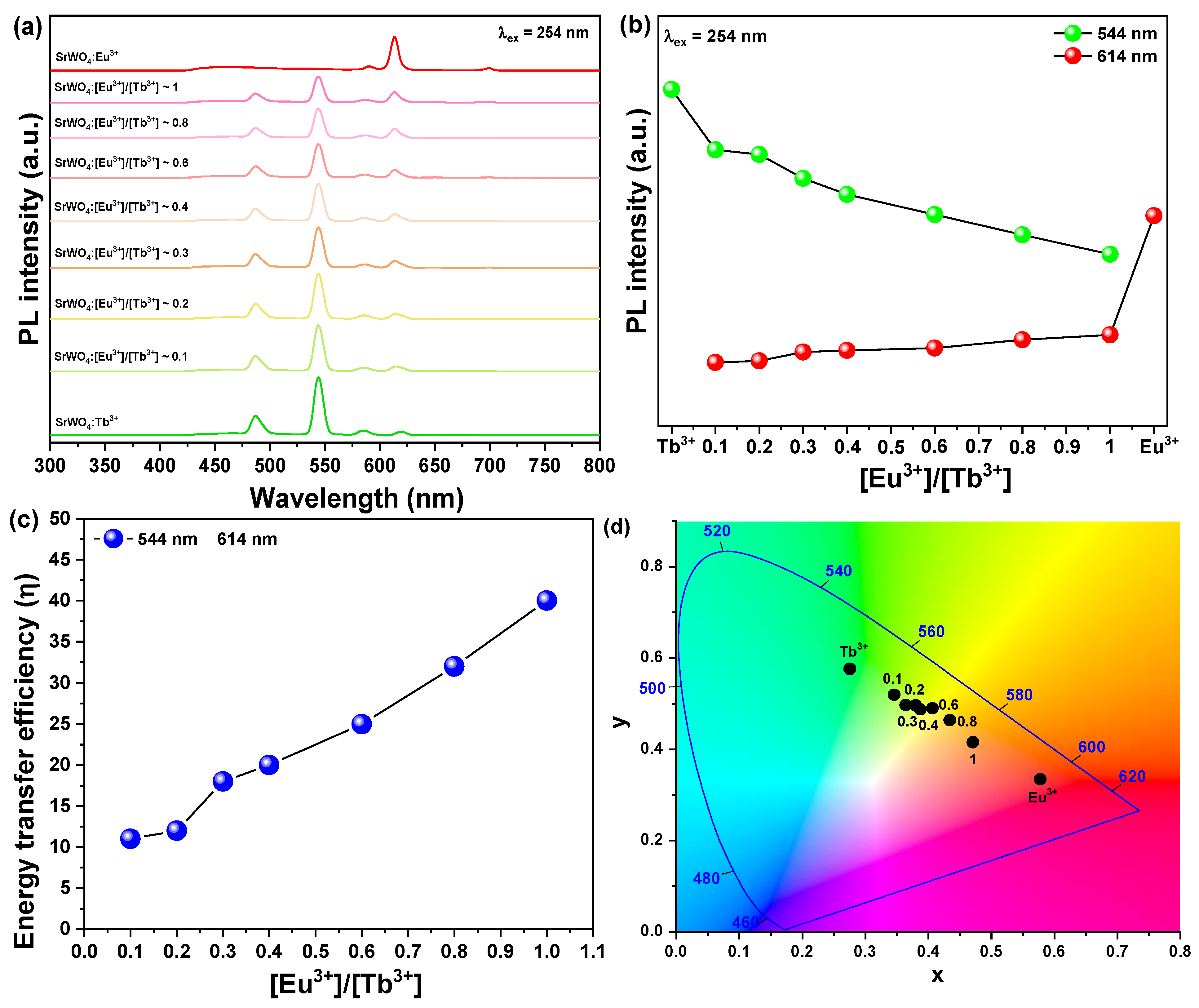
Figure 5a shows the X-ray diffraction peak of SrWO4 co-doped with rare earth ions Tb3+ and Eu3+ as a white light-emitting phosphor. In the XRD pattern of the synthesized samples, a secondary phase caused by rare earth doping was not found, and the diffraction signal of the main peak (112) was clearly observed. Figure 5b shows the lattice constant change of the (112) phase, which is the main peak of the rare earth co-doped SrWO4:[Eu3+]/[Tb3+] samples. Previously, the lattice constants of the SrWO4:Tb3+ and SrWO4:Eu3+ samples doped with a single rare earth decreased, but the lattice constants of the samples doped with both increased. It is believed that the crystal lattice is distorted, or the structure is changed by the amount of added rare earth ions, which have a relatively large ionic radius.
Figure 6 shows the FE-SEM images and energy dispersive X-ray spectroscopy (EDS) mapping component analysis results of the synthesized SrWO4:[Eu3+][Tb3+] phosphor. The shape of the particles grew in the longitudinal direction with a cylindrical shape close to the shape of a dumbbell. The particles were about 3.18 μm in the longitudinal direction and about 1.45 μm in the transverse direction. In the EDS component analysis, Sr, W, O, Tb, and Eu components were detected, which confirmed that rare earth ions had been successfully doped.
Figure 7a shows the emission spectrum of the SrWO4:[Eu3+]/[Tb3+] phosphor powder co-doped with changing Eu3+ ion concentrations, while the Tb3+ ion concentration remained fixed. The emission spectra of the two rare earth ions were shown when excited at a wavelength of 254 nm, as the doping concentration of Eu3+ increased. Green at 544 nm and orange–red at 614 nm were simultaneously observed. As the concentration of Eu3+ ions increased, the intensity of the green emission by Tb3+ ions decreased, which means that the emission energy was converted from Tb3+ ions in the host lattice to Eu3+ ions (Figure 7b). The energy transfer efficiency from Tb3+ to Eu3+ ions can be expressed by Equation (1) [31].
η = 1 I / I 0
Figure 7. (a) Pl spectra under 254 nm, (b) change in PL intensity, (c) energy transfer efficiency, and (d) CIE coordination SrWO4:[Eu3+]/[Tb3+].
Crystals 12 00915 g007
Here, I is the emission intensity of the Tb3+ ions in the SrWO4:[Eu3+]/[Tb3+] phosphors, and I0 is the emission intensity of Tb3+ ions in the SrWO4:Tb3+ phosphors. As shown in Figure 7c, as the amount of added Eu3+ ions increases, the energy transfer efficiency tends to increase. However, the emission intensity decreased, which is a concentration-quenching phenomenon due to excessive rare earth doping [23]. In the CIE color coordinates, as the doping concentration of Eu3+ ions increased, the green coordinates moved to the yellow region (Figure 7d). According to Zhu et al. [32], among the Tb3+ and Eu3+ rare earth ions co-doped with the CaCO3 cubic structure, the green emission of Tb3+ decreases and the intensity of the red emission of Eu3+ increases as the doping concentration of Eu3+ increases. Regarding the energy from Tb3+ to Eu3+ ions, it was reported that a transfer occurred. In this study, as the doping concentration of Eu3+ ions increased in the Tb3+ and Eu3+ ions co-doped with SrWO4, the intensity of green emission decreased, and the intensity of red emission increased as the energy transfer occurred.
Figure 8 shows the schematic energy diagram of terbium and europium ions luminescence mechanisms in SrWO4:[Eu3+]/[Tb3+] phosphors. The phosphors under 254 nm were excited, and 5D3 states of Tb3+ can luminesce non-radiatively to the energetically lower 3D4 excited states. Since the 5D4 states of the Tb3+ and the 5D3 states of Eu3+ are energetically closed to each other, excitation energy availability transferred to the 5D3 states from Eu3+ by the path of resonance transmission. Thus, Tb3+ mainly emitted green peaks due to 5D47F5, and Eu3+ emitted red due to 5D07FJ [32].
Table 1 shows a reported phosphor synthesized by adding various rare earth ions, with tungsten oxide as a host. Several types of phosphors have been reported, such as up-conversion pre-conversion phosphors co-doped with Yb3+ and Er3+ and red phosphors co-doped with Eu3+ and Sm3+. A phosphor was synthesized, and a yellow-emitting phosphor was synthesized by co-doping with Tb3+ and Eu3+ ions to produce various light-emitting materials as in the previously reported research [33,34,35,36,37].

3.3. Applied for Fingerprint Identification

To clearly observe the fingerprint of the author’s thumb on the glass substrate, the synthesized phosphor powder was scattered, and then the shape of the fingerprint was visualized by illuminating it with a UV lamp. The phosphor doped with rare earth ions revealed the fingerprints in emissions of green and red, which are their own colors, and the specimens co-doped with Eu3+ and Tb3+ revealed the fingerprints by emitting yellow light. The results suggest that the synthesized phosphor can be used for fingerprint identification (Figure 9).

4. Conclusions

Crystalline SrWO4 was synthesized at room temperature by co-precipitation. Then, green and red phosphors were synthesized by doping with rare earth ions, Tb3+ and Eu3+, respectively. The synthesized samples clearly exhibited the (112) phase, which was the main peak in the X-ray diffraction pattern, and the lattice constant was changed by doping with rare earth ions. The synthesized specimens had a size of several microns and a cylindrical shape. In addition, when each specimen was excited using an absorption peak in the ultraviolet region, SrWO4:Tb3+ exhibited green, and SrWO4:Eu3+ emitted red due to the doped rare earth. In the specimen co-doped with Tb3+ and Eu3+ to obtain various color emissions, the color coordinates shifted to the yellow region as the doping concentration of Eu3+ ions increased. The synthesized phosphor was scattered on the glass substrate on which the fingerprint was printed, and when a UV lamp was lit, the green, red, and yellow emission colors were visualized so that the fingerprint could be clearly recognized.

Author Contributions

Conceptualization, J.-Y.J.; methodology, J.-Y.J.; software, J.-Y.J.; validation, J.-Y.J. and S.-S.Y.; formal analysis, J.-Y.J.; investigation, J.-Y.J.; resources, J.-Y.J.; data curation, J.-Y.J.; writing—original draft preparation, J.-Y.J. and S.-S.Y.; writing—review and editing, J.-Y.J. and S.-S.Y.; visualization, J.-Y.J.; supervision, J.-Y.J.; project administration, S.-S.Y. and J.-Y.J.; funding acquisition, S.-S.Y. All authors have read and agreed to the published version of the manuscript.

Funding

This research was supported by the Basic Research Program through the National Research Foundation of Korea (NRF) funded by the Ministry of Education (NRF-2020R1F1A1072676).

Data Availability Statement

The data presented in this study are available on request from the corresponding author.

Conflicts of Interest

The authors declare no conflict of interest.

References

  1. Do, Y.R.; Huh, Y.D. Optical Properties of Potassium Europium Tungstate Phosphors. J. Electrochem. Soc. 2000, 147, 4385–4388. [Google Scholar] [CrossRef]
  2. Zhao, Y.; Wang, X.; Zhang, Y.; Li, Y.; Yao, X. Winning wide-temperature-range and high-sensitive thermometry by a multichannel strategy of dual-lanthanides in the new tungstate phosphors. J. Alloys Compd. 2020, 834, 154998. [Google Scholar] [CrossRef]
  3. LEE, G.; KIM, T.; YOON, C.; KANG, S. Effect of local environment and Sm3+-codoping on the luminescence properties in the Eu3+-doped potassium tungstate phosphor for white LEDS. J. Lumin. 2008, 128, 1922–1926. [Google Scholar] [CrossRef]
  4. Hua, Y.; Yu, J.S. Double-excited states of charge transfer band and 4f-4f in single-phase K3Gd(VO4)2:Tb3+/Sm3+ phosphors with superior sensing sensitivity for potential luminescent thermometers. J. Mater. Sci. Technol. 2021, 91, 148–159. [Google Scholar] [CrossRef]
  5. Qin, X.; Liu, X.; Huang, W.; Bettinelli, M.; Liu, X. Lanthanide-Activated Phosphors Based on 4f-5d Optical Transitions: Theoretical and Experimental Aspects. Chem. Rev. 2017, 117, 4488–4527. [Google Scholar] [CrossRef]
  6. Li, L.; Yang, P.; Xia, W.; Wang, Y.; Ling, F.; Cao, Z.; Jiang, S.; Xiang, G.; Zhou, X.; Wang, Y. Luminescence and optical thermometry strategy based on emission and excitation spectra of Pr3+ doped SrMoO4 phosphors. Ceram. Int. 2021, 47, 769–775. [Google Scholar] [CrossRef]
  7. Wu, H.; Niu, P.; Pei, R.; Zheng, Y.; Jin, W.; Li, X.; Jiang, R. Tb3+ and Sm3+ co-doped CaWO4 white light phosphors for plant lamp synthesized via solid state method: Phase, photoluminescence and electronic structure. J. Lumin. 2021, 236, 118146. [Google Scholar] [CrossRef]
  8. Pollnau, M.; Romanyuk, Y.E.; Gardillou, F.; Borca, C.N.; Griebner, U.; Rivier, S.; Petrov, V. Double Tungstate Lasers: From Bulk Toward On-Chip Integrated Waveguide Devices. JSTQE 2007, 13, 661–671. [Google Scholar] [CrossRef] [Green Version]
  9. Semenov, P.A.; Meshchanin, A.P.; Davidenko, A.M.; Kormilitsin, V.A.; Batarin, V.A.; Goncharenko, Y.M.; Stone, S.; Kravtsov, V.I.; Matulenko, Y.A.; Semenov, V.K.; et al. Design and performance of LED calibration system prototype for the lead tungstate crystal calorimeter. Nucl. Instrum. Methods Phys. Res. Sect. A Accel. Spectrometers Detect. Assoc. Equip. 2005, 556, 94–99. [Google Scholar] [CrossRef] [Green Version]
  10. Jung, J.-Y. Luminescent Color-Adjustable Europium and Terbium Co-Doped Strontium Molybdate Phosphors Synthesized at Room Temperature Applied to Flexible Composite for LED Filter. Crystals 2022, 12, 552. [Google Scholar] [CrossRef]
  11. Feng, L.; Chen, X.; Mao, C. A facile synthesis of SrWO4 nanobelts by the sonochemical method. Mater. Lett. 2010, 64, 2420–2423. [Google Scholar] [CrossRef]
  12. Thongtem, T.; Phuruangrat, A.; Thongtem, S. Preparation and characterization of nanocrystalline SrWO4 using cyclic microwave radiation. Curr. Appl. Phys. 2008, 8, 189–197. [Google Scholar] [CrossRef]
  13. Gopal, R.; Kumar, A.; Manam, J. Enhanced photoluminescence and abnormal temperature dependent photoluminescence property of SrWO4:Dy3+ phosphor by the incorporation of Li+ ion. Mater. Chem. Phys. 2021, 272, 124960. [Google Scholar] [CrossRef]
  14. Long, Q.; Xia, Y.; Huang, Y.; Liao, S.; Gao, Y.; Huang, J.; Liang, J.; Cai, J. Na+ induced electric-dipole dominated transition (5D07F2) of Eu3+ emission in AMgPO4:Eu3+ (A = Li+, Na+, K+) phosphors. Mater. Lett. 2015, 145, 359–362. [Google Scholar] [CrossRef]
  15. Roh, H.; Lee, S.; Caliskan, S.; Yoon, C.; Lee, J. Luminescence and electric dipole in Eu3+ doped strontium phosphate: Effect of SiO4. J. Alloys Compd. 2019, 772, 573–578. [Google Scholar] [CrossRef]
  16. Blasse, G.; Bril, A. Luminescence of Phosphors Based on Host Lattices ABO4 (A is Sc, In; B is P, V, Nb). J. Chem. Phys. 1969, 50, 2974–2980. [Google Scholar] [CrossRef]
  17. Song, X.; Wang, X.; Xu, X.; Liu, X.; Ge, X.; Meng, F. Crystal structure and magnetic-dipole emissions of Sr2CaWO6: RE3+ (RE = Dy, Sm and Eu) phosphors. J. Alloys Compd. 2018, 739, 660–668. [Google Scholar] [CrossRef]
  18. Yu, R.; Wang, C.; Chen, J.; Wu, Y.; Li, H.; Ma, H. Photoluminescence Characteristics of Eu3+-Doped Double-Perovskite Phosphors. ECS J. Solid State Sci. Technol. 2014, 3, R33–R37. [Google Scholar] [CrossRef]
  19. Tian, L.; Yu, B.; Pyun, C.; Park, H.L.; Mho, S. New red phosphors BaZr(BO3)2 and SrAl2B2O7 doped with Eu3+ for PDP applications. Solid State Commun. 2004, 129, 43–46. [Google Scholar] [CrossRef]
  20. Yu, P.; Su, L.; Xu, J. Synthesis and Luminescence Properties of Eu3+, Bi3+-Doped BaWO4 Phosphors. Opt. Rev. 2014, 21, 455–460. [Google Scholar] [CrossRef]
  21. Jung, J.; Kim, J.; Shim, Y.; Hwang, D.; Son, C.S. Structure and Photoluminescence Properties of Rare-Earth (Dy3+, Tb3+, Sm3+)-Doped BaWO4 Phosphors Synthesized via Co-Precipitation for Anti-Counterfeiting. Materials 2020, 13, 4165. [Google Scholar] [CrossRef]
  22. Shinde, K.N.; Dhoble, S.J.; Kumar, A. Combustion synthesis of Ce3+, Eu3+ and Dy3+ activated NaCaPO4 phosphors. J. Rare Earths 2011, 29, 527–535. [Google Scholar] [CrossRef]
  23. Qi, G.; Yang, R.T.; Chang, R. MnOx-CeO2 mixed oxides prepared by co-precipitation for selective catalytic reduction of NO with NH3 at low temperatures. Appl. Catal. B Environ. 2004, 51, 93–106. [Google Scholar] [CrossRef]
  24. Thongtem, T.; Kungwankunakorn, S.; Kuntalue, B.; Phuruangrat, A.; Thongtem, S. Luminescence and absorbance of highly crystalline CaMoO4, SrMoO4, CaWO4 and SrWO4 nanoparticles synthesized by co-precipitation method at room temperature. J. Alloys Compd. 2010, 506, 475–481. [Google Scholar] [CrossRef]
  25. Shu, Y.; Travert, A.; Schiller, R.; Ziebarth, M.; Wormsbecher, R.; Cheng, W. Effect of Ionic Radius of Rare Earth on USY Zeolite in Fluid Catalytic Cracking: Fundamentals and Commercial Application. Top. Catal. 2015, 58, 334–342. [Google Scholar] [CrossRef]
  26. Krishna Bharat, L.; Lee, S.H.; Yu, J.S. Synthesis, structural and optical properties of BaMoO4:Eu3+ shuttle like phosphors. Mater. Res. Bull. 2014, 53, 49–53. [Google Scholar] [CrossRef]
  27. Liao, J.; Qiu, B.; Wen, H.; Chen, J.; You, W. Hydrothermal synthesis and photoluminescence of SrWO4:Tb3+ novel green phosphor. Mater. Res. Bull. 2009, 44, 1863–1866. [Google Scholar] [CrossRef]
  28. Barja, B.; Baggio, R.; Garland, M.T.; Aramendia, P.F.; Peña, O.; Perec, M. Crystal structures and luminescent properties of terbium(III) carboxylates. Inorg. Chim. Acta 2003, 346, 187–196. [Google Scholar] [CrossRef]
  29. Tseng, T.; Choi, J.; Davidson, M.; Holloway, P.H. Synthesis and luminescent characteristics of europium dopants in SiO2/Gd2O3 core/shell scintillating nanoparticles. J. Mater. Chem. 2010, 20, 6111–6115. [Google Scholar] [CrossRef]
  30. Dhara, S.; Imakita, K.; Mizuhata, M.; Fujii, M. Europium doping induced symmetry deviation and its impact on the second harmonic generation of doped ZnO nanowires. Nanotechnology 2014, 25, 225202. [Google Scholar] [CrossRef]
  31. Sun, S.; Guo, R.; Zhang, Q.; Lv, X.; Leng, P.; Wang, Y.; Huang, Z.; Wang, L. Efficient deep-blue thermally activated delayed fluorescence emitters based on diphenylsulfone-derivative acceptor. Dye. Pigment. 2020, 178, 108367. [Google Scholar] [CrossRef]
  32. Zhu, H.; Qian, B.; Zhou, X.; Song, Y.; Zheng, K.; Sheng, Y.; Zou, H. Tunable luminescence and energy transfer of Tb3+/Eu3+ co-doped cubic CaCO3 nanoparticles. J. Lumin. 2018, 203, 441–446. [Google Scholar] [CrossRef]
  33. Pandey, A.; Rai, V.K.; Kumar, V.; Kumar, V.; Swart, H.C. Upconversion based temperature sensing ability of Er3+–Yb3+codoped SrWO4: An optical heating phosphor. Sens. Actuators B Chem. 2015, 209, 352–358. [Google Scholar] [CrossRef]
  34. Song, H.; Wang, C.; Han, Q.; Tang, X.; Yan, W.; Chen, Y.; Jiang, J.; Liu, T. Highly sensitive Tm3+/Yb3+ codoped SrWO4 for optical thermometry. Sens. Actuators A Phys. 2018, 271, 278–282. [Google Scholar] [CrossRef]
  35. Li, G.; Gao, S.; He, W. Preparation and photoluminescence properties of the Sm3+, Eu3+ co-doped CaWO4 phosphors. Optik 2015, 126, 3272–3275. [Google Scholar] [CrossRef]
  36. Kang, F.; Hu, Y.; Wu, H.; Mu, Z.; Ju, G.; Fu, C.; Li, N. Luminescence and red long afterglow investigation of Eu3+–Sm3+ CO-doped CaWO4 phosphor. J. Lumin. 2012, 132, 887–894. [Google Scholar] [CrossRef]
  37. Ren, Y.; Liu, Y.; Yang, R. A series of color tunable yellow–orange–red-emitting SrWO4:RE (Sm3+, Eu3+–Sm3+) phosphor for near ultraviolet and blue light-based warm white light emitting diodes. Superlattices Microstruct. 2016, 91, 138–147. [Google Scholar] [CrossRef]
Figure 1. Procedure for co-precipitation.
Crystals 12 00915 g001
Figure 2. (a) XRD patterns, (b) d(112) spacing of SrWO4 and SrWO4:RE3+.
Crystals 12 00915 g002
Figure 3. FE-SEM images of (a) SrWO4, (b) SrWO4:Tb3+, and (c) SrWO4:Eu3+ samples.
Crystals 12 00915 g003
Figure 4. Photoluminescence spectra of (a) SrWO4, (b) SrWO4:Tb3+, and (c) SrWO4:Eu3+ samples.
Crystals 12 00915 g004
Figure 5. (a) XRD patterns of SrWO4:[Eu3+]/[Tb3+] and (b) change of d(112) spacing.
Crystals 12 00915 g005
Figure 6. FE-SEM images and EDS mapping analysis of SrWO4:[Eu3+]/[Tb3+].
Crystals 12 00915 g006
Figure 8. Schematic of Tb3+ and Eu3+ energy levels indicating the energy transfer processes in the SrWO4 phosphors.
Crystals 12 00915 g008
Figure 9. Images of fingerprint coated with phosphors under a UV lamp.
Crystals 12 00915 g009
Table 1. Comparison of previous work in tungsten oxide phosphors.
No.HostRare EarthTypeWavalength (nm)
1 [33]SrWO4Er3+/Yb3+Up conversion489, 525
2 [34]SrWO4Tm3+/Yb3+Up conversion684, 814
3 [35]CaWO4Sm3+/Eu3+Down conversion592, 615
4 [36]CaWO4Eu3+/Sm3+Down conversion622, 630
5 [37]SrWO4Eu3+/Sm3+Down conversion590, 613
This workSrWO4Eu3+/Tb3+Down conversion544, 614
Publisher’s Note: MDPI stays neutral with regard to jurisdictional claims in published maps and institutional affiliations.