Article

Thermal Spike Responses and Structure Evolutions in Lithium Niobate on Insulator (LNOI) under Swift Ion Irradiation

1
Key Laboratory of Particle Physics and Particle Irradiation (MOE), Institute of Frontier and Interdisciplinary Science, Shandong University, Qingdao 266237, China
2
Shanghai Institute of Applied Physics, Chinese Academy of Sciences (CAS), Shanghai 201800, China
*
Author to whom correspondence should be addressed.
Crystals 2022, 12(7), 943; https://doi.org/10.3390/cryst12070943
Submission received: 12 June 2022 / Revised: 30 June 2022 / Accepted: 2 July 2022 / Published: 5 July 2022

Abstract

:
Irradiating solid materials with energetic ions are extensively used to explore the evolution of structural damage and specific properties in structural and functional materials under natural and artificial radiation environments. Lithium niobate on insulator (LNOI) technology is revolutionizing the lithium niobate industry and has been widely applied in various fields of photonics, electronics, optoelectronics, etc. Based on 30 MeV 35Cl and 40Ar ion irradiation, thermal spike responses and microstructure evolution of LNOI under the action of extreme electronic energy loss are discussed in detail. Combining experimental transmission electron microscopy characterizations with numerical calculations of the inelastic thermal spike model, discontinuous and continuous tracks with a lattice disorder structure in the crystalline LiNbO3 layer and recrystallization in the amorphous SiO2 layer are confirmed, and the ionization process via energetic ion irradiation is demonstrated to inherently connect energy exchange and temperature evolution processes in the electron and lattice subsystems of LNOI. According to Rutherford backscattering/channeling spectrometry and the direct impact model, the calculated track damage cross–section is further verified, coinciding with the experimental observations, and the LiNbO3 layer with a thickness of several hundred nanometers presents track damage behavior similar to that of bulk LiNbO3. Systematic research into the damage responses of LNOI is conducive to better understanding and predicting radiation effects in multilayer thin film materials under extreme radiation environments, as well as to designing novel multifunctional devices.

1. Introduction

With the rapid development of nanotechnology, nanomultilayer film systems have attracted more attention due to overcoming the disadvantages of monolithic films and having much better comprehensive properties [1,2]. Irradiation effects on various thin film systems have been reported, including nanocrystal films [3,4], metal films [5,6], polymer multilayer films [7], etc. Among the numerous thin film materials, lithium niobate on insulator (LNOI) technology has achieved tremendous progress in revolutionizing the surface structuring and engineering field, enabling higher performance, lower cost, and entirely new devices and applications, especially in domain wall nanoelectronics and optoelectronics [8,9,10]. The high contrast of the refractive index between the LiNbO3 film and the SiO2 layer allows the realization of integrated optical devices based on photonic wires and bandgap structures for various nonlinear optical applications, such as highly effective low–loss waveguides, electro–optical modulators, and wavelength converters [11,12,13,14]. In addition, the creation of periodic domain patterns with submicron periods in LNOI produced by irradiation etching can be used for second–harmonic generation, backscattering optical parametric oscillators, etc. [15,16]. In view of the structural advantage, micro/nanostructured studies of LNOI wafers under various conditions are still indispensable, and new methods to modify the micro/nanostructure to improve properties are being continually investigated. More recently, as a unique modification technique, swift heavy ion irradiation could deposit energy into the target materials via the intense interaction between incident ions and target electrons (electronic energy loss process), leading to a wide variety of local microstructure and stoichiometry changes. Notably, the induced high–aspect–ratio latent tracks with tailored morphologies enhance structural properties for photonic and optoelectronic applications, such as quantum dots [17], nanowires [18], nanotubes [19], etc. Thus, thermal spike responses in and microstructure evolution of LNOI under the action of extreme electronic energy loss (Eele) are attracting extensive attention, especially for the different damage behaviors in the varying crystalline and amorphous regions [20,21,22].
In this work, by utilizing 30 MeV 35Cl– and 40Ar–ion irradiation to introduce intense electronic energy losses, the microstructure evolution of LNOI is discussed in detail. Combining experimental characterizations, including high–resolution transmission electron microscopy (HRTEM) and Rutherford backscattering/channeling (RBS/channeling) spectrometry, with numerical calculations of the inelastic thermal spike (iTS) model, irradiation–induced track formation in the crystalline LiNbO3 layer and recrystallization behavior in the amorphous SiO2 layer are demonstrated, underlining the structural evolution of layered nanostructure films under the action of swift heavy ion irradiation with intense Eele.

2. Materials and Methods

2.1. Sample Preparation and Ion Irradiation Process

The LNOI wafer obtained from the NanoLN company consists of a Z–cut crystalline LiNbO3 layer with 300 nm thickness, an amorphous SiO2 layer with 1700 nm thickness, and a crystalline Si substrate (Figure 1a), and the relevant preparation processes (physical vapor deposition, lift–off, etc.) are clarified in the references [9,23]. The LNOI samples with dimensions of 10 × 10 × 1 mm3 were cut from the wafer for Cl and Ar ion irradiation at 300 K with a fluence ranging from 3 × 10¹² to 3 × 1013 cm−2. In order to ensure the appropriate ion flux and irradiation time, a focused and scanned Cl ion beam (beam rastering) with an energy of 30 MeV was performed utilizing a 6 MV tandem accelerator within the State Key Laboratory of Nuclear Physics and Technology, Peking University; accordingly, for Ar ion irradiation, a continuous and defocused Ar ion beam with an energy of 30 MeV was performed utilizing the Heavy Ion Research Facility in Lanzhou (HIRFL), Institute of Modern Physics, Chinese Academy of Sciences. During the ion irradiation process, a relatively low beam current density and an ion flux of 1.9 × 109 cm−2 s−1 for Cl ion and 6.5 × 1010 cm−2 s−1 for Ar ion were maintained.

2.2. Experimental Characterizations and Numerical Simulations

The irradiation–induced damage behaviors of LNOI samples were analyzed through TEM observations and RBS/channeling spectrometry. Cross–sectional TEM samples were prepared by focused ion beam (FIB) milling with lift–out processing performed on an FEI Helios NanoLab 600 Dual Beam system, and the observations were performed utilizing an FEI Tecnai G2 F20 transmission electron microscope. In the RBS/channeling analysis, a 2.0 MeV He+ beam with a probe size of 2 × 2 mm2 was extracted from a 1.7 MV tandem accelerator and used to quantitatively assess the irradiation–induced damage level. A Si detector located at a scattering angle of 160° relative to the incoming beam was used to collect the backscattered He+ signal from the pristine and irradiated samples. The Eele and nuclear energy loss (Enucl) distributions in LNOI samples induced by 30 MeV 35Cl and 40Ar ion irradiations were determined through the stopping and range of ions in matter (SRIM) 2013 [24], in which densities of 4.65 g cm−3, 2.62 g cm−3, and 2.32 g cm−3 were used for the LiNbO3 layer, SiO2 layer, and Si substrate, respectively. Under current irradiation conditions, the Enucl will not cause significant lattice damage and could be considered negligible owing to the relatively low fluence and dpa, even at the end of the ion range. For the different regions in the LNOI sample, the Eele distributions are as follows: 7.0 keV/nm and 7.5 keV/nm in the surface LiNbO3 layer, 5.0 keV/nm and 5.4 keV/nm in the buried SiO2 layer, and, subsequently, 3.8 keV/nm and 3.9 keV/nm in the Si substrate, which steadily decrease along the ion penetration depth (Figure 1b,c). It is worth noting that the velocity of the incident ion has a direct impact on the interaction cross–section between the irradiating ions and target electrons and the subsequent electron cascade, which determines the radial distribution of the energy deposited in the electronic subsystem via the electronic energy loss process. Based on the classical heat diffusion equations described in the iTS model [25,26], with varying Eele and ion velocities, the deposited energy exchange and diffusion and the temperature evolution processes in the electron and lattice subsystems were numerically calculated, further demonstrating the damage evolution behaviors in different regions of LNOI samples.

3. Results and Discussion

3.1. TEM Observations of Ion Irradiation Regions

Under 30 MeV Cl and Ar ion irradiation with a fluence of 3 × 1012 cm−2, the HRTEM images with corresponding fast Fourier transform (FFT) diffraction patterns of the LNOI samples provide atomic–level structural damage information, further elaborating internal ion track production in the crystalline LiNbO3 layer and recrystallization in the amorphous SiO2 layer. Focusing on the LiNbO3 layer, under 0.85 MeV/u ion irradiation with an Eele of 7.0 keV/nm, a series of discontinuous tracks with diameters of 4.0~4.5 nm consisting of a partially disordered structure occurred (Figure 2a); the diffraction spots partly disappeared and deformed, and ring patterns appeared. Under 0.75 MeV/u ion irradiation with an Eele of 7.5 keV/nm, the track morphologies evolved into continuous tracks with diameters of 5.0~5.5 nm (Figure 2b), and the corresponding diffraction spots seriously disappeared, further confirming the enhanced latent track damage in the surface LiNbO3 layer. Focusing on the SiO2 layer, under 0.80 MeV/u ion irradiation with an Eele of 5.0 keV/nm, the amorphous region remained almost unchanged (Figure 2c); however, under 0.70 MeV/u ion irradiation with an Eele of 5.4 keV/nm, a series of cylindrical recrystallization tracks with a transition from amorphous to crystalline SiO2 was generated (Figure 2d) with a diameter of 3~5 nm, which were in contrast with the track surrounding area, indicating an improvement in crystallinity (Figure 2d). For the Si substrate, within a range of 0.46~0.54 MeV/u and an Eele of 3.8~3.9 keV/nm, no irradiation defect was discovered, maintaining a perfect lattice structure (Figure 2e,f). The irradiation–induced track damage level and recrystallization occurrence depend on the varying Eele and ion velocities, which actually determine the thermal spike response and melting state formation.

3.2. Numerical Calculations of the iTS Model

Electronic energy deposition produces localized electronic excitations and thermal spike responses, which can be numerically calculated based on the iTS model [25,26], and, therefore, provides a significant aspect for understanding the ionization effects on atomic–level defects. By employing 0.75 MeV/u ion irradiation with an Eele of 7.5 keV/nm as an example (Figure 3a), the iTS–calculated radial dissipation of electronic energy deposition occurs as follows: The inelastic energy transfer from incident ions, as well as secondary recoils, to target electrons via ionization (Eele) induces a cascade of electron–electron energy transfers within a subfemtosecond time frame (~10−15 s). Subsequently, depending on the difference between local electronic temperature (Te) and atomic temperature (Ta), the excited electrons transfer energy to the target atom through electron–phonon (eph) coupling, leading to highly localized thermal spike responses and lattice heating accompanied by an increase in Ta, and then Te and Ta attain an equilibrium within several picoseconds (10−12 s). The subsequent quenching and annealing processes occur over picosecond to nanosecond (10−9 s) timescales, further resulting in track and recrystallization effects. As shown in Figure 3b,c, the fraction of irradiation energy deposited into the electronic subsystem could be derived from the Katz model and MC calculations [27,28]. The absorption radius (αe), indicating the position of irradiation energy deposition up to 66%, increases from 2.36 to 2.45 nm, corresponding to ion velocities from 0.75 to 0.85 MeV/u in the LiNbO3; from 2.86 to 3.04 nm, corresponding to ion velocities from 0.70 to 0.80 MeV/u in the SiO2; and from 2.53 to 2.70 nm, corresponding to ion velocities from 0.46 to 0.54 MeV/u in the Si. The radial distribution of the energy deposition density in the electronic subsystem is concentrated in a cylindrical region within the αe (Figure 3d); the spatiotemporal evolution profiles of the energy deposition to atoms (peak: 0.71 eV/atom) and the atomic temperature (peak: 2886 K) are determined (Figure 3e), which could drive the local lattice atoms far from the equilibrium position, finally causing local atomic rearrangement in the LNOI sample.
The numerical calculations of the thermal spike response induced by intense electronic energy loss demonstrate the track damage evolution and thermal annealing effect existing in different irradiation regions of the LNOI samples (Figure 4): (i) Track formation and evolution—once the irradiation energy transferred to the lattice atoms exceeds a certain threshold Eth, local melting occurs, eventually leading to permanent damage via a subsequent cooling process. Under 0.85 MeV/u Cl ion irradiation with an Eele of 7.0 keV/nm, the energy deposition of the 0.66 eV/atom to the LiNbO3 lattice exceeds the criterion Em (0.47 eV/atom) for melting phase formation [29], and the atomic temperature of 2580 K exceeds the melting point Tm (1523 K), resulting in the diameter in the cylindrical melting zone reaching to ~4.0 nm. Under 0.75 MeV/u Ar ion irradiation with an Eele of 7.5 keV/nm, the energy deposited to the lattice atoms further increases to 0.71 eV/atom, and the corresponding atomic temperature increases to 2886 K, leading to more serious track damage with a diameter of ~5.0 nm. Obviously, the iTS calculations are consistent with TEM observations (Figure 2a,b). (ii) Recrystallization effect—comparing 0.80 MeV/u Cl ion irradiation with an Eele of 5.0 keV/nm and 0.70 MeV/u Ar ion irradiation with an Eele of 5.4 keV/nm, the energy deposition to the atoms of amorphous SiO2 increases from 0.55 eV/atom to 0.60 eV/atom, and the corresponding atomic temperature increases from 2724 K to 3004 K. Thus, this effects leads to the transition from a completely amorphous state to a partially crystalline state at the melting region, similar to [30,31,32], demonstrating the appearance of the cylindrical recrystallization track in Figure 2d. For the Si substrate (Figure 4e,f), the atomic temperatures (715 K and 740 K) in the irradiation regions are significantly lower than the melting point Tm (1420 K), and the irradiation energy deposited to atoms is lower than the critical threshold for damage production originating from the thermal spike process. Thus, no melting phase or lattice damage would occur (Figure 2e,f).

3.3. RBS/Channeling Spectrometry of LNOI Samples

The measured RBS/channeling spectra of LNOI samples corresponding to 30 MeV Ar ion irradiation with different ion fluences are shown in Figure 5. The experimentally measured spectra combined with the SIMNRA fitting curve indicate the signal positions of the atoms in LNOI (Nb with channel numbers from 365 to 425; Si with channel numbers below 205; O with channel numbers below 160). Accompanying the Ar ion fluence increasing from 3 × 1012 cm−2 to 3 × 1013 cm−2, the Nb sublattice disorder in the LiNbO3 layer is enhanced owing to ion track accumulation, and, finally, a completely amorphous region is formed. The damage cross–section of a single ion track could be numerically determined utilizing the classical approximate expression f d = ( χ i χ v ) / ( χ r χ v ) [33] and the direct impact model f d = 1 exp ( σ t ϕ ) [34], where f d , χ i , χ v , and χ r are the relative disorder fraction in the surface region, the backscattering yields of the irradiated sample under channeling conditions, and the virgin sample along channeling direction and with random orientation, respectively; ϕ is the ion fluence, and σ t is the damage cross–section of a latent ion track. Considering an irradiation fluence of 3 × 1012 cm−2 as an example, the calculated damage cross–section of the isolated track produced by a single ion is ~17.0 nm2, and the corresponding diameter is ~5 nm, consistent with the TEM observations (Figure 2b). Notably, the backscattering yield of the channeling spectrum measured in the LiNbO3 layer of LNOI is approximately consistent with that of the bulk LiNbO3, demonstrating that they present similar radiation damage behaviors.

4. Conclusions

With the advantages of LNOI technology and the progress of associated surface structuring and engineering, novel LNOI–based devices significantly promote the development of integrated photonics. The damage responses of multilayer structures in the LNOI under the action of extreme electronic energy loss are discussed in this work by utilizing 30 MeV 35Cl and 40Ar ion irradiation. Based on TEM observations and iTS calculations, discontinuous and continuous ion tracks with lattice disorder in the crystalline LiNbO3 layer and recrystallization in the amorphous SiO2 layer are confirmed, which are interrelated with the dominant effects of energy deposition and temperature evolution in electronic and atomic subsystems. In addition, RBS/channeling measurements also indicate that the LiNbO3 layer with a thickness of several hundred nanometers presents track damage behavior similar to that of bulk LiNbO3. Combining a classical approximate expression with the direct impact model, the calculated damage cross–section of the isolated track is consistent with experimental observations. Exploring and controlling the complex electronic and atomic interactions in multilayer thin film materials are critical to understanding and predicting the radiation effects under extreme radiation environments, as well as further developing novel material functionalities for advanced technology applications, such as novel on–chip micro/nanoscale photonic devices, high–quality surface domain engineering, advanced heterogeneous integration technology, etc.

Author Contributions

P.L. and X.W. conceived and designed the experiments; X.H. and Q.H. carried out the ion irradiations, TEM observations, RBS/channeling measurements, and iTS model calculations; X.H., C.L. and M.Z. performed the data analysis; X.H. wrote the paper in consultation with all authors. All authors have read and agreed to the published version of the manuscript.

Funding

This research was funded by the National Natural Science Foundation of China, grant number 11875038.

Institutional Review Board Statement

Not applicable.

Informed Consent Statement

Not applicable.

Data Availability Statement

Not applicable.

Acknowledgments

P.L. acknowledges the support from the National Laboratory of Heavy Ion Accelerator in Lanzhou and the State Key Laboratory of Nuclear Physics and Technology, Peking University.

Conflicts of Interest

The authors declare no conflict of interest.

References

  1. Carr, J.M.; Langhe, D.S.; Ponting, M.T. Confined crystallization in polymer nanolayered films: A review. J. Mater. Res. 2012, 27, 1326–1350. [Google Scholar] [CrossRef] [Green Version]
  2. Sáenz-Trevizo, A.; Hodge, A.M. Nanomaterials by design: A review of nanoscale metallic multilayers. Nanotechnology 2020, 31, 292002. [Google Scholar] [CrossRef] [PubMed]
  3. Balamurugan, B.; Mehta, B.R.; Avasthi, D.K. Modifying the nanocrystalline characteristics-structure, size, and surface states of copper oxide thin films by high-energy heavy-ion irradiation. J. Appl. Phys. 2002, 92, 3304–3310. [Google Scholar] [CrossRef]
  4. Zhang, Y.; Jiang, W.; Wang, C. Grain growth and phase stability of nanocrystalline cubic zirconia under ion irradiation. Phys. Rev. B 2010, 82, 184105. [Google Scholar] [CrossRef]
  5. Yu, K.Y.; Chen, Y.; Li, J. Measurement of heavy ion irradiation induced in-plane strain in patterned face-centered-cubic metal films: An in situ study. Nano Lett. 2016, 16, 7481–7489. [Google Scholar] [CrossRef] [PubMed]
  6. Chen, Y.; Li, N.; Bufford, D.C. In situ study of heavy ion irradiation response of immiscible Cu/Fe multilayers. J. Nucl. Mater. 2016, 475, 274–279. [Google Scholar] [CrossRef] [Green Version]
  7. Rück, D.M. Ion induced modification of polymers at energies between 100 keV and 1 GeV applied for optical waveguides and improved metal adhesion. Nucl. Instrum. Methods Phys. Res. Sect. B Beam Interact. Mater. Atmos 2000, 166, 602–609. [Google Scholar] [CrossRef]
  8. Jia, Y.; Wang, L.; Chen, F. Ion-cut lithium niobate on insulator technology: Recent advances and perspectives. Appl. Phys. Rev. 2021, 8, 011307. [Google Scholar] [CrossRef]
  9. Poberaj, G.; Hu, H.; Sohler, W. Lithium niobate on insulator (LNOI) for micro-photonic devices. Laser Photonics Rev. 2012, 6, 488–503. [Google Scholar] [CrossRef]
  10. Boes, A.; Corcoran, B.; Chang, L. Status and potential of lithium niobate on insulator (LNOI) for photonic integrated circuits. Laser Photonics Rev. 2018, 12, 1700256. [Google Scholar] [CrossRef]
  11. Cai, L.; Zhang, S.; Hu, H. A compact photonic crystal micro-cavity on a single-mode lithium niobate photonic wire. J. Opt. 2016, 18, 035801. [Google Scholar] [CrossRef]
  12. Volk, M.F.; Suntsov, S.; Rüter, C.E. Low loss ridge waveguides in lithium niobate thin films by optical grade diamond blade dicing. Opt. Express 2016, 24, 1386–1391. [Google Scholar] [CrossRef] [PubMed]
  13. Mahmoud, M.; Cai, L.; Bottenfield, C. Lithium niobate electro-optic racetrack modulator etched in Y-cut LNOI platform. IEEE Photonics J. 2018, 10, 1–10. [Google Scholar] [CrossRef]
  14. Niu, Y.; Lin, C.; Liu, X.; Chen, Y.; Hu, X.; Zhang, Y. Optimizing the efficiency of a periodically poled LNOI waveguide using in situ monitoring of the ferroelectric domains. Appl. Phys. Lett. 2020, 116, 101104. [Google Scholar] [CrossRef]
  15. Slautin, B.N.; Turygin, A.P.; Greshnyakov, E.D. Domain structure formation by local switching in the ion sliced lithium niobate thin films. Appl. Phys. Lett. 2020, 116, 152904. [Google Scholar] [CrossRef] [Green Version]
  16. Slautin, B.N.; Zhu, H.; Shur, V.Y. Submicron periodical poling by local switching in ion sliced lithium niobate thin films with a dielectric layer. Ceram. Int. 2021, 47, 32900–32904. [Google Scholar] [CrossRef]
  17. Akcoltekin, E.; Peters, T.; Meyer, R.; Duvenbeck, A. Creation of multiple nanodots by single ions. Nat. Nanotechnol. 2007, 2, 290–294. [Google Scholar] [CrossRef] [Green Version]
  18. Singhal, R.; Tripathi, A.; Avasthi, D.K. Synthesis of carbon nanowires by SHI irradiation of fullerene C70 thin film. Adv. Mater. Lett. 2013, 4, 413–417. [Google Scholar] [CrossRef]
  19. Gómez-Navarro, C.; De Pablo, P.J.; Gómez-Herrero, J. Tuning the conductance of single-walled carbon nanotubes by ion irradiation in the Anderson localization regime. Nat. Mater. 2005, 4, 534–539. [Google Scholar] [CrossRef]
  20. Park, S.; Lang, M.; Tracy, C.L. Response of Gd2Ti2O7 and La2Ti2O7 to swift-heavy ion irradiation and annealing. Acta Mater. 2015, 93, 1–11. [Google Scholar] [CrossRef] [Green Version]
  21. Han, X.; Liu, Y.; Crespillo, M.L. Latent tracks in ion-irradiated LiTaO3 crystals: Damage morphology characterization and thermal spike analysis. Crystals 2020, 10, 877. [Google Scholar] [CrossRef]
  22. Han, X.; Liu, Y.; Huang, Q. Swift heavy ion tracks in alkali tantalate crystals: A combined experimental and computational study. J. Phys. D Appl. Phys. 2019, 53, 105304. [Google Scholar] [CrossRef]
  23. Hu, H.; Yang, J.; Gui, L. Lithium niobate-on-insulator (LNOI): Status and perspectives. Silicon Photonics Photonic Integr. Circuits III SPIE 2012, 8431, 268–275. [Google Scholar]
  24. Ziegler, J.F.; Ziegler, M.D.; Biersack, J.P. SRIM—The stopping and range of ions in matter (2010). Nucl. Instrum. Methods Phys. Res. Sect. B Beam Interact. Mater. Atmos 2010, 268, 1818–1823. [Google Scholar] [CrossRef] [Green Version]
  25. Xue, H.; Zarkadoula, E.; Liu, P.; Jin, K.; Zhang, Y.; Weber, W.J. Amorphization due to electronic energy deposition in defective strontium titanate. Acta Mater. 2017, 127, 400–406. [Google Scholar] [CrossRef] [Green Version]
  26. Toulemonde, M.; Assmann, W.; Dufour, C.; Meftah, A.; Studer, F.; Trautmann, C. Experimental phenomena and thermal spike model description of ion tracks in amorphisable inorganic insulators. Mat. Fys. Medd. 2006, 52, 263–292. [Google Scholar]
  27. Waligorski, M.P.R.; Hamm, R.N.; Katz, R. The radial distribution of dose around the path of a heavy ion in liquid water. Int. J. Radiat. Appl. Instrumentation. Part D Nucl. Tracks Radiat. Meas. 1986, 11, 309–319. [Google Scholar] [CrossRef] [Green Version]
  28. Toulemondea, M.; Assmannb, W.; Dufoura, C.; Meftahc, A.; Trautmann, C. Nanometric transformation of the matter by short and intense electronic excitation: Experimental data versus inelastic thermal spike model. Nucl. Instrum. Methods Phys. Res. Sect. B Beam Interact. Mater. Atmos 2012, 277, 28–39. [Google Scholar] [CrossRef]
  29. Han, X.; Huang, Q.; Crespillo, M.L. Electronic energy loss and ion velocity correlation effects in track production in swift-ion-irradiated LiNbO3: A quantitative assessment between structural damage morphology and energy deposition. J. Mater. Sci. Technol. 2022, 116, 30–40. [Google Scholar] [CrossRef]
  30. Zhang, Y.; Sachan, R.; Pakarinen, O.H. Ionization-induced annealing of pre-existing defects in silicon carbide. Nat. Commun. 2015, 6, 8049. [Google Scholar] [CrossRef] [Green Version]
  31. Zhang, Y.; Weber, W.J.; Jiang, W.; Halle’n, A.; Possnert, G. Damage evolution and recovery on both Si and C sublattices in Al-implanted 4H-SiC studied by RBS and NRA. J. Appl. Phys. 2002, 91, 6388. [Google Scholar] [CrossRef]
  32. Ishikawa, N.; Taguchi, T.; Ogawa, H. Comprehensive understanding of hillocks and ion tracks in ceramics irradiated with swift heavy ions. Quantum Beam Sci. 2020, 4, 43. [Google Scholar] [CrossRef]
  33. Liu, P.; Zhang, Y.; Xue, H. A coupled effect of nuclear and electronic energy loss on ion irradiation damage in lithium niobate. Acta Mater. 2016, 105, 429–437. [Google Scholar] [CrossRef] [Green Version]
  34. Sellami, N.; Crespillo, M.L.; Zhang, Y. Two-stage synergy of electronic energy loss with defects in LiTaO3 under ion irradiation. Mater. Res. Lett. 2018, 6, 339–344. [Google Scholar] [CrossRef] [Green Version]
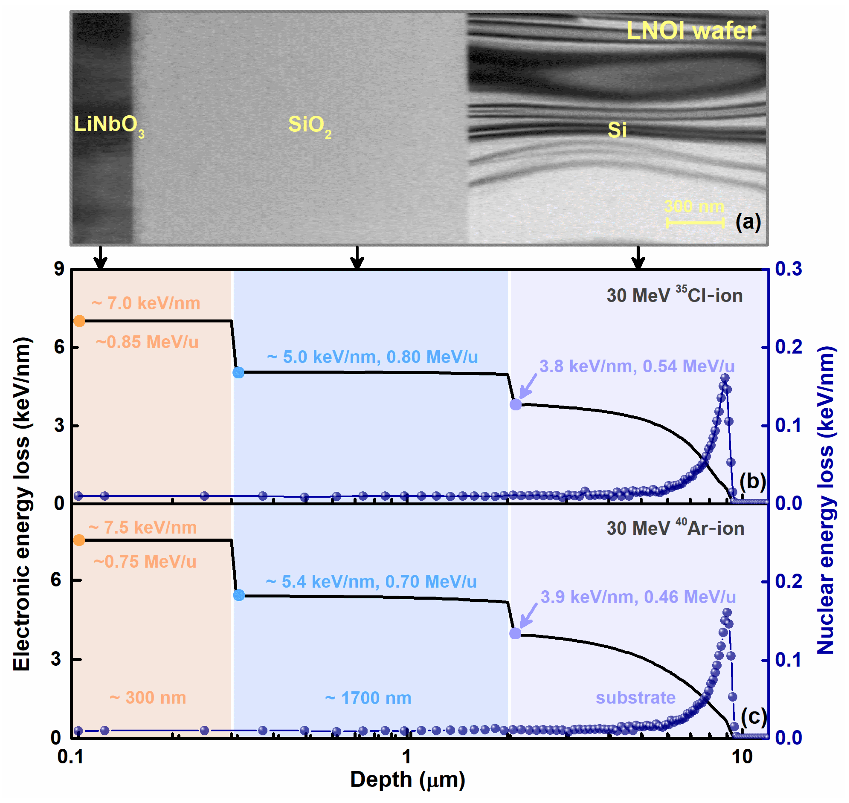
Figure 1. (a) TEM image of the focused ion beam (FIB)–fabricated LNOI sample. (b,c) Distributions of electronic and nuclear energy losses for LNOI samples irradiated with 30 MeV Cl ion and Ar ion, simulated by SRIM 2013, respectively.
Crystals 12 00943 g001
Figure 2. Bright–field (BF) cross–sectional TEM images of (a,b) LiNbO3 layer, (c,d) SiO2 layer, and (e,f) Si substrate in the LNOI samples irradiated with 30 MeV Cl ion and Ar ion with different ion velocities and electronic energy losses.
Crystals 12 00943 g002
Figure 3. (a) Deposited energy exchange between the electronic and atomic subsystems in LiNbO3 under 0.75 MeV/u ion irradiation with an Eele of 7.5 keV/nm. (b,c) Fraction of energy deposited into the electronic subsystem and absorption radius αe in LiNbO3, SiO2, and Si layers under ion irradiation with different ion velocities. For 0.75 MeV/u ion irradiation with an Eele of 7.5 keV/nm, (d) the density distribution of energy deposited into the electronic subsystem and (e) energy deposition to the atomic subsystem and the corresponding atomic temperature calculated by the iTS model.
Crystals 12 00943 g003
Figure 4. Under the action of Cl and Ar ion irradiation, the evolution of energy deposited to atoms and corresponding atomic temperature at different radii versus time in (a,b) LiNbO3 layer, (c,d) SiO2 layer, and (e,f) Si substrate.
Crystals 12 00943 g004
Figure 5. RBS/channeling spectra of LNOI samples irradiated with 30 MeV Ar ion to different ion fluences. The channeling spectrum of bulk LiNbO3 under 30 MeV Ar ion irradiation with a fluence of 3 × 1012 cm−2 is also indicated for comparison.
Crystals 12 00943 g005
Publisher’s Note: MDPI stays neutral with regard to jurisdictional claims in published maps and institutional affiliations.