Article

In Situ Growth of Mg-Fe Layered Double Hydroxides (LDH) Film on Titanium Dental Implant Substrates for pH Regulation in Oral Environments

1
Department of Industrial Engineering, University of Rome Tor Vergata, Via del Politecnico 1, 00133 Rome, Italy
2
Department of System Medicine, University of Rome Tor Vergata, Via del Politecnico 1, 00133 Rome, Italy
3
Independent Researcher, 20019 Milano, Italy
4
Department of Experimental Medicine, University of Rome Tor Vergata, Via Montpellier 1, 00133 Rome, Italy
*
Author to whom correspondence should be addressed.
Crystals 2023, 13(12), 1636; https://doi.org/10.3390/cryst13121636
Submission received: 10 November 2023 / Revised: 19 November 2023 / Accepted: 23 November 2023 / Published: 26 November 2023

Abstract

:
Layered double hydroxides (LDHs) consist of two-dimensional, positively charged lamellar structures with the ability to host various anions in the interlayer spaces, which grants them unique properties and tunable characteristics. LDHs, a class of versatile inorganic compounds, have recently emerged as promising candidates for enhancing osseointegration. A suitable alkaline microenvironment is thought to be beneficial for stimulating osteoblasts’ differentiation (responsible for bone matrix formation) while suppressing osteoclast generation (responsible for bone matrix disintegration). LDHs are prone to adjusting their alkalinity and thus offering us the chance to study how pH affects cellular behavior. LDHs can indeed modulate the local pH, inflammatory responses, and oxidative stress levels, factors that profoundly influence the behavior of osteogenic cells and their interactions with the implant surface. Herein, we deposited Mg–Fe LDH films on titanium substrates for dental implants. The modified Ti substrates was more alkaline in comparison to the bare ones, with a pH higher than 8 after hydrolysis in an aqueous environment.

1. Introduction

Elemental titanium (Ti) and Ti alloys have been used as materials for dental and orthopedic implants since the early 1950s due to their stable chemical properties, good mechanical characteristics, low density, and good bone-contact biocompatibility [1,2,3]. However, most Ti-based implants can fail due to the poor antibacterial activity and osteoinductive properties. In fact, standard metal orthopedic implants with inert surfaces are ineffective for the formation of bone and its integration in the early stages [4]. The adhesion of osteoblasts is strongly affected by the composition of the surface while the oxide layer tends to have favorable biological properties. For these reasons, after initial manufacture, titanium implants typically have their surfaces modified to assure uniform oxidation and the elimination of contaminants [5,6]. The resultant surfaces are intended to enhance biological properties and encourage cell adhesion and proliferation, two processes that contribute to bone bonding [5,6]. It was shown that the behaviors of osteoclasts and osteoblasts are strongly influenced by the local microenvironment that occurs between the natural bone and implant, thus affecting new bone formation [7]. In particular, mineralization processes are enhanced in a microenvironment that has suitable conditions of alkalinity [4,8,9]. As a result, an appropriate design to modify pure titanium surfaces and move the pH to values higher than 7.4 for a more alkaline environment [3] may be favorable to improve the adhesion of fibroblasts and contribute to the sealing of soft tissue. Therefore, it is of great interest to deposit a modified film that can control the pH of the local microenvironment. However, this still represents a great challenge for biomedical research and applications.
The development of nanoroughened surfaces has gained prominence in recent times, achieved through various techniques such as applying TiO2 nanotubes, hydroxyapatite coatings, and utilizing UV photo-functionalization, among others. These nanostructures are advantageous as they enhance protein adhesion from bodily fluids and improve osteoblast growth on titanium implants compared to micro-rough surfaces [10,11]. Moreover, surface characteristics, including hydrophilicity, are influenced by the purity and physicochemical properties of the titanium surface, which naturally has a titanium dioxide layer that aids in attracting essential ions from the bone matrix. Nevertheless, the biological aging of titanium can lead to reduced hydrophilicity and biological integration due to atmospheric carbon contamination [12]. To maintain hydrophilicity over time, methods like photo-functionalization and argon plasma treatment are employed. Another innovative technique involves drying the implants under nitrogen to shield them from the air and storing them in isotonic NaCl solution [13]. There is also a process for the dry storage of hydrophilicity on sand-blasted and acid-etched surfaces involving the application of a salt layer, followed by air-drying, which was found effective in preserving hydrophilicity without relying on a physical barrier against atmospheric contamination [13].
Layered double hydroxides (LDHs), as inorganic materials with controllable alkalinity, provide the chance to adjust microenvironmental pH values. LDHs are two-dimensional (2D) layered materials that are made of common elements, exist in nature (hydrotalcite), and are nontoxic. Their lattice structure consists of positively charged brucite-type layers, where trivalent cations partially substitute divalent cations, separated by an interlayer space hosting water and several types of possible anions. The general formula is [MII1 − xMIIIx(OH)2]x+(An−)x/n∙yH2O, with MII, MIII, and An− representing divalent metal cations, trivalent metal cations, and interlayer anions, respectively. This material has high tunability since both metal cations and interlayer anions can be modified over a wide range of elements. Common divalent cations (M2+) are Zn2+, Mg2+, Cu2+, and Ni2+. Differently, the trivalent cations (M3+) include, but are not limited to, Cr3+, Fe3+, and Al3+. Replacing divalent ions with trivalent cations makes the hydroxide layers positively charged and must be balanced with the intercalation of n valent anions to be inserted in between the lamellas to keep the structure electrically neutral. Frequently used inorganic anions are Br, Cl, OH, NO3, CO32−, SO42−, PO43−, etc. [10]. The stability of the system requires that the molar ratio be in the range 0.2 < x < 0.4. The upper limit originates from the electrostatic repulsion between trivalent neighboring cations and Pauling’s rules, while a minimum concentration of M3+ is needed because the resulting imbalance of charge maintains both the layer intercalation and the whole 3D structure. In addition, it presents good ion-exchange capability, excellent stability, and biocompatibility. Indeed, LDHs are widely applied in biomedical applications such as bioimaging, cancer therapy, osteogenesis, antibacterial, and biodegradation control [11]. Their remarkable capacity to easily exchange the interlayer anions permits the use as drug delivery systems, biological coating materials [12], corrosion-resistant films [13], and a microenvironment pH regulator [14]. All these experimental findings and investigations suggest that LDHs are promising materials for the coating and surface biofunctionalization of Ti-based implants. In fact, LDH coatings could be used as a protective shield for these systems, even enhancing the cell adhesion and the osseointegration capacity [15,16]. However, in most related studies, the LDHs films that are investigated as biomaterials generally contain harmful elements to human body, such as Al, which is related to skeletal, neurological, and hematological risks. For this reason, and considering the general safety requirements of any equipment to be used in biological and medical environment, we selected Mg and Fe, two metals that are essential trace elements in human body, to synthetize LDHs. Magnesium (Mg) is an element that is generally biologically compatible with good biodegradability and nontoxicity. Specifically, magnesium is one trace element of the human body that plays a fundamental role in promoting bone formation, bone regeneration, and retaining the density and strength of bones and teeth [17]. On the other hand, iron is a mineral that the body needs for growth and development, and is essential for the structure and biological activity of hemoglobin and myoglobin since it permits oxygen binding, delivery, and storage. Iron also contributes to the production of some hormones [18]. It is difficult to make Mg-Fe LDH films on inert substrates, such as pure Ti surfaces, due to the large mismatch between the radius of Fe3+ and that of Mg2+. To best of our knowledge, only a few references available reported the synthesis of Mg-Fe LDHs films onto pure titanium substrates [3,19,20]. The work by Li et al. [3] demonstrated that the deposition of Mg-Fe LDHs films on bare titanium surfaces can effectively improve the proliferation, multiplication, and osteogenic differentiation of stem cells. This confirmed that the establishment of a suitable alkaline microenvironment can be useful for boosting the proliferation and osteogenic activity through the deposition of Mg-Fe layered double hydroxides (LDHs) on Ti substrates. The LDH coating improved the osteo immune-modulatory property of Ti [19]. In Ref. [20], coating the titanium surface using Mg–Fe LDHs films enhanced hGF cell adhesion, providing a new approach for the design of multifunctional biomaterials to be used to seal soft tissue around dental implants. However, all the reported studies used pure titanium substrates derived from machine polished plates, while real dental implants are made of titanium alloy with specifically treated surfaces that present a certain degree of roughness to improve the interaction with the bone.
In this work, Mg-Fe LDH films were deposited on the titanium surface of real dental implants via the hydrothermal treatment method. Specifically, the titanium disks that were used for the LDH growth were made of titanium alloy, usually named Ti 6Al-4V [2], an alpha–beta alloy with a content of 6% Al and 4% V. It is one of the most commonly used titanium alloys and exhibits a very good combination of corrosion resistance, toughness, and strength. In addition, this alloy has shown good biological compatibility. However, it contains both aluminum and vanadium located at the surface layers, usually in the form of their respective oxides. Since the release of both aluminum and vanadium can cause biological problems, it should be prevented, especially in the oral environment. Aluminum is known to interfere with bone mineralization, and this can lead to structural deficiencies, while the presence of vanadium is cytotoxic and can cause type IV allergic reactions. On the other hand, many studies confirmed that this alloy produces satisfactory osseointegration, especially when treated so as to enhance the formation of an oxide layer on the surface [21]. Ti-6A1-4V is a bioactive material and can improve the formation of bone directly on the metal surface. Moreover, the oxide layer on the surface plays a major role in the success of osseointegration. In addition, the surface roughness is an important feature because it affects the quality of the interaction of titanium implant with the bone.
By increasing the thickness and roughness of the oxide layer, the osteointegration can be encouraged to occur quickly and reliably, at least in the first stage of the process.
The passivation of the metal substrate is also a result of the presence of the oxide layer, which thus inhibits corrosion and minimizes the release of Ti ions [2]. The Mg-Fe LDH-modified titanium surface demonstrated the capability to increase the pH in an aqueous environment, thus forming an alkaline microenvironment that is more suitable for osteoblast growth and osseointegration [22]. This result has great potential applications for the enhancement of the osteogenesis of implant materials and provides a new path towards the design and tuning of alkaline environments.

2. Materials and Methods

2.1. Materials

We investigated titanium alloy disks with a diameter of 0.9 cm and a thickness of 0.2 cm. The disks are made of the same Ti6Al4V alloy as that one used for real dental implants. The samples were provided by Alpha-Bio Tec Ltd. (Petach Tikva, Israel) and presented the same surface treatment and roughness as the implant material. The detailed composition of the disks is given in Table 1. The disks were cleaned with distilled water three times before use to remove the original NaCl coating from the surface. This coating was deposited on the substrates to maintain their hydrophilicity and avoid titanium aging due to atmospheric CO2 adsorption on the surface [13,23]. No further cleaning procedures were performed in order to simulate the usual handling of real implants as much as possible. Magnesium chloride hexahydrate (MgCl2∙6H2O), ferric trichloride hexahydrate (FeCl3∙6H2O), sodium hydroxide (NaOH), sodium bicarbonate (NaHCO3), and sodium carbonate (Na2CO3) were obtained from Sigma Aldrich, and they were used without further processing. Deionized water (DI water, Milli-Q deionized 18.25 MΩ cm, Millipore, Milford, MA, USA) was used for all experiments.

2.2. LDH Synthesis

Mg-Fe LDH films on titanium substrates were prepared via hydrothermal growth. The hydrothermal method was applied as described in the paper by Li. et al. [3]. Briefly, a basic aqueous solution containing 3 M NaOH and 1 M NaHCO3 was first prepared for the pH regulation of the main growth solution. This basic solution was then added dropwise to the growth salt solution that had been prepared separately by dissolving 20.54 mM MgCl2 and 6.25 mM FeCl3 in deionized (DI) water. The addition was performed while continuously stirring the growth solution at room temperature until the pH reached the value 9.5. The growth solution was further stirred for 30 min and then left still for 2 h for the formation of precipitate. Afterwards, the supernatant (around 300 mL) was taken in order to obtain only nanoparticles of Mg-Fe LDH to cover the Ti alloy and avoid bigger particles and by-products. The supernatant was transferred to the autoclave to cover the Ti disks that were immersed upside-down. The autoclave was sealed and put in the oven for an 18 h reaction time at 105 °C. The following reactions (Equations (1)–(3)) can be considered to take place:
Mg(H2O)62+ + H2O → Mg(OH)(H2O)5+ + H3O+
Fe(H2O)63+ + H2O → Fe(OH)(H2O)52+ + H3O+
(1 − x)Mg(H2O)62+ + x Fe(H2O)63+ + 2OH + xCl → Mg(1−x)Fex(OH)2Clx(H2O)6
The reaction is affected by pH: low pH values can cause the incomplete processes of precipitation of the different cations; basic conditions can lead to the undesired dissolution of metal ions [24].
After cooling down to room temperature naturally, the disks were taken out, washed by using deionized water three times, and afterwards dried in the oven in normal atmosphere at 60 °C for 30 min.

2.3. Surface Characterization

The morphology of the deposited Mg-Fe LDH was investigated by using scanning electron microscopy (SEM) investigations. A Tescan Mira 3 field-emission scanning electron microscope (FE-SEM, TESCAN, Brno, Czech Republic) equipped with an energy dispersive X-ray (EDX) spectrometer (OXFORD XPLORE 30) was used. Energy dispersive spectrometry was used to obtain the elemental compositions of samples. For both imaging and EDX analysis, the FE-SEM was operated at 20 keV electron energy and 1 nA beam current with the working distance set to 9 mm during image acquisitions and to an analytical distance of 15 mm during EDX analysis. During imaging, the samples did not show any charging effect, removing the necessity of depositing a conductive layer on their surface.

2.4. pH Measurement

Both the Mg-Fe LDH-coated Ti disks and the uncoated Ti disks, i.e., without LDH deposition, were immersed in 400 μL of DI water for 5 min, then the tip of the pH meter (Apera instruments, PH60S-E, Columbus, OH, USA) was immersed and located 1 mm above the sample surface to measure the local pH. To ensure that no further liquid convection occurred during the pH measurement process, the pH meter probe was inserted extremely slowly. Therefore, during the whole testing period, there was essentially no liquid vibration. The final pH result is the average of N = 4 tests, each one lasting 1 min to ensure the accuracy of the measurements.

3. Results and Discussion

3.1. Mechanism for the Growth of LDH Films

Co-precipitation in NaOH solution is a very common method for the preparation of LDHs. It is based on a simple procedure and allows the operator to control several parameters during the precipitation phase, such as the pH of the medium, the concentration of the different metallic species, and the type of anions. For these reasons, co-precipitation is also chosen to introduce inorganic or organic moieties into the interlayer space. The pH can either be kept constant or be allowed to change during the reaction [25]. In particular, LDHs with high degree of crystallinity can be obtained when the M2+/M3+ ratio is 2. Higher ratios can lead to phase segregation and a loss of crystallinity [26].
In the Ti alloys used to make implantable devices, namely commercially pure titanium (cpTi), Ti-6A1-4V, the surfaces are mainly composed of the oxide TiO2 [3]. This oxide layer also contains hydroxyl groups in addition to the oxide. As shown in Figure 1, Mg2+ and Fe3+ reacted in the alkaline aqueous solution and then formed the Mg-Fe LDHs precursor. In the hydrothermal process, the Mg-Fe LDHs grew on the surface of the Ti and connected with the titanium substrate by the Ti-O bond produced in the dissolution of titanium.
The hydrothermal crystallization method is effective for obtaining more desirable structural properties of LDHs. In fact, among other synthesis methods, the hydrothermal procedure is simple and effective in creating adhesive LDH coatings by exploiting in situ processes and the existence of chemical bonds of the substrate. In addition, the chemistry of the growth solution and the hydrothermal parameters can be well regulated to control the stoichiometry and induce the adhesion of yielded coatings. For instance, hydrothermal growth can be implemented by using sacrificial substrates that act as both substrate and reagent in the synthesis [27].

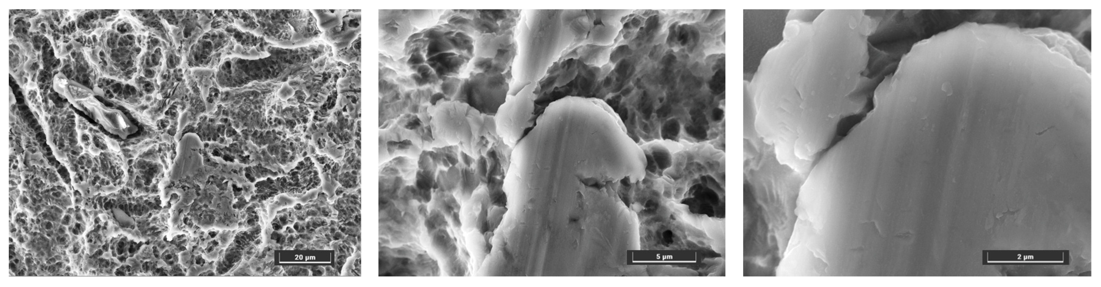
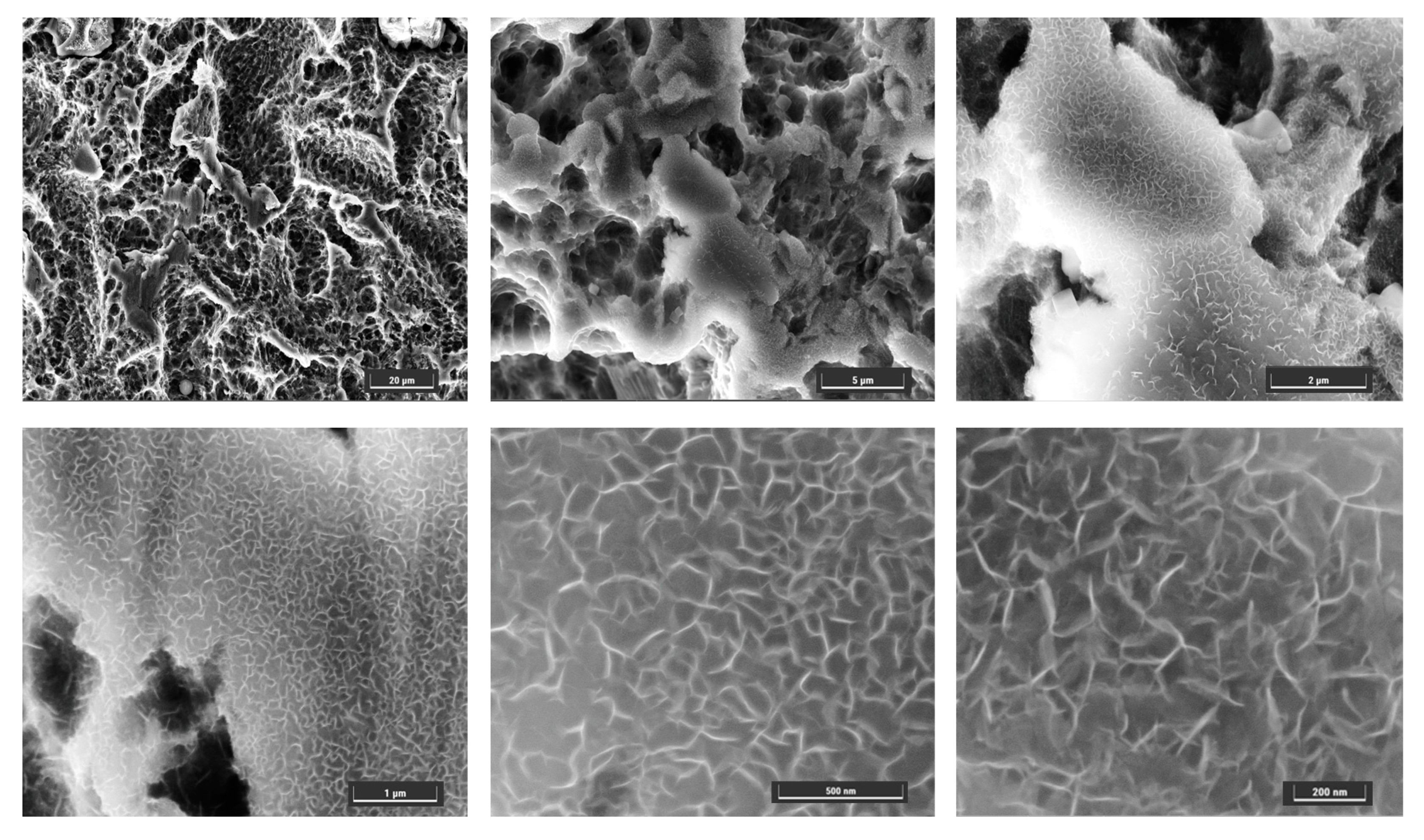
3.2. Surface Characterization

Figure 2 shows the SEM surface topography of uncoated and Mg-Fe LDH-deposited Ti disks. The uncoated disk surface shows a porous nanostructure with small depressions and indentations in the lower micrometric range.
The Mg-Fe LDH coating was attained by immersing Ti disks in the suspension of MgCl2 and FeCl3 (Mg:Fe molar ratio = 3.29) at 105 °C for 18 h. After the reaction, the entire Ti disk surfaces were homogeneously and densely coated with Mg-Fe LDH films (Figure 3). The LDH films were made of the typical nanoplatelets, mainly perpendicular to the substrate surface, with curved shapes generally observed during in situ growth [28,29]. The lateral dimensions of nanoplatelets are in the range of 100–200 nm while the platelets’ thickness seems on the low end of the nanometric scale, below 10 nm. It should be noted that this morphology is quite different from the one that can be achieved by using hydrothermal growth on sacrificial substrate. For instance, by growing Zn-Al LDH on aluminum plates that act as both substrate and reactant in the synthesis, lateral dimensions in the range of 2–5 μm and thicknesses of 200 nm were obtained. This difference can be due to the different elements and growth mechanism used in the present investigation.
Growth duration times of 16 and 24 h were also tested with worse results in terms of density and purity of morphology of the nanoplatelets.
The nanoroughness of the LDH-modified surfaces showcased a unique topography with a higher density of nanostructures. These alterations could potentially enhance protein adhesion, a crucial initial step for cellular interactions with the implant surface. A recent study demonstrated how such a kind of surface may favor early stage osseointegration by facilitating protein adsorption and subsequent cell adhesion, migration, and proliferation [30].
EDX spectra and elemental compositions of the samples investigated are shown in Figure 4. In the uncoated Ti surface, the expected composition with the right percentage of Ti, Al, and V was found. In the Ti substrate with LDH deposition, EDX analysis provided a value of the atomic ratio of 2.62, while the stoichiometric ratio of the growth solution was 3.29. However, it should be noted that the ratio of the metal ions in the supernatant might have changed during the precipitation step since the precipitate could contain unreacted ions or by-products.
In addition, EDX maps (Figure 5) show that Mg and Fe are uniformly distributed over the selected area.
Therefore, morphological and chemical analyses confirm that Mg-Fe LDH films were successfully grown on the Ti surface.

3.3. pH Measurement

Hydrolysis of alkali films can occur in a water environment and lead to the release of metal ions along with OH, thus increasing the local pH value near the surface of the sample. As regards Mg–Fe LDHs films, according to reaction equations (see Equations (4)–(7)) [20], OH accumulate on the interface, thus increasing the value of pH near surface.
Mg(OH)2 → Mg2+ + 2OH
MgO + H2O → Mg2+ + 2OH
Fe(OH)3 → Fe3+ + 3OH
Fe2O3 + 3H2O → 2Fe3+ + 6OH
Figure 6 reports the measurement of local pH near the surface of an uncoated Ti substrate in comparison with the results of three different LDH-deposited samples obtained with the same growth procedure.
Experimental results confirmed that there was a shift of pH towards the basic range in the LDH samples. Specifically, an average value of pH = 8.4 was found in coated samples, while the uncoated Ti substrate resulted in the value of pH = 6.3, virtually identical to the value of pH in ultrapure water.
Enhanced osseointegration has been correlated with high pH environments, which are believed to positively influence the surface characteristics and cellular responses essential for bone integration. The pH level is a critical parameter of the surface chemistry of an implant, directly influencing cellular behaviors critical to bone regeneration. Elevated pH conditions may promote the deposition of a carbonate apatite layer on the implant surface, mirroring the inorganic phase of bone and consequently facilitating bone healing [31]. Moreover, the aging of titanium implants from a biological point of view can lead to the accretion of hydrocarbons due to atmospheric carbon, diminishing the material’s hydrophilicity and its capacity to attract osteoprogenitor and integrins cells [32]. Thus, this pilot study is aimed at developing a bioactive surface that employs layered double hydroxides (LDH), with the inclusion of Mg and Fe ions to raise the pH and evaluate the necessity of LDH salts for maintaining hydrophilicity [32].
It must be noted that, in the measurements of pH, the influence of air or other factors possibly influencing the results was not taken into account even though all measurements were carried out in comparison with the reference sample. This is one of the limitations of this study, and it will be addressed in a future study that will try to consider the complex oral environment. In addition, no statistical concern was applied in relation to sample size calculation. From this point of view, this can be considered a pilot observation on a small sample.

4. Conclusions

Osseointegration, a pivotal process for the stability and longevity of implants, is significantly influenced by the topographical and chemical characteristics of implant surfaces [33]. This investigation probed into the surface modifications induced by the application of a Mg-Fe LDH coating and the consequential effects these modifications have on surface pH and nanotopography. A local environment with a suitable level of alkalinity can be favorable to the multiplication and differentiation of osteoblast cells. In order to control the microenvironmental pH, we have grown Mg-Fe LDHs films on titanium surfaces via the hydrothermal treatment method. The LDH film presented a nanostructured morphology characterized by nanoplatelets with a lateral size in the range 100–200 nm and a thickness below 10 nm. This morphology is quite different from the one obtained in Zn-Al LDH on an aluminum substrate, where the nanoplatelets presented lateral dimensions in the range of 2–5 μm and thicknesses of 200 nm. This difference could be due to the different elements and growth mechanism used in the present investigation. The pH value near the surface of the Mg-Fe LDH-modified Ti substrates was more alkaline (i.e., pH = 8.4) than the control uncoated surface (pH = 6.2), possibly due to an OH- exchange mechanism. The synthesized Mg-Fe LDH films could thus have applications in the field of materials for medical implants, while the related results could provide a new approach for the surface design. In addition, these results are quite promising since they suggest the applicability of LDHs to other interesting fields in the domain of osseointegration. This represents a compelling avenue for enhancing the integration of implants with host bone tissue. LDHs can modulate the local pH and possibly the inflammatory response and oxidative stress levels, factors that profoundly influence the behavior of osteogenic cells and their interactions with the implant surface [34]. Furthermore, LDHs can be functionalized with bioactive molecules or drugs, providing a versatile platform for tailored therapeutic interventions at the implant site. Nevertheless, the clinical relevance of induced nanotopographies remains ambiguous. While ex vitro experiments and in vivo studies suggest the potential advantages of nanorough surfaces in promoting osteogenic cell adhesion and proliferation, translating these findings to clinical scenarios warrants further rigorous investigation [35,36].
In conclusion, the LDH-induced surface modification method delineated in this study offers promising avenues for altering surface topographies, potentially favoring the osseointegration process. The quest for conclusive evidence on the clinical significance of nanotopographical features continues, urging further explorative studies in this domain.

Author Contributions

Conceptualization, E.N., F.G. and R.P.; methodology, E.N.; formal analysis, E.N.; investigation, Y.L. and E.N.; data curation, E.N. and R.P.; writing—original draft preparation, E.N. and R.P.; review, A.E. and P.B.; material supply, C.D.; supervision, E.N. and R.P. All authors have read and agreed to the published version of the manuscript.

Funding

This research received no external funding.

Data Availability Statement

Data is contained within the article.

Conflicts of Interest

The authors declare no conflict of interest.

References

  1. Geetha, M.; Singh, A.K.; Asokamani, R.; Gogia, A.K. Ti Based Biomaterials, the Ultimate Choice for Orthopaedic Implants—A Review. Prog. Mater. Sci. 2009, 54, 397–425. [Google Scholar] [CrossRef]
  2. Nicholson, J.W. Titanium Alloys for Dental Implants: A Review. Prosthesis 2020, 2, 100–116. [Google Scholar] [CrossRef]
  3. Li, Q.; Wang, D.; Qiu, J.; Peng, F.; Liu, X. Regulating the Local PH Level of Titanium: Via Mg-Fe Layered Double Hydroxides Films for Enhanced Osteogenesis. Biomater. Sci. 2018, 6, 1227–1237. [Google Scholar] [CrossRef] [PubMed]
  4. Chen, L.; Yin, Y.; Jian, L.; Han, X.; Zhao, X.; Wang, D. Enhanced Bactericidal Effect of Calcinated Mg–Fe Layered Double Hydroxide Films Driven by the Fenton Reaction. Int. J. Mol. Sci. 2023, 24, 272. [Google Scholar] [CrossRef]
  5. Kligman, S.; Ren, Z.; Chung, C.H.; Perillo, M.A.; Chang, Y.C.; Koo, H.; Zheng, Z.; Li, C. The Impact of Dental Implant Surface Modifications on Osseointegration and Biofilm Formation. J. Clin. Med. 2021, 10, 1641. [Google Scholar] [CrossRef] [PubMed]
  6. Chouirfa, H.; Bouloussa, H.; Migonney, V.; Falentin-Daudré, C. Review of Titanium Surface Modification Techniques and Coatings for Antibacterial Applications. Acta Biomater. 2019, 83, 37–54. [Google Scholar] [CrossRef]
  7. Shen, Y.; Liu, W.; Wen, C.; Pan, H.; Wang, T.; Darvell, B.W.; Lu, W.W.; Huang, W. Bone Regeneration: Importance of Local PH—Strontium-Doped Borosilicate Scaffold. J. Mater. Chem. 2012, 22, 8662–8670. [Google Scholar] [CrossRef]
  8. Tan, J.; Wang, D.; Cao, H.; Qiao, Y.; Zhu, H.; Liu, X. Effect of Local Alkaline Microenvironment on the Behaviors of Bacteria and Osteogenic Cells. ACS Appl. Mater. Interfaces 2018, 10, 42018–42029. [Google Scholar] [CrossRef]
  9. Liu, W.; Wang, T.; Yang, C.; Darvell, B.W.; Wu, J.; Lin, K.; Chang, J.; Pan, H.; Lu, W.W. Alkaline Biodegradable Implants for Osteoporotic Bone Defects—Importance of Microenvironment PH. Osteoporos. Int. 2016, 27, 93–104. [Google Scholar] [CrossRef]
  10. Wennerberg, A.; Jimbo, R.; Stübinger, S.; Obrecht, M.; Dard, M.; Berner, S. Nanostructures and hydrophilicity influence osseointegration—A biomechanical study in the rabbit tibia. Clin. Oral. Implant. Res. 2014, 25, 1041–1050. [Google Scholar] [CrossRef]
  11. Yamamura, K.; Miura, T.; Kou, I.; Muramatsu, T.; Furusawa, M.; Yoshinari, M. Influence of various superhydrophilic treatments of titanium on the initial attachment, proliferation, and differentiation of osteoblast-like cells. Dent. Mater. J. 2015, 34, 120–127. [Google Scholar] [CrossRef] [PubMed]
  12. Delgado-Ruiz, R.; Romanos, G. Potential Causes of Titanium Particle and Ion Release in Implant Dentistry: A Systematic Review. Int. J. Mol. Sci. 2018, 19, 3585. [Google Scholar] [CrossRef]
  13. Gianfreda, F.; Antonacci, D.; Raffone, C.; Muzzi, M.; Pistilli, V.; Bollero, P. Microscopic Characterization of Bioactivate Implant Surfaces: Increasing Wettability Using Salts and Dry Technology. Materials 2021, 14, 2608. [Google Scholar] [CrossRef] [PubMed]
  14. Zhang, D.; Cheng, S.; Tan, J.; Xie, J.; Zhang, Y.; Chen, S.; Du, H.; Qian, S.; Qiao, Y.; Peng, F.; et al. Black Mn-Containing Layered Double Hydroxide Coated Magnesium Alloy for Osteosarcoma Therapy, Bacteria Killing, and Bone Regeneration. Bioact. Mater. 2022, 17, 394–405. [Google Scholar] [CrossRef]
  15. Daniel, S.; Thomas, S. Layered double hydroxides: Fundamentals to applications. In Layered Double Hydroxide Polymer NanoComposites; Woodhead Publishing: Sawston, UK, 2020; pp. 1–76. ISBN 9780081019030. [Google Scholar]
  16. Yu, S.; Choi, G.; Choy, J.-H. Multifunctional Layered Double Hydroxides for Drug Delivery and Imaging. Nanomaterials 2023, 13, 1102. [Google Scholar] [CrossRef]
  17. Al Alawi, A.M.; Majoni, S.W.; Falhammar, H. Magnesium and Human Health: Perspectives and Research Directions. Int. J. Endocrinol. 2018, 2018, 9041694. [Google Scholar] [CrossRef] [PubMed]
  18. Abbaspour, N.; Hurrell, R.; Kelishadi, R. Review on Iron and Its Importance for Human Health. J. Res. Med. Sci. 2014, 19, 164–174. [Google Scholar]
  19. Cao, Y.; Zheng, D.; Zhang, F.; Pan, J.; Lin, C. Layered Double Hydroxide (LDH) for Multi-Functionalized Corrosion Protection of Metals: A Review. J. Mater. Sci. Technol. 2022, 102, 232–263. [Google Scholar] [CrossRef]
  20. Izbudak, B.; Cecen, B.; Anaya, I.; Miri, A.K.; Bal-Ozturk, A.; Karaoz, E. Layered Double Hydroxide-Based Nanocomposite Scaffolds in Tissue Engineering Applications. RSC Adv. 2021, 11, 30237–30252. [Google Scholar] [CrossRef]
  21. Rojas, R.; Mosconi, G.; Zanin, J.P.; Gil, G.A. Layered Double Hydroxide Applications in Biomedical Implants. Appl. Clay Sci. 2022, 224, 106514. [Google Scholar] [CrossRef]
  22. Ding, X.; Wu, L.; Chen, J.; Zhang, G.; Xie, Z.; Sun, D.; Jiang, B.; Atrens, A.; Pan, F. Enhanced Protective Nanoparticle-Modified MgAl-LDHs Coatings on Titanium Alloy. Surf. Coat. Technol. 2020, 404, 126449. [Google Scholar] [CrossRef]
  23. Gianfreda, F.; Raffone, C.; Antonacci, D.; Mussano, F.; Genova, T.; Chinigò, G.; Canullo, L.; Bollero, P. Early Biological Response of an Ultra-Hydrophilic Implant Surface Activated by Salts and Dry Technology: An In-Vitro Study. Appl. Sci. 2021, 11, 6120. [Google Scholar] [CrossRef]
  24. Othman, M.R.; Helwani, Z.; Martunus, W.; Fernando, J.N. Synthetic hydrotalcites from different routes and their application as catalysts and gas adsorbents: A review. Appl. Organo-Met. Chem. 2009, 23, 335. [Google Scholar] [CrossRef]
  25. Crepaldi, E.L.; Pavan, P.C.; Valim, J.B. Comparative study of the coprecipitation methods for the preparation of layered double hydroxides. Braz. Chem. Soc. 2000, 11, 64. [Google Scholar] [CrossRef]
  26. Silva, C.G.; Bouizi, Y.; Fornes, V.; Garcia, H. Layered double hydroxides as highly efficient photocatalysts for visible light oxygen generation from water. JACS 2009, 131, 13833. [Google Scholar] [CrossRef] [PubMed]
  27. Scarpellini, D.; Leonardi, C.; Mattoccia, A.; Giamberardino, L.D.; Medaglia, P.G.; Mantini, G.; Gatta, F.; Giovine, E.; Foglietti, V.; Falconi, C.; et al. Solution-grown Zn/Al layered double hydroxide nanoplatelets onto Al thin films: Fine control of position and lateral thickness. J. Nanomater. 2015, 16, 178. [Google Scholar] [CrossRef]
  28. Shan, R.R.; Yan, L.G.; Yang, Y.M.; Yang, K.; Yu, S.J.; Yu, H.Q.; Zhu, B.C.; Du, B. Highly efficient removal of three red dyes by adsorption onto Mg-Al-layered double hydroxide. J. Ind. Eng. Chem. 2015, 21, 561–568. [Google Scholar] [CrossRef]
  29. Li, Y.; Narducci, R.; Varone, A.; Kaciulis, S.; Bolli, E.; Pizzoferrato, R. Zn–Al Layered Double Hydroxides Synthesized on Aluminum Foams for Fluoride Removal from Water. Processes 2021, 9, 2109. [Google Scholar] [CrossRef]
  30. Moses, O.; Bengazi, F.; Ferri, M.; Gianfreda, F.; Velez, J.U.; Botticelli, D.; Canullo, L. Bioactivated Implant Surfaces Placed in Healed Sites or Extraction Sockets: A Preliminary Experimental Study in Dogs. Int. J. Oral Maxillofac. Implants 2022, 37, 963–970. [Google Scholar] [CrossRef]
  31. Wang, Z.; Wang, J.; Wu, R.; Wei, J. Construction of functional surfaces for dental implants to enhance osseointegration. Front. Bioeng. Biotechnol. 2023, 11, 1320307. [Google Scholar] [CrossRef]
  32. Hori, N.; Att, W.; Ueno, T.; Sato, N.; Yamada, M.; Saruwatari, L.; Suzuki, T.; Ogawa, T. Age-dependent Degradation of the Protein Adsorption Capacity of Titanium. J. Dent. Res. 2009, 88, 663–667. [Google Scholar] [CrossRef]
  33. Albrektsson, T.; Johansson, C. Osteoinduction, osteoconduction and osseointegration. Eur. Spine J. 2001, 10, S96–S101. [Google Scholar]
  34. Oh, J.M.; Park, D.H.; Choi, S.J.; Choy, J.H. LDH nanocontainers as bio-reservoirs and drug delivery carriers. Recent Pat. Nanotechnol. 2012, 6, 200–217. [Google Scholar] [CrossRef] [PubMed]
  35. Buser, D.; Broggini, N.; Wieland, M.; Schenk, R.K.; Denzer, A.J.; Cochran, D.L.; Hoffmann, B.; Lussi, A.; Steinemann, S.G. Enhanced bone apposition to a chemically modified SLA titanium surface. J. Dent. Res. 2004, 83, 529–533. [Google Scholar] [CrossRef] [PubMed]
  36. Schupbach, P.; Glauser, R.; Rocci, A.; Martignoni, M.; Sennerby, L.; Lundgren, A.; Gottlow, J. The human bone-oxidized titanium implant interface: A light microscopic, scanning electron microscopic, back-scatter scanning electron microscopic, and energy-dispersive X-ray study of clinically retrieved dental implants. Clin. Implant. Dent. Relat. Res. 2005, 7, S36–S43. [Google Scholar] [CrossRef] [PubMed]
Figure 1. Illustration of the possible mechanism for the enhanced adhesion of osteoblasts on Mg–Fe LDHs via microenvironment pH regulation.
Crystals 13 01636 g001
Figure 2. SEM images of uncoated Ti surface at different magnifications.
Crystals 13 01636 g002
Figure 3. SEM images of LDH-deposited Ti surfaces at different magnifications.
Crystals 13 01636 g003
Figure 4. EDX spectrum and quantitative calculation of uncoated (a) and LDH-deposited Ti surface (b).
Crystals 13 01636 g004
Figure 5. SEM image of LDH-deposited surface and EDX maps of the selected area.
Crystals 13 01636 g005
Figure 6. pH measurement of three Mg-Fe LDH-coated Ti surfaces obtained with the same method of synthesis (namely: LDH #1, LDH #2, LDH #3) and the uncoated surface (namely: Uncoated). N = 4 and the error bars are ±σ.
Crystals 13 01636 g006
Table 1. Composition and properties of titanium alloy implants.
CompositionTi-6A1-4V
Titanium 90%
Aluminum6%
Vanadium4%
UTS (MPa)900
Yields
Strength (MPa)850
Elongation at failure (%)10
Note: UTS is the abbreviation of ultimate tensile strength.
Disclaimer/Publisher’s Note: The statements, opinions and data contained in all publications are solely those of the individual author(s) and contributor(s) and not of MDPI and/or the editor(s). MDPI and/or the editor(s) disclaim responsibility for any injury to people or property resulting from any ideas, methods, instructions or products referred to in the content.

