Article

Angular-Dependent Back-Reflection of Chiral-Nematic Liquid Crystal Microparticles as Multifunctional Optical Elements

Department of Applied Chemistry, Ritsumeikan University, 1-1-1 Nojihigashi, Kusatsu 525-8577, Japan
*
Authors to whom correspondence should be addressed.
Present address: Laboratory for Chemistry and Life Science, Institute of Innovative Research, Tokyo Institute of Technology, 4259 Nagatsuta, Midori-ku, Yokohama 226-8501, Japan.
Crystals 2023, 13(12), 1660; https://doi.org/10.3390/cryst13121660
Submission received: 1 November 2023 / Revised: 23 November 2023 / Accepted: 1 December 2023 / Published: 3 December 2023
(This article belongs to the Special Issue Advances in Liquid Crystal Optical Devices)

Abstract

:
The development of multifunctional optical elements capable of controlling polarization, wavelength, and propagation direction is pivotal for the miniaturization of optical devices. However, designing the spatial distribution of the refractive index for the fabrication of such elements remains challenging. This study demonstrates the spectroscopic function of microparticles composed of chiral-nematic liquid crystals (N* LC), which inherently selectively reflect circularly polarized light. The measurement of the reflection spectra with fiber probes revealed angular-dependent back-reflection of the single layer of the N* LC particles. These results indicate that our N* LC microparticles possess multiple optical functions, enabling the separation of incident light polarization and wavelength within a single material. This suggests broad applications of N* LC particles as compact optical elements.

1. Introduction

The control of light properties, including polarization, wavelength, and propagation, is a fundamental technology with applications across various fields [1,2,3,4]. This technology is used in spectroscopy [5], sensing [6], imaging [7], three-dimensional scanning [8,9], and photovoltaics [10], which play pivotal roles in daily life and science and technology. Recently, a demand for the miniaturization of these optical devices has emerged. One approach to achieving miniaturization involves reducing the number of components. Typically, optical devices with specific functionalities are assembled using various optical elements, such as diffraction gratings, prisms, filters, polarizers, lenses, and mirrors [1]. Rational design of the spatial distribution of the refractive index, such as liquid crystal holograms, is a promising approach for realizing multifunctional optical elements. However, developing multifunctional materials for microscale components remains a significant challenge. Consequently, innovative designs for optical elements are imperative to expedite the further miniaturization of optical devices.
One potential material with such multifunctionality is a chiral-nematic liquid crystal (N* LC), which exhibits wavelength-selective reflection of circularly polarized light (CPL) [11,12,13,14,15,16,17]. N* LC is a liquid–crystalline phase that emerges upon the introduction of chiral molecules into nematic liquid crystals, spontaneously forming a helical molecular alignment. Owing to its helical structure and refractive index anisotropy, this phase exhibits a periodic distribution of the refractive index and demonstrates selective reflection, following Bragg’s law. The reflection wavelength λ r e f depends on their helical pitch P in which the molecules rotate 360°, as per the following equation:
λ r e f = n N *   L C · P sin θ ,
where n N *   L C is the average refractive index of the N* LC used and θ is the angle between the incident light and surface plane. By manipulating the molar ratio of the chiral molecules, we can modulate the helical pitch (P) of the N* LC, allowing us to attain N* LC with a specified reflection wavelength. Moreover, the N* LC exhibited selective reflection of CPL with the same handedness as its helical structure [18]. The dual functionality of N* LC, which enables simultaneous control over the circular polarization and wavelength of reflected light, has generated considerable interest in its application as a multifunctional optical element.
In addition to these functionalities, several studies have successfully controlled the light propagation direction [11,12,13,14,15]. Recently, Ozaki et al. proposed an innovative route to diffuse or focus reflected light by manipulating the phase of the helical alignment along the helical axis within the film [11]. This new capability to control the light direction holds a significant potential for advancing the application of N* LC as a multifunctional holographic device. However, the propagation direction of light depends on the incidence angle. This is because of the film-type N* LC with a unidirectionally aligned helical axis. Furthermore, these functionalities were exclusively observed under monochromatic light. Achieving back-reflection capable of changing the reflection wavelength based on the incidence angle remains challenging for N* LC materials.
We recently fabricated N* LC particles through the dispersion polymerization of monomers [19,20]. This technology enables the production of monodisperse microsized polymer particles in a single step [21,22]. The resulting N* LC particles exhibited vivid reflection colors that varied with the molar ratio of the chiral monomer. These polymer particles can serve as reflective materials with superior environmental stability compared with low-molecular-weight LC droplets [23,24,25,26]. In a previous study, molecular alignments revealed the presence of radial helical axis alignments [19,20]. Because the helical axis alignment is not unidirectional, the incidence angle against the helical axis varies according to the illuminated area of the N* LC particles [27]. We anticipated that this heterogeneous reflection behavior would induce angular-dependent back-reflection. This is because the reflection wavelength and angle were altered based on the orientation of the incident beam.
Herein, we conducted experimental investigations into the angular-dependent reflective properties of the N* LC particles to assess their potential as versatile optical elements. We observed variations in the back-reflection wavelength corresponding to changes in the incidence angle. These multifunctional N* LC particles hold promise for applications as ultracompact optical elements at the micrometer scale. The N* LC particles exhibit reflective properties at specific wavelengths and CPL selectivity. In addition, these particles can produce diverse reflection colors, enabling the straightforward creation of colorant materials with a range of reflection modes [19,20]. Thus, our microsized N* LC particles represent a substantial advancement in developing multifunctional optical elements, significantly contributing to the miniaturization of optical devices.

2. Materials and Methods

2.1. Preparation of N* LC Particles Using Dispersion Polymerization

The N* LC particles were synthesized following the procedure outlined in our previous report [19,28]. The molecular structures of the monomers and dispersion stabilizer (polyvinylpyrrolidone (PVP)) are shown in Figure 1. The base liquid crystal monomer (LCM) was generously provided by Osaka Organic Chemical Industry Ltd (Osaka, Japan). and was purified by recrystallization from methanol before use. The chiral monomer (CM) was synthesized and purified according to previously reported methods [28].
The monomers, LCM and CM, along with PVP and polymerization initiator (2,2′-azobis(isobutyronitrile), AIBN), were dissolved in a mixed solvent of N,N-dimethylformamide (DMF) and methanol in a 30 mL Schlenk flask (U-1013, Sugiyama-Gen Co., Ltd., Tokyo, Japan). The composition of the polymerization mixture is listed in Table S1. The solution was subjected to several freeze–pump–thaw cycles to remove dissolved oxygen, backfilled with Ar gas, and then stirred at 55 °C for 20 h. The resulting N* LC particles were collected by filtration through a membrane filter (T080A025A, ADVANTEC, Tokyo, Japan) with a pore size of 0.8 µm to obtain the polymer particles.

2.2. Characterization of N* LC Particles

Size-exclusion chromatography (SEC) was performed using LC-20AD (Shimadzu, Kyoto, Japan) equipped with a KF805 column (Shodex, Tokyo, Japan) and UV-vis detector (254 nm) at 40 °C, using tetrahydrofuran (THF) as an eluent at a flow rate of 1.0 mL min−1. Molecular weights were calibrated using polystyrene standards. 1H NMR spectra were recorded on a JEOL ECS-400 spectrometer (400 MHz) in CDCl3 for the monomers and CD2Cl2 for the polymer. Chemical shifts were reported in parts per million (ppm) using the residual protons in the NMR solvent. The thermodynamic properties were determined using differential scanning calorimetry (DSC, SII X-DSC7000) at heating and cooling rates of 10 °C min−1. Representative results of the P2 measurements are shown in Figures S1, S2 and Figure 2.

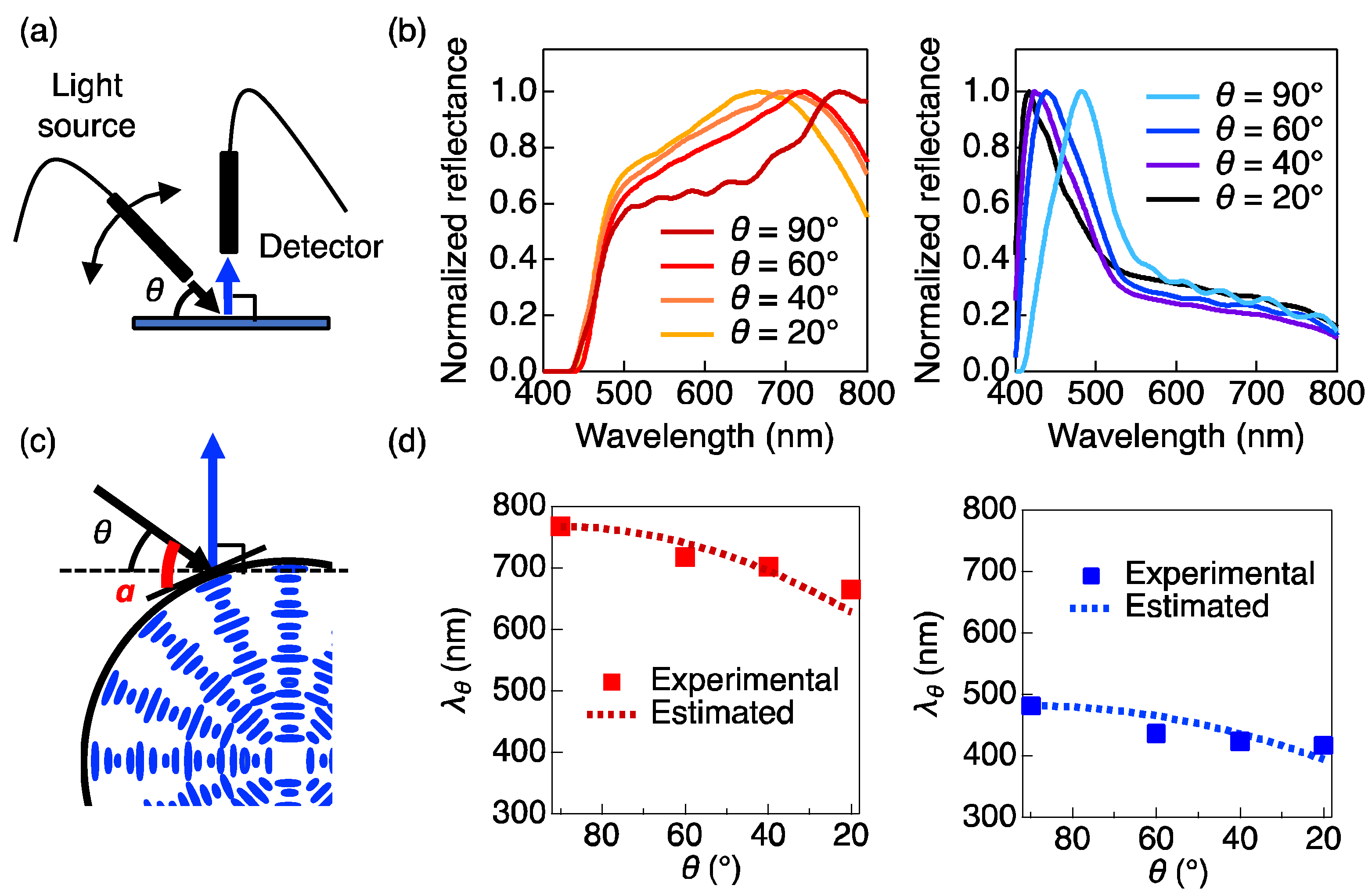
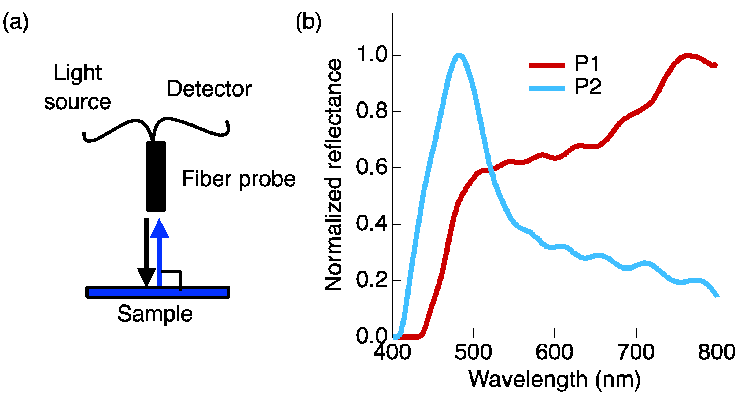
2.3. Evaluation of Reflection Functionalities

Single layer particles were fabricated on the carbon tape as follows. A suspension of the N* LC polymer particles (1.0 mg mL−1, 0.2 mL) in water was dropcasted onto a pre-cleaned glass substrate (15 mm × 13 mm) and slowly evaporated at 5 °C overnight. Subsequently, a carbon tape was adhered to this substrate, and a single layer of N* LC particles was transferred onto the carbon tape.
Reflection spectra were measured using a diode array spectrometer (BLUE-WaveUVN, StellarNet, Tampa, FL, USA) equipped with two types of fiber probes. A coaxial fiber probe (R600, StellarNet, Tampa, FL, USA) was employed to measure the normal reflection. To investigate the incidence angle dependence, two uniaxial fiber probes (F400, StellarNet, Tampa, FL, USA) were used. In this study, the incidence angle (θ) was defined as the angle between the substrate surface plane and the incident light, which was controlled by the fiber probe connected to the light source. The fiber probe connected to the detector was fixed perpendicular to the substrate surface. A tungsten lamp (SL-1, StellarNet, Tampa, FL, USA) served as the incident light source for unpolarized white light. To evaluate the differences in wavelengths, each spectrum was normalized by dividing the original data by the corresponding intensity value at the maximum reflection wavelength in the measured ranges.

3. Results and Discussion

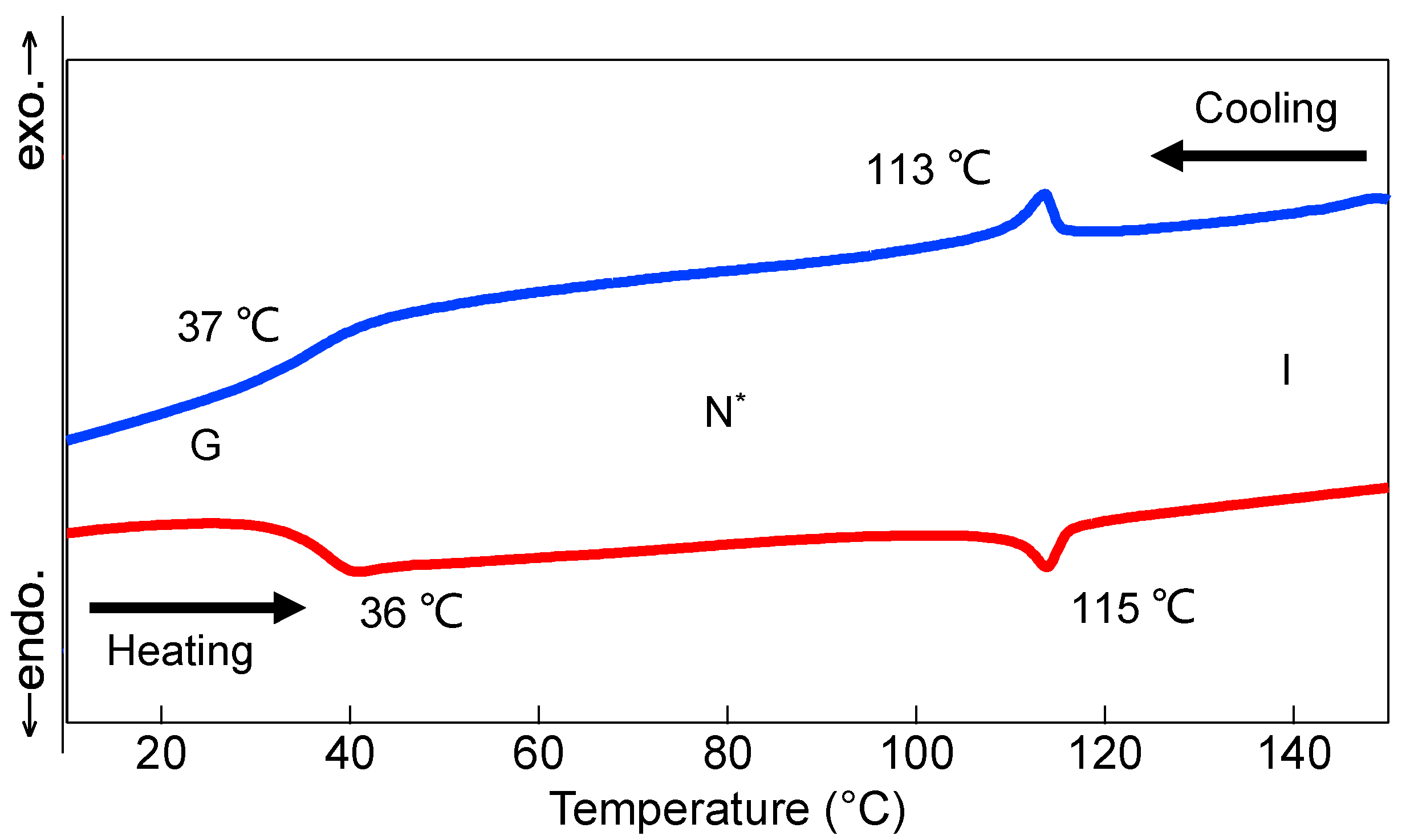
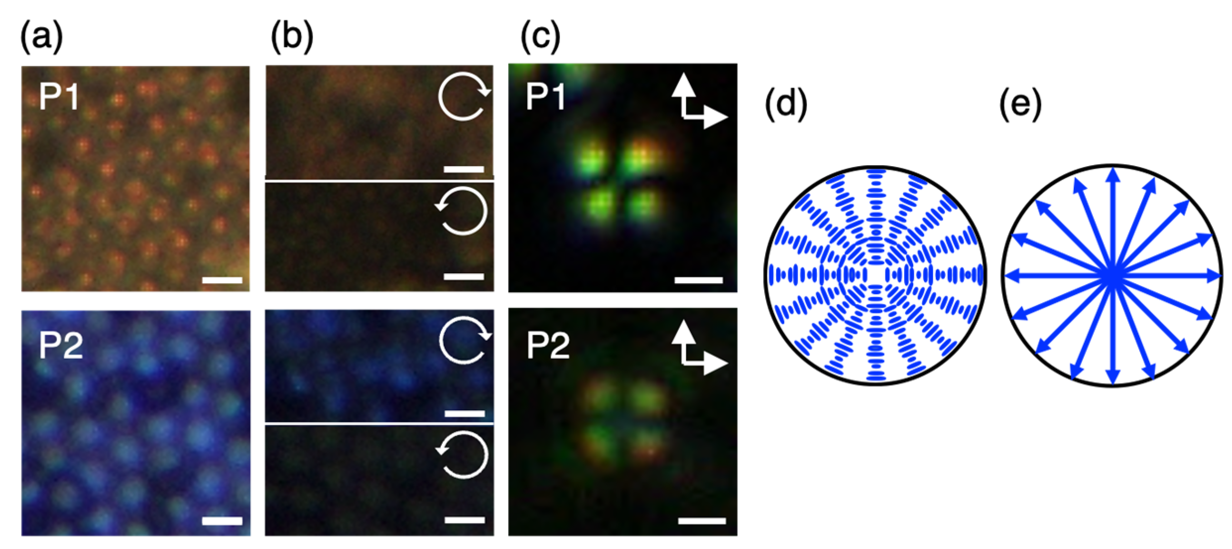
3.1. Synthesis and Characterization of N* LC Polymer Particles

We successfully obtained monodisperse polymer particles with an average diameter (d) of ~2.5 μm and a standard deviation of 0.1 μm at ~50% conversion (Table 1). SEC analysis revealed a number-average molecular weight (Mn) of 15,000 Da and a polydispersity index of 2.6 (Table 1). The copolymer composition was determined using 1H NMR spectroscopy, which confirmed that the desired copolymer with the same composition as that of the monomer mixture for polymerization was achieved (Table 1 and Figure S2). DSC measurement indicates that the synthesized polymers had a glass transition temperature (Tg) at 36 °C and showed a liquid–crystalline phase up to 115 °C in the heating process (Figure 2). These results demonstrate that our N* LC particles were solid at room temperature, wherein the helical alignment of the mesogens in the N* LC phase was fixed within the solid particles.
Microparticles with red (P1) and blue (P2) reflection colors were prepared by controlling the molar ratio of the CM (Figure 3a). Observations through CPL filters revealed that right-handed CPL was reflected by N* LC particles (Figure 3b) [18]. As in our previous report, we considered that the helical axes aligned radially within the particles, as evidenced by the Maltese cross-pattern observed using polarized optical microscopy (POM, Figure 3c) [19]. The idealized molecular and helical axis alignments are schematically illustrated in Figure 3d and Figure 3e, respectively.

3.2. Retro-Reflection Behavior

We measured the retro-reflection spectra from a single layer of N* LC particles with an incidence angle normal to the substrate surface using a coaxial optical fiber probe, as shown in Figure 4a. In the obtained spectra, prominent bands were observed at 760 nm for P1 and 480 nm for P2, along with several smaller bands at baseline (Figure 4b). The wavelength of the major reflection peak varied with the molar ratio of the chiral agent, indicating its association with the selective reflection from the particles. The bandwidth of P1 was wider than that of P2 because the bandwidth of the reflection from the N* LC was proportional to the helical pitch. The reflection bandwidth Δ λ r e f is determined by the following equation when light is normally incident to the helical axis: Δ λ r e f = Δ n N *   L C ·P, where Δ n N *   L C is a birefringence of mesogen. Assuming that the birefringence is constant in the wavelength range of visible light (400–800 nm), the Δ λ r e f depends on the value of P. Therefore, P1 with the longer helical pitch, i.e., longer reflection wavelength, should possess a wider reflection bandwidth than P2 with the shorter helical pitch. Thus, nearly identical reflection wavelengths were previously reported in reflection spectroscopy using an integration sphere (Figure S3) [19,20]. However, a detailed comparison between the spectra obtained with the fiber probe and integration sphere revealed that the reflection peak detected with the fiber probe was narrower, with a slight red shift. We attributed this discrepancy to the characteristics of the optical setup employed for the measurements.
In measurements using the coaxial fiber probe, only the light reflected perpendicular to the tangent of the particle, essentially parallel to the helical axis, can be detected. This indicated that only specular reflections from the particles were captured, resulting in the observation of narrow reflection bands. Conversely, omnidirectional reflections from the particles at all solid angles were amalgamated in the measurements using the integrating sphere. Consequently, the shorter reflection wavelengths stemming from the tilted helical axis caused the reflection bands to broaden and shift toward the shorter end of the spectrum. These findings strongly imply that the wavelength of light reflected by the particles is contingent on the direction of the incident light.
The small bands observed at the baseline of the reflection spectra are interpreted as interference fringes resulting from the densely packed single microparticle layer [29]. The peak wavelengths of the neighboring interference fringes ( λ i and λ i + 1 , where i is a consecutive number of fringes) allow us to calculate the thickness of the layer, denoted as d′, which can be theoretically estimated as follows:
d = λ i · λ i + 1 2 n e f f λ i λ i + 1 ,
where n e f f is the effective refractive index of the packed structure, given by the following equation [29]:
n e f f 2 = ψ   n N *   L C 2 + 1 ψ   n a i r 2 ,
where ψ represents the packing density of the particles and n a i r is the refractive index of air. When the particles are arranged in a single layer with a two-dimensional closest packing structure, ψ is 0.61. n N *   L C can be the same as that of a low-molecular-weight liquid crystal with a chemical structure similar to monomer LCM, namely, 4′-pentyl-4-biphenylcarbonitrile (n = 1.6) [30]. This implies that the n e f f can be estimated as 1.4. Using this value and considering the periods of neighboring interference fringes in the reflection spectra of P2, d′ was calculated to be 2.4 µm. This value closely corresponds to d, supporting our hypothesis that the small peaks observed at the baseline are indeed interference fringes from the microparticle layer. Based on these results, we confirmed that the microparticles formed a single layer with the closest packing arrangement on the substrate.

3.3. Optical Functionalities under Different Incidence Angles

To investigate the dependence of the incidence angle on the wavelength of reflection normal to the substrate, we conducted reflection spectra measurements using two uniaxial fiber probes while varying the angle θ, as shown in Figure 5a. θ is defined as the angle between the substrate and incident light. The fiber probe connected to the detector was fixed perpendicular to the substrate. In the generally planarly aligned N* LC film, the reflection angle corresponds to the incidence angle owing to specular reflection following Bragg’s law. This implies that the back-reflection is not observed; thus, for the optical setup in Figure 5a with discordance between incident and reflection angles against the sample plane, the reflection peak should be extremely small or not be recorded, except at θ = 90°. However, as shown in Figure 5b, a reflection band was observed at all angles, even when the incidence angle differed from the reflection angle. Furthermore, the reflection wavelength shifted towards shorter wavelengths as θ decreased. According to Bragg’s law, the reflection wavelength is determined by the angle between the incident light and helical axis [27]. This geometric relationship is shown in Figure 5c. We anticipated that the radial alignment of the helical axis would result in a distinctive reflection pattern, where the incident and reflection angles would match at the tangent of the particle surface. Following this expectation, the reflection wavelength is determined by the angle α between the incident light and particle tangent (Figure 5c). In this illustration, the angle between the incident light and the reflected light is 90° − θ. The angle between the normal to helical axis and the reflected light is also α. Therefore, the relationship between θ and α can be expressed as α + 90° − θ + α = 180°. The relationship between α and θ is best described as follows:
α = 45° + θ/2.
From this relationship and Bragg’s law, the reflection wavelength at each θ, λ θ , was estimated using the following equation [31]:
λ θ = λ 90 °   sin α .
The estimated and experimental values of λ θ are plotted in Figure 5d, demonstrating a close match between them. These findings indicate that the back-reflection wavelength of N* LC particles varies depending on the incidence angle. Given that each particle exhibits this angular-dependent back-reflection, it can be used as an ultracompact optical element. In addition, the N* LC particles showed right-handed CPL-selective reflections (Figure 3b). Consequently, our N* LC particles can control the circular polarization, wavelength, and direction of the incident light, even with a single element. Therefore, they can be used as multifunctional optical elements, contributing to the miniaturization of optical devices, including MEMS and microsensors.
Figure 5. (a) Optical setup for measuring reflection spectra with two uniaxial optical fiber probes. Black and blue arrows indicate the incident light and reflection light, respectively. The θ (°), an angle between the incident light and substrate, varied while the fiber probe of the detector side was fixed perpendicular to the substrate. (b) Reflection spectra of the N* LC polymer particles P1 (left) and P2 (right) when θ changed. (c) Schematic of the reflection manner of the N* LC polymer particles. α (°) is an angle between the incident light and particle tangent. (d) θ dependence of the peak top of reflection spectra in (b). The dashed line indicates the peak top estimated from the equation λθ = λ90° sin(45° + θ/2).
Crystals 13 01660 g005

4. Conclusions

In this study, we synthesized N* LC microparticles via dispersion polymerization and characterized their unique reflective functionalities. We measured the CPL reflection wavelength, which depended on the incidence angle, revealing the CPL-selective angular-dependent back-reflection of the N* LC particles. This underscores their potential as multifunctional optical elements for controlling the circular polarization, wavelength, and direction of incident light. Notably, compared to the low-molecular-weight N* LC droplets developed in recent years, our N* LC particles exhibited stability in the solid state [23,31,32,33,34], augmenting their utility as ultrasmall optical elements. Furthermore, liquid crystal materials have the potential to expand functionalities through stimuli responsiveness [35,36]. As such, our N* LC particles represent a significant advancement in the field of versatile optical elements and offer a new avenue for designing compact optical devices.

5. Patents

The methodology for generating the N* LC particles outlined in this paper was filed as the following patents by Ritsumeikan University: JP 2020-139135A (2020) and PCT/JP2022/019448 (2022).

Supplementary Materials

The following supporting information can be downloaded from https://www.mdpi.com/article/10.3390/cryst13121660/s1: Figure S1: Size-exclusion chromatogram of P2.; Figure S2: 1H NMR spectra of the monomers and P2.; Figure S3: Reflection spectra of the N* LC polymer particles of P1 and P2 were measured with an integration sphere.; Table S1: Polymerization composition for synthesis of the N* LC particles.

Author Contributions

Conceptualization, K.H. and O.T.; methodology, T.S.; investigation, T.S., K.M. and K.H.; writing—original draft preparation, T.S., K.M., K.H. and O.T.; supervision, K.H. and O.T.; project administration, K.H. and O.T.; funding acquisition, T.S., K.H. and O.T. All authors have read and agreed to the published version of the manuscript.

Funding

This research was funded by the Japan Science and Technology Agency (JST) A-STEP (JPMJTR22T1, for O.T.), the Toshiaki Ogasawara Memorial Foundation (for O.T.), the Japan Society for the Promotion of Science (JSPS) KAKENHI (22K14737, for K.H.), JST SPRING (JPMJSP2101, for T.S.), and the Cooperative Research Program of the Network Joint Research Center for Materials and Devices.

Data Availability Statement

The data presented in this study are available in this article and supplementary materials.

Acknowledgments

We thank Osaka Organic Chemical Industry, Ltd. for generously providing the base liquid crystal monomer (LCM).

Conflicts of Interest

The authors declare no conflict of interest.

References

  1. Li, L.; Bryant, D.; Bos, P.J. Broadband Liquid crystal lens with concentric electrodes and inter-electrode resistors. Liq. Cryst. Rev. 2014, 2, 130–154. [Google Scholar] [CrossRef]
  2. Ishiguro, M.; Sato, D.; Shishido, A.; Ikeda, T. Bragg-Type Polarization Gratings Formed in Thick Polymer Films Containing Azobenzene and Tolane Moieties. Langmuir 2007, 23, 332–338. [Google Scholar] [CrossRef]
  3. Provenzano, C.; Pagliusi, P.; Cipparrone, G. Highly Efficient Liquid Crystal Based Diffraction Grating Induced by Polarization Holograms at the Aligning Surfaces. Appl. Phys. Lett. 2006, 89, 121105. [Google Scholar] [CrossRef]
  4. Foelen, Y.; Schenning, A.P.H.J. Optical Indicators Based on Structural Colored Polymers. Adv. Sci. 2022, 9, 2200399. [Google Scholar] [CrossRef] [PubMed]
  5. Eichert, D.; Gregoratti, L.; Kaulich, B.; Marcello, A.; Melpignano, P.; Quaroni, L.; Kiskinova, M. Imaging with Spectroscopic Micro-Analysis Using Synchrotron Radiation. Anal. Bioanal. Chem. 2007, 389, 1121–1132. [Google Scholar] [CrossRef] [PubMed]
  6. Ferreira, M.F.S.; Castro-Camus, E.; Ottaway, D.J.; López-Higuera, J.M.; Feng, X.; Jin, W.; Jeong, Y.; Picqué, N.; Tong, L.; Reinhard, B.M. Roadmap on Optical Sensors. J. Opt. 2017, 19, 083001. [Google Scholar] [CrossRef]
  7. Zhu, X.; Xia, Y.; Wang, X.; Si, K.; Gong, W. Optical Brain Imaging: A Powerful Tool for Neuroscience. Neurosci. Bull. 2017, 33, 95–102. [Google Scholar] [CrossRef]
  8. Talbot, B.; Astrup, R. A Review of Sensors, Sensor-Platforms and Methods Used in 3D Modelling of Soil Displacement after Timber Harvesting. Croat. J. For. Eng. 2021, 42, 149–164. [Google Scholar] [CrossRef]
  9. Raj, T.; Hashim, F.H.; Huddin, A.B.; Ibrahim, M.F.; Hussain, A. A Survey on LiDAR Scanning Mechanisms. Electronics 2020, 9, 741. [Google Scholar] [CrossRef]
  10. Shanks, K.; Senthilarasu, S.; Mallick, T.K. Optics for Concentrating Photovoltaics: Trends, Limits and Opportunities for Materials and Design. Renew. Sustain. Energy Rev. 2016, 60, 394–407. [Google Scholar] [CrossRef]
  11. Kobashi, J.; Yoshida, H.; Ozaki, M. Planar Optics with Patterned Chiral Liquid Crystals. Nat. Photonics 2016, 10, 389–392. [Google Scholar] [CrossRef]
  12. Yue, Y.; Kurokawa, T.; Haque, M.A.; Nakajima, T.; Nonoyama, T.; Li, X.; Kajiwara, I.; Gong, J.P. Mechano-Actuated Ultrafast Full-Colour Switching in Layered Photonic Hydrogels. Nat. Commun. 2014, 5, 4659. [Google Scholar] [CrossRef] [PubMed]
  13. Kim, D.Y.; Nah, C.; Kang, S.W.; Lee, S.H.; Lee, K.M.; White, T.J.; Jeong, K.U. Free-Standing and Circular-Polarizing Chirophotonic Crystal Reflectors: Photopolymerization of Helical Nanostructures. ACS Nano 2016, 10, 9570–9576. [Google Scholar] [CrossRef]
  14. Chen, P.; Ma, L.L.; Duan, W.; Chen, J.; Ge, S.J.; Zhu, Z.H.; Tang, M.J.; Xu, R.; Gao, W.; Li, T. Digitalizing Self-Assembled Chiral Superstructures for Optical Vortex Processing. Adv. Mater. 2018, 30, 1705865. [Google Scholar] [CrossRef]
  15. Hisano, K.; Kimura, S.; Ku, K.; Shigeyama, T.; Akamatsu, N.; Shishido, A.; Tsutsumi, O. Mechano-Optical Sensors Fabricated with Multilayered Liquid Crystal Elastomers Exhibiting Tunable Deformation Recovery. Adv. Funct. Mater. 2021, 31, 2104702. [Google Scholar] [CrossRef]
  16. White, T.J.; McConney, M.E.; Bunning, T.J. Dynamic Color in Stimuli-Responsive Cholesteric Liquid Crystals. J. Mater. Chem. 2010, 20, 9832–9847. [Google Scholar] [CrossRef]
  17. Mitov, M. Cholesteric Liquid Crystals with a Broad Light Reflection Band. Adv. Mater. 2012, 24, 6260–6276. [Google Scholar] [CrossRef]
  18. Shin, S.; Park, M.; Ku Cho, J.; Char, J.; Gong, M.; Jeong, K.U. Tuning Helical Twisting Power of Isosorbide-Based Chiral Dopants by Chemical Modifications. Mol. Cryst. Liq. Cryst. 2011, 534, 19–31. [Google Scholar] [CrossRef]
  19. Shigeyama, T.; Hisano, K.; Tsutsumi, O. Control of Helical-Axis Orientation of Chiral Liquid Crystals in Monodispersed Polymer Particles. Proc. SPIE 2021, 11807, 118070F. [Google Scholar] [CrossRef]
  20. Shigeyama, T.; Hisano, K.; Matsumoto, K.; Tsutsumi, O. Tunable Reflection through Size Polydispersity of Chiral-Nematic Liquid Crystal Polymer Particles. Molecules 2023, 28, 7779. [Google Scholar] [CrossRef]
  21. Kawaguchi, S.; Ito, K. Dispersion Polymerization. Adv. Polym. Sci. 2005, 175, 299–328. [Google Scholar] [CrossRef]
  22. Paine, A.J.; Luymes, W.; McNulty, J. Dispersion Polymerization of Styrene in Polar Solvents. 6. Influence of Reaction Parameters on Particle Size and Molecular Weight in Poly(N-Vinylpyrrolidone)-Stabilized Reactions. Macromolecules 1990, 23, 3104–3109. [Google Scholar] [CrossRef]
  23. Chen, H.Q.; Wang, X.Y.; Bisoyi, H.K.; Chen, L.J.; Li, Q. Liquid Crystals in Curved Confined Geometries: Microfluidics Bring New Capabilities for Photonic Applications and Beyond. Langmuir 2021, 37, 3789–3807. [Google Scholar] [CrossRef] [PubMed]
  24. Balenko, N.; Shibaev, V.; Bobrovsky, A. Mechanosensitive polymer-dispersed cholesteric liquid crystal composites based on various polymer matrices. Polymer 2023, 281, 126119. [Google Scholar] [CrossRef]
  25. Pan, Y.; Xie, S.; Wang, H.; Huang, L.; Shen, S.; Deng, Y.; Ma, Q.; Liu, Z.; Zhang, M.; Jin, M.; et al. Microfluidic Construction of Responsive Photonic Microcapsules of Cholesteric Liquid Crystal for Colorimetric Temperature Microsensors. Adv. Opt. Mater. 2023, 11, 2202141. [Google Scholar] [CrossRef]
  26. Froyen, A.A.F.; Debije, M.G.; Schenning, A.P.H.J. Polymer Dispersed Cholesteric Liquid Crystal Mixtures for Optical Time–Temperature Integrators. Adv. Opt. Mater. 2022, 10, 2201648. [Google Scholar] [CrossRef]
  27. Agha, H.; Zhang, Y.S.; Geng, Y.; Lagerwall, J.P.F. Pixelating Structural Color with Cholesteric Spherical Reflectors. Adv. Photonics Res. 2023, 4, 2200363. [Google Scholar] [CrossRef]
  28. Ku, K.; Hisano, K.; Kimura, S.; Shigeyama, T.; Akamatsu, N.; Shishido, A.; Tsutsumi, O. Environmentally Stable Chiral-nematic Liquid-crystal Elastomers with Mechano-optical Properties. Appl. Sci. 2021, 11, 5037. [Google Scholar] [CrossRef]
  29. Jiang, P.; Bertone, J.F.; Hwang, K.S.; Colvin, V.L. Single-Crystal Colloidal Multilayers of Controlled Thickness. Chem. Mater. 1999, 11, 2132–2140. [Google Scholar] [CrossRef]
  30. Mamuk, A.E.; Nesrullajev, A.; Mukherjee, P.K. Refractive and Birefringent Properties of 4-Alkyl-4′-Oxycyanobiphenyls at Direct and Reverse Phase Transitions. Mol. Cryst. Liq. Cryst. 2017, 648, 168–181. [Google Scholar] [CrossRef]
  31. Noh, J.H.; Liang, H.-L.; Olenikcd, I.D.; Lagerwall, J.P.F. Tuneable multicoloured patterns from photonic cross-communication between cholesteric liquid crystal droplets. J. Mater. Chem. C 2014, 2, 806–810. [Google Scholar] [CrossRef]
  32. He, J.; Liu, S.; Gao, G.; Sakai, M.; Hara, M.; Nakamura, Y.; Kishida, H.; Seki, T.; Takeoka, Y. Particle Size Controlled Chiral Structural Color of Monodisperse Cholesteric Liquid Crystals Particles. Adv. Opt. Mater. 2023, 11, 2300296. [Google Scholar] [CrossRef]
  33. Humar, M.; Muševič, I.; Sullivan, K.G.; Hall, D.G. 3D Microlasers from Self-Assembled Cholesteric Liquid-Crystal Microdroplets. Opt. Express 2010, 18, 26995–27003. [Google Scholar] [CrossRef] [PubMed]
  34. Fan, J.; Li, Y.; Bisoyi, H.K.; Zola, R.S.; Yang, D.; Bunning, T.J.; Weitz, D.A.; Li, Q. Light-Directing Omnidirectional Circularly Polarized Reflection from Liquid-Crystal Droplets. Angew. Chem. Int. Ed. 2015, 54, 2160–2164. [Google Scholar] [CrossRef] [PubMed]
  35. Belmonte, A.; Pilz da Cunha, M.; Nickmans, K.; Schenning, A.P.H.J. Brush-Paintable, Temperature and Light Responsive Triple Shape-Memory Photonic Coatings Based on Micrometer-Sized Cholesteric Liquid Crystal Polymer Particles. Adv. Opt. Mater. 2020, 8, 2000054. [Google Scholar] [CrossRef]
  36. Belmonte, A.; Ussembayev, Y.Y.; Bus, T.; Nys, I.; Neyts, K.; Schenning, A.P.H.J. Dual Light and Temperature Responsive Micrometer-Sized Structural Color Actuators. Small 2020, 16, 1905219. [Google Scholar] [CrossRef]
Figure 1. Molecular structures of liquid crystal monomer (LCM), chiral monomer (CM), and dispersion stabilizer (PVP).
Crystals 13 01660 g001
Figure 2. DSC thermograms of P2 (second scan cycle). Scanning rate was 10 °C min−1. Abbreviations: G, glassy; N*, chiral-nematic; I, isotropic.
Crystals 13 01660 g002
Figure 3. (a,b) Epi-illuminated micrographs of P1 (upper) and P2 (lower) without (a) and with CPL filter (b). In (b), white circle-arrows show the handedness of light transmitted through the CPL filter. Scale bars represent 5 µm. (c) POM images of P1 and P2 dispersed in water. White arrows show the direction of polarizers. Scale bars represent 1 µm. (d,e) Schematics of the idealized molecular alignment (d) and the helical axis alignment (e) in the N* LC particles.
Crystals 13 01660 g003
Figure 4. (a) Optical setup for measuring reflection spectra using the coaxial fiber probe. Black and blue arrows indicate the incident and reflection light, respectively. (b) Reflection spectra of the single-layered N* LC polymer particles, P1 and P2.
Crystals 13 01660 g004
Table 1. General properties of the N* LC polymer particles.
ParticleMolar ratio of CM (mol%)d (µm)Mn (Mw/Mn)Tg (°C)
P12.42.515,000 (2.6)36
P23.82.615,000 (2.6)37
Abbreviations: d, average diameter; Mn, number-average molecular weight; Mw, weight-average molecular weight; Tg, glass transition temperature during heating.
Disclaimer/Publisher’s Note: The statements, opinions and data contained in all publications are solely those of the individual author(s) and contributor(s) and not of MDPI and/or the editor(s). MDPI and/or the editor(s) disclaim responsibility for any injury to people or property resulting from any ideas, methods, instructions or products referred to in the content.

