Article

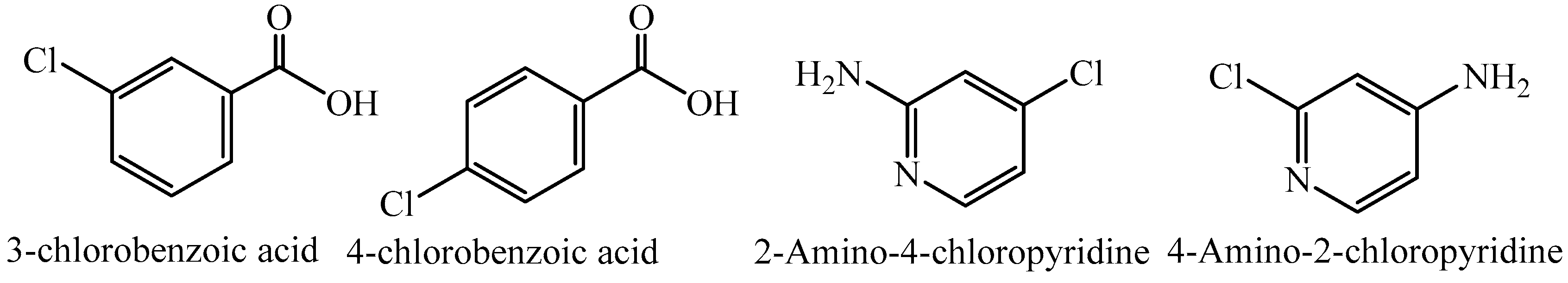
Supramolecular Assemblies of 3/4-Chlorobenzoic Acid and Amino-Chloropyridine Derivatives: Synthesis, X-ray Diffraction, DFT Calculations, and Biological Screening

1
Department of Chemistry, University of Malakand, Chakdara 18800, Pakistan
2
Department of Chemistry, University of Management and Technology (UMT), Johar Town, Lahore 54770, Pakistan
3
Materials Chemistry Laboratory, Institute of Chemistry, The Islamia University of Bahawalpur, Baghdad ul Jaded Campus, Bahawalpur 63100, Pakistan
4
Department of Chemistry, College of Science, University of Bahrain, Main Campus, Zallaq 32038, Bahrain
5
Department of Chemistry, COMSATS University Islamabad, Abbottabad Campus, Abbottabad 22060, Pakistan
*
Author to whom correspondence should be addressed.
Crystals 2023, 13(12), 1663; https://doi.org/10.3390/cryst13121663
Submission received: 16 September 2023 / Revised: 28 November 2023 / Accepted: 30 November 2023 / Published: 5 December 2023

Abstract

:
Organic acids (3-chlorobenzoic acid and 4-chlorobenzoic acid) were treated with 4-amino-2-chloropyridine and 2-amino-4-chloropyridine as coformers for cocrystallization. Acid/base pairs afforded a cocrystal (3-chlorobenzoic acid and 4-amino-2-chloropyridine, compound 1) and molecular salts (2-amino-4-chloropyridinium 3-chlorobenzoate, 2; 2-amino-4-chloropyridinium 4-chlorobenzoate, 3). The products were characterized with the help of FT-IR, UV/visible spectroscopy, PXRD, and SC-XRD. The position of the Cl-substituent on the phenyl ring was explored with respect to proton transfer between acid/base pairs, and its role in supramolecular chemistry is discussed in detail. The existence of supramolecular assemblies was further reproduced using DFT calculations. Further, frontier molecular orbital (FMO), molecular electrostatic potential (MEP), and noncovalent interaction index (NCI) analyses were performed to gain insight into the electronic properties and nature of noncovalent interactions. The prepared compounds were examined for their biological activities against selected Gram-positive and Gram-negative bacterial strains and were also tested as antioxidant agents (DPPH free radical scavenging). Structural parameters determined experimentally and theoretically are within the expected range, and the biological activities of the resultant compounds are moderate.

1. Introduction

Molecular salts and cocrystals are competitive products obtained via reactions between weak organic acids and bases. The resultant materials are collectively considered multicomponent crystals which can be distinguished from each other on the basis of the transfer of at least one proton [1,2]. The formation of salts and/or cocrystals is carried out for the enhancement of physicochemical properties like solubility, dissolution, bioavailability, and stability and for pharmaceutical purposes [3]. The ionization constant gives important information about the degree of proton transfer or the ability of salt formation to resist the disproportionation of the involved synthons [4]. The formulation of new supramolecular assemblies without breaking existing covalent bonds is the beauty of crystal engineering in which intermolecular interactions are responsible for holding synthons together [5,6]. Various non-covalent interactions like hydrogen bonding [7,8,9,10], halogen bonds [11,12,13,14,15], van der Waals interactions, and π–π stacking [16,17,18] play an important role in this regard. Similar to hydrogen bonding, halogen bonding is also considered significant in the packing of crystal structures [19,20,21]. Resnati and Merangolo et al. briefly explained the importance of halogen bonding in crystal packing and nature in the multicomponent structure of an iodoperflurocarbon with aliphatic amines [22,23]. Both hydrogen and halogen bonds coexist within the same crystal structure, which tunes the properties in an engineered way in the cocrystals/salts of thiophene carboxylic acids and nitrogenous bases [24]. Halogen bonds play a role in designing functional materials in the fields of optics, photonics, biomimetics, photo-switchable and functional surface supramolecules [25].
The non-covalent interactions between halogen atoms and donor atoms like N, O, and S are considered efficient for holding synthons together [26,27]. From both theoretical and experimental studies, it can be determined that electron clouds are anisotropically distributed along with covalently held halogen atoms [28,29]. This causes a minute localized positive potential region known as the σ-hole [30]. Thus, a nucleophilic atom can interact with the σ-hole, and a halogen bond is formed [31,32]. Sometimes, halogen–halogen interactions appear when both electrophilic and nucleophilic interactions coexist on a halogen atom; these are also termed σ-hole–σ-hole interactions [33,34]. Halogen roles are vital in pharmaceutical products wherein the concept of halogen-based supramolecular assemblies is worth exploring. In this regard, the first example of a pharmaceutical cocrystal based on halogen bonds was reported by Baldrighi et al. in 2013 [35]. The halogen atoms in the drug molecules may result in increased permeability which, in turn, leads to an increase in oral bioavailability [36].
Pyridine is a well-known heterocycle which plays a role as a central ring in many organic compounds [37]. Its effects against various ailments as medicine have been shown, and is a good Lewis base, causing its derivatives to have attractive ligands to accept protons and bind to metal centers [38,39,40,41]. Several halogenated derivatives of pyridine and organic acids have been well characterized, and their properties have been explored [42]. The multicomponent structure as a product can be efficiently predicted from Rule of 3. According to this rule, molecular salt formation is dominant over cocrystals if ΔpKa > 3 (ΔpKa is the difference between pKa (base) and pKa (acid)), while cocrystals are formed if ΔpKa < 3 [43]. If this difference in pKa is closer to or equal to 3, either product can be obtained [44,45]. Carboxylic functionality shows efficiency in establishing hydrogen bonds with a suitable coformer (base) like pyridine and amine derivatives [46].
Here, we report the molecular salts of 3-chlorobenzoic acid/4-amino-2-chloropyridine, 3-chlorobenzoic acid/2-amino-4-chloropyridine, and 4-chlorobenzoic acid/2-amino-4-chloropyridine (Scheme 1). The incorporation of a chloro-group led to weak Van der Waals interactions and interesting supramolecular chemistry, but the final products as molecular salts/cocrystals were obtained as expected for the difference in pKa of the starting material. The supramolecular chemistry of isolated synthons was discussed, and their biological applications were explored.

2. Results and Discussion

In the current work, heterosynthons were selected according to the availability of donor–accepter sites for noncovalent interactions. Chloro functionality at various positions of the principal aromatic rings of both acids and bases in molecules was considered in which the position of the halogen affects its role in secondary interactions. In a continuation of previous work on non-covalent interactions and multicomponent structure design [39,47,48], new products are hereby presented from the reaction of benzoic acid derivatives and pyridine derivatives. The pKa values of 3-chlorobenzoic acid, 4-chlorobenzoic acid, 4-Amino-2-cholorpyridine, and 2-Amino-4-cholorpyridine are 3.85, 3.98, 4.73 and 5.72, respectively [49]. In case of compounds 1 (3-chlorobenzoic acid/4-amino-2-chloropyridine) and 2 (2-amino-4-chloropyridinium 3-chlorobenzoate) and 3 (2-amino-4-chloropyridinium 4-chlorobenzoate), the difference in the value of pKa is less than 3 (ΔpKa = 0.88, 1.87, and 1.74, respectively) so, according to Rule 3, cocrystal formation dominates. The role of the structure of coformers is more active in comparison with the “Rule of 3,” particularly the presence of the pyridine ring, which is more basic and has a greater affinity to accept H+ [47].

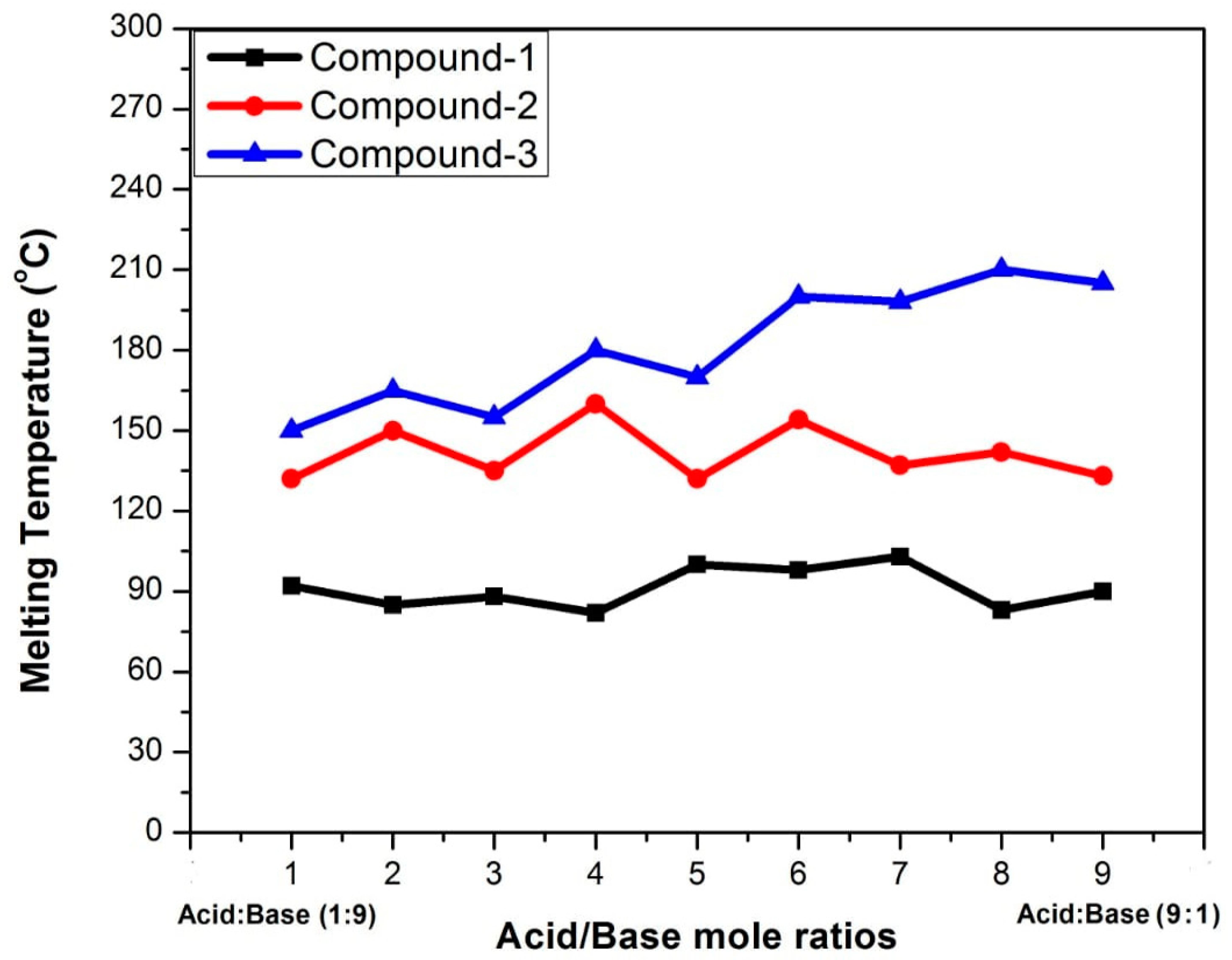
2.1. Physical Phase Diagram of the Starting Materials of Compounds 1, 2, and 3

The physical mixture of the starting materials of compound 1 (3-chlorobenzoic acid and 4-Amino-2-chloropyridine) were prepared in different molar ratios, 1:9, 2:8, 3:7, 4:6, 5:5, 6:4, 7:3, 8:2, and 9:1, in a solid state. The nine different physical mixtures of the components of 3-chlorobenzoic acid (in the case of compound 2) and 4-chlorobenzoic acid (in the case of compound 3) with 2-amino-4-chloropyridine were prepared in the same way as discussed above. A zigzag pattern (W-type) in the melting points of the physical mixtures is an indication of physical interactions. It is a well-established fact that cocrystals and the physical mixture of their coformers have different melting points [50,51]. In a similar way, binary phase and ternary phase diagrams are easy methods of determining solubility as a function of physical interactions. In this regard, a phase diagram of a physical mixture of the coformers of cinnamic acid and nicotinamide is reported as a case study [52]. The melting point of mixture does not follow a regular pattern, as shown in Figure 1, which confirms that both conformers interact with each other via physical interactions like hydrogen bonding, halogen bonding, or other non-covalent interactions.

2.2. FT-IR Studies of Compounds 1, 2 and 3

The FT-IR spectra of both starting materials (synthons) and their resultant multicomponent structures were recorded and correlated (shown in Figures S1–S3). In the spectrum of compound 1, a weak broad absorption at 3350 cm−1 can be assigned to an OH group. Extensive hydrogen bonding in the cocrystal is evident from a shift toward a lower wavenumber and the broadness of the peak when compared with the starting acid. The same phenomenon was observed with the N-H bond of aminopyridine, for which a shift occurs from 3300 cm−1 to a weak and broad peak at 3200–3300 cm−1. Other peaks of the starting materials and resultant products appear with slight shifts because of the involvements of some of the groups in secondary interactions. In the spectrum of compound 2, the O-H bond 3560 cm−1 disappears, indicating the transfer of a proton from the acid molecule, which clearly supports the formation of a molecular salt. Also, the N-H bonds of aminopyridine are involved in noncovalent interactions with coformers. The aromatic C-H bonds appear in their expected region.

2.3. Powder and Single X-Ray Diffraction Studies of Compounds 13

The powder X-ray diffraction patterns of compounds 1 (cocrystal), 2, and 3 (molecular salts) are given in Figures S5–S7. From the regularity of the peaks in the pattern, the crystalline nature of the compounds is confirmed. The same pattern of peak intensities can also be achieved from single-crystal X-ray diffraction using a simulated powder pattern. These spectra reveal that the bulk of the compounds obtained in this study are reasonably pure, with negligible side reactions and side products. Further confirmation of the structural characterization of the compounds was carried out for single crystals of all the compounds.

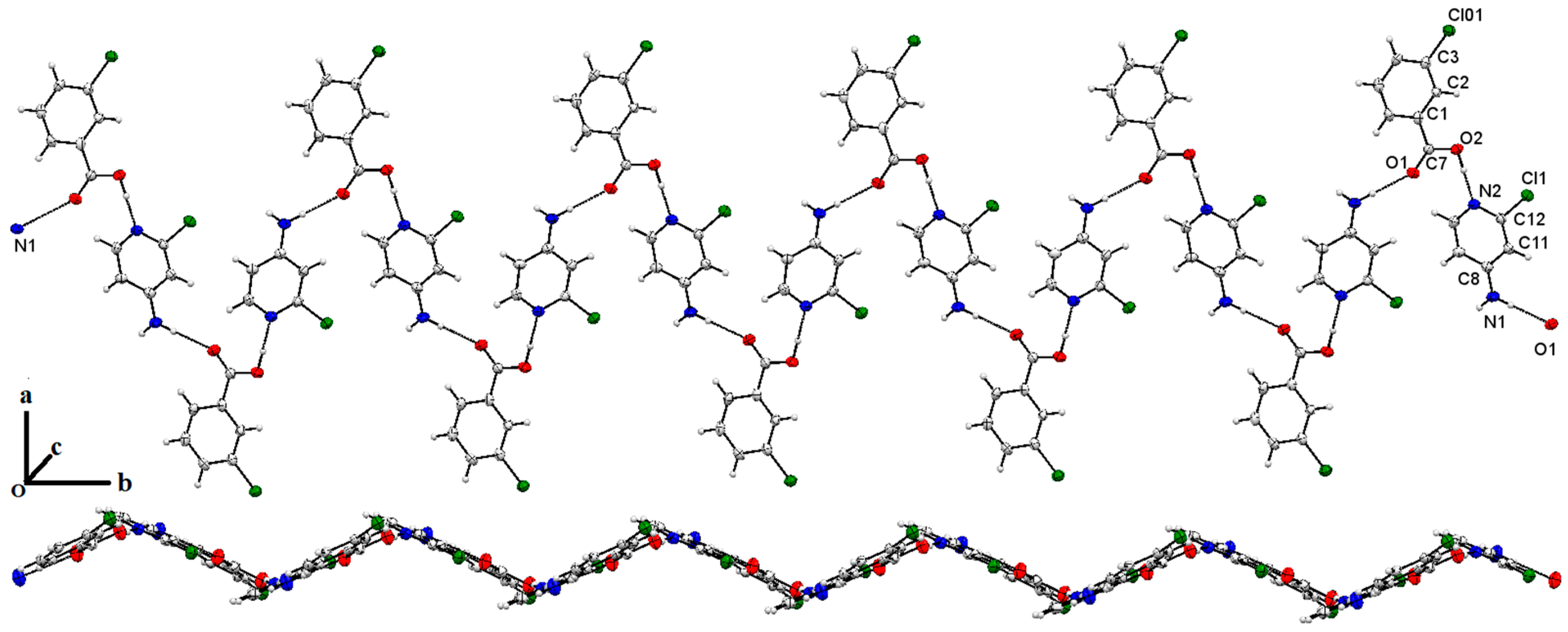
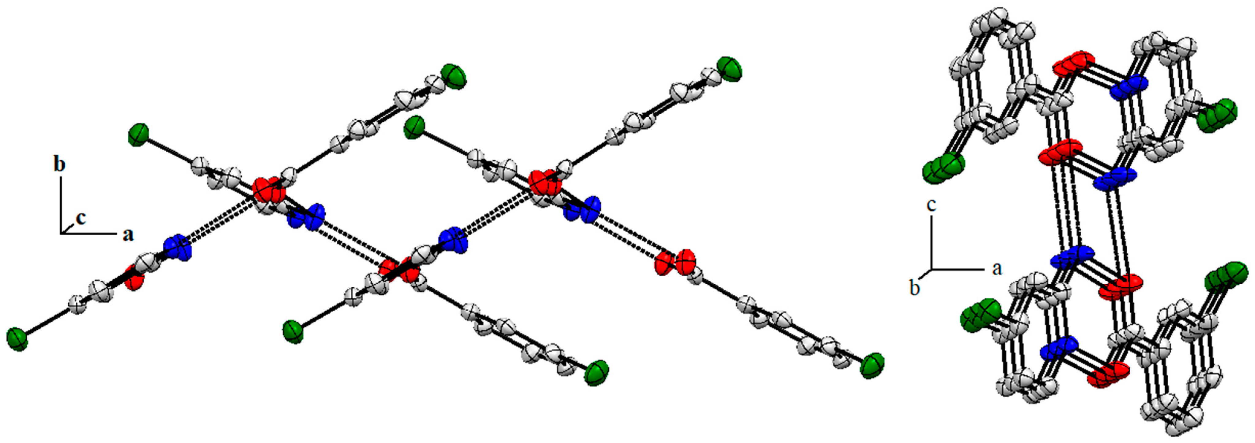
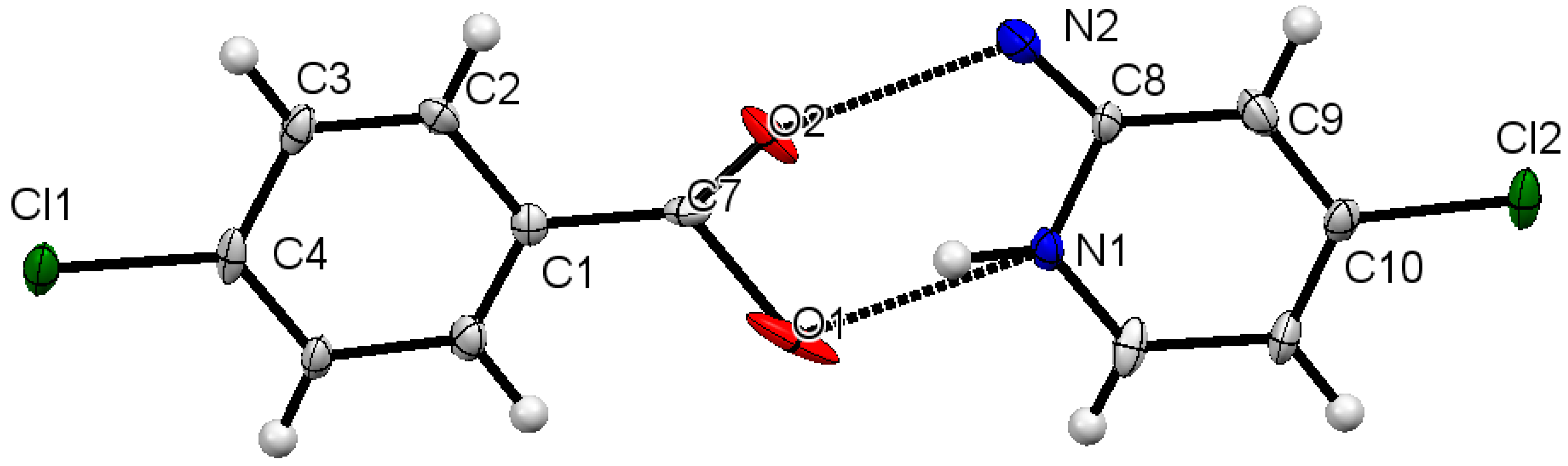
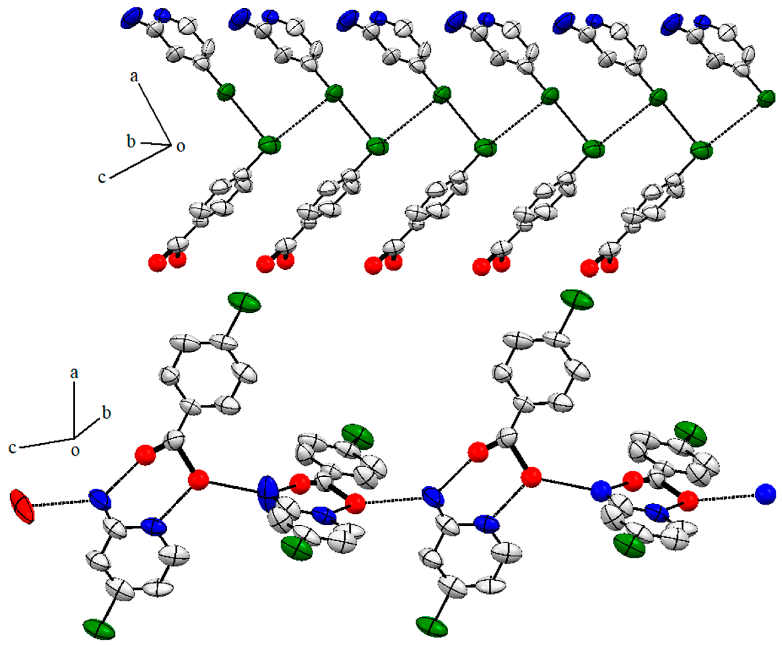
2.4. Single X-Ray Structural Description of Cocrystal of 3-Chlorobenzoic Acid:4-Amino-2-Chloropyridine, Compound 1

The molecular structure of compound 1 is shown in Figure 2; it crystallizes in the P21 space group. It is evident from the single-crystal analysis of compound 1 that secondary interactions give extra stability to the compound in a solid state. Interactions between OH---N (where the OH of the acid and the N of the pyridine ring) appear with a separation distance between N(2) and H(2) equal to 1.633 Å. This is the only H-bonding interaction with the help of which an acid/base pair is held together. H+ transfer in an acid/base pair does not take place under the provided conditions. The pyridine and phenyl ring of the acid are twisted with respect to each other at approximately 45°. Further, each NH2 and C=O group is involved in intermolecular H-bonding and stabilizing the supramolecular structure of the co-crystal. The distance between N(1)H and O(1) is 2.019 Å. As a result of H-bonding, the chair structure extends in a 1D fashion, as shown in Figure 3. The chloro functionalities of both the heterosynthons’ acids and bases interact with each other through van der Waals forces with a separation distance of Cl(1)---Cl(01) 3.497 Å, and at the same time, Cl(01), which belongs to benzoic acid moiety, is associated with an ammine group of the base, and Cl(01)---HN is equal to 2.683 Å. In contrast to H-bonding (discussed above), Cl---Cl interactions stabilize the 2D extension of the supramolecular structure. There are some other interactions which, collectively with Cl---Cl and H-bonding, afford a 3D structure in a solid state. The main role played by the aromatic ring of the acidic moiety is obvious in stabilizing the 3D supramolecular structure of the molecule. The C=O---HN bonds are the main forces which hold molecules together in a solid state (Figure 2), while halogen interactions can be observed among all nearby halogen atoms (Cl atoms), as shown in Figure 3. Data pertaining to crystal structure refinements and solutions are summarized in Table 1. In the crystal structure of the compound, each acid molecule is attached to four pyridine derivatives, one of which is attached with the help of a halogen–halogen interaction. In the case of 4-Amino-2-chloropyridine, four different acid molecules are attached, one of which is because of halogen bonding, while the remaining are attached with hydrogen bonds. The C(1) of benzoic acid is linked to the C(6) of another acidic moiety through π–π interactions (C(1)---C(6); 3.397 Å), extending the straight chain of acidic groups. Two parallel chains of acids are associated with each other via C(5)H---C(2) interactions which are separated by 3.781 Å, while the H(5)---C(2) distance is 2.855 Å (Figure 4). Such interactions are not present within base molecules.

2.5. Single X-ray of Compound 2 (2-Amino-4-Chloropyridinium 3-Chlorobenzoate)

A 1:1 acid/base unit of the molecule of compound 2 (monoclinic, space group P21) is shown in Figure 5 with a partial numbering scheme. The NH2 group in the base is at appropriate position to afford an R 2 2 8 loop, as has been observed in 2-aminopridine derivatives [38]. Here, the incorporation of a chloride function in the base was expected to adversely affect the basicity of ring N to hinder it from accepting H+. Based on the difference in the pH of the pair, a cocrystal was anticipated, but the actual product was a molecular salt. It indicates the role of the structure of the acid/base unit in the co-crystal/molecular salt formation and not only the pH. The C(1)-O(1) and C(1)-O(2) distances in the acid are 1.264(5) and 1.247(6) Å, which reveal a difference in bond order between the C and oxygen atoms. Hydrogen bonding between O(1)---HN(1) link acid/base pairs, with a distance between O and H of 1.998 Å, making a stronger interaction when compared with the H-bonding in compound 1. As a result of this interaction, acid/base pairs extend in 1D fashion along the b-axis, as shown in Figure 6 (left). Among short contacts, C(1)---C(8), C(11)---O(2), and C(11)---H(3) are linked together with distances of 3.376, 3.165, and 2.832 Å, respectively. These short interactions stabilize the supramolecular structure along the b and c axes, as shown in Figure 6 (right).

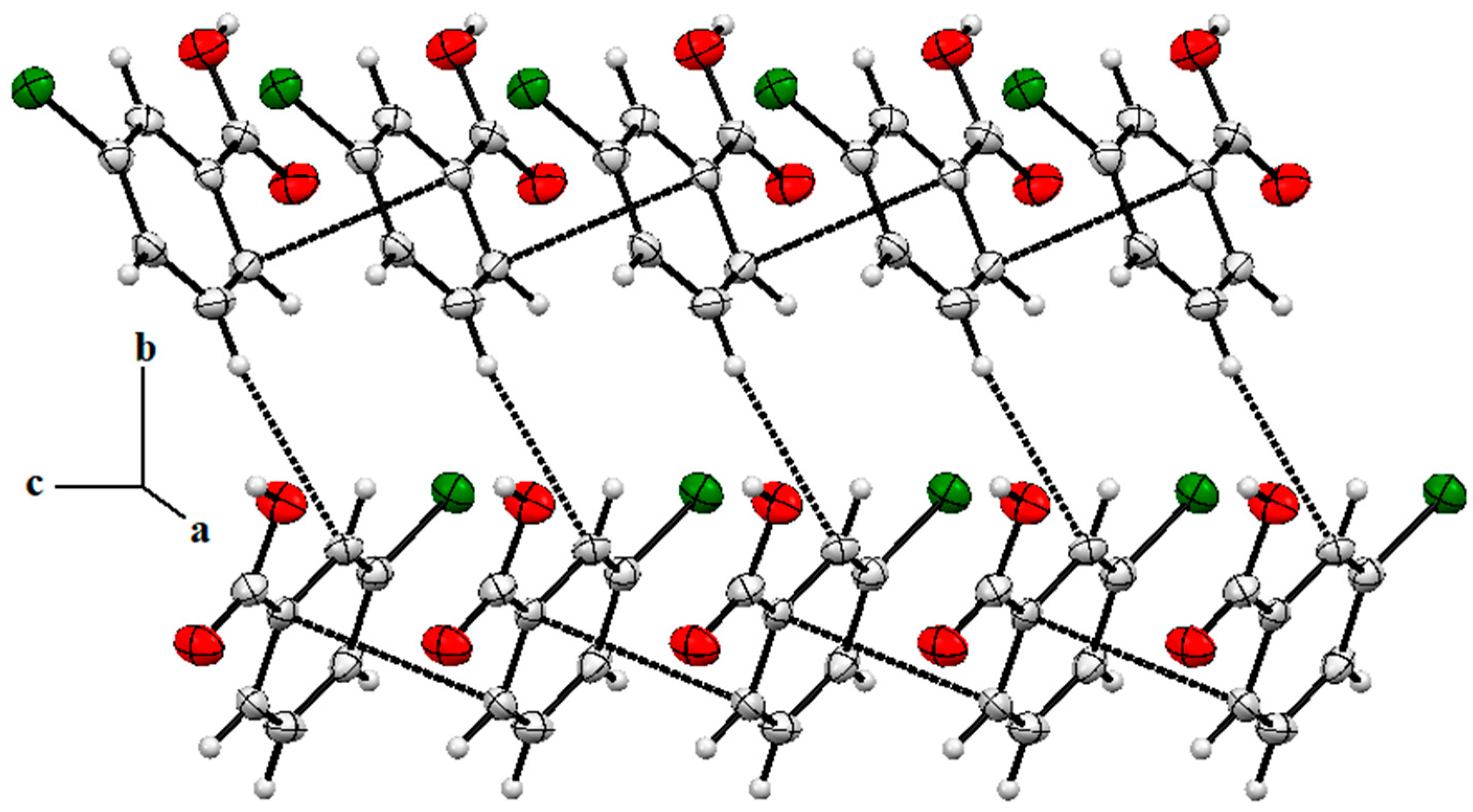
2.6. Structure Description of Compound 3 (2-Amino-4-Chloropyridinium 4-Chlorobenzoate)

The crystalline nature of compound 3 is monoclinic with the space group Pc. Its molecular structure is shown in Figure 7 with a partial numbering scheme. The oxygen atoms of the compound suffer from slight disorder in which the data pertaining to structural features such as bond lengths can be misleading. The structure provides enough information to establish the proposed connectivity of an acid/base pair like compound 2. The structure indicates that proton transfer takes place from the acid to the base in a similar way as discussed for compound 2 to afford a molecular salt, making the ring system R 2 2 ( 8 ) . Proton transfer is evident from the difference in the C-O bond length in the COO group. The C(7)-O(1) and C(7)-O(2) bond lengths are 1.658(19) and 1.072(19) Å. The electron density is localized within the C=O bond rather than delocalized along the O-C-O fragment. The acid/base pair is coplanar, with negligible deviation from planarity. Both coformers are linked with the help of H-bonding where distance between O(2) and N(2) is relatively short (2.518 Å) as compared to compound 2. As discussed above, the disordered O(1) interacts with N(1)H with separation distances of 1.689 (O(1)---H) and 2.529 Å (O(1)---N(2)). The O(1) and N(2) are potential centers for H-bonding, and they interact with neighboring salt molecules, stabilizing a 1D straight chain (Figure 8).
An overview of short contacts indicates that Cl---Cl interactions are present in the molecules. The acid moiety through Cl(1) links two base synthons through Cl(2) and similarly, each Cl(2) interacts with two Cl(1). The distance between Cl(1) and Cl(2) is slightly different due to different orientations of the molecules in the space, i.e., 3.453 and 3.461 Å (Figure 9). Other types of short contacts were not observed in the molecules of compound 3.

3. Supramolecular Interactions via DFT Calculations

DFT studies of compounds 13 were performed to analyze insights into supramolecular assemblies in a solid state, and relaxed geometries are given in Figure 9. Important nonbonding interactions such as hydrogen bonding, halogen bonding, and van der Waals interactions are prominent in crystal packing. Such interactions were also observed in optimized gaseous-phase structures of all these compounds (13). In the crystal structures of compounds 1 and 3, hydrogen bonding as well as halogen bonding can be seen between acid–base pairs on opposite sites. To examine such interactions [53,54,55], two types of orientations were considered for the dimeric compounds 1 (1a for hydrogen bonding and 1b for halogen bonding) and 3 (3a for hydrogen bonding and 3b for halogen bonding). These compounds (1a, 1b, 2, 3a, and 3b) were optimized at the same B3LYP-D3/6-311++G(d,p) level of theory. As a result of these interactions, thermodynamically stable clusters were formed. Thermodynamic stability was confirmed using the counterpoise-corrected energies (Ecp) and interaction energies (Eint) of these compounds (13), and the values are given in Table 1. The Ecp values of dimers of the compounds 1a, 1b, 2, 3a, and 3b are −12.91, −2.10, −96.45, 36.37, and 52.63 kcal/mol, respectively. The highest Ecp (−96.45 kcal/mol) is seen for compound 2, and the lowest Ecp (−2.10 kcal/mol) is obtained for compound 1b. The negative signs represent the feasibility of dimerization; ultimately, all the reactions are exothermic in nature except compounds 3a and 3b, which show endothermic behavior. The hydrogen-bonded dimers (1a, 2, and 3a) are more stable compared to the halogen-bonded dimers (1b and 3b). The Eint values of dimers of the compounds 1a, 1b, 2, 3a, and 3b are −17.81, −8.20, −101.32, −29.99, and −46.12 kcal/mol, respectively. The Eint values of the respective dimers are comparable to the Ecp values, which agrees with previously reported similar work on complexation of different chemical species [56]. Compounds 1a and 1b are neutral species, whereas compounds 2, 3a, and 3b are charged species. In the case of compound 2, electrostatic interactions are responsible for strong interactions. In compounds 1a and 1b, hydrogen and halogen bonding play dominant roles. Compounds 3a and 3b also show weak electrostatic intaeractions compared to other compounds.

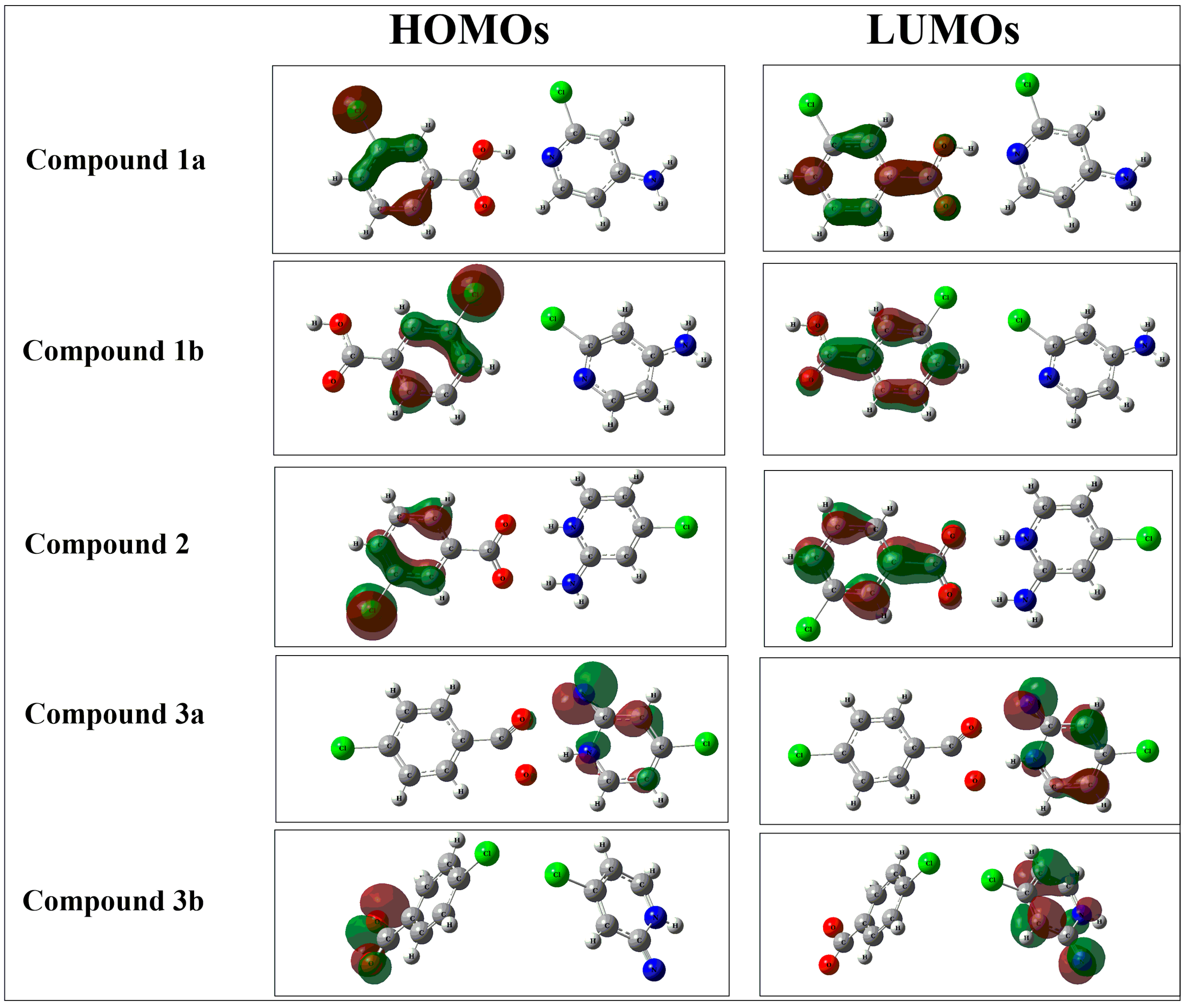
3.1. Frontier Molecular Orbital (MO) Analysis

A frontier molecular orbital (MO) analysis was performed to evaluate the electronic properties of compounds 13. During this analysis, the energies of the highest occupied molecular orbitals (EHOMO), the lowest unoccupied molecular orbitals (ELUMO), and the HOMO-LUMO gap (EL-H) were calculated. The EHOMO values of compounds 1a, 1b, 2, 3a, and 3b are −6.76, −6.72, −6.10, −5.93, and −5.82 eV, respectively. The ELUMO values of compounds 1a, 1b, 2, 3a and 3b are −1.38 eV, −1.72 eV, −3.01 eV, −0.47, and −0.65 eV respectively. The EL-H gaps of compounds 1a, 1b, 2, 3a, and 3b were 5.38, 5.00, 3.09, 5.46, and 5.17 eV, respectively (Table 2). These values reflect the semiconductor behavior of these compounds, along with high chemical and kinetic stabilities. The same type of behavior was noticed in compounds recently reported in [57]. Electronic density distributions illustrate that in compounds 1a and 1b, the HOMO densities reside on the chloro-group of the 3-chlorobenzoic acid fragment and its nearest carbon (Figure 10). The reason is the electron-withdrawing effect of the halogen. The electronic densities of the LUMOs are localized on the acetate group and the carbon connected to this group. Partially, the densities also reside on the C-C bonds of the benzene ring, which is away from the chloro-group. Similarly, in compound 2, the HOMO densities reside on the chloro-group of the 3-chlorobenzoic acid fragment. Some of the densities are also found on the carbon atoms attached to this group. The LUMO densities reside on the acetate group of the 3-chlorobenzoic acid fragment. Some of the densities are also found on the carbon–carbon bond attached to the acetate group. The reason might be because of the hydrogen transfer between the acetate group of the 3-chlorobenzoic acid and the amino group of the 2-amino-4-chloropyridine. Like compound 1, the HOMO densities are localized on all atoms of the 2-amino-4-chloropridine except the chloro-group. The LUMO densities in compound 3a are localized on the amino-group and the carbon–carbon bonds of 2-amino-4-chloropyridine. This is due to the fact that a strong noncovalent interaction exists between the amino group of 2-amino-4-chloropyridine and the oxygen atom of the acetate group in compound 3a. Some of the density also resides on the oxygen atoms of the 4-chlorobenzoic acid. The HOMO densities are localized on the acetate group of the 3-chlorobenzoic acid fragment in compound 3b. The LUMO densities are localized on the amino group and carbon–carbon bonds of the 2-amino-4-chloropyridine.

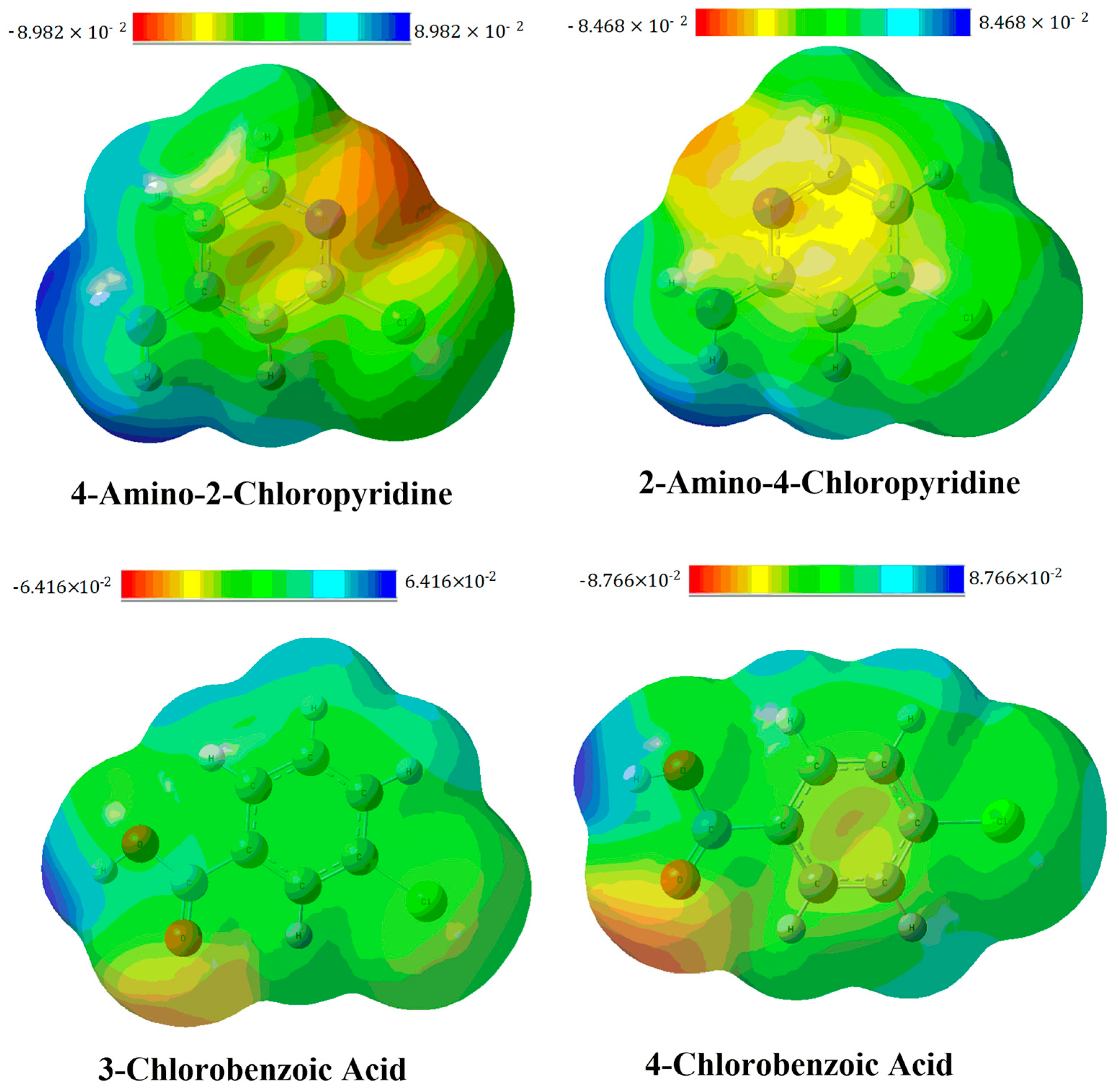
3.2. Molecular Electrostatic Potential (MEP) Analysis

A molecular electrostatic potential (MEP) analysis can be used for the prediction of reactive site compounds. The potentials on the surfaces of complexes are represented by different colors. The increasing order of electrophilicity is given as red < orange < yellow < green < blue. This range represents that the blue color shows electrophilic sites and red color shows nucleophilic sites. The green color surface depicts a neutral region of the respective compound [56]. To investigate these active sites in isolated organic acids and bases used for the synthesis of our compounds (1a, 1b, 2, 3a and 3b), the MEP surfaces of the acids and bases were constructed as shown in Figure 11. The red surfaces, seen on the oxygen atoms of the acetate groups in each of the organic acids, represent nucleophilic sites. On the other hand, the hydrogen of the hydroxyl group in the organic acid shows an electrophilic site. The blue surfaces seen on amino groups reflect electrophilic sites in organic bases, and the nitrogen in the ring with a red surface indicates a nucleophilic site.
Non-covalent interactions are also explained through sigma holes. These holes represent regions of lower electronic density (maxima) on the surface of a covalently bonded atom (along the extension of the bond), illustrating the anisotropies of atoms bonded through a covalent bond. In the region of these covalently bonded atoms, a positive electrostatic potential is generated, and the sites with these potentials are responsible for the interaction of the molecule with the negative sites (i.e., atoms with lone pairs of electrons, anions, and pi electrons) of another molecule. These holes are responsible for self-assembly in crystal growth through noncovalent interactions [58]. Contrary to sigma holes, regions of higher electronic density (minima) are also generated on the atoms with a negative electrostatic potential. Molecules can easily interact with positive sites through these negative potential sites [59]. In compounds 1–3, sigma holes are generated on the hydrogen atoms of the amino group, which are responsible for hydrogen-bonding with a nucleophile during crystal growth. The energies of the maxima points are 52.19 and 51.72 kcal/mol for the hydrogens of the amino group, while the nitrogen of pyridine has a critical point energy of −30.51 kcal/mol.
In addition to amino groups, sigma holes are seen in chloro-groups in compounds 13, which are responsible for halogen bonding during crystal growth. The chloro-group acts as an electrophilic site for the incoming nucleophile (Figure 12). The positive potentials on the chloro-group of the pyridine ring have maximum energies of approximately 35.65 kcal/mol. For 3-chlorobenzoic acid and 4-chlorobenzoic acid, the oxygen atoms of the carbonyl groups have minimum points on the global energy surface with energies of −30.81 and −30.50 kcal/mol, respectively. These sites act as nucleophiles and have negative potential due to the presence of lone pairs of electrons of the oxygen atoms in the carbonyl group. Thus, the hydroxyl site acts as an electrophilic site, and oxygen acts as a nucleophilic site. Overall, benzoic acid moieties withdraw electron density from pyridine in complexation, leaving it electron-deficient in all compounds 13. The most prominent sigma hole is seen in compound 2. The greater polarizability of the bonds between benzoic acid and pyridine moieties causes an increase in the electropositive regions of the sigma holes, resulting in stronger non-covalent interactions. In addition to polarizability, the strong electron-withdrawing group in benzoic acid increases the positive potential on the chlorine group of the pyridine moiety, strengthening the noncovalent interactions. The presence of sigma holes in our compounds confirms the occurrence of noncovalent interactions in compounds 13.

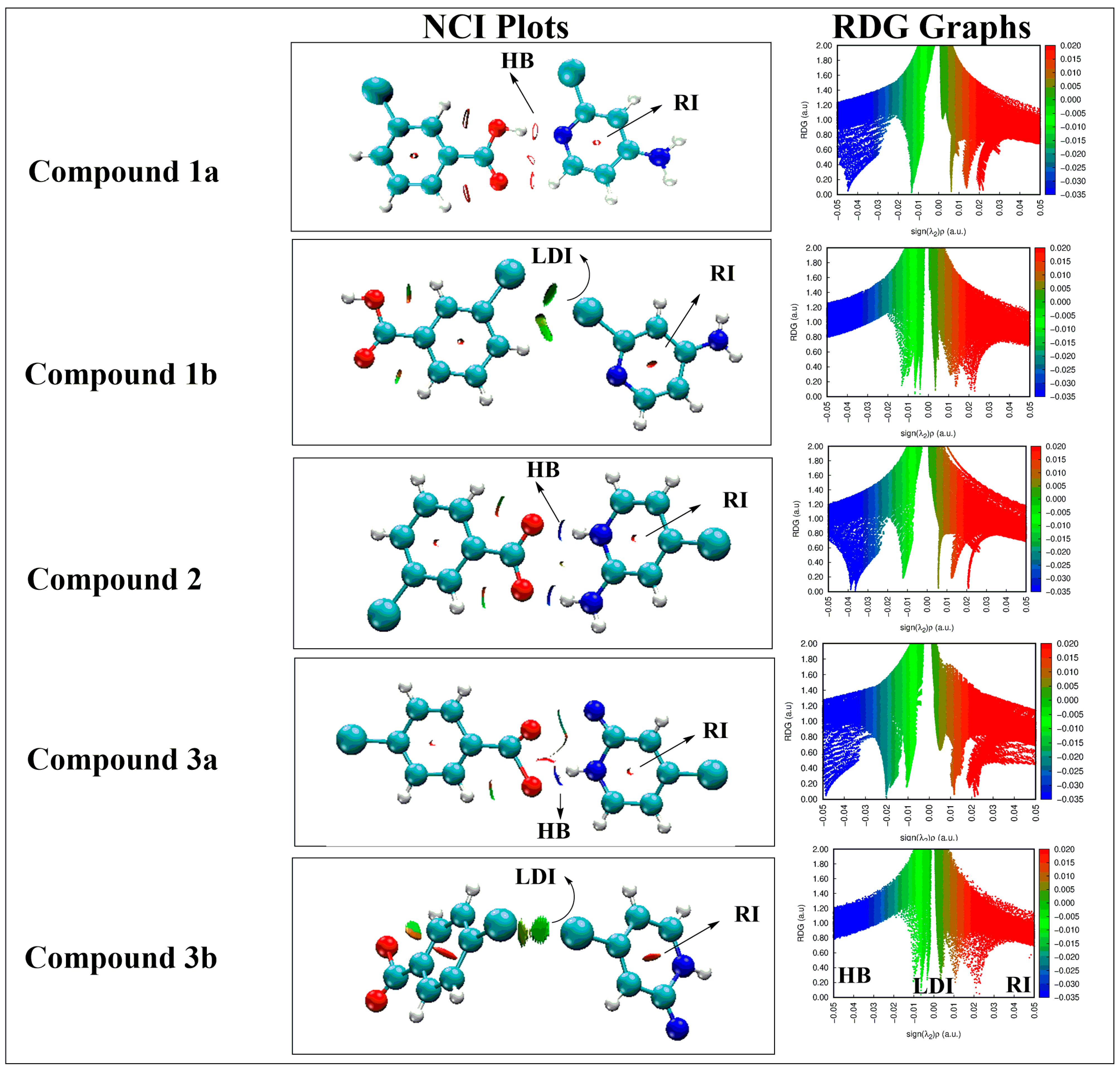
3.3. Noncovalent Interaction (NCI) Analysis

For a further deep visualization of noncovalent interactions, an NCI analysis was carried out. Noncovalent interactions including weak Van der Waals interactions, hydrogen bonding, and steric repulsion were studied as observed in real space [56]. Three-dimensional NCI isosurfaces and two-dimensional reduced density gradient (RDG) graphs were constructed, and data are given in Figure 13. In the three-dimensional NCI plots, the nature of different interactions is estimated from three different colours of the isosurfaces [60]. Strong hydrogen-bonding (HB) and weak London dispersion interactions (LDI) are observed as blue and green isosurfaces, respectively. Repulsive interactions (RI) appear in red between interacting monomers of a complex. Moreover, the strength and intensity of the interactive forces are proportional to the thickness of the isosurfaces. The NCI plots represent electrostatic and hydrogen bonding between the benzoic-acid- and amino-pyridine-containing fragments in each of these compounds (1a, 1b, 2, 3a, and 3b). But hydrogen bonding is a dominating interaction in all compounds 1a, 2, and 3a, whereas halogen bonding (Cl-Cl) is the dominant interaction in compounds 1b and 3b. The thickness of the isosurfaces represents strong interactions between two fragments which justify the stability of all these compounds.
In the two-dimensional RDG graphs, the sign (λ2)ρ values are in the range of 0.05 to −0.05 au in scatter graphs which exhibit three types of interactions including hydrogen bonding, weak van der Waals forces, and repulsive forces. Various spikes appeared in these graphs, with blue, green, and red colors for strong van der Waals forces (such as hydrogen bonding), weak van der Waals forces, and repulsive forces, respectively. Furthermore, the deep RDG spikes and more negative (λ2)ρ values (−0.03 to −0.05 au) of spikes reflect the presence of strong hydrogen bonding in compounds 2, 1a, and 3a, respectively. Blue patches are absent in this region, but a few blue spikes in isosurfaces are obtained in the (λ2)ρ region of −0.02 to −0.03 au. In addition to blue, green color spikes in the 0.01 to −0.01 au range represent weak van der Waals interactions in all compounds 13 but are more prominent in 1b and 3b. The dark red-green spikes between the 0.00 and 0.01 au regions in the RDG graphs of compounds 13 represent C-H---π interactions between two fragments. The red spikes in the 0.02 to 0.03 au regions of the RDG graphs reflect repulsive forces between the two fragments in each of the five compounds 13. These blue, red, and green spikes indicate the presence of hydrogen, weak van der Waals, and electrostatic repulsive interactions.

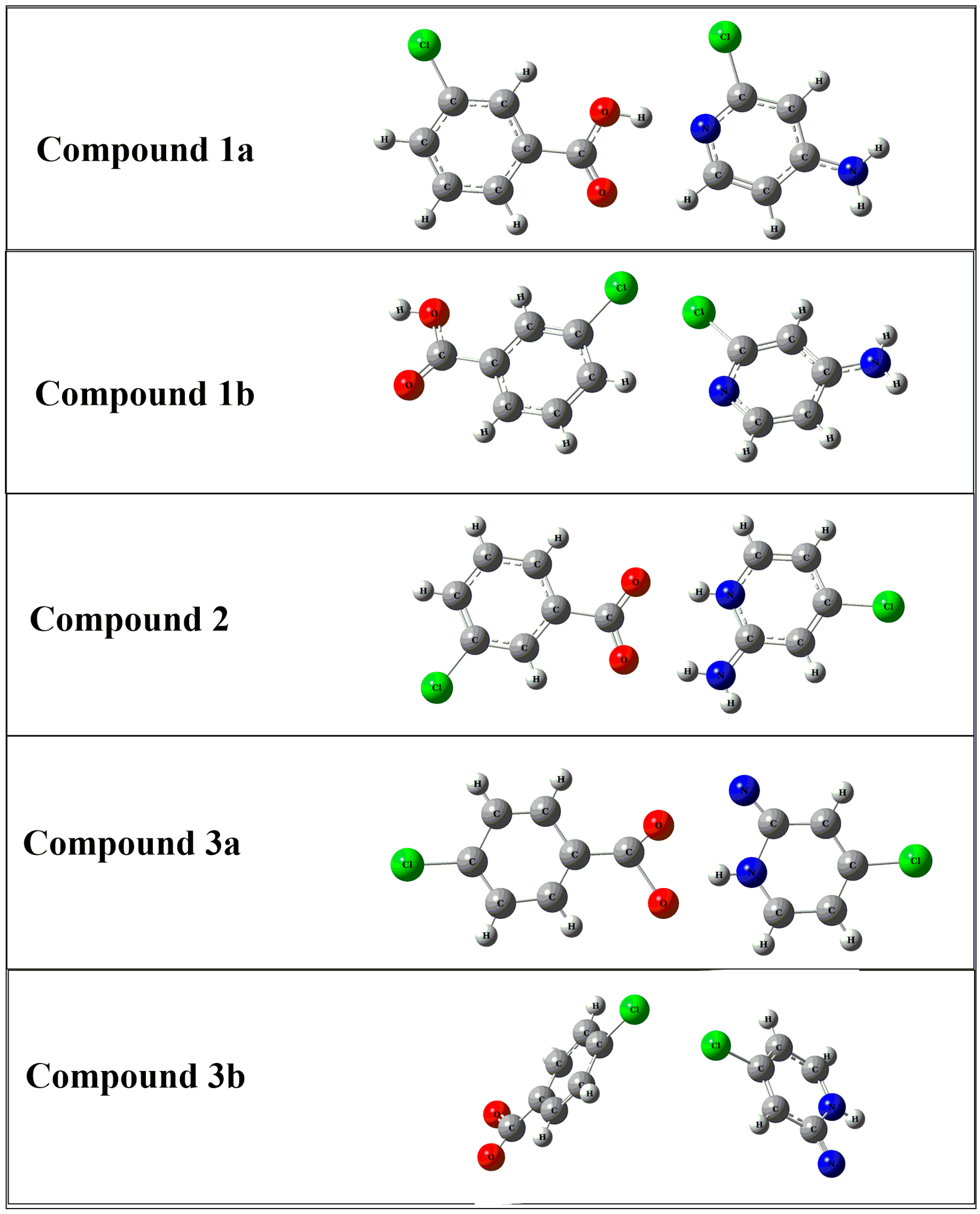
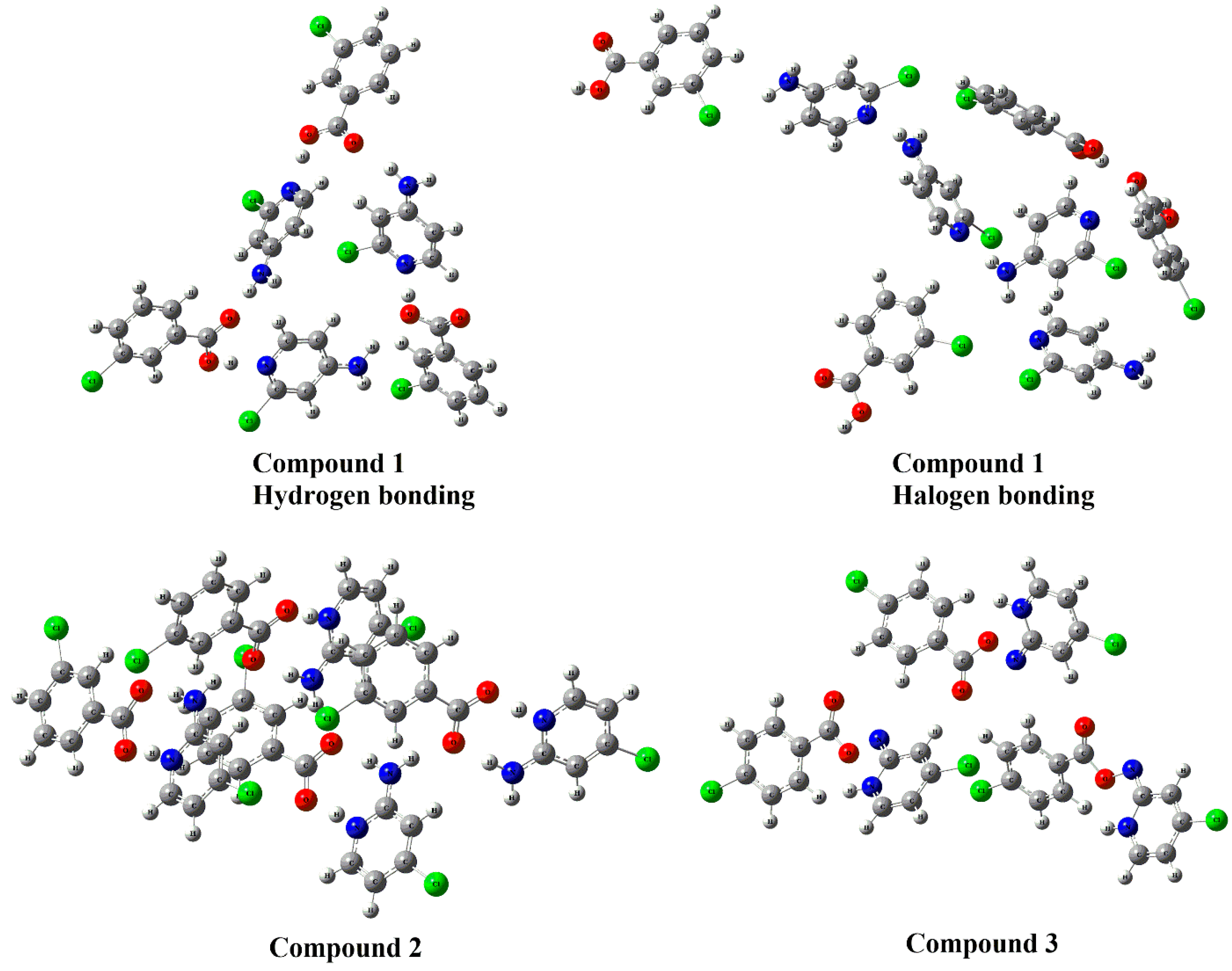
3.4. DFT Studies for Clusters of Compounds 13

The large clusters of compounds 13 were also modeled (in the orientations obtained from their crystal structures) and optimized at the same B3LYP-D3/6-311++G(d,p) level of theory (Figure 14). For compound 1, two types of orientations are considered, one in which halogen bonding is prominent and a second in which hydrogen bonding is seen. These two types of orientations are named compound 1-hydrogen bonding and compound 1 halogen-bonding (Figure 14). All these clusters are thermodynamically highly stable, as is evident from their interaction energies which illustrate that all reactions are exothermic in nature and that the cluster formation of these compounds is highly feasible. The HOMO and LUMO densities were also constructed, and it can be observed that localizations of densities on fragments are similar to the initial dimeric compounds 13. The HOMO densities are localized on the acetate group of the benzoic-acid-containing fragment, and the LUMOs reside on the amino-pyridine fragment in all packing clusters of 13. Furthermore, MEP surfaces were also constructed on all four trimeric compounds 13. Similar to the isolated dimeric compounds 13, again, the amino group represents an electrophilic site, whereas the acetate groups represent nucleophilic sites as we observe a red color on acetate groups and a blue color on amino groups, respectively. The chloro-groups seem to have neutral behavior which is evident from the green color on the MEP surfaces.
NCI plots and RDG graphs were also constructed for clusters of compounds 13 (Figure S9 and Supporting Information Figure S4). The NCI plots represent electrostatic and hydrogen bonding between the two benzoic-acid- and amino-pyridine-containing fragments in each of the trimeric compounds 13. But hydrogen bonding is the dominating interaction in all three trimeric compounds 13. The thickness of the green isosurfaces represents strong interactions between the two fragments which justifies the stability of all these compounds. The blue, red, and green spikes in the RDG graphs of clusters of 13 (Figure 13) indicate the presence of hydrogen, weak van der Waals, and electrostatic/repulsive interactions between different fragments of the compounds.

3.5. UV Absorbance Spectra

Compounds 13 were scanned in the UV/visible region between 200 and 800 nm with a fast scanning speed. The Beer–Lambert law was used for the determination of the molar extinction coefficient (ε) of each of the compounds. The results and molar absorptivity values are given in Table 3. For the determination of the molar absorptivity coefficient, measured absorbance maxima were divided by the sample concentrations for an optical path length of 1 cm. For obtaining UV-visible spectra, 0.5 μM solutions in methanol (cutoff value = 205 nm) were prepared in which the intensities of solvent blanks were noted at 0.014, 0.013, and 0.017 for 130, respectively. The absorption intensities for compounds 13 were 0.39, 0.30, and 0.31, respectively, from which the molar absorptivity of each sample was calculated, as given in Table 3.

4. Biological Applications of Compounds

4.1. Antioxidant Essay (DPPH)

Antioxidant materials are classified into different classes according to their mode of action, like chelators, free-radical scavengers, and oxygen scavengers. Several available methods exist to determine antioxidant activates. A DPPH free radical scavenging assay was used to evaluate the antioxidant potential of these compounds. 2-2-diphenyl-1-picrylhydrazal (DPPH) is a stable free radical and is commercially available. Mostly, compounds have been tested for free radical scavenging relative to of DPPH by following the method described in [61]. The method was followed with a slight modification for the determination of the DPPH free radical scavenging ability of the synthesized cocrystal and molecular salts and their coformers. As per the literature, DPPH absorbs UV light at 517 nm, but the absorption is dependent on the concentration of the solution (Figure S8). The central nitrogen accepts an electron or a hydrogen atom, resulting in decolorization from dark purple to yellow and then colorless, which can be measured using the decreasing absorbance in UV light.
A solution of DPPH was prepared in an ethanol/water (1:1, v/v) solvent system by dissolving 0.348 g of DPPH per 100 mL of solution. Various concentrations of solutions (50, 100, 150, 200, and 250 µg/mL) were prepared for both starting precursors and the synthesized compounds from a stock solution of each sample (0.001g in 10 mL). To each solution, 1 mL of DPPH was added and incubated for 30 min. Changes in color were observed from purple to yellow and then to colorless upon increasing the concentration. The IC50 values for DPPH free radical scavenging are given in Table 4. It is evident that the molecular salts (2 and 3) are more efficient than the cocrystal (1). The percent inhibition values were determined using the below formula:
% Inhibition = A c o n t r o l A s a m p l e A c o n t r o l × 100

4.2. Anti-Bacterial Study of Compounds 1, 2 and 3 and Their Comparison with Coformers

One of the best available strategies to enhance anti-bacterial efficiency and other properties of compounds are salt/cocrystal formulations. For enhancing the in vitro anti-bacterial activities of pharmaceutical drugs, this strategy is very efficient [62]. The antibacterial activity and zones of inhibition of the synthesized compounds and their coformers were determined against both Gram-positive and Gram-negative bacteria. Staphylococus aureus and Staphylococcus epidermis are Gram-positive, while Klebsiella pneumonia and Pneumonia mirabilis are Gram-negative bacteria. From the average zone of inhibition, it is evident that the cocrystal and salt formulations both increase antibacterial activity. Compounds 2 and 3 show efficient anti-bacterial activity over compound 1 (cocrystal) against both Gram-positive and Gram-negative bacterial strains. The coformers of the compounds are less efficient compared to their respective heterosynthons against the selected bacterial strains (Table 5).

5. Materials and Methods

5.1. General Considerations and Instrumentations

The benzoic acid and pyridine derivatives used in this study were depicted in Scheme 1 above. Chemicals, 3-chlorobenzoic acid, 4-chlorobenzoic acid, 2-amino-4-chloropyridine, and 4-amino-2-chloropyridine, were purchased from TCI, Japan, and were used as received. Melting points, uncorrected, were determined with the help of Stuart SMP-10, Japan. The melting points of the final products were compared with their respective coformers (157 ± 1 °C (3-chlorobenzoic acid), 241 ± 1 °C (4-chlorobenzoic acid), 153 ± 1 °C (4-amino-2-chloropyridine), and 126 ± 1 °C (2-amino-4-chloropyridine)); the difference in values was taken as a positive sign of a successful reaction. The FT-IR spectra of the resultant compounds were recorded using a SHIMADZU FT-IR Model 18,400, and single X-ray diffraction data were collected with the help of a Bruker D8 VENTURE diffractometer with a PHOTON II detector and dual-wavelength Mo/Cu and Mo-Kα, λ = 0.71073 Å. Cell refinement and data reduction were carried out using SAINT V7.06A software [63]. The crystal structure solution and refinements were accomplished using SIR97, [64] SHELXL97, [65] WinGX31 [66], and PLATON [67].

5.2. Synthesis of Compound 1, (Cocrystal of 3-Chlorobenzoic Acid:4-Amino-2-Chloropyridine)

The solution of 3-chlorobenzoic acid (0.1 g; 0.638 mmol) was prepared in 5 mL of acetonitrile. The acid solution was added dropwise to a separately prepared solution of 4-amino-2-chloropyridine (0.08 g; 0.638 mmol). The reaction was executed according to the reported procedure in [38]. The mixture of both coformers was refluxed for 4 h with constant stirring at ca. 300 rpm. The solution mixture was stored and placed for slow evaporation; after few days, transparent (white) crystals appeared. The crystals were isolated from the mother liquor and studied (m. p. = 110–112 °C; FT-IR data, ν (cm−1) = 3773, 3144, 3000, 2919, 2820, 2170, 1980, 1720, 1632, 1450, 1344).

5.3. Synthesis of 2-Amino-4-Chloropyridinium 3-Chlorobenzoate (2) and 2-Amino-4-Chloropyridinium 4-Chlorobenzoate (3)

Molecular salts 2 and 3 were prepared under reflux conditions using equimolar ratios (1:1) of acids and bases. Solution of 3-Chlorobenzoic acid (for compound 2), 4-chlrobenzoic acid (for compound 3) (0.1 g; 0.063 mmol each), and 2-amino-4-chloropyrdine (0.082 g; 0.063 mmol) were separately prepared in acetonitrile. The acid–base solutions were slowly mixed, and the reactions were processed as discussed for compound 1. The crystals of both compounds were grown in the same solution at room temperature via slow evaporation. The melting points of the starting precursors/coformers are 157 ± 1 °C (3-chlorobenzoic acid), 241 ± 1 °C (4-chlorobenzoic acid), and 126 °C (2-amino-4-chloropyridine).
Compound 2:
m. p. = 210–211 °C; FT-IR data, ν (cm−1) = 3750, 3355, 3134, 3090, 2960, 2940, 2790, 1728, 1640, 1580, 1400, 1330, 1021, 798, 712.
Compound 3:
m. p. = 180–182 °C; FT-IR data, ν (cm−1) = 3700, 3080, 3043, 2944, 2910, 2840, 1740, 1600, 1580, 1540, 1400, 1180, 810, 760.

5.4. Computational Methodology

All required simulations were performed using Gaussian 09 software [68], and the results were visualized using GaussView 05 software [69]. A B3LYP-D3 with 6-311++G(d,p) basis set was used for the computational modeling of compounds 13. B3LYP better explains the geometry of a system, and D3 was added to study the dispersion forces inside the designed structures. The larger basis set was used in this study due to the fact that the geometries and energies of systems are sensitive to basis sets. A higher basis set provides more accurate results for a desired data set than a lower basis set [70]. After structure elucidation, a frontier molecular orbital (FMO) analysis was also performed.
Molecular electrostatic potential was analyzed at the same level of theory (B3LYP-D3/6-311++G(d,p)), providing information about electrophilic and nucleophilic sites [70]. The thermodynamic stability of the compounds was estimated using interaction energies (Eint). This interaction energy was corrected for basis set superposition error (BSSE) [71]. Whenever two isolated molecules interact through van der Waals forces, the basis functions of one molecule interact with those of the other, resulting in BSS. This error was corrected using the Bernardi and Boys method of counterpoise correction (CP). In this method, counterpoise-corrected energies (Ecp) are calculated by subtracting BSSE energies from the Eint. The energies of the lowest unoccupied molecular orbitals (EL), the energies of the highest occupied molecular orbitals (EH), and the energy gap between these two orbitals (EL-H) were also calculated at the same level of DFT. These data were used to obtain information about the stability of the compounds. Noncovalent interaction (NCI) plots and RDG graphs were constructed using MultiWfn V3.80 software [59] that provide detailed information related to noncovalent interactions between the individual monomers of compounds (13) and also between trimers of all compounds (13) [72].

5.5. In Vitro Antibacterial Study

Following the reported procedure, an agar solution and Petri dishes were sterilized in an autoclave [73,74]. About 20 mL of sterile agar solution was added to each Petri dish, followed by incubation at 37 °C for 24 h. After incubation, the plates were inspected, and the contamination-free plates were selected for further study. The bacterial strains (S. Epidermis, P. Mirabilis, S. aureus, and K. pneumonia) were carefully spread with the help of sterile swabs on the surfaces of the agar plates. Standard azithromycin and an equivalent amount of synthesized material (500 µg/mL) were dissolved to prepare a solution. A volume of about 5 µL from each solution was added to 6 mm disk filter paper. The sample-loaded disk filters were kept at a certain distance on the surfaces of the inoculated agar plates and then incubated at 37 °C for 24 h. After incubation, the zone of inhibition values were noted manually.

5.6. Determination of Antioxidant Potential

The 2,2-diphenyl-1-picrylhydrazyl (DPPH) assay was used to determine the antioxidant potential (free radical scavenging activity) of the prepared materials and the respective pure starting materials [57]. Samples of about 20 µL and a DPPH reagent volume of 180 µL were loaded into microplate wells. Afterwards, the samples were incubated for 1 h. Ascorbic acid was used as a positive control at different concentrations (0.75, 1.5, 3, 6, and 15 µg/mL). The absorbance of each sample and control were recorded using a Uv-Visible spectrophotometer. All experiments were performed in triplicate, and the results were obtained as the average of three readings. The percentage of free radical scavenging activity was calculated using the following equation [75]:
P e r c e n t   i n h i b i t i o n = C o n t r o l   a b s o r b a n c e     s a m p l e   a b s o r b a n c e C o n t r o l   a b s o r b a n c e × 100

6. Conclusions

In this study, the chemical reagents 3-chlorobenzoic acid and 4-amino-2-chloropyridine and 4-chlorobenzoic acid with 2-amino-4-chloropyridine were treated for the formulation of multicomponent structures. The differences in pH between the acids and bases in the respective pairs for compounds 1, 2, and 3 are 0.88, 1.87, and 1.74, respectively. The resultant materials were obtained as expected. For the acid/base pair in compound 1 (3-chlorobenzoic acid/4-amino-2-chloropyridine), a cocrystal was obtained, while compound 2 (3-chlorobenzoic acid/2-amino-4-chloropyridine) and 3 (4-chlorobenzoic acid/2-amino-4-chloropyridine) are molecular salts. Chlorine moieties at different positions in acids as well as in bases did not alter the formation of cocrystals or salts; however, they contributed to a minor extent to supramolecular chemistry. The products were characterized using FT-IR, PXRD, and SC-XRD analyses. The SC-XRD analysis clearly depicted that halogen bonding and hydrogen bonding played very important roles in the crystal design of the compounds. DFT studies provided evidence of the thermodynamic and electronic stabilities of the synthesized dimers (13) and their clusters. The highest Ecp (−96.45 kcal/mol) was observed for compound 2, while the lowest Ecp (−2.10 kcal/mol) was obtained for compound 1b. This study conclusively demonstrated the existence of weak noncovalent interactions between organic acids and bases which are responsible for their stabilities, as supported by an NCI analysis. Furthermore, noncovalent interactions confirm the presence of hydrogen bonding and halogen bonding. A MEP analysis offers detailed insights into the nucleophilicity and electrophilicity of the acetate and amino groups, respectively, during complexation. The DPPH free radical scavenging efficiency of compound 1 increases with an increase in concentration while in the case of compound 3, a decrease occurred initially; then, it became independent of concentration. The antibacterial study shows that the cocrystal and molecular salts obtained in this study exhibited moderate efficiency. A mutual comparison of the three compounds (1, 2 and 3) indicates that the molecular salts are relatively more active than the cocrystal against both Gram-positive and Gram-negative bacteria. These compounds are also more efficient than their respective starting precursors. The strategy of cocrystal or salt formation can lead to more efficient products for other derivatives.

Supplementary Materials

The following are available online at https://www.mdpi.com/article/10.3390/cryst13121663/s1. The following are the supplementary data for this article. Crystallographic data were deposited at the Cambridge Crystallographic Data Centre as Supplementary Publications no. 2117438-2117439 (Compounds 1 and 2) and 2244702 (Compound 3) Copies of the data can be obtained, free of charge, upon application to CCDC, 12 Union Road, Cambridge, CB2 1EZ, UK (fax: +44-1223-336033; e-mail: [email protected]). The supporting files contain FT-IR spectra (Figure S1: FT-IR spectra of compound 1 and its starting precursors (a) 3-chlorobenzoic acid, (b) 4-amino-2-chloropyridine, and (c) the 1:1 cocrystal of 3-chlorobenzoic acid:4-amino-2-chloropyridine; Figure S2: FT-IR spectra of compound 2 and (a) 3-chlorobenzoic acid and (b) the 1:1 molecular salt of 3-chlorobenzoic acid:2-amino-4-chloropyridine; Figure S3: FT-IR spectra of compound 3 and its starting materials (a) 4-chlorobenzoic acid, (b) 2-amino-4-chloropyridine, and (c) the 1:1 molecular salt of 4-chlorobenzoic acid:2-amino-4-chloropyridine). Figure S4 shows the 2D reduced density gradient (RDG) graphs and 3D noncovalent interactions (NCI plots) for the clusters of compounds, 1 halogen bonding, and 2 and 3. Powder XRD patterns of compounds 1, 2, and 3 (Figures S5–S7), Figure S8a,c UV-visible spectra of compounds 1 and 2, showing a decreasing pattern upon an increase in concentration, and Figure S8b is compound 2, showing a smaller or limited decrease in absorbance at 517 nm upon increasing the concentration. Figure S9, The 2D reduced density gradient (RDG) and 3D noncovalent interactions (NCI plots) for cluster of compounds 1 having dominant hydrogen bonding. The three types of interactions including hydrogen bonding, London dispersion interaction and repulsive interactions are represented by blue, green and red color, respectively.

Author Contributions

Conceptualization, T.A. and E.K.; methodology, T.A.; software, N.K., M.A. and T.M.; validation, M.A., T.M. and E.K.; investigation, T.A.; resources, E.K.; writing—original draft preparation, T.A., N.K., M.S. and E.K; writing—review and editing, E.K.; visualization, N.K, T.M. and E.K.; supervision, E.K.; project administration, E.K. All authors have read and agreed to the published version of the manuscript.

Funding

This research received no funding.

Institutional Review Board Statement

Not applicable.

Informed Consent Statement

Not applicable.

Data Availability Statement

All data pertaining to the instant publication is included in the main text and a Supporting Information file.

Conflicts of Interest

The authors declare that they have no known competing interest or personal relationship that could have appeared to influence the work reported in this paper.

References

  1. Childs, S.L.; Stahly, G.P.; Park, A. The salt−cocrystal continuum: The influence of crystal structure on ionization state. Mol. Pharm. 2007, 4, 323–338. [Google Scholar] [CrossRef]
  2. Stahly, G.P. Diversity in single-and multiple-component crystals. The search for and prevalence of polymorphs and cocrystals. Cryst. Growth Des. 2007, 7, 1007–1026. [Google Scholar] [CrossRef]
  3. Stahl, P.H.; Wermuth, C.G. Handbook of pharmaceutical salts: Properties, selection and use. Chem. Int. 2002, 24, 21. [Google Scholar]
  4. Brittain, H.G. Strategy for the Prediction and Selection of Drug Substance Salt Forms. Pharm. Technol. 2007, 31, 78–84. [Google Scholar]
  5. Lemmerer, A.; Esterhuysen, C.; Bernstein, J. Synthesis characterization molecular modeling of a pharmaceutical co-crystal:(2-chloro-4-nitrobenzoic acid):(nicotinamide). J. Pharm. Sci. 2010, 99, 4054–4071. [Google Scholar] [CrossRef] [PubMed]
  6. Santra, R.; Ghosh, N.; Biradha, K. Crystal engineering with acid and pyridine heteromeric synthon: Neutral and ionic co-crystals. New J. Chem. 2008, 32, 1673–1676. [Google Scholar] [CrossRef]
  7. Bosch, E. Chain-link hydrogen-bonded capsules. CrystEngComm 2007, 9, 191–198. [Google Scholar] [CrossRef]
  8. Childs, S.L.; Hardcastle, K.I. Cocrystals of piroxicam with carboxylic acids. Cryst. Growth Des. 2007, 7, 1291–1304. [Google Scholar] [CrossRef]
  9. Wenger, M.; Bernstein, J. Designing a Cocrystal of γ-Amino Butyric Acid. Angew. Chem. Int. Ed. 2006, 45, 7966–7969. [Google Scholar] [CrossRef]
  10. Aakeröy, C.B.; Salmon, D.J. Building co-crystals with molecular sense and supramolecular sensibility. CrystEngComm 2005, 7, 439–448. [Google Scholar] [CrossRef]
  11. Metrangolo, P.; Neukirch, H.; Pilati, T.; Resnati, G. Halogen bonding based recognition processes: A world parallel to hydrogen bonding. Acc. Chem. Res. 2005, 38, 386–395. [Google Scholar] [CrossRef]
  12. Metrangolo, P.; Meyer, F.; Pilati, T.; Proserpio, D.M.; Resnati, G. Highly Interpenetrated Supramolecular Networks Supported by N I Halogen Bonding. Chem. Eur. J. 2007, 13, 5765–5772. [Google Scholar] [CrossRef] [PubMed]
  13. Cincic, D.; Friscic, T.; Jones, W. A stepwise mechanism for the mechanochemical synthesis of halogen-bonded cocrystal architectures. J. Am. Chem. Soc. 2008, 130, 7524–7525. [Google Scholar] [CrossRef] [PubMed]
  14. Cinčić, D.; Friščić, T.; Jones, W. Isostructural Materials Achieved by Using Structurally Equivalent Donors and Acceptors in Halogen-Bonded Cocrystals. Chem. A Eur. J. 2008, 14, 747–753. [Google Scholar] [CrossRef] [PubMed]
  15. Shirman, T.; Freeman, D.; Posner, Y.D.; Feldman, I.; Facchetti, A.; van der Boom, M.E. Assembly of crystalline halogen-bonded materials by physical vapor deposition. J. Am. Chem. Soc. 2008, 130, 8162–8163. [Google Scholar] [CrossRef] [PubMed]
  16. Barooah, N.; Sarma, R.J.; Baruah, J.B. Solid-state hydrogen bonded assembly of N, N′-bis (glycinyl)-pyromellitic diimide with aromatic guests. CrystEngComm 2006, 8, 608–615. [Google Scholar] [CrossRef]
  17. Nishio, M.; Hirota, M.; Umezawa, Y. The CH/π Interaction: Evidence, Nature, and Consequences; John Wiley & Sons: Hoboken, NJ, USA, 1998; Volume 21. [Google Scholar]
  18. Fedorov, A.Y.; Rychkov, D. Comparison of different computational approaches for unveiling the high-pressure behavior of organic crystals at a molecular level. Case study of tolazamide polymorphs. J. Struct. Chem. 2020, 61, 1356–1366. [Google Scholar] [CrossRef]
  19. Metrangolo, P.; Milani, R.; Pilati, T.; Priimagi, A.; Resnati, G.; Terraneo, G. The Halogen Bond. Chem. Rev. 2016, 116, 2478. [Google Scholar]
  20. Aakeroy, C.B.; Chopade, P.D.; Desper, J. Establishing a hierarchy of halogen bonding by engineering crystals without disorder. Cryst. Growth Des. 2013, 13, 4145–4150. [Google Scholar] [CrossRef]
  21. Karanam, M.; Choudhury, A.R. Study of halogen-mediated weak interactions in a series of halogen-substituted azobenzenes. Cryst. Growth Des. 2013, 13, 4803–4814. [Google Scholar] [CrossRef]
  22. Metrangolo, P.; Resnati, G. Halogen versus hydrogen. Science 2008, 321, 918–919. [Google Scholar] [CrossRef]
  23. Cardillo, P.; Corradi, E.; Lunghi, A.; Meille, S.V.; Messina, M.T.; Metrangolo, P.; Resnati, G. The N···I intermolecular interaction as a general protocol for the formation of perfluorocarbon–hydrocarbon supramolecular architectures. Tetrahedron 2000, 56, 5535–5550. [Google Scholar] [CrossRef]
  24. Jennifer, S.J.; Muthiah, P.T. Design of co-crystals/salts of some Nitrogenous bases and some derivatives of thiophene carboxylic acids through a combination of hydrogen and halogen bonds. Chem. Cent. J. 2014, 8, 1–22. [Google Scholar] [CrossRef]
  25. Prri, A.; Cabriella, G.; Metrabgolo, P.; Resbati, G. The halogen bond in the design of functional supramolecular materials: Recent advances. Acc. Chem. Res. 2013, 46, 2686–2695. [Google Scholar]
  26. De Santis, A.; Forni, A.; Liantonio, R.; Metrangolo, P.; Pilati, T.; Resnati, G. N···Br Halogen Bonding: One-Dimensional Infinite Chains through the Self-Assembly of Dibromotetrafluorobenzenes with Dipyridyl Derivatives. Chem.—A Eur. J. 2003, 9, 3974–3983. [Google Scholar] [CrossRef] [PubMed]
  27. Auffinger, P.; Hays, F.A.; Westhof, E.; Ho, P.S. Halogen bonds in biological molecules. Proc. Natl. Acad. Sci. USA 2004, 101, 16789–16794. [Google Scholar] [CrossRef] [PubMed]
  28. Politzer, P.; Murray, J.S.; Clark, T. Halogen bonding and other σ-hole interactions: A perspective. Phys. Chem. Chem. Phys. 2013, 15, 11178–11189. [Google Scholar] [CrossRef] [PubMed]
  29. Bilewicz, E.; Rybarczyk-Pirek, A.J.; Dubis, A.T.; Grabowski, S.J. Halogen bonding in crystal structure of 1-methylpyrrol-2-yl trichloromethyl ketone. J. Mol. Struct. 2007, 829, 208–211. [Google Scholar] [CrossRef]
  30. Wang, H.; Wang, W.; Jin, W.J. σ-Hole bond vs π-hole bond: A comparison based on halogen bond. Chem. Rev. 2016, 116, 5072–5104. [Google Scholar] [CrossRef]
  31. Clark, T.; Hennemann, M.; Murray, J.S.; Politzer, P. Halogen bonding: The σ-hole. J. Mol. Model. 2007, 13, 291–296. [Google Scholar] [CrossRef]
  32. Politzer, P.; Murray, J.S.; Clark, T. σ-Hole bonding: A physical interpretation. In Halogen Bonding I; Springer International Publishing A.G. Switzerland: Cham, Switzerland, 2014; pp. 19–42. [Google Scholar]
  33. Bui, T.T.T.; Dahaoui, S.; Lecomte, C.; Desiraju, G.R.; Espinosa, E. The Nature of Halogen···Halogen Interactions: A Model Derived from Experimental Charge-Density Analysis. Angew. Chem. 2009, 121, 3896–3899. [Google Scholar] [CrossRef]
  34. Ibrahim, M.A.A.; Moussa, N.A.M. Unconventional Type III Halogen···Halogen Interactions: A Quantum Mechanical Elucidation of σ-Hole···σ-Hole and Di-σ-Hole Interactions. ACS Omega 2020, 5, 21824–21835. [Google Scholar] [CrossRef] [PubMed]
  35. Baldrighi, M.; Cavallo, G.; Chierotti, M.R.; Gobetto, R.; Metrangolo, P.; Pilati, T.; Resnati, G.; Terraneo, G. Halogen bonding and pharmaceutical cocrystals: The case of a widely used preservative. Mol. Pharm. 2013, 10, 1760–1772. [Google Scholar] [CrossRef] [PubMed]
  36. La Motta, C.; Sartini, S.; Mugnaini, L.; Salerno, S.; Simorini, F.; Taliani, S.; Marini, A.M.; Da Settimo, F.; Lavecchia, A.; Novellino, E. Exploiting the pyrazolo [3, 4-d] pyrimidin-4-one ring system as a useful template to obtain potent adenosine deaminase inhibitors. J. Med. Chem. 2009, 52, 1681–1692. [Google Scholar] [CrossRef] [PubMed]
  37. Khan, E. Pyridine Derivatives as Biologically Active Precursors; Organics and Selected Coordination Complexes. ChemistrySelect 2021, 6, 3041–3064. [Google Scholar] [CrossRef]
  38. Ullah, I.; Khan, E.; Zhang, Z.; Xiang, S.; Chen, C.; Li, L. Synthesis, crystal growth and supramolecular chemistry of 4-dimethylaminopyridinium salts of benzoates and a phenolate ion. Z. Phys. Chem. 2023, 237, 1381–1408. [Google Scholar] [CrossRef]
  39. Khan, E.; Khan, A.; Gul, Z.; Ullah, F.; Tahir, M.N.; Khalid, M.; Asif, H.M.; Asim, S.; Braga, A.A.C. Molecular salts of terephthalic acids with 2-aminopyridine and 2-aminothiazole derivatives as potential antioxidant agents; Base-Acid-Base type architectures. J. Mol. Struct. 2020, 1200, 127126. [Google Scholar] [CrossRef]
  40. Tolstoy, P.M.; Smirnov, S.N.; Shenderovich, I.G.; Golubev, N.S.; Denisov, G.S.; Limbach, H.-H. NMR studies of solid state—Solvent and H/D isotope effects on hydrogen bond geometries of 1: 1 complexes of collidine with carboxylic acids. J. Mol. Struct. 2004, 700, 19–27. [Google Scholar] [CrossRef]
  41. Ishida, H.; Fukunaga, T. 3-Cyanopyridine–2-chloro-4-nitrobenzoic acid (1/1). Acta Crystallogr. Sect. E Struct. Rep. Online 2004, 60, o1664–o1665. [Google Scholar] [CrossRef]
  42. Oruganti, M.; Nechipadappu, S.K.; Khade, P.A.; Trivedi, D.R. Solid-state versatility of the molecular salts/cocrystals of 2-chloro-4-nitrobenzoic acid: A case study on halogen bonds. ACS Omega 2017, 2, 7146–7162. [Google Scholar] [CrossRef]
  43. Cruz-Cabeza, A.J. Acid–base crystalline complexes and the p K a rule. CrystEngComm 2012, 14, 6362–6365. [Google Scholar] [CrossRef]
  44. Delori, A.; Galek, P.T.; Pidcock, E.; Patni, M.; Jones, W. Knowledge-based hydrogen bond prediction and the synthesis of salts and cocrystals of the anti-malarial drug pyrimethamine with various drug and GRAS molecules. CrystEngComm 2013, 15, 2916–2928. [Google Scholar] [CrossRef]
  45. Morissette, S.L.; Almarsson, Ö.; Peterson, M.L.; Remenar, J.F.; Read, M.J.; Lemmo, A.V.; Ellis, S.; Cima, M.J.; Gardner, C.R. High-throughput crystallization: Polymorphs, salts, co-crystals and solvates of pharmaceutical solids. Adv. Drug Deliv. Rev. 2004, 56, 275–300. [Google Scholar] [CrossRef] [PubMed]
  46. Patra, R.; Titi, H.M.; Goldberg, I. Crystal engineering of molecular networks: Tailoring hydrogen-bonding self-assembly of tin-tetrapyridylporphyrins with multidentate carboxylic acids as axial ligands. Cryst. Growth Des. 2013, 13, 1342–1349. [Google Scholar] [CrossRef]
  47. Hanif, M.; Khan, E.; Khalid, M.; Tahir, M.N.; de Alcântara Morais, S.F.; Braga, A.A.C. 2-Amino-3-methylpyridinium, 2-amino-4-methylbenzothiazolium and 2-amino-5-chloropyridinium salts. Experimental and theoretical findings. J. Mol. Struct. 2020, 1222, 128914. [Google Scholar] [CrossRef]
  48. Khan, E.; Khalid, M.; Gul, Z.; Shahzad, A.; Tahir, M.N.; Asif, H.M.; Asim, S.; Braga, A.A.C. Molecular structure of 1, 4-bis (substituted-carbonyl) benzene: A combined experimental and theoretical approach. J. Mol. Struct. 2020, 1205, 127633. [Google Scholar] [CrossRef]
  49. Khan, S.; Zahoor, M.; Rahman, M.U.; Gul, Z. Cocrystals; basic concepts, properties and formation strategies. Z. Für Phys. Chem. 2023, 237, 273–332. [Google Scholar] [CrossRef]
  50. Yamashita, H.; Hirakura, M.Y.; Yuda, T.; Teramura, K. Terada, Detection of cocrystal formation based on binary phase diagrams using thermal analysis. Pharm. Res. 2013, 30, 70–80. [Google Scholar] [CrossRef]
  51. Islam, N.U.; Umar, M.N.; Khan, E.; Al-Joufi, F.A.; Abed, S.N.; Said, M.; Ullah, H.; Iftikhar, M.; Zahoor, M.; Khan, F.A. Levofloxacin Cocrystal/Salt with Phthalimide and Caffeic Acid as Promising Solid-State Approach to Improve Antimicrobial Efficiency. Antibiotics 2022, 11, 797. [Google Scholar] [CrossRef]
  52. Chiarella, R.A.; Davey, R.J.; Peterson, M.L. Making co-crystals the utility of ternary phase diagrams. Cryst. Growth Des. 2007, 7, 1223–1226. [Google Scholar] [CrossRef]
  53. Rychkov, D.A.; Stare, J.; Boldyreva, E.V. Pressure-driven phase transition mechanisms revealed by quantum chemistry: L-serine polymorphs. Phys. Chem. Chem. Phys. 2017, 19, 6671–6676. [Google Scholar] [CrossRef] [PubMed]
  54. Rychkov, D.; Arkhipov, S.; Boldyreva, E. Structure-forming units of amino acid maleates. Case study of l-valinium hydrogen maleate. Acta Crystallogr. Sect. B 2016, 72, 160–163. [Google Scholar] [CrossRef] [PubMed]
  55. Spackman, P.R.; Turner, M.J.; McKinnon, J.J.; Wolff, S.K.; Grimwood, D.J.; Jayatilaka, D.; Spackman, M.A. CrystalExplorer: A program for Hirshfeld surface analysis, visualization and quantitative analysis of molecular crystals. J. Appl. Crystallogr. 2021, 54, 1006–1011. [Google Scholar] [CrossRef] [PubMed]
  56. Kosar, N.; Gul, S.; Ayub, K.; Bahader, A.; Gilani, M.A.; Arshad, M.; Mahmood, T. Significant nonlinear optical response of alkaline earth metals doped beryllium and magnesium oxide nanocages. Mater. Chem. Phys. 2020, 242, 122507. [Google Scholar] [CrossRef]
  57. Hanif, M.; Kosar, N.; Mahmood, T.; Muhammad, M.; Ullah, F.; Tahir, M.N.; Ribeiro, A.I.; Khan, E. Schiff Bases Derived from 2-Amino-6-methylbenzothiazole, 2-Amino-5-chloropyridine and 4-Chlorobenzaldehyde: Structure, Computational Studies and Evaluation of Biological Activity. Chem. Sel. 2022, 7, e202203386. [Google Scholar] [CrossRef]
  58. Politzer, P.; Murray, J.S. An Overview of Strengths and Directionalities of Noncovalent Interactions: σ-Holes and π-Holes. Crystals 2019, 9, 165. [Google Scholar] [CrossRef]
  59. Lu, T.; Chen, F. Multiwfn: A multifunctional wavefunction analyzer. J. Comput. Chem. 2012, 33, 580–592. [Google Scholar] [CrossRef] [PubMed]
  60. Tan, S.L.; Jotani, M.M.; Tiekink, E.R.T. Utilizing Hirshfeld surface calculations, non-covalent interaction (NCI) plots and the calculation of interaction energies in the analysis of molecular packing. Acta Crystallogr. Sect. E 2019, 75, 308–318. [Google Scholar] [CrossRef]
  61. Molyneux, P. The use of the stable free radical diphenylpicrylhydrazyl (DPPH) for estimating antioxidant activity. Songklanakarin. J. Sci. Technol. 2004, 26, 211–219. [Google Scholar]
  62. Liu, L.; Zou, D.; Zhang, Y.; Zhang, Q.; Feng, Y.; Guo, Y.; Liu, Y.; Zhang, X.; Cheng, G.; Wang, C. Pharmaceutical salts/cocrystals of enoxacin with dicarboxylic acids: Enhancing in vitro antibacterial activity of enoxacin by improving the solubility and permeability. Eur. J. Pharm. Biopharm. 2020, 154, 62–73. [Google Scholar] [CrossRef]
  63. Bruker. APEX3, SAINT and SADABS; Crystallography Software Suite, Bruker AXS, Inc.: Madison, WI, USA, 2016. [Google Scholar]
  64. Altomare, A.; Burla, M.C.; Camalli, M.; Cascarano, G.L.; Giacovazzo, C.; Guagliardi, A.; Moliterni, A.G.G.; Polidori, G.; Spagna, R. SIR97: A new tool for crystal structure determination and refinement. J. Appl. Crystallogr. 1999, 32, 115–119. [Google Scholar] [CrossRef]
  65. Sheldrick, G. Crystal structure refinement with SHELXL. Acta Crystallogr. Sect. C 2015, 71, 3–8. [Google Scholar] [CrossRef] [PubMed]
  66. Farrugia, L. WinGX suite for small-molecule single-crystal crystallography. J. Appl. Crystallogr. 1999, 32, 837–838. [Google Scholar] [CrossRef]
  67. Spek, A. Structure validation in chemical crystallography. Acta Crystallogr. Sect. D 2009, 65, 148–155. [Google Scholar] [CrossRef]
  68. Frisch, M.J.; Trucks, G.W.; Schlegel, H.B.; Scuseria, G.E.; Robb, M.A.; Cheeseman, J.R.; Scalmani, G.; Barone, V.; Petersson, G.A.; Nakatsuji, H.; et al. Fox Gaussian 09, Revision A.02; Gaussian, Inc.: Wallingford, CT, USA, 2016. [Google Scholar]
  69. John, D. Roy, Keith, Todd, Millam, GaussView 5.0; Semichem Inc.: Shawnee Mission, KS, USA, 2009. [Google Scholar]
  70. Tretyakova, I.S.; Rychkov, D.A.; Lomovskiy, I.O. Computational study of chemical phenol glycosylation mechanism in the gas phase for modeling direct glycoconjugate formation in raw plant material. Comput. Theor. Chem. 2023, 1225, 114182. [Google Scholar] [CrossRef]
  71. Bursch, M.; Mewes, J.M.; Hansen, A.; Grimme, S. Best-Practice DFT Protocols for Basic Molecular Computational Chemistry. Angew. Chem. Int. Ed. 2022, 61, e202205735. [Google Scholar] [CrossRef]
  72. Hirshfeld, F.L. Bonded-atom fragments for describing molecular charge densities. Theor. Chim. Acta 1977, 44, 129–138. [Google Scholar] [CrossRef]
  73. Balouiri, M.; Sadiki, M.; Ibnsouda, S.K. Methods for in vitro evaluating antimicrobial activity: A review. J. Pharm. Anal. 2016, 6, 71–79. [Google Scholar] [CrossRef]
  74. Asghar, A.; Tan, Y.C.; Zahoor, M.; Zainal Abidin, S.A.; Yow, Y.Y.; Khan, E.; Lahiri, C. A scaffolded approach to unearth potential antibacterial components from epicarp of Malaysian Nephelium lappaceum L. Sci. Rep. 2021, 11, 13859. [Google Scholar] [CrossRef]
  75. Baliyan, S.; Mukherjee, R.; Priyadarshini, A.; Vibhuti, A.; Gupta, A.; Pandey, R.P.; Chang, C.M. Determination of Antioxidants by DPPH Radical Scavenging Activity and Quantitative Phytochemical Analysis of Ficus religiosa. Molecules 2022, 27, 1326. [Google Scholar] [CrossRef]
Scheme 1. Organic acids and bases used in this study.
Crystals 13 01663 sch001
Figure 1. Physical phase diagram revealing the melting temperatures of mixtures of the starting precursors used in the synthesis of compounds 13. It indicates physical interactions between the components in various molar ratios. Different colour are used to distinguish compounds from each other as shown at the top of the figure.
Crystals 13 01663 g001
Figure 2. One-dimensional Molecular extension of a 1:1 cocrystal of 3-chlorobenzoic acid:4-Amino-2-chloropyridine. Acid-base are made as a result of NH-O interactions and the respective pairs are linked through NH2---O=C bonds (Uniform color scheme is used for all molecules as; red = oxygen, blue = nitrogen and green = chlorine atoms).
Crystals 13 01663 g002
Figure 3. Halogen bonding among heterosynthons of compound 1.
Crystals 13 01663 g003
Figure 4. Acid-acid associations via ππ (C(1)---C(6)) and H(5)-π(C(2)) interactions, making a 2D network along the a and b axes.
Crystals 13 01663 g004
Figure 5. Molecular unit of compound 2, with a partial numbering scheme; thermal ellipsoids are drawn at a 50% probability level. Proton transfer in the molecule is clear, and the acid–base pair makes an R 2 2 8 loop.
Crystals 13 01663 g005
Figure 6. Two-dimensional structure of the compound stabilized via short-range interactions (left); the molecules of compound 2 associated via H-bonding (right); contacts are deleted for clarity. In both structures, hanging contacts are deleted for clarity.
Crystals 13 01663 g006
Figure 7. Solid state structure of a 1:1 molecular salt of 4-chlorobenzoic acid:2-Amino-4-chloropridine, compound 3.
Crystals 13 01663 g007
Figure 8. One-dimensional ((lower), along c-axis) and partial three-dimensional (upper) extensions of the molecules of compound 3, stabilized by H-bonding and short contacts including Cl---Cl interactions, respectively.
Crystals 13 01663 g008
Figure 9. Dimers of compounds 13, calculated via the B3LYP-D3/6-311++G(d,p) method.
Crystals 13 01663 g009
Figure 10. The isodensities of the highest occupied (HOMOs, green) and the lowest unoccupied molecular (LUMOs, dark red) orbitals of compounds 13.
Crystals 13 01663 g010
Figure 11. The molecular electrostatic potentials (MEPsr) of organic acids and bases.
Crystals 13 01663 g011
Figure 12. Graphical representation of prototypical sigma hole related to halogen bonding in compounds 13. Blue and red colours represent electropositive and electronegative regions, respectively, Different orientations of molecules of compounds 1 and 3 are shown as (a,b) for better visualization of sigma holes.
Crystals 13 01663 g012
Figure 13. The 3D noncovalent interactions (NCI plots) and 2D reduced density gradient (RDG) graphs of compounds 13. The three types of interactions, hydrogen bonding (HB), London dispersion interaction (LDI), and repulsive interactions (RI), are represented by blue, green, and red, respectively.
Crystals 13 01663 g013
Figure 14. The clusters of compounds 13, calculated using the B3LYP-D3/6-311++G(d,p) method.
Crystals 13 01663 g014
Table 1. Experimental details of compounds 13.
Compound 1Compound 2Compound 3
Chemical formulaC7H5ClO2·C5H5ClN2C7H4ClO2·C5H6ClN20.5(C7H4ClO2)·0.5(C5H4ClN2)
Mr285.13285.13141.56
Crystal system, space groupMonoclinic, P21Monoclinic, P21Monoclinic, Pc
Temperature (K)150150150
a, b, c (Å)3.7979 (4), 11.4738 (11), 14.0722 (15)8.0024 (16), 6.8306 (12), 11.1631 (18)10.0269 (10), 5.1405 (6), 12.1494 (15)
β (°)92.088 (4)93.656 (5)90.678 (4)
V3)612.81 (11)608.95 (19)626.18 (12)
Z224
Radiation typeMo KαMo KαMo Kα
µ (mm−1)0.520.530.51
Crystal size (mm)0.17 × 0.16 × 0.110.24 × 0.16 × 0.120.27 × 0.33 × 0.37
Data collection
DiffractometerBruker D8 VENTURE PHOTON II with Oxford COBRA Cryosystem
Absorption correctionMulti-scanMulti-scanMulti-scan
No. of measured, independent, and
observed [I ≥ 2u(I)] reflections
2460, 2460, 204812465, 2473, 16539501, 2433, 1516
Rint0.0220.1260.059
(sin θ/λ)max−1)0.6240.6240.626
R[F2 > 2σ(F2)], wR(F2), S0.046, 0.110, 1.080.051, 0.112, 1.080.125, 0.445, 1.71
No. of reflections246024732433
No. of parameters167173163
No. of restraints112
H-atom treatmentH-atom parameters constrained
Δρmax, Δρmin (e Å−3)0.58, −0.330.50, −0.560.85, −0.91
Absolute structure parameter−0.04 (10)0.02 (12)0.56 (6)
Table 2. Interaction energies (Eint), counterpoise-corrected energies (Ecp), and the energies of the highest occupied molecular orbitals (EHOMO) and the lowest unoccupied molecular orbitals (ELUMO) and the HOMO-LUMO gaps (EL-H) of compounds 13.
CompoundsEintEcpELUMOEHOMOEL-H
1a−17.81−12.91−1.38−6.765.38
1b−8.20−2.10−1.72−6.725.00
2−101.32−96.45−3.01−6.103.09
3a29.9936.37−0.47−5.935.46
3b46.1252.63−0.65−5.825.17
Table 3. UV/visible data of compounds 13.
CompoundλmaxMolar Absorptivity Coefficient (ε)
1345 ± 1782
2243 ± 1.5604
3344 ± 1632
Table 4. DPPH free radical scavenging efficiency of compounds 13 and their coformers, where ascorbic acid was used as standard.
CompoundsConc. µg/mLSampleControl%RSAIC50
3-Chlorobenzoic acid500.3550.5231.734.85
1000.31639.23
1500.28944.42
2000.26948.27
2500.2159.62
4-Amino-2-chloropyridine500.4550.5212.59.53
1000.43616.15
1500.39923.27
2000.38925.19
2500.2846.15
2-Amino-4-chloropyridine500.450.5213.468.27
1000.41620
1500.36929.04
2000.31539.42
2500.2650
1500.350.5232.694.92
1000.3238.46
1500.2944.23
2000.2650
2500.2159.62
2500.310.5240.384.05
1000.2846.15
1500.2453.85
2000.2159.62
2500.1865.38
3500.310.5240.383.55
1000.2944.23
1500.2650
2000.2257.69
2500.2159.62
Table 5. Anti-bacterial study of compounds 13 and their comparison with coformers.
Compound No.Average Zone of Inhibition (mm)
Gram PositiveGram Negative
S. AureusS. EpidermisK. PneumoniaP. Mirabilis
AcidBaseProductAcidBaseProductAcidBaseProductAcidBaseProduct
1121014111813110616111413
2120916110814110517110714
3110915100814150516130715
Standard Azithromycin28273027
Disclaimer/Publisher’s Note: The statements, opinions and data contained in all publications are solely those of the individual author(s) and contributor(s) and not of MDPI and/or the editor(s). MDPI and/or the editor(s) disclaim responsibility for any injury to people or property resulting from any ideas, methods, instructions or products referred to in the content.

