Review

Biomedical Applications of Titanium Alloys Modified with MOFs—Current Knowledge and Further Development Directions

Institute of Chemical Technology and Engineering, Poznan University of Technology, ul. Berdychowo 4, 60-965 Poznań, Poland
*
Author to whom correspondence should be addressed.
Crystals 2023, 13(2), 257; https://doi.org/10.3390/cryst13020257
Submission received: 8 January 2023 / Revised: 20 January 2023 / Accepted: 28 January 2023 / Published: 2 February 2023

Abstract

:
MOFs (Metal–Organic Frameworks) are so-called coordination polymers with a porous crystalline structure. In this review, the main emphasis was placed on these compounds’ use in modifying titanium implants. The article describes what MOFs are, gives examples of ligands used in the synthesis of MOFs, and describes a subgroup of these materials, i.e., Zeolitic imidazolate frameworks. The article also lists the basic biomedical applications of these compounds. This review shows the significant impact of titanium surface modification with Metal–Organic Frameworks. These modifications make it possible to obtain layers with antibacterial properties, better corrosion resistance, increasing cell proliferation, faster bone growth in vivo, and much more. The presented work shows that the modification of titanium with MOFs is a very promising method of improving their properties. We hope that the prepared review will help research groups from around the world in the preparation of implants modified with Metal–Organic Frameworks with enhanced properties and utility applications.

1. Metal-Organic Frameworks (MOFs)

Metal–organic frameworks (MOFs) or porous coordination polymers are crystalline materials that have attracted the interest of a large number of scientists from around the world in recent years. It is one of the fastest-growing groups of materials, with about 100,000 structures obtained so far [1]. They consist of metal ions (e.g., Zn2+, Co2+, Ni2+, Fe3+, Cr3+, and Zr4+) and organic ligands with various functional groups (e.g., carboxyl or amino groups). Most of the ligands used are of synthetic origin (e.g., terephthalic acid, 2-amino terephthalic acid, 4,4′-biphenyl dicarboxylic acid, 1,1′,2′,1″-terphenyl-4,4′,4″,5′-tetracarboxylic acid, 6-(4-carboxylphenyl)nicotinic acid, 5-propoxy-isophthalic acid), while some are also of natural origin (e.g., gallic acid, L-glutamic acid, adenine or porphyrins) [2,3,4,5,6,7,8,9,10]. Examples of ligands used in the synthesis of MOFs are shown in Figure 1. Such great interest in this class of materials is due to their unique properties. They have a large specific surface area. There are some MOFs with a specific surface area greater than 7000 m2 · g−1. It is possible to obtain networks with different porosity from 3 to 100 Angstroms. Many existing MOFs have excellent thermal stability, up to 600 °C, and chemical stability in solutions of strong acids or bases [11,12,13]. In addition, the physicochemical properties of Metal–Organic Frameworks can be easily modified. This can be done by, e.g., modification with silanizing agents, creation of open metal sites, or chemical modification of the ligand [14,15]. The presented properties and a large number of available MOFs have led to an increase in their application potential in various fields. One of the fields of their application is the storage of gases such as hydrogen or methane. They can also be used as molecular sieves for separating gas mixtures such as C2H4/C2H2 [16]. Some of these structures are studied for application in electrochemistry. They can be used, for example, as positive and negative electrodes in Li-ion batteries. Another application in electrochemistry is electrocatalysis where MOFs can be used in, e.g., hydrogen evolution reactions [17]. MOFs can also be used to construct electrochemical sensors, e.g., to detect pesticides or heavy metal ions [18,19]. Another application of these materials is photocatalysis; MOFs based on titanium ions have special properties in this aspect. According to the literature, they have outstanding photocatalytic and optoelectronic properties [20]. They are also used as “traditional” catalysts for chemical reactions [21].
The last mentioned application of MOFs is the adsorption of impurities such as toxic dyes from aqueous solutions [22]. These structures also have many biomedical applications, which will be discussed later in the article. To date, many review articles have been published on the biomedical applications of MOFs. For example, articles published by Chen and Keskin et al. [23,24]. The first article describes the applications of Ti-based MOFs in the biomedical field, however, it focuses mainly on drug delivery systems and the development of antibacterial materials. The second paper also focuses on other applications such as drug delivery systems and the use of MOFs as imaging agents. A recently published paper by Sharabati et al. also focused mainly on drug delivery and imaging in diseases such as cancer, viral infections, diabetes, bacterial infections, and lung diseases [25]. In the next article on the biomedical applications of MOFs, only MOFs based on porphyrins were discussed, and so far they have not been used in the modification of titanium. The last mentioned work published by Sun et al. also applies only to the delivery of drugs [26,27].

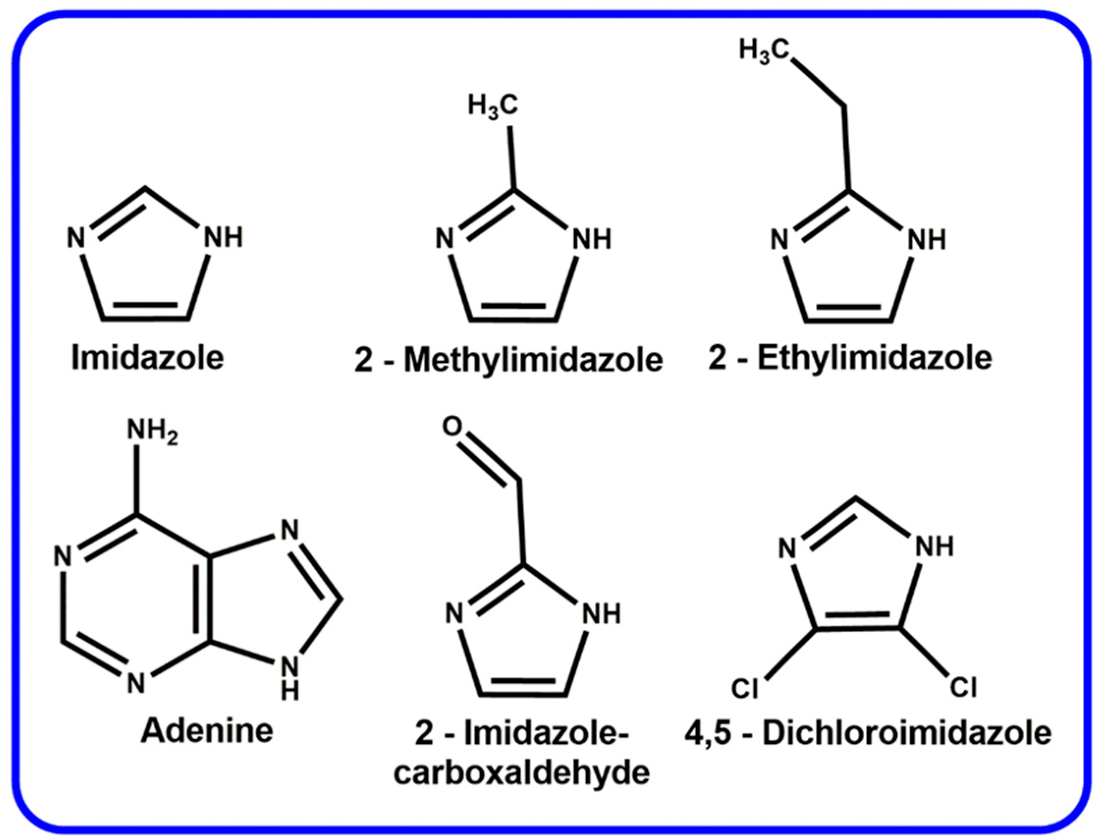
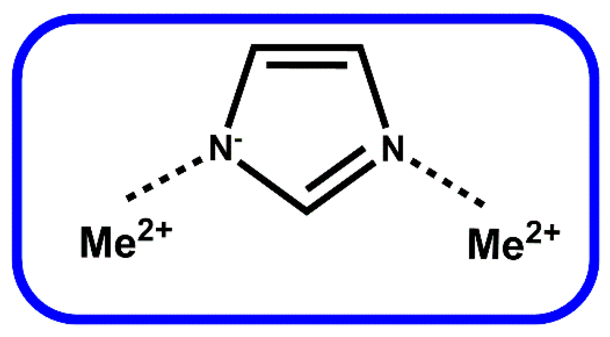
2. Zeolitic Imidazolate Frameworks (ZIFs)

Zeolite imidazolate frameworks (ZIFs) are one of the subgroups of MOFs that are of great interest to the scientific community. These materials, like all MOFs, consist of an organic ligand and an inorganic metal cation. The ligand is based on an imidazole skeleton, and the metal cation is most often Zn2+ or Co2+ [28,29,30,31]. Examples of the ligands used to synthesize various ZIFs are presented in Figure 2. The scheme of combining metal ions and ligands can be schematically presented as follows: Me2+-IM–Me2+. This bond has an angle of 145° The bonding scheme is shown in Figure 3.
Such a bonding angle makes these materials similar in structure to zeolites. This is why they are so popular. In this material, metal cations, e.g., zinc, play the role of silicon atoms, while the linker plays the role of oxygen atoms [33,34]. This type of connection of atoms makes them have unique properties, as in the case of MOFs. They have a large specific surface, high porosity, and tunable surface properties. They are also characterized by good chemical stability (especially in an alkaline environment) and thermal stability [34]. Like MOFs, ZIFs have many applications in various branches of chemistry [28]. They are used, for example, in the separation of gas mixtures such as H2/CO2 or N2/H2 [35,36]. ZIFs are also used to prepare so-called mixed matrix membranes (MMMs). These membranes are used for gas or liquid separation. ZIFs can also be used to create catalysts, for example, transesterification or acylation reactions [28]. ZIFs as materials with a large surface areas and porosity are also a great support for obtaining catalysts by incorporating, for example, active metal oxides [37]. As in the case of MOFs, ZIFs also possess many biomedical applications, which will be discussed in the next section.

3. Biomedical Applications of MOFs

As mentioned earlier, due to their excellent properties, MOFs have many biomedical applications. These are, for example, drug delivery, gene delivery, and delivery of gasses (nitric oxide) necessary for many biochemical processes, bioimaging, biosensors, and scaffold materials [24,38]. Some of these applications will be briefly explained in this review.
The first application described will be for drug delivery and gene delivery. MOFs can retain various drugs in their structure, which undergo intelligent, slowed release in the body. This often happens under the influence of appropriate conditions, such as reduced pH of the tumor or the presence of glutathione. For this purpose, various MOFs are employed. One of the most commonly used networks for this purpose is ZIF-8. For example, Kaur et al. prepared ZIF-8 containing an encapsulated anticancer drug—6-mercaptopurine [39]. Drug release studies have shown that the drug is released under the influence of reduced tumor pH. Another example of using this network in drug delivery is presented by Zheng et al. [40]. In this work, authors prepared ZnO@Zif-8 core-shell nanoparticles loaded with an important anticancer agent—doxorubicin. The work also shows that this system has the ability to release the drug in an environment with a reduced pH. Another network that has been proposed for drug delivery applications is the UiO-66. In this work, Gong et al. prepared MOF containing free SH2 groups in the linker structure [41]. These groups were used to attach 6-mercaptopurine to it via a covalent disulfide bond. The results showed that the release of the drug is possible only in the presence of glutathione, which is present in increased amounts in cancer cells. MOFs can also be used to deliver protein drugs and modify the genome. In the work presented by Yang et al., authors synthesized ZIF-90 loaded with cytotoxic protein for cancer therapy and genome-editing protein Cas9 [42]. The results of their research showed that both proteins are released under the influence of adenosine triphosphate (ATP), which is present in large amounts in the intracellular fluid. All these results suggest that MOFs can be used for the delivery of various types of drugs.
Properly constructed MOFs are also used in photodynamic therapy. Photodynamic therapy consists of the fact that photosensitizers under the influence of light radiation generate various reactive oxygen species (ROS), such as singlet oxygen or hydroxide radicals [43]. For instance, Lu et al. prepared a MOF consisting of Hf4+ ions and a porphyrine-based ligand [44]. The ability of porphyrins to generate ROS is well known, however, their combination with metal ions increases the amount of ROS generated. In this work, it was almost twice as large. In the next paper, the authors show that the use of an MOF based on Mn2+ ions has the ability to generate oxygen from H2O2 present in cells [45]. In a paper published by Sharma et al., the possibility of delivering the photosensitizer through encapsulation in the MOF structure is also shown [46]. In their work, a MOF based on Cu2+ ions and gallic acid was used for this purpose. Their work shows that material loaded with methylene blue has the ability to generate more ROS than material without it.
MOFs can also be used as imaging agents. For instance, Ryu et al. prepared two types of MOFs named UiO-67 and MOF-801 loaded with two fluorescent dyes, Resorufin and Rhodoamine-6G, respectively [47]. Both frameworks were functionalized with the targeting agent galactosamine. The test results showed that the prepared particles have high biocompatibility towards two human cell lines and are excellent as fluorescent imaging agents. In the next work, Rieter et al. prepared an MOF consisting of gadolinium ions and 1,4-benzenedicarboxylic acid [48]. The synthesis was carried out in the inverted microemulsion system, which resulted in obtaining a material with the morphology of nanorods. The obtained materials were tested for use as a contrast agent in nuclear magnetic resonance imaging. The obtained results showed that the prepared material has high values of R1 and R2 relaxivities per mM of material and is suitable for use as a contrast agent. Zeolitic imidazolate frameworks are also used for the preparation of imaging agents. Zhao et al. presented the synthesis of ZIF-8 doped with manganese ions [49]. The obtained material was tested for use as a contrast agent in nuclear magnetic resonance. The results of the study showed that such material has the ability to act as a contrast agent and is additionally characterized by low cytotoxicity against the human 4T1 cell line.
MOFs are also used to obtain scaffolds with different properties. For example, Guerrero et al. prepared a kidney scaffold consisting of ZIF-8 and (poly[isobutylene-alt-maleic anhydride]-graft-dodecyl) [50]. The prepared material was tested for retention of two uremic toxins, p-cresyl sulfate, and indoxyl sulfate. The results of these studies showed a high retention rate for p-cresyl sulfate and less for indoxyl sulfate. However, they also showed the great potential of using organometallic lattices in kidney scaffold construction. In another paper, Karakeçili et al. prepared a chitosan scaffold loaded with ZIF-8 and encapsulated with the antibiotic vancomycin [51]. The test results showed an excellent antibacterial effect of the prepared material. They also showed that the release of the antibiotic is to some extent pH-dependent as in an acidic environment more percentage of the drug is released. The authors also conducted biocompatibility studies on the MC3T3-E preosteoblast cell line. The results of these studies showed that the prepared material increases cell proliferation and alkaline phosphatase activity. This means that this material has great potential in the treatment of bone diseases. Han et al. prepared a bio-glass scaffold also functionalized with ZIF-8 loaded with vancomycin [52]. The prepared scaffold, as in the previous work, also showed pH-dependent release. In this work, the authors also managed to confirm that the prepared scaffold increases cell proliferation and has strong antibacterial properties.
MOFs can also be used to create biosensors. Biosensors are devices consisting of a biological recognition element, which can be, for example, enzymes or DNA fragments, in close contact with the transducer [53]. For instance, Sheta et al. prepared an electrochemical biosensor consisting of a composite that consisted of polyaniline and an Ni-based MOF [54]. The material was also modified with DNA aptamers capable of detecting the hepatitis-C virus. The authors managed to obtain a sensor characterized by a low detection level (0.75 fM) and the ability to detect the virus in real biological samples. In another work, the authors prepared a fluorescent biosensor consisting of zirconium porphyrin-based MOF (PCN-222) for the detection of the antibiotic chloramphenicol. This material was also functionalized with appropriate aptamers. The prepared sensor was characterized by a low detection limit of 0.08 pg · mL−1 and a wide measurement range of 0.1 pg · mL−1–10 ng · mL−1. This sensor also could detect the antibiotic in real milk samples [55].

4. MOFs in Modification of Titanium Alloy

Bone diseases are one of the most common diseases in the world. Examples of bone diseases are osteoporosis, rheumatoid arthritis, and bone cancer. Elderly people and postmenopausal women, in the case of osteoporosis, are particularly at risk. The presence of any of these diseases can lead to increased bone fragility, which often leads to serious fractures. In some cases, the fusion of the bone is impossible, which leads to the fact that the bone must be replaced with an implant [56]. To date, many different materials have been proposed for this purpose: metals such as tantalum or titanium, ceramics, or polymers. The most frequently chosen material, however, is a biomedical titanium alloy with the designation Ti6Al4V. This material has excellent mechanical properties, good biocompatibility, and is practically completely resistant to corrosion in human body fluids. Literature reports show that this material additionally has a very high survival rate. Like any material, this also has several disadvantages. Despite this, titanium is bioinert, i.e., it does not cause allergic reactions and is not toxic, although it is still recognized by the body as a foreign body. This action causes inflammation in the body, which negatively affects the process of osseointegration, and thus bone reconstruction [57,58]. In addition, there is a possibility of bacterial or fungal infection after the implantation operation, which is another serious disadvantage [59,60].
All these disadvantages cause scientists around the world to modify the surface of titanium implants in order to obtain a material with better properties such as the increased proliferation of osteoblasts, and antibacterial or accelerated growth of hydroxyapatite. The properties of the resulting layers depend on many different factors such as surface energy, hydrophilicity, surface topography, and porosity [61]. To date, many different materials have been used for this purpose. For instance, titanium dioxide is modified with different alkali earth metal ions, titanates layer with different cations or zeolites [62,63,64,65,66]. MOFs are also used for this purpose, and layers prepared with their use will be discussed in this review. Examples of applications of titanium implants covered with MOFs are presented in Figure 4. As mentioned, many parameters affect osseointegration, such as wettability, surface porosity, and roughness. Scientific research proves that osseointegration is faster when the surface of the implant is hydrophilic [67]. Modification of metallic and polymer surfaces with MOFs allows to an increase in their water contact angle and thus hydrophilicity [68]. In addition, high porosity and surface roughness are needed for effective and fast osseointegration [61]. These are the parameters that MOFs also provide. Unfortunately, MOFs can also have disadvantages such as ion leakage and the ligands used for their synthesis.
In the first paper presented, Zhang et al. prepared a titanium alloy modified with ZIF-8 by a simple hydrothermal approach [68]. In their research, the authors proved that the modified material is biocompatible with the MC3T3-E1 cell line. The work also examined the release of zinc ions from the prepared layer, and it was found that only amounts of zinc ions are released. The effect of the synthesized layer on extracellular matrix mineralization (ECM) and collagen production was also investigated. The results show that ZIF-8-coated titanium materials significantly increased extracellular matrix mineralization and collagen production. To confirm the positive effect of the modification on accelerated osseointegration, alkaline phosphatase activity and the expression of osteo-related genes were also tested. In all the tests performed, the titanium coated with ZIF-8 showed an increase in the above-mentioned parameters. The authors also performed in vivo studies using mice. The study confirmed the results of in vitro tests. It was proved that in mice implanted with ZIF-8-modified titanium, more mature collagen and more mineralized bone matrix were formed. Figure 5 shows the scheme of the surgery and results of the in vivo study of the collagen and mineral matrix formation.
In another work, Chen et al. prepared titanium alloy plates modified with nano and micro ZIF-8 films [69]. Both layers were synthesized by different methods. In order to obtain the ZIF-8 nanolayer, the secondary growth method was used, while the in situ synthesis method was used to obtain the microscale ZIF. Various parameters such as the morphology of the obtained layers, MG63 cell proliferation, alkaline phosphatase activity, osteocalcin production, and cell adhesion were investigated in the work. SEM photos of both films confirmed the receipt of layers of the assumed size. It was found that particle size in the nanolayer ranged from 200–300 nm while the particle size in the microlayer was found to be over 10 um. Biocompatibility studies have shown that the ZIF-8 microlayer has cytotoxic properties while the nanolayer is biocompatible. This is due to the fact that on a micro-scale, the ZIF-8 releases much larger amounts of zinc ions, which in too high concentrations cause a cytotoxic effect, which has been confirmed by the authors. The results of biocompatibility tests and zinc release from both layers are shown in Figure 6. The study of ALP activity showed that the nanolayer significantly increases its activity in relation to unmodified titanium. The work also examined the antibacterial activity against the S. Mutans strain. It was found that the prepared layer shows remarkable antibacterial activity against this strain.
Teng et al. prepared titanium also modified with ZIF-8 with immobilized iodine [70]. The scheme of this work is shown in the Figure 7. Prior to modification, the material was subjected to the micro-arc oxidation (MAO) process, on which ZIF-8 was then synthesized in situ. Iodine release studies show a dependence on pH; in an acidic environment, more iodine is released. In addition, the authors also showed that the prepared material can release immobilized iodine under the influence of near-infrared (NIR) light. It was found that NIR exposure act as an ON/OFF switch for iodine release. The material has also been subjected to antibacterial tests. It has been proven that it has antibacterial properties against the S. Aureus strain, especially when the samples were irradiated with NIR light. In vitro biocompatibility studies have shown that the material has no cytotoxic properties. In addition, in vivo studies were carried out. The bacteria-infected implants were implanted in mice. It was observed that, despite the material being infected, the post-operative wounds of the mice that had the modified implant healed without any complications, while the wound swelling was seen in the mice that had the infected material without the modification.
As you can see, great efforts are being made to obtain implant surfaces with antibacterial properties. One of the methods of achieving such an effect is the use of silver ions. Li et al. prepared a ZIF-8 layer modified with Ag+ ions on a titanium implant [59]. Studies using various techniques such as scanning electron microscopy, X-ray diffraction, or X-ray photoelectron spectroscopy, have proven the effective synthesis of the presented layer. The tests performed by the authors also showed that the modification is biocompatible, and increases the corrosion resistance and hydrophilicity of the surface. In addition, the layer loaded with silver ions significantly supports antibacterial properties.
Titanium implant modifications can also be used as local drug delivery systems. For example, ZIF-8-modified Ti6Al4V titanium alloy can be used as a local drug delivery system for an osteoporotic drug, risedronate (RSD). In this work, the authors proposed a new approach to the synthesis of MOFs on a titanium surface. Prior to modification, alloy was treated with NaOH to produce a layer of sodium titanate. Then, thanks to ion exchange, zinc titanate was obtained, which was modified with a linker (2-methylimidazole) to obtain a monolayer on the surface. The surface prepared in this way ensures excellent adhesion of ZIF-8 crystals, which were synthesized by the hydrothermal method. The material was used as an RSD carrier; it was proven that the drug is released from the surface of the material in constant amounts for 16 hours. Such material can be of great importance immediately after surgery. In addition, the uniformity of the occurrence of ZIF and RSD on the surface was confirmed by FT-IR microscopy [71].
Titanium coated with ZIF-8 loaded with the antibiotic levofloxacin was prepared by Tao et al. [72]. Figure 8 shows the scheme of the coating preparation and possible antibacterial pathways of the modified implant.
The layer was prepared using electrophoretic deposition. Covering titanium with this layer clearly increased the hydrophilicity of the surface. Zinc and drug release studies have shown that it is released gradually in a controlled manner over 240 h. In addition, titanium with a modified surface showed the highest biocompatibility and cell adhesion. The authors of this paper also studied the expression of osteo-related genes such as Runx2, Col1, OCN, and OPN. The expression values of all genes were higher for the samples modified with the ZIF layer. The samples were also antibacterial against E. Coli and S. Aureus bacterial strains. In vivo tests have shown that the material retains its antibacterial properties in these conditions, and additionally reduces the formation of inflammation around the implant.
As also mentioned earlier, other MOFs can also be used to modify titanium. Shen et al. modified the implant with MOF-74, which had mixed metal cations [73]. The cations used were Mg2+ and Zn2+. Using X-ray diffraction, it was possible to confirm the effective synthesis of the MOF. The hydrophilicity of the surface was also tested; it turned out to be very hydrophilic, with a water contact angle value below 10°. The prepared material was obtained with different ion ratios in the MOF. It was found that as the content of zinc ions increases, the thickness of the obtained coating decreases. This phenomenon is shown in Figure 9. The coating containing the most Zn2+ was also the most stable. Antibacterial tests showed a significant increase in the antibacterial effect compared to unmodified material. Tests on cells and in vivo were also performed. Studies have shown that the modified material increases ALP activity, collagen secretion, and mRNA expression of some osteo-related genes. The modified material was able to maintain its antibacterial properties in vivo and additionally increased the growth of healthy bone on the implant.
In another work, authors prepared Co2+-based ZIF-67 modification on Ti implant. They used this MOF as a local delivery system for osteogenic growth peptide (OGP) [74]. As in the previous cases, the hydrophilicity of the implant increases after covering it with a layer of MOF. The prepared layer killed almost all bacteria from E. coli and S. Aureus strains. The authors proved that such a strong antibacterial effect is due to the presence of cobalt ions in the prepared layer. Biocompatibility studies have shown that the material does not cause cytotoxicity and even increases cell proliferation in relation to unmodified titanium. It was also found that it increased the expression of osteo-related genes. Thanks to the modification of the ZIF-67 alloy, it was also possible to increase ALP activity and collagen secretion. In vivo studies confirmed the results of in vitro studies. The implant retained its antibacterial properties and additionally increased the rate of bone growth and bone–implant contact ratio.
MIL-125 doped with rare earth Cerium ions was also used as a modification for the implant surface. The coating containing this MOF and hydroxyapatite was prepared using the galvanostatic method on titanium with a layer of TiO2 nanotubes on the surface. Scanning electron microscopy images confirmed the formation of a uniform layer while energy dispersive X-ray spectroscopy and X-ray photoelectron spectroscopy confirmed the presence of cerium ions in the material. In this work, the corrosion properties were also assessed. The unmodified titanium had the lowest corrosion potential and the highest corrosion current density, while the titanium coated with a layer of MOF combined with HA had the highest corrosion potential and the lowest corrosion current density. Antibacterial tests showed almost complete inhibition of bacterial growth. The prepared layer was biocompatible and could slowly release bioactive calcium and phosphorus ions [75].
The last modification described in this article will be the work published by Wu et al. [76]. In their work, they synthesized a MOF on the surface of titanium called bio-MOF-1. It has a natural linker in its structure—adenine. Using scanning electron microscopy, X-ray diffraction, and FT-IR spectroscopy, it was possible to confirm the effective synthesis of the layer. Biological properties were also investigated in this work. Biocompatibility studies have shown that titanium modified with this MOF increases cell proliferation. It also significantly increases the activity of alkaline phosphatase and the expression of osteogenic genes. In vivo studies conducted additionally by the authors also show that the presented modification accelerates bone growth.
All methods of titanium alloy modification and their influence on the final properties are summarized in Table 1.

5. Conclusions and Possible Development Directions

This article shows that MOFs are a great material for modifying titanium implants. The prepared modifications enable the implant to acquire new properties. These are excellent biocompatibility, enhanced alkaline phosphatase activity, enhanced collagen production, better cell adhesion, and release of bioactive ions. In addition, the prepared layers also have the ability to increase the expression of osteo-related genes. The obtained coatings also have strong antibacterial properties against various strains of bacteria and better corrosion resistance. This review also shows that the modification of titanium with MOFs can be used as a carrier in the controlled release of drugs, e.g., antibiotics or anti-osteoporotic drugs. Numerous in vivo studies have also shown that the modifications accelerate bone growth in mice. Despite such good properties, there are still some challenges that need to be solved. One of the biggest concerns when using MOFs in medicine is ligand leakage. It is not entirely clear what amounts of ligand will be released by a titanium implant placed in the body for many years. Additionally, almost all papers cited in this review show that the ligand and ion are released. It should be remembered that the materials in these works were prepared on a small scale. On the other hand, the hip implant is much larger, which will result in the fact that the total amount of released substances will be greater. This shows that further research is needed on this topic.
Despite the high degree of research on the modification of titanium with MOFs, there are further possible directions of development. So far, MOFs synthesized on the surface of implants contain mainly synthetic ligands. The only one containing a natural linker is bio-MOF-1. However, this ligand also contains a second ligand (4,4’-biphenyl dicarboxylic acid) of synthetic origin in its structure. Thus, the next step in the research on the formation of MOF coatings on implants may be the synthesis of those containing only natural linkers. An example of a linker that would be suitable for this purpose is, for example, gallic acid, which has a proven ability to form MOFs and has antimicrobial properties. As can also be seen, the only metal ions used in the synthesis of MOFs are zinc and cobalt. However, there are many more metal ions with the ability to improve osseointegration, such as calcium, magnesium, or strontium [79]. These metals also have a proven ability to form MOFs [80,81]. These networks have, for example, anti-oxidative properties and are biocompatible. One example is MOF synthesized from Mg2+ ions and gallic acid. It was biocompatible against HL-60, RAW 264.7, and NCI-H460 macrophage cell lines [6]. Another element that affects osseointegration is lanthanum [82]. It also has the ability to create MOFs. This could be another new line of research. We hope that this review will help scientists from around the world create new modifications of titanium implants using MOFs.

Author Contributions

Writing—original draft preparation, M.J.; Writing—original draft preparation, A.D.; writing—review and editing, supervision, A.V.; writing—review and editing, supervision, M.S. All authors have read and agreed to the published version of the manuscript.

Funding

This research was funded by the Ministry of Education and Science (Poland).

Data Availability Statement

Not applicable.

Acknowledgments

Mariusz Sandomierski was supported by the Foundation for Polish Sciences (FNP).

Conflicts of Interest

The authors declare no conflict of interest.

References

  1. Li, A.; Bueno-Perez, R.; Wiggin, S.; Fairen-Jimenez, D. Enabling Efficient Exploration of Metal-Organic Frameworks in the Cambridge Structural Database. CrystEngComm 2020, 22, 7152–7161. [Google Scholar] [CrossRef]
  2. Bohrman, J.A.; Carreon, M.A. Synthesis and CO2/CH4 Separation Performance of Bio-MOF-1 Membranes. Chem. Commun. 2012, 48, 5130–5132. [Google Scholar] [CrossRef]
  3. Gan, Y.-L.; Huang, K.-R.; Li, Y.-G.; Qin, D.-P.; Zhang, D.-M.; Zong, Z.-A.; Cui, L.-S. Synthesis, Structure and Fluorescent Sensing for Nitrobenzene of a Zn-Based MOF. J. Mol. Struct. 2021, 1223, 129217. [Google Scholar] [CrossRef]
  4. Zha, Q.; Yuan, F.; Qin, G.; Ni, Y. Cobalt-Based MOF-on-MOF Two-Dimensional Heterojunction Nanostructures for Enhanced Oxygen Evolution Reaction Electrocatalytic Activity. Inorg. Chem. 2020, 59, 1295–1305. [Google Scholar] [CrossRef] [PubMed]
  5. Rojas, S.; Devic, T.; Horcajada, P. Metal Organic Frameworks Based on Bioactive Components. J. Mater. Chem. B 2017, 5, 2560–2573. [Google Scholar] [CrossRef] [PubMed]
  6. Cooper, L.; Hidalgo, T.; Gorman, M.; Lozano-Fernández, T.; Simón-Vázquez, R.; Olivier, C.; Guillou, N.; Serre, C.; Martineau, C.; Taulelle, F.; et al. A Biocompatible Porous Mg-Gallate Metal-Organic Framework as an Antioxidant Carrier. Chem. Commun. 2015, 51, 5848–5851. [Google Scholar] [CrossRef]
  7. Can, M.; Demirci, S.; Sunol, A.K.; Sahiner, N. An Amino Acid, l-Glutamic Acid-Based Metal-Organic Frameworks and Their Antibacterial, Blood Compatibility, Biocompatibility, and Sensor Properties. Microporous Mesoporous Mater. 2020, 309, 110533. [Google Scholar] [CrossRef]
  8. Dong, X.; Shi, Z.; Li, D.; Li, Y.; An, N.; Shang, Y.; Sakiyama, H.; Muddassir, M.; Si, C. The Regulation Research of Topology and Magnetic Exchange Models of CPs through Co(II) Concentration Adjustment. J. Solid State Chem. 2023, 318, 123713. [Google Scholar] [CrossRef]
  9. Qin, L.; Li, Y.; Liang, F.; Li, L.; Lan, Y.; Li, Z.; Lu, X.; Yang, M.; Ma, D. A Microporous 2D Cobalt-Based MOF with Pyridyl Sites and Open Metal Sites for Selective Adsorption of CO2. Microporous Mesoporous Mater. 2022, 341, 112098. [Google Scholar] [CrossRef]
  10. Qin, L.; Liang, F.; Li, Y.; Wu, J.; Guan, S.; Wu, M.; Xie, S.; Luo, M.; Ma, D. A 2D Porous Zinc-Organic Framework Platform for Loading of 5-Fluorouracil. Inorganics 2022, 10, 202. [Google Scholar] [CrossRef]
  11. Hönicke, I.M.; Senkovska, I.; Bon, V.; Baburin, I.A.; Bönisch, N.; Raschke, S.; Evans, J.D.; Kaskel, S. Balancing Mechanical Stability and Ultrahigh Porosity in Crystalline Framework Materials. Angew. Chem. Int. Ed. 2018, 57, 13780–13783. [Google Scholar] [CrossRef] [PubMed]
  12. Furukawa, H.; Cordova, K.E.; O’Keeffe, M.; Yaghi, O.M. The Chemistry and Applications of Metal-Organic Frameworks. Science 2013, 341, 1230444. [Google Scholar] [CrossRef] [PubMed]
  13. Wang, Z.-S.; Li, M.; Peng, Y.-L.; Zhang, Z.; Chen, W.; Huang, X.-C. An Ultrastable Metal Azolate Framework with Binding Pockets for Optimal Carbon Dioxide Capture. Angew. Chem. Int. Ed. 2019, 58, 16071–16076. [Google Scholar] [CrossRef] [PubMed]
  14. Almáši, M.; Zeleňák, V.; Palotai, P.; Beňová, E.; Zeleňáková, A. Metal-Organic Framework MIL-101(Fe)-NH2 Functionalized with Different Long-Chain Polyamines as Drug Delivery System. Inorg. Chem. Commun. 2018, 93, 115–120. [Google Scholar] [CrossRef]
  15. Trushina, D.B.; Sapach, A.Y.; Burachevskaia, O.A.; Medvedev, P.V.; Khmelenin, D.N.; Borodina, T.N.; Soldatov, M.A.; Butova, V.V. Doxorubicin-Loaded Core-Shell UiO-66@SiO2 Metal-Organic Frameworks for Targeted Cellular Uptake and Cancer Treatment. Pharmaceutics 2022, 14, 1325. [Google Scholar] [CrossRef] [PubMed]
  16. Li, B.; Cui, X.; O’Nolan, D.; Wen, H.-M.; Jiang, M.; Krishna, R.; Wu, H.; Lin, R.-B.; Chen, Y.-S.; Yuan, D.; et al. An Ideal Molecular Sieve for Acetylene Removal from Ethylene with Record Selectivity and Productivity. Adv. Mater. 2017, 29, 1704210. [Google Scholar] [CrossRef]
  17. Sun, Y.; Xue, Z.; Liu, Q.; Jia, Y.; Li, Y.; Liu, K.; Lin, Y.; Liu, M.; Li, G.; Su, C.-Y. Modulating Electronic Structure of Metal-Organic Frameworks by Introducing Atomically Dispersed Ru for Efficient Hydrogen Evolution. Nat. Commun. 2021, 12, 1369. [Google Scholar] [CrossRef]
  18. Umapathi, R.; Ghoreishian, S.M.; Sonwal, S.; Rani, G.M.; Huh, Y.S. Portable Electrochemical Sensing Methodologies for On-Site Detection of Pesticide Residues in Fruits and Vegetables. Coord. Chem. Rev. 2022, 453, 214305. [Google Scholar] [CrossRef]
  19. Venkateswara Raju, C.; Hwan Cho, C.; Mohana Rani, G.; Manju, V.; Umapathi, R.; Suk Huh, Y.; Pil Park, J. Emerging Insights into the Use of Carbon-Based Nanomaterials for the Electrochemical Detection of Heavy Metal Ions. Coord. Chem. Rev. 2023, 476, 214920. [Google Scholar] [CrossRef]
  20. Ratnamala, A.; Reddy, G.D.; Noorjahaan, M.; Manjunatha, H.; Janardan, S.; Kumar, N.S.; Chandra Babu Naidu, K.; Khan, A.; Asiri, A.M. Chapter 3—Titanium-Based Metal-Organic Frameworks for Photocatalytic Applications. In Metal-Organic Frameworks for Chemical Reactions; Khan, A., Verpoort, F., Asiri, A.M., Hoque, M.E., Bilgrami, A.L., Azam, M., Naidu, K.C.B., Eds.; Elsevier: Amsterdam, The Netherlands, 2021; pp. 37–63. [Google Scholar]
  21. Yang, D.; Gates, B.C. Catalysis by Metal Organic Frameworks: Perspective and Suggestions for Future Research. ACS Catal. 2019, 9, 1779–1798. [Google Scholar] [CrossRef]
  22. Beydaghdari, M.; Hooriabad Saboor, F.; Babapoor, A.; Karve, V.V.; Asgari, M. Recent Advances in MOF-Based Adsorbents for Dye Removal from the Aquatic Environment. Energies 2022, 15, 2023. [Google Scholar] [CrossRef]
  23. Chen, J.; Cheng, F.; Luo, D.; Huang, J.; Ouyang, J.; Nezamzadeh-Ejhieh, A.; Khan, M.S.; Liu, J.; Peng, Y. Recent Advances in Ti-Based MOFs in Biomedical Applications. Dalton Trans. 2022, 51, 14817–14832. [Google Scholar] [CrossRef] [PubMed]
  24. Keskin, S.; Kızılel, S. Biomedical Applications of Metal Organic Frameworks. Ind. Eng. Chem. Res. 2011, 50, 1799–1812. [Google Scholar] [CrossRef]
  25. Al Sharabati, M.; Sabouni, R.; Husseini, G.A. Biomedical Applications of Metal-Organic Frameworks for Disease Diagnosis and Drug Delivery: A Review. Nanomaterials 2022, 12, 277. [Google Scholar] [CrossRef] [PubMed]
  26. Chen, J.; Zhu, Y.; Kaskel, S. Porphyrin-Based Metal-Organic Frameworks for Biomedical Applications. Angew. Chem. Int. Ed. 2021, 60, 5010–5035. [Google Scholar] [CrossRef]
  27. Sun, Y.; Zheng, L.; Yang, Y.; Qian, X.; Fu, T.; Li, X.; Yang, Z.; Yan, H.; Cui, C.; Tan, W. Metal-Organic Framework Nanocarriers for Drug Delivery in Biomedical Applications. Nano-Micro Lett. 2020, 12, 103. [Google Scholar] [CrossRef]
  28. Chen, B.; Yang, Z.; Zhu, Y.; Xia, Y. Zeolitic Imidazolate Framework Materials: Recent Progress in Synthesis and Applications. J. Mater. Chem. A 2014, 2, 16811–16831. [Google Scholar] [CrossRef]
  29. McCarthy, M.C.; Varela-Guerrero, V.; Barnett, G.V.; Jeong, H.-K. Synthesis of Zeolitic Imidazolate Framework Films and Membranes with Controlled Microstructures. Langmuir 2010, 26, 14636–14641. [Google Scholar] [CrossRef]
  30. Shieh, F.-K.; Wang, S.-C.; Leo, S.-Y.; Wu, K.C.-W. Water-Based Synthesis of Zeolitic Imidazolate Framework-90 (ZIF-90) with a Controllable Particle Size. Chem.–Eur. J. 2013, 19, 11139–11142. [Google Scholar] [CrossRef]
  31. Huang, X.-C.; Lin, Y.-Y.; Zhang, J.-P.; Chen, X.-M. Ligand-Directed Strategy for Zeolite-Type Metal-Organic Frameworks: Zinc(II) Imidazolates with Unusual Zeolitic Topologies. Angew. Chem. Int. Ed. 2006, 45, 1557–1559. [Google Scholar] [CrossRef]
  32. Phan, A.; Doonan, C.J.; Uribe-Romo, F.J.; Knobler, C.B.; O’Keeffe, M.; Yaghi, O.M. Synthesis, Structure, and Carbon Dioxide Capture Properties of Zeolitic Imidazolate Frameworks. Acc. Chem. Res. 2010, 43, 58–67. [Google Scholar] [CrossRef] [PubMed]
  33. Nune, S.K.; Thallapally, P.K.; Dohnalkova, A.; Wang, C.; Liu, J.; Exarhos, G.J. Synthesis and Properties of Nano Zeolitic Imidazolate Frameworks. Chem. Commun. 2010, 46, 4878–4880. [Google Scholar] [CrossRef] [PubMed]
  34. Park, K.S.; Ni, Z.; Côté, A.P.; Choi, J.Y.; Huang, R.; Uribe-Romo, F.J.; Chae, H.K.; O’Keeffe, M.; Yaghi, O.M. Exceptional Chemical and Thermal Stability of Zeolitic Imidazolate Frameworks. Proc. Natl. Acad. Sci. USA 2006, 103, 10186–10191. [Google Scholar] [CrossRef] [PubMed]
  35. Li, Y.; Liang, F.; Bux, H.; Yang, W.; Caro, J. Zeolitic Imidazolate Framework ZIF-7 Based Molecular Sieve Membrane for Hydrogen Separation. J. Membr. Sci. 2010, 354, 48–54. [Google Scholar] [CrossRef]
  36. Schulte, Z.M.; Kwon, Y.H.; Han, Y.; Liu, C.; Li, L.; Yang, Y.; Jarvi, A.G.; Saxena, S.; Veser, G.; Johnson, J.K.; et al. H2/CO2 Separations in Multicomponent Metal-Adeninate MOFs with Multiple Chemically Distinct Pore Environments. Chem. Sci. 2020, 11, 12807–12815. [Google Scholar] [CrossRef] [PubMed]
  37. Jiang, H.-L.; Liu, B.; Akita, T.; Haruta, M.; Sakurai, H.; Xu, Q. Au@ZIF-8: CO Oxidation over Gold Nanoparticles Deposited to Metal-Organic Framework. J. Am. Chem. Soc. 2009, 131, 11302–11303. [Google Scholar] [CrossRef] [PubMed]
  38. Yang, J.; Yang, Y.-W. Metal-Organic Frameworks for Biomedical Applications. Small 2020, 16, 1906846. [Google Scholar] [CrossRef]
  39. Kaur, H.; Mohanta, G.C.; Gupta, V.; Kukkar, D.; Tyagi, S. Synthesis and Characterization of ZIF-8 Nanoparticles for Controlled Release of 6-Mercaptopurine Drug. J. Drug Deliv. Sci. Technol. 2017, 41, 106–112. [Google Scholar] [CrossRef]
  40. Zheng, C.; Wang, Y.; Phua, S.Z.F.; Lim, W.Q.; Zhao, Y. ZnO-DOX@ZIF-8 Core–Shell Nanoparticles for PH-Responsive Drug Delivery. ACS Biomater. Sci. Eng. 2017, 3, 2223–2229. [Google Scholar] [CrossRef]
  41. Gong, M.; Yang, J.; Li, Y.; Gu, J. Glutathione-Responsive Nanoscale MOFs for Effective Intracellular Delivery of the Anticancer Drug 6-Mercaptopurine. Chem. Commun. 2020, 56, 6448–6451. [Google Scholar] [CrossRef]
  42. Yang, X.; Tang, Q.; Jiang, Y.; Zhang, M.; Wang, M.; Mao, L. Nanoscale ATP-Responsive Zeolitic Imidazole Framework-90 as a General Platform for Cytosolic Protein Delivery and Genome Editing. J. Am. Chem. Soc. 2019, 141, 3782–3786. [Google Scholar] [CrossRef] [PubMed]
  43. Lan, G.; Ni, K.; Lin, W. Nanoscale Metal-Organic Frameworks for Phototherapy of Cancer. Coord. Chem. Rev. 2019, 379, 65–81. [Google Scholar] [CrossRef] [PubMed]
  44. Lu, K.; He, C.; Lin, W. Nanoscale Metal-Organic Framework for Highly Effective Photodynamic Therapy of Resistant Head and Neck Cancer. J. Am. Chem. Soc. 2014, 136, 16712–16715. [Google Scholar] [CrossRef]
  45. Lu, J.; Yang, L.; Zhang, W.; Li, P.; Gao, X.; Zhang, W.; Wang, H.; Tang, B. Photodynamic Therapy for Hypoxic Solid Tumors via Mn-MOF as a Photosensitizer. Chem. Commun. 2019, 55, 10792–10795. [Google Scholar] [CrossRef]
  46. Sharma, S.; Mittal, D.; Verma, A.K.; Roy, I. Copper-Gallic Acid Nanoscale Metal-Organic Framework for Combined Drug Delivery and Photodynamic Therapy. ACS Appl. Bio Mater. 2019, 2, 2092–2101. [Google Scholar] [CrossRef]
  47. Ryu, U.; Yoo, J.; Kwon, W.; Choi, K.M. Tailoring Nanocrystalline Metal-Organic Frameworks as Fluorescent Dye Carriers for Bioimaging. Inorg. Chem. 2017, 56, 12859–12865. [Google Scholar] [CrossRef]
  48. Rieter, W.J.; Taylor, K.M.L.; An, H.; Lin, W.; Lin, W. Nanoscale Metal-Organic Frameworks as Potential Multimodal Contrast Enhancing Agents. J. Am. Chem. Soc. 2006, 128, 9024–9025. [Google Scholar] [CrossRef] [PubMed]
  49. Zhao, G.; Wu, H.; Feng, R.; Wang, D.; Xu, P.; Wang, H.; Guo, Z.; Chen, Q. Bimetallic Zeolitic Imidazolate Framework as an Intrinsic Two-Photon Fluorescence and PH-Responsive MR Imaging Agent. ACS Omega 2018, 3, 9790–9797. [Google Scholar] [CrossRef]
  50. Guerrero, F.; Pulido, V.; Hamad, S.; Aljama, P.; Martín-Malo, A.; Carrillo-Carrión, C. Incorporating Zeolitic-Imidazolate Framework-8 Nanoparticles into Kidney Scaffolds: A First Step towards Innovative Renal Therapies. Nanoscale 2022, 14, 17543–17549. [Google Scholar] [CrossRef]
  51. Karakeçili, A.; Topuz, B.; Korpayev, S.; Erdek, M. Metal-Organic Frameworks for on-Demand PH Controlled Delivery of Vancomycin from Chitosan Scaffolds. Mater. Sci. Eng. C 2019, 105, 110098. [Google Scholar] [CrossRef]
  52. Han, L.; Huang, Z.; Zhu, M.; Zhu, Y.; Li, H. Drug-Loaded Zeolite Imidazole Framework-8-Functionalized Bioglass Scaffolds with Antibacterial Activity for Bone Repair. Ceram. Int. 2022, 48, 6890–6898. [Google Scholar] [CrossRef]
  53. Soldatkina, O.V.; Kucherenko, I.S.; Soldatkin, O.O.; Pyeshkova, V.M.; Dudchenko, O.Y.; Akata Kurç, B.; Dzyadevych, S.V. Development of Electrochemical Biosensors with Various Types of Zeolites. Appl. Nanosci. 2019, 9, 737–747. [Google Scholar] [CrossRef]
  54. Sheta, S.M.; El-Sheikh, S.M.; Osman, D.I.; Salem, A.M.; Ali, O.I.; Harraz, F.A.; Shousha, W.G.; Shoeib, M.A.; Shawky, S.M.; Dionysiou, D.D. A Novel HCV Electrochemical Biosensor Based on a Polyaniline@Ni-MOF Nanocomposite. Dalton Trans. 2020, 49, 8918–8926. [Google Scholar] [CrossRef] [PubMed]
  55. Liu, S.; Bai, J.; Huo, Y.; Ning, B.; Peng, Y.; Li, S.; Han, D.; Kang, W.; Gao, Z. A Zirconium-Porphyrin MOF-Based Ratiometric Fluorescent Biosensor for Rapid and Ultrasensitive Detection of Chloramphenicol. Biosens. Bioelectron. 2020, 149, 111801. [Google Scholar] [CrossRef] [PubMed]
  56. Kurup, A.; Dhatrak, P.; Khasnis, N. Surface Modification Techniques of Titanium and Titanium Alloys for Biomedical Dental Applications: A Review. Mater. Today Proc. 2021, 39, 84–90. [Google Scholar] [CrossRef]
  57. Jaafar, A.; Hecker, C.; Árki, P.; Joseph, Y. Sol-Gel Derived Hydroxyapatite Coatings for Titanium Implants: A Review. Bioengineering 2020, 7, 127. [Google Scholar] [CrossRef] [PubMed]
  58. Souza, J.C.M.; Sordi, M.B.; Kanazawa, M.; Ravindran, S.; Henriques, B.; Silva, F.S.; Aparicio, C.; Cooper, L.F. Nano-Scale Modification of Titanium Implant Surfaces to Enhance Osseointegration. Acta Biomater. 2019, 94, 112–131. [Google Scholar] [CrossRef] [PubMed]
  59. Olmo, J.A.-D.; Ruiz-Rubio, L.; Pérez-Alvarez, L.; Sáez-Martínez, V.; Vilas-Vilela, J.L. Antibacterial Coatings for Improving the Performance of Biomaterials. Coatings 2020, 10, 139. [Google Scholar] [CrossRef]
  60. Stewart, P.S.; Bjarnsholt, T. Risk Factors for Chronic Biofilm-Related Infection Associated with Implanted Medical Devices. Clin. Microbiol. Infect. 2020, 26, 1034–1038. [Google Scholar] [CrossRef]
  61. Wang, Q.; Zhou, P.; Liu, S.; Attarilar, S.; Ma, R.L.-W.; Zhong, Y.; Wang, L. Multi-Scale Surface Treatments of Titanium Implants for Rapid Osseointegration: A Review. Nanomaterials 2020, 10, 1244. [Google Scholar] [CrossRef]
  62. Yan, Y.; Sun, J.; Han, Y.; Li, D.; Cui, K. Microstructure and Bioactivity of Ca, P and Sr Doped TiO2 Coating Formed on Porous Titanium by Micro-Arc Oxidation. Surf. Coat. Technol. 2010, 205, 1702–1713. [Google Scholar] [CrossRef]
  63. Guo, S.; Yu, D.; Xiao, X.; Liu, W.; Wu, Z.; Shi, L.; Zhao, Q.; Yang, D.; Lu, Y.; Wei, X.; et al. A Vessel Subtype Beneficial for Osteogenesis Enhanced by Strontium-Doped Sodium Titanate Nanorods by Modulating Macrophage Polarization. J. Mater. Chem. B 2020, 8, 6048–6058. [Google Scholar] [CrossRef]
  64. Sandomierski, M.; Jakubowski, M.; Ratajczak, M.; Voelkel, A. Drug Distribution Evaluation Using FT-IR Imaging on the Surface of a Titanium Alloy Coated with Zinc Titanate with Potential Application in the Release of Drugs for Osteoporosis. Spectrochim. Acta Part A Mol. Biomol. Spectrosc. 2022, 281, 121575. [Google Scholar] [CrossRef]
  65. Sandomierski, M.; Zielińska, M.; Buchwald, T.; Patalas, A.; Voelkel, A. Controlled Release of the Drug for Osteoporosis from the Surface of Titanium Implants Coated with Calcium Titanate. J. Biomed. Mater. Res. B Appl. Biomater. 2022, 110, 431–437. [Google Scholar] [CrossRef]
  66. Sandomierski, M.; Zielińska, M.; Voelkel, A. A Long-Term Controlled Release of the Drug for Osteoporosis from the Surface of Titanium Implants Coated with Calcium Zeolite. Mater. Chem. Front. 2021, 5, 5718–5725. [Google Scholar] [CrossRef]
  67. Gittens, R.A.; Scheideler, L.; Rupp, F.; Hyzy, S.L.; Geis-Gerstorfer, J.; Schwartz, Z.; Boyan, B.D. A Review on the Wettability of Dental Implant Surfaces II: Biological and Clinical Aspects. Acta Biomater. 2014, 10, 2907–2918. [Google Scholar] [CrossRef] [PubMed]
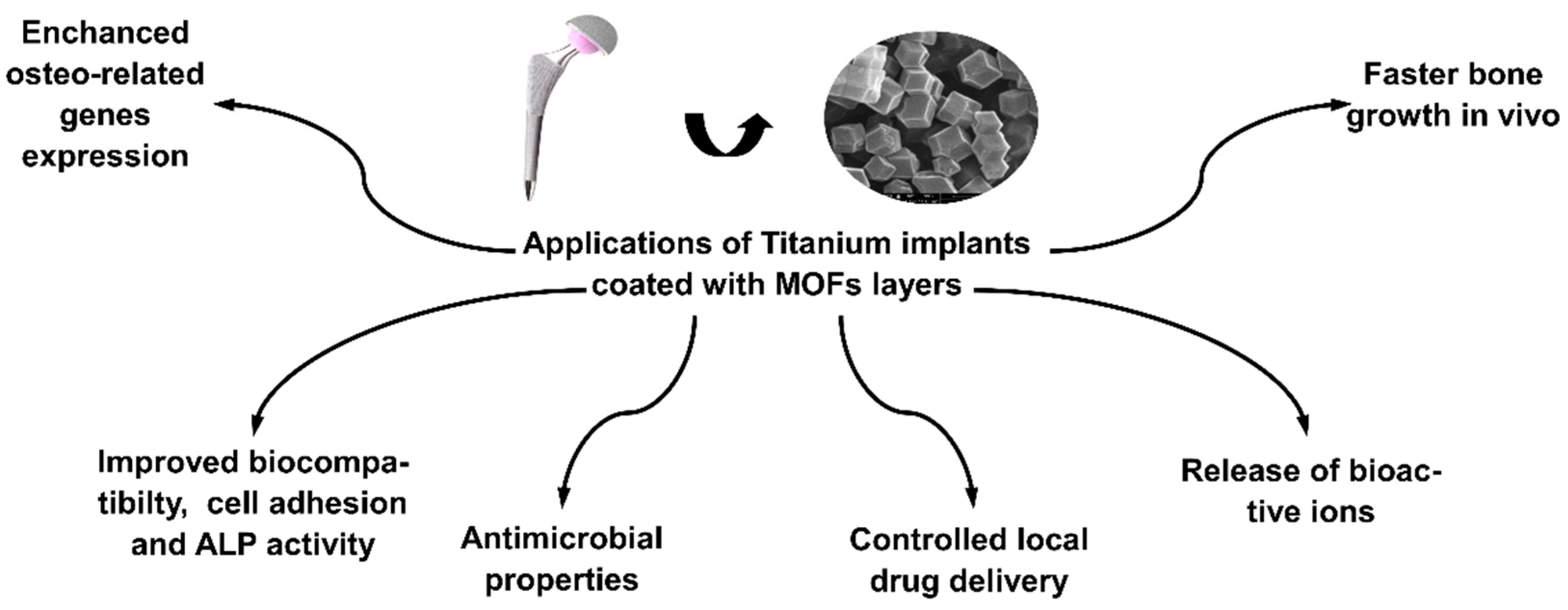
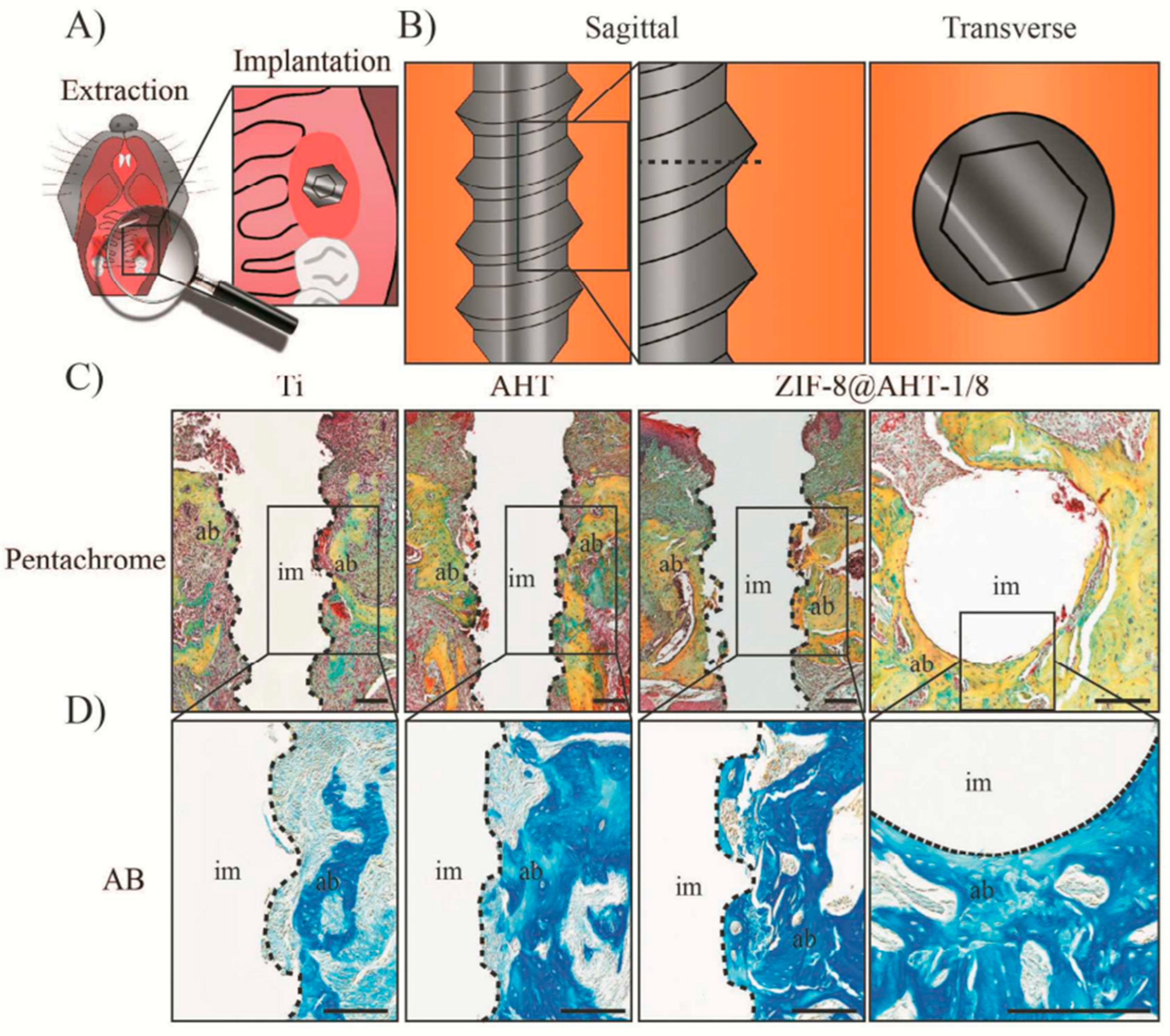
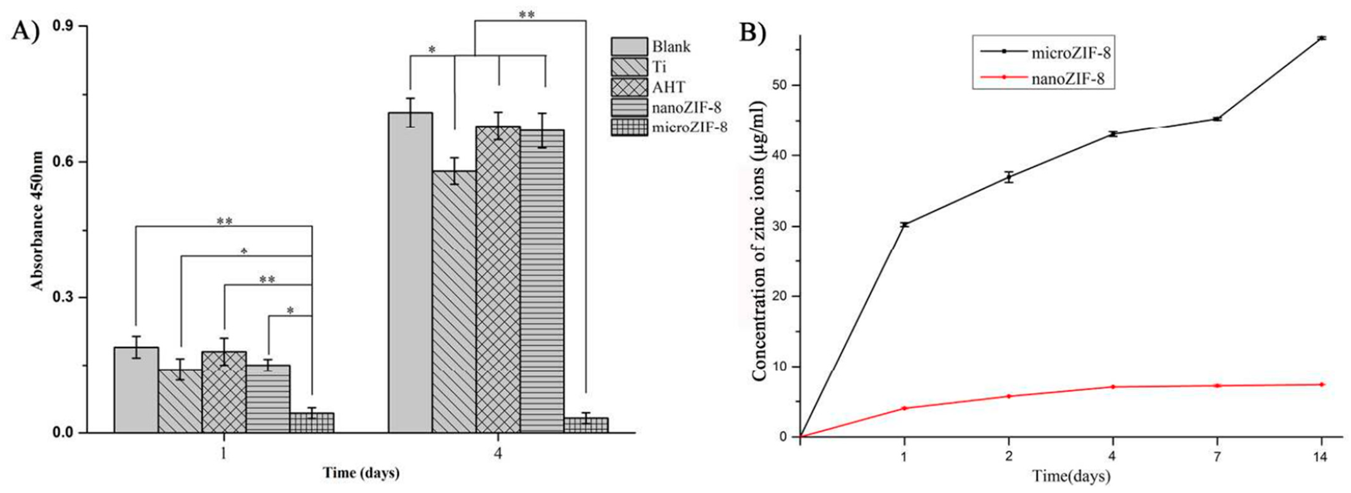
  68. Zhang, X.; Chen, J.; Pei, X.; Wang, J.; Wan, Q.; Jiang, S.; Huang, C.; Pei, X. Enhanced Osseointegration of Porous Titanium Modified with Zeolitic Imidazolate Framework-8. ACS Appl. Mater. Interfaces 2017, 9, 25171–25183. [Google Scholar] [CrossRef] [PubMed]
  69. Chen, J.; Zhang, X.; Huang, C.; Cai, H.; Hu, S.; Wan, Q.; Pei, X.; Wang, J. Osteogenic Activity and Antibacterial Effect of Porous Titanium Modified with Metal-Organic Framework Films. J. Biomed. Mater. Res. A 2017, 105, 834–846. [Google Scholar] [CrossRef]
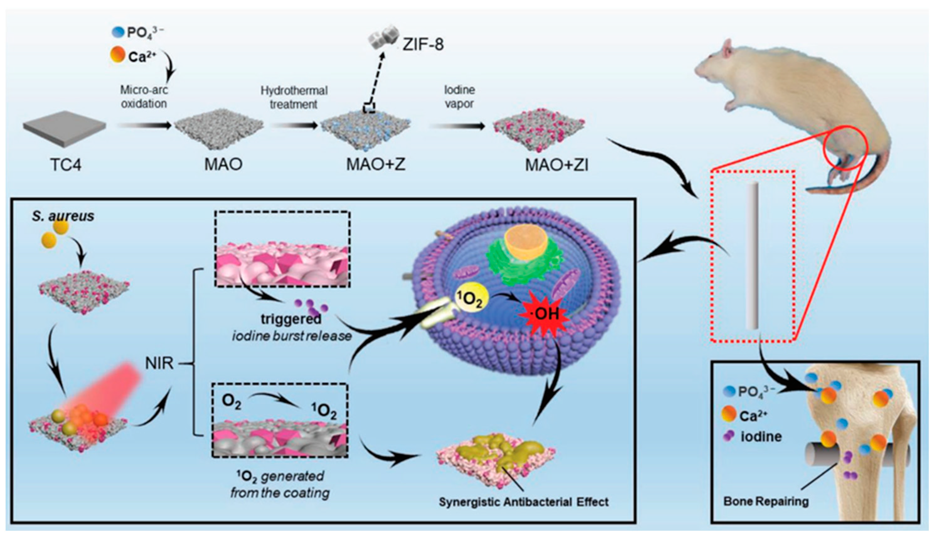
  70. Teng, W.; Zhang, Z.; Wang, Y.; Ye, Y.; Yinwang, E.; Liu, A.; Zhou, X.; Xu, J.; Zhou, C.; Sun, H.; et al. Iodine Immobilized Metal-Organic Framework for NIR-Triggered Antibacterial Therapy on Orthopedic Implants. Small 2021, 17, 2102315. [Google Scholar] [CrossRef]
  71. Sandomierski, M.; Jakubowski, M.; Ratajczak, M.; Voelkel, A. Zeolitic Imidazolate Framework-8 (ZIF-8) Modified Titanium Alloy for Controlled Release of Drugs for Osteoporosis. Sci. Rep. 2022, 12, 9103. [Google Scholar] [CrossRef]
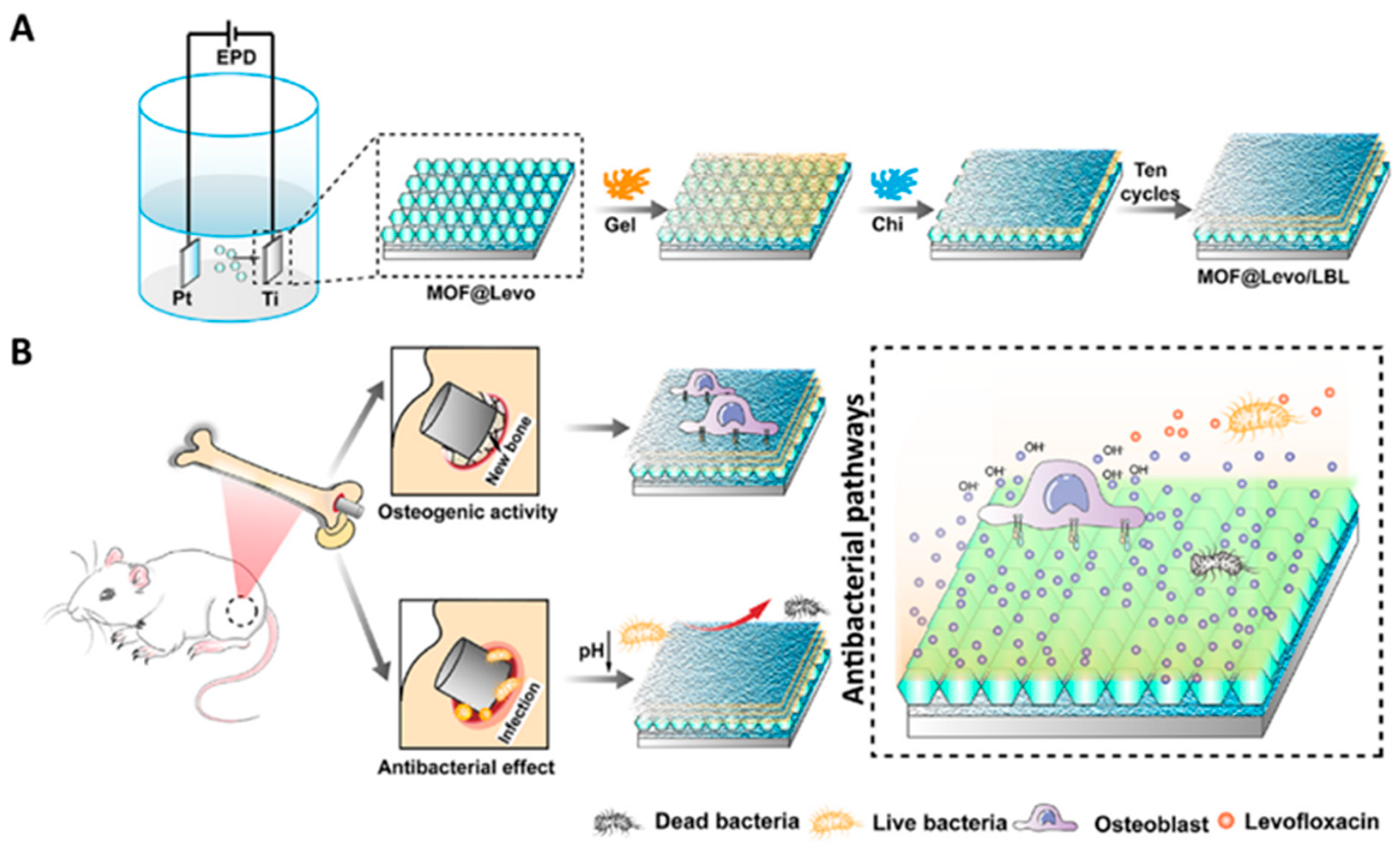
  72. Tao, B.; Zhao, W.; Lin, C.; Yuan, Z.; He, Y.; Lu, L.; Chen, M.; Ding, Y.; Yang, Y.; Xia, Z.; et al. Surface Modification of Titanium Implants by ZIF-8@Levo/LBL Coating for Inhibition of Bacterial-Associated Infection and Enhancement of In Vivo Osseointegration. Chem. Eng. J. 2020, 390, 124621. [Google Scholar] [CrossRef]
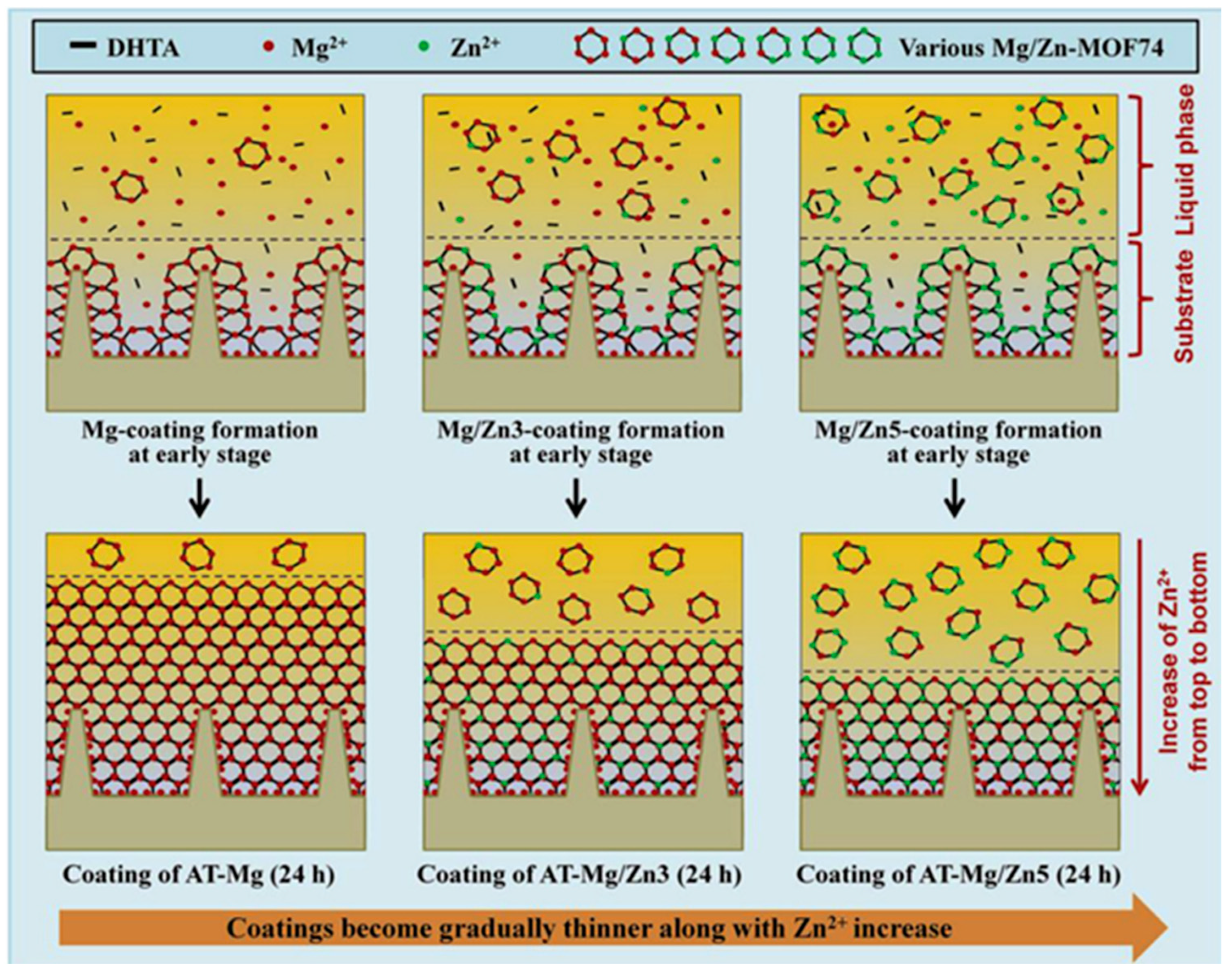
  73. Shen, X.; Zhang, Y.; Ma, P.; Sutrisno, L.; Luo, Z.; Hu, Y.; Yu, Y.; Tao, B.; Li, C.; Cai, K. Fabrication of Magnesium/Zinc-Metal Organic Framework on Titanium Implants to Inhibit Bacterial Infection and Promote Bone Regeneration. Biomaterials 2019, 212, 1–16. [Google Scholar] [CrossRef] [PubMed]
  74. Tao, B.; Lin, C.; He, Y.; Yuan, Z.; Chen, M.; Xu, K.; Li, K.; Guo, A.; Cai, K.; Chen, L. Osteoimmunomodulation Mediating Improved Osteointegration by OGP-Loaded Cobalt-Metal Organic Framework on Titanium Implants with Antibacterial Property. Chem. Eng. J. 2021, 423, 130176. [Google Scholar] [CrossRef]
  75. Zhang, Z.; Zhang, Y.; Zhang, S.; Yao, K.; Sun, Y.; Liu, Y.; Wang, X.; Huang, W. Synthesis of Rare Earth Doped MOF Base Coating on TiO2nanotubes Arrays by Electrochemical method Using as Antibacterial Implant Material. Inorg. Chem. Commun. 2021, 127, 108484. [Google Scholar] [CrossRef]
  76. Wu, J.; Jiang, S.; Xie, W.; Xue, Y.; Qiao, M.; Yang, X.; Zhang, X.; Wan, Q.; Wang, J.; Chen, J.; et al. Surface Modification of the Ti Surface with Nanoscale Bio-MOF-1 for Improving Biocompatibility and Osteointegration in Vitro and in Vivo. J. Mater. Chem. B 2022, 10, 8535–8548. [Google Scholar] [CrossRef]
  77. Li, M.; Wei, Y.; Ma, B.; Hu, Y.; Li, D.; Cui, X. Synthesis and Antibacterial Properties of ZIF-8/Ag-Modified Titanium Alloy. J. Bionic Eng. 2022, 19, 507–515. [Google Scholar] [CrossRef]
  78. Ran, J.; Zeng, H.; Cai, J.; Jiang, P.; Yan, P.; Zheng, L.; Bai, Y.; Shen, X.; Shi, B.; Tong, H. Rational Design of a Stable, Effective, and Sustained Dexamethasone Delivery Platform on a Titanium Implant: An Innovative Application of Metal Organic Frameworks in Bone Implants. Chem. Eng. J. 2018, 333, 20–33. [Google Scholar] [CrossRef]
  79. Li, M.; He, P.; Wu, Y.; Zhang, Y.; Xia, H.; Zheng, Y.; Han, Y. Stimulatory Effects of the Degradation Products from Mg-Ca-Sr Alloy on the Osteogenesis through Regulating ERK Signaling Pathway. Sci. Rep. 2016, 6, 32323. [Google Scholar] [CrossRef]
  80. Matlinska, M.A.; Ha, M.; Hughton, B.; Oliynyk, A.O.; Iyer, A.K.; Bernard, G.M.; Lambkin, G.; Lawrence, M.C.; Katz, M.J.; Mar, A.; et al. Alkaline Earth Metal-Organic Frameworks with Tailorable Ion Release: A Path for Supporting Biomineralization. ACS Appl. Mater. Interfaces 2019, 11, 32739–32745. [Google Scholar] [CrossRef]
  81. Hidalgo, T.; Cooper, L.; Gorman, M.; Lozano-Fernández, T.; Simón-Vázquez, R.; Mouchaham, G.; Marrot, J.; Guillou, N.; Serre, C.; Fertey, P.; et al. Crystal Structure Dependent in Vitro Antioxidant Activity of Biocompatible Calcium Gallate MOFs. J. Mater. Chem. B 2017, 5, 2813–2822. [Google Scholar] [CrossRef]
  82. Hu, H.; Zhao, P.; Liu, J.; Ke, Q.; Zhang, C.; Guo, Y.; Ding, H. Lanthanum Phosphate/Chitosan Scaffolds Enhance Cytocompatibility and Osteogenic Efficiency via the Wnt/β-Catenin Pathway. J. Nanobiotechnol. 2018, 16, 98. [Google Scholar] [CrossRef] [PubMed]
Figure 1. Commonly used synthetic and natural linkers in the synthesis of MOFs.
Crystals 13 00257 g001
Figure 2. Examples of imidazolate-based ligands for the synthesis of Zeolitic imidazolate frameworks [32].
Crystals 13 00257 g002
Figure 3. Bonding scheme of metal ions with imidazole ligands.
Crystals 13 00257 g003
Figure 4. Examples of the applications of MOFs as a coating on titanium alloy.
Crystals 13 00257 g004
Figure 5. (A) Schematic of the surgery; (B) Sagittal and transverse direction of the implants placed into osteotomies; (C) Pentachrome staining, and (D) Aniline blue staining of Ti, AHT, and ZIF-8@AHT-1/8. Scale bar representing 100 µm; n = 6 per group. Reprinted with permission from [68]. Copyright 2023 American Chemical Society.
Crystals 13 00257 g005
Figure 6. (A) Cell proliferation examined with a CCK-8 assay after MG63 cells were cultured for 1 and 4 days. * p < 0.05; ** p < 0.01; (B) Biodegradation of nanoZIF-8 and microZIF-8 films in 10% FBScontaining α-MEM. Reproduced with permission from [69].
Crystals 13 00257 g006
Figure 7. Schematic illustration of the synthesis process, hierarchical structure of MAO+ZI coating system, the antibacterial process (S. aureus) guided by NIR-triggered iodine burst release combined with one O2 generated from the surface, and effective osseointegration enhanced by the Ca2+, PO4 3− and iodine in vitro and in vivo. Reproduced with permission from [70].
Crystals 13 00257 g007
Figure 8. (A) Schematic illustration of ZIF-8@Levo coating onto Ti implant and (B) potential antibacterial pathways of the ZIF-8@Levo/LBL implant for infected femur treatment. Reproduced with permission from [72].
Crystals 13 00257 g008
Figure 9. Illustration scheme of the formation of various Mg/Zn-MOF74 coatings on AT surfaces. Reproduced with permission from [73].
Crystals 13 00257 g009
Table 1. The influence of MOF layers on the properties of titanium alloys is described so far in the literature.
Type of MOFInfluence of Modification on Material PropertiesRef.
ZIF-8Biocompatibility, Zn2+ release, increased collagen production, improved extracellular matrix mineralization and alkaline phosphatase activity, faster bone growth in vivo. [68]
ZIF-8Biocompatibility, Zn2+ release, better cell adhesion, antibacterial activity[69]
ZIF-8Biocompatibility, NIR triggered iodine release, antibacterial effect[70]
ZIF-8Local controlled risedronate delivery[71]
ZIF-8Controlled levofloxacin delivery, improved osteo-related genes expression, biocompatibility, antibacterial[72]
ZIF-8Ag+ release, improved antibacterial effect, biocompatibility better corrosion resistance[77]
ZIF-8Controlled dexamethasone delivery, biocompatibility, enhanced ALP activity[78]
MOF-74Antibacterial, Zn2+ release, enhanced osteo-related genes expression, biocompatibility[73]
ZIF-67Osteogenic growth peptide delivery, Co2+ release, biocompatibility, antibacterial[74]
Bio-MOF-1Enhanced osteo-related genes expression, improved ALP activity, better cell proliferation, and faster bone growth in vivo. [76]
MIL-125-TiImproved corrosion resistance, biocompatibility, Cerium release, Ca and P release, antibacterial[75]
Disclaimer/Publisher’s Note: The statements, opinions and data contained in all publications are solely those of the individual author(s) and contributor(s) and not of MDPI and/or the editor(s). MDPI and/or the editor(s) disclaim responsibility for any injury to people or property resulting from any ideas, methods, instructions or products referred to in the content.

