Article

The Wannier-Mott Exciton, Bound Exciton, and Optical Phonon Replicas of Single-Crystal GaSe

1
Institute of Materials Science, Vietnam Academy of Science and Technology, Hanoi 100000, Vietnam
2
Department of Physics, Kyung Hee University, Seoul 02447, Republic of Korea
3
Department of Physics, Nha Trang University, Nha Trang 650000, Vietnam
4
Department of Physics and Energy Harvest Storage Research Center, University of Ulsan, Ulsan 44610, Republic of Korea
*
Authors to whom correspondence should be addressed.
Crystals 2024, 14(6), 539; https://doi.org/10.3390/cryst14060539
Submission received: 17 May 2024 / Revised: 4 June 2024 / Accepted: 5 June 2024 / Published: 8 June 2024
(This article belongs to the Topic Optoelectronic Materials, 2nd Volume)

Abstract

:
We report the absorption and photoluminescence spectra of GaSe single crystals in the near-edge region. The temperatures explored the range from 17 to 300 K. Specifically, at a temperature of 17 K, the photoluminescence spectrum reveals an interesting phenomenon: the Wannier-Mott exciton separates into two states. These states are a triplet state with an energy of 2.103 eV and a singlet state with an energy of 2.109 eV. The energy difference between these two states is 6 meV. Furthermore, the bound exciton (BX) can be localized at an energy of 2.093 eV. It is worth noting that its phonon replicas (BX-nLO) can be clearly distinguished up to the fourth order. Interestingly, the energy gaps between these replicas exhibit a consistent spacing of 7 ± 0.5 meV. This intriguing finding suggests a high-quality crystalline structure as well as a strong coupling between the phonon and BX-nLO. Additionally, at low temperatures, both the ground state (n = 1) at 2.11 eV and the excited state (n = 2) at 2.127 eV of free excitons can be observed.

1. Introduction

Gallium selenide (GaSe) is a member of a group of compounds known as group layered AIIIBVI binary chalcogenides. It consists of vertically stacked tetralayers of Se-Ga-Ga-Se, which are held together by van der Waals forces. This compound possesses exceptional properties that have contributed to advancements in photodetectors [1,2,3,4,5,6], water splitting [6,7,8], lasers [9,10,11,12], and nonlinear optics [13,14,15]. The various stacking sequences of the tetralayers give rise to different polytypes, including β-, ε-, γ-, and δ-phase structures. Among these, the ε- and γ-phases are the most common and have a pseudodirect optical bandgap. In these phases, the indirect bandgap is only 25 meV lower than the direct bandgap [16].
A comprehensive understanding of the optical properties of materials near the fundamental band edge is essential for effectively designing photonic and photovoltaic devices. Several studies have investigated the optical properties of this material using different techniques, including Raman scattering [17,18,19,20], photoluminescence (PL) [21,22,23,24,25], absorption [26,27,28], and spectroscopic ellipsometry [29,30,31,32]. In 1974, Voitchosky and Mercier [33] conducted a study on PL in various pure and doped single crystals of GaSe. The study covered a temperature range of 4.2 K to 300 K, with excitation intensities ranging from 4.10−5 to 3.102 KWcm−2. The results indicated that there were no specific emission lines associated with any particular impurity in the crystals. However, the spectra at 4.2 K could be classified based on the number of impurities added. Above 40 K, all crystals exhibited the same spectra. This temperature dependence of the spectra is due to the rapid decrease in luminescence between 20 and 40 K, which is caused by recombination at trap states. Furthermore, at high temperatures, the significance of free-exciton recombination increases. Antonioli et al. [34] (1979) performed a study to measure the optical absorption coefficient for E ⊥ c (where the c-axis is perpendicular to the cleavage plane) in GaSe. The measurements were taken between 480 and 700 nm and at temperatures ranging from 65 K to room temperature. The results of the study indicated that the oscillator strength of the first exciton level remained constant regardless of temperature, which aligns with the expected behavior according to the theory of the Wannier exciton. Additionally, the electrons exhibited a strong coupling with the fully symmetric A1 phonon mode, which had a polarization parallel to the c-axis and an energy of 16.7 meV. A year later, Toullec et al. [35] observed the excited (n = 2 and n = 3) levels of the exciton in the absorption spectra at 10 K for GaSe with a thickness of 97.5 µm. In contrast to the results obtained by Ref. [33], in 1981, Capozzi and Minafra [36] carried out PL measurements on both undoped and Cu-doped crystals of GaSe, a layered semiconductor. These measurements were carried out at temperatures ranging from 80 K to room temperature. The main distinction observed in the doped crystals, as opposed to the undoped samples, was the appearance of two new bands and an enhancement in light emission. In 2015, O Del Pozo-Zamudio et al. [37] reported on the low-temperature micro-PL of GaSe films with thicknesses ranging from 200 nm to a single unit cell. The results showed a dramatic decrease of 104–105 when the film thickness was reduced from 200 to 10 nm. Usman et al. [24] recently used micro-PL to study the changes in exciton bands and structural characteristics of a few layers of GaSe. Their temperature-dependent investigation, ranging from 100 to 380 K, showed that the PL redshifts due to a narrowing band gap. Additionally, the intensity of the PL decreases as a result of thermally stimulated nonradiative recombination, which is caused by an increase in the electron-phonon interaction. Even if there are a few studies on the optical properties of GaSe single crystals as mentioned above, there has not been a comprehensive study of the temperature dependence of these properties, including both absorption and photoluminescence methods.
In this study, we present a systematic investigation on the absorption and photoluminescence spectra in the near-edge region of GaSe single crystals across a temperature range of 17 to 300 K. At 17 K, we observed a splitting of the Wannier-Mott exciton into triplet (2.103 eV) and singlet (2.109 eV) states, as seen in the PL spectrum. The energy difference between these states is 6 meV. The bound exciton (BX) is localized at 2.093 eV, and its phonon replicas (BX-nLO) up to the fourth order are clearly distinguishable in this work. The energy gaps between the replicas are consistently spaced at 7 ± 0.5 meV, indicating high crystalline quality and a strong coupling between phonon and BX. This coupling originates from the thermal activation of the bound exciton. Additionally, at low temperatures, we observed both the ground state (n = 1) at 2.11 eV and excited state (n = 2) at 2.127 eV of free excitons. It should be emphasized that we could observe absorption and photoluminescence of the excited state of excitons (n = 2) in the GaSe thin slab. This information is valuable for device engineering and improves our understanding of the fundamental optical properties of GaSe.

2. Materials and Methods

2.1. Sample Growth and Preparation

GaSe single crystal was grown using the temperature-gradient method [32]. Gallium (99.99%) and selenium (99.99%) powders were weighed at a molecular ratio of 1:1 and used as precursor substances. These substances were loaded into a quartz ampoule, which was then evacuated to a pressure of <10−4 Torr (<10−2 Pa) and sealed. To protect the ampoule, it was inserted into another quartz ampoule before annealing in a vertical furnace. The temperature was gradually increased to 980 °C and held for 16 h to homogenize the melt. Finally, the temperature was slowly cooled from 980 °C to 960 °C at a rate of 1 °C per hour, and then rapidly cooled from 960 °C to room temperature at a rate of 20 °C per hour. To perform the absorption measurements, a GaSe thin slab was prepared by mechanical exfoliation using Scotch tape.

2.2. Characterization

X-ray diffraction (XRD) analysis was carried out with an XD8 Advance (Bruker, Billerica, MA, USA) instrument using Cu-Kα radiation. XRD data were collected in the 2θ range of 10 to 70 degrees, with a scanning rate of 2.4 degrees per minute. Raman scattering measurements were conducted using the XploRA PLUS Raman spectroscopy instrument from Horiba, Kyoto Japan. A 532 nm excitation wavelength was used, along with a 1% neutral-density filter and a 2400 grooves/mm grating. The laser was focused on a spot of approximately 1 μm diameter on the sample using a 100× objective lens (N.A. = 0.8), and the scattered light was collected.
To conduct morphological analysis, we utilized a JEM-2010 (JEOL, Kyoto, Japan) transmission electron microscope with an accelerating voltage of 200 kV. The analysis involved high-resolution transmission electron microscopy (HRTEM) and selected-area electron diffraction (SAED). GaSe flakes, obtained through ultrasonic exfoliation in ethanol, were directly transferred onto a TEM grid. It is crucial to complete the transfer rapidly to minimize GaSe oxidation in ambient conditions.
For temperature-dependent transmission and PL measurements, the prepared sample was then loaded into a cryostat (APD cryogenics, Allentown, PA, USA) and evacuated using a turbomolecular pump with a base pressure of approximately 10−6 mbar (10−4 Pa). The transmission measurements were performed with illumination by a halogen lamp (50 W) passed through a 2 mm diameter pinhole on the cold finger. A 355-nm laser diode (Teem Photonics, Grenoble, France) was used as the excitation source for PL measurements under an incident angle of 45°. The excitation power of 0.225 W/cm2 was carried out according to the PL temperature dependence. The signal was collected by an optical fiber and recorded by a QE65 Pro spectrometer (grating density: 900 grooves/mm, bandpass: 400–650 nm, QE65 Pro, Ocean Optics, Delray Beach, FL, USA).

3. Results and Discussion

Figure 1a displays the XRD pattern of the GaSe single crystal in the c-plane. The peaks at 11.14, 22.34, 33.77, 45.50, and 57.80° correspond to the (002), (004), (006), (008), and (202) planes, respectively. These peaks were clearly observed and are consistent with previous reports on the hexagonal structure of GaSe [38,39]. The lattice constants of GaSe can be determined using the hexagonal system, yielding values of a = 3.76 Å and c = 15.93 Å. Further investigation of the crystal quality was conducted using Raman scattering and HRTEM for structural examination and high-resolution imaging, respectively. In Figure 1b, the Raman spectrum of the sample reveals peaks E 1 g 1 , A 1 g 1 , E 2 g 1 , E 1 g 1 , and A 1 g 2 at 60.2, 134.8, 213.4, 248.7 and 308.5 cm−1, respectively. These values align closely with previously reported results [19,20,25,40,41]. Both XRD and Raman scattering results indicate that the GaSe single crystal was formed in either the ε or β polytypes. The ε-GaSe is the most common polytype of GaSe. The thickness of the sample was then checked by an optical microscope, as shown in Figure 1c. The thickness is about 40 μm. Additionally, Figure 1d presents the HRTEM image of the c-plane of the GaSe structures. The SAED pattern confirms the six-fold rotational symmetry of the hexagonal structure of the GaSe single crystal.
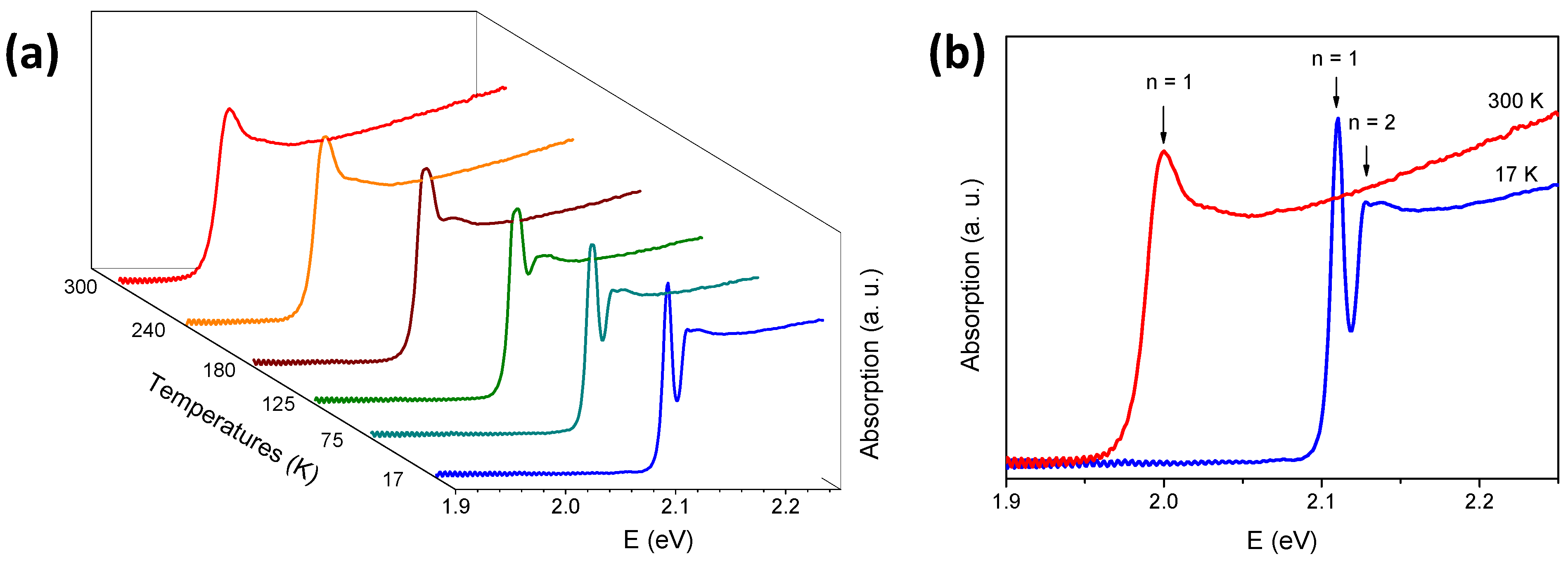
Figure 2a illustrates the change in absorption in a GaSe thin slab with the temperature increasing from 17 to 300 K. The absorption coefficient, α, is determined by converting the transmission spectra using the relation α = 1 / d l o g ( I 0 / I ) ; here d represents the sample thickness, I0 is the transmission without the sample, and I is the transmission with the sample. It is clear that as the temperature decreases, there is a blue shift and sharpening of the excitonic feature and band edge. Figure 2b depicts the presence of the ground state (n = 1) and excited state (n = 2) of free excitons at lower temperatures. According to Figure 2b, the optical band gaps are approximately 2.11 and 2.0 eV at 17 and 300 K, respectively. These values are consistent with previous studies [26,27,34,35,41]. We note that the thickness of the GaSe slab used in this study is comparable to that reported by Bourdon and Khelladi [42]. However, in Ref. [42], the excited exciton state was not evident. We conjecture that reducing the thickness of a GaSe slab further may allow for the observation of higher excited states of excitons (n = 3, 4, …).
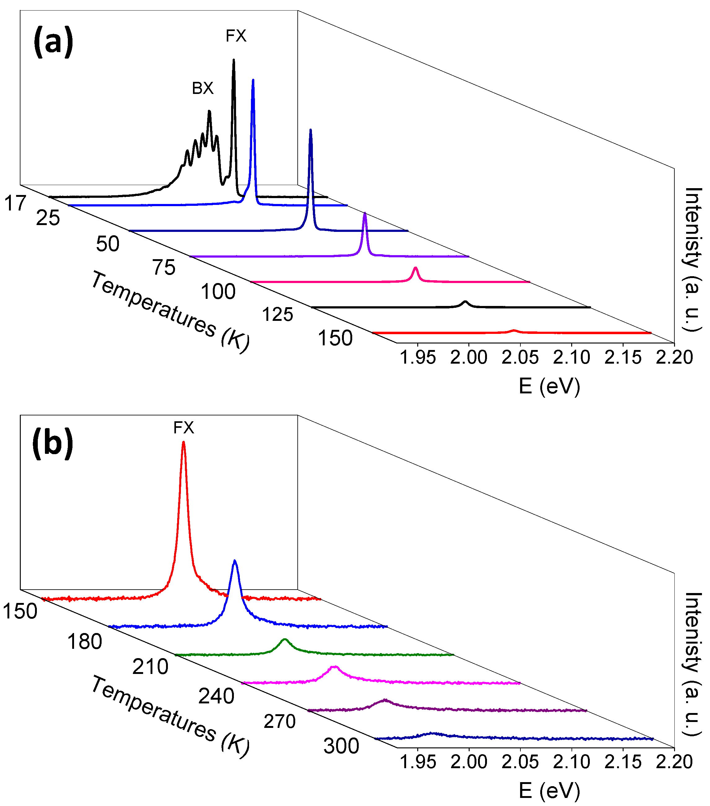
The temperature dependence of the PL spectra, excited by a 355 nm laser, is shown in Figure 3a,b. The PL intensity increases rapidly as the temperature decreases. The dominant peak can be attributed to the recombination of free excitons (FX), specifically to the n = 1 exciton state associated with the direct gap. This observation is consistent with earlier reports [21,24,25,26,29,32,33,43,44]. At low temperatures, other peaks occur below the FX, which may be due to bound excitons (BX) or ground states of excitons with large binding energy (Frenkel excitons). The high-energy FX is undoubtedly caused by the ground states of Wannier-Mott excitons. These Wannier-Mott excitons are formed by electrons from the bottom conduction band and holes from the top valence band at the Γ-point [32].
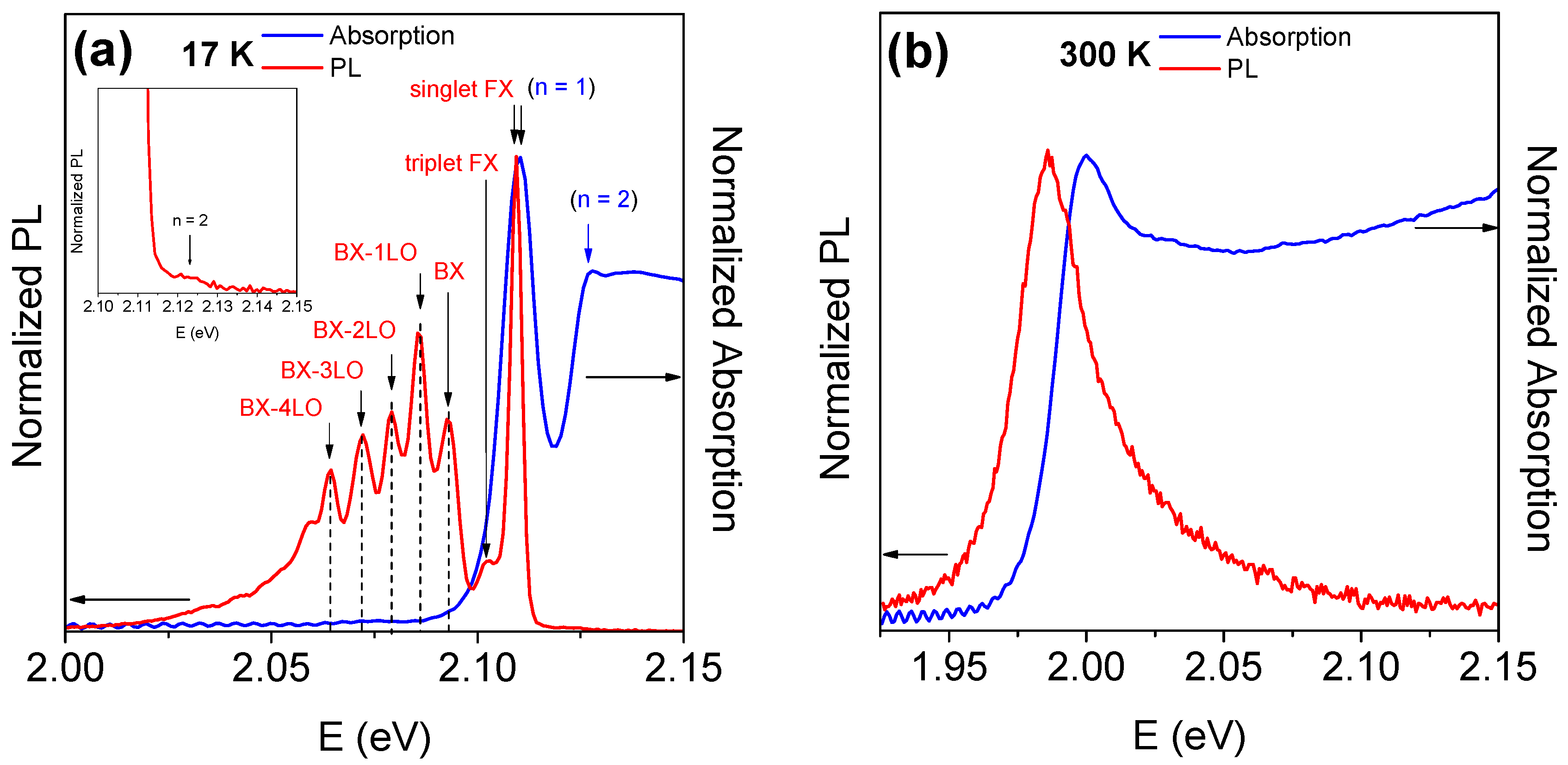
To further investigate the optical properties of GaSe single crystal at low temperatures, Figure 4a,b presents the PL and absorption spectra at 17 and 300 K, respectively. In Figure 4a, the exciton ground state (FX) splits into the triplet (2.103 eV) and singlet (2.109 eV) states, with an energy deviation of 6 meV. This deviation is comparable to the values (4 meV) obtained at 4.2 K by Mercier et al. [45]. The difference may be attributed to different light source excitations, as reported by Zalamai et al. [23]. The bound exciton (BX) is localized at 2.093 eV, and its phonon replicas are clearly distinguishable. The associated peaks are equally spaced in energy with an interval of 7 ± 0.5 meV, which corresponds to the E 1 g 1 mode in the Raman scattering measurement [33]. This value is similar to the phonon replica on the higher energy side of the first exciton state reported by Ishii et al. [46] using transverse electroreflectance at 4.2 K. Furthermore, observations of up to the fourth-order phonon replicas of free-exciton emission were possible, indicating the high crystalline quality and strong coupling of phonon and BX. It is worth emphasizing that both the splitting of the exciton ground state and the phonon replicas in the PL spectra of GaSe occur at low temperatures. As temperature increases, the intensities of BX-related emissions rapidly decrease due to the thermal activation of the bound exciton. This phenomenon is well-known because free excitons can bind to structural defects or impurities, leading to suppressed emission at low temperatures in favor of the bound excitons [21,22,43,47]. The Stokes shift between the ground exciton state (n = 1) and the singlet FX is approximately 1 meV at 17 K, while it is 14 meV at 300 K.
The shift of the exciton depending on the temperature can be described by the following expression:
E ( T ) = E 0 + A T + B 1 e ω p h / k B T 1 ,
where the linear term accounts for the thermal expansion of the lattice and the other for the phonon contribution to the electron’s energy [34].
Figure 5a,b depicts the temperature dependence of the ground and excited exciton states of the absorption and PL spectra, respectively, along with the corresponding best-fit parameters of Equation (1) listed in Table 1. The dots represent the data, while the solid lines depict the fitted curves. The fitting results reveal that the phonon energy ω p h of the ground exciton states for both absorption and PL are 18.6 ± 3.5 and 20.4 ± 2 meV, respectively. These values are consistent with those obtained by Antonioli et al. [34] (18.8 ± 1 meV) and are very close to the optical mode at 16.7 meV, as shown in Figure 1b. Moreover, the phonon energy for the excited exciton states is nearly identical to the phonon mode (30.8 ± 1 meV).
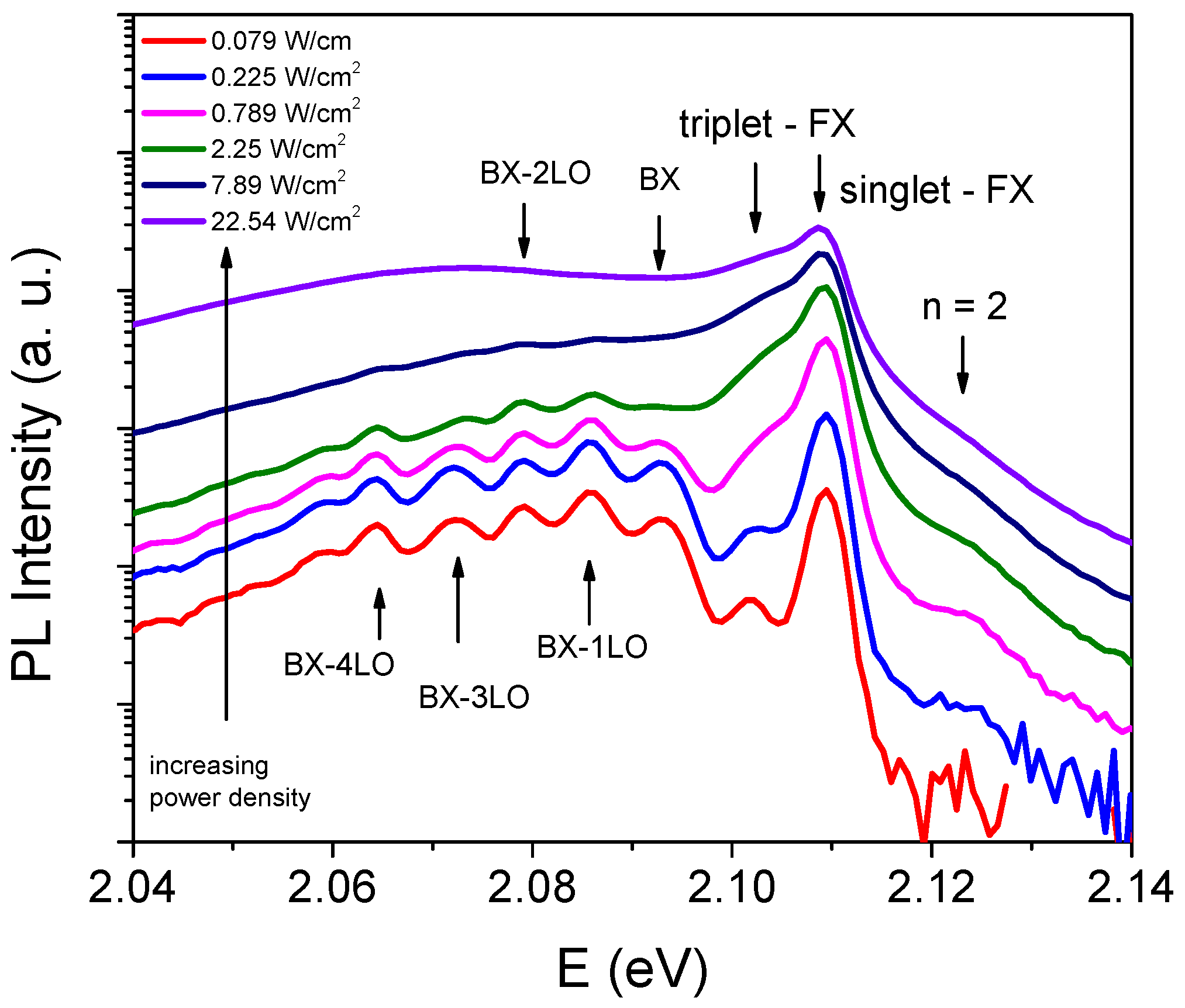
Figure 6 illustrates the evolution of the PL spectra at 17 K for various power excitation densities. As the excitation intensity increases, the peaks become broader. When the excitation power reaches its highest level, the BX-nLO peaks broaden to the extent that the emission appears continuous within our spectral resolution. Furthermore, as the excitation power densities increase, the band-filling effect becomes more prominent. With a rise in excitation power density, a greater number of electron-hole pairs are generated, and the electrons and holes located at k ≠ 0 also recombine, thus contributing to the observed peak width [48]. This phenomenon is observed in numerous materials [49,50].

4. Conclusions

In conclusion, this study presents the absorption and photoluminescence spectra of the GaSe single crystal near the band-edge covering temperatures ranging from 17 to 300 K. At a temperature of 17 K, an intriguing phenomenon can be observed in the photoluminescence spectrum: the Wannier-Mott exciton splits into two distinct states. These states consist of a triplet state with an energy of 2.103 eV and a singlet state with an energy of 2.109 eV. Additionally, there is a bound exciton (BX) that can be localized at an energy of 2.093 eV. It is important to note that its phonon replicas (BX-nLO) can be easily distinguished up to the fourth order. Notably, there is a consistent spacing of 7 ± 0.5 meV between these replicas. This finding suggests that there is a strong interaction between the phonon and BX, indicating an excellent quality of the crystalline structure. Furthermore, at low temperatures, we can observe both the ground state (n = 1) at 2.11 eV and the excited state (n = 2) at 2.127 eV of free excitons. It is important to highlight that the absorption and photoluminescence of the excited state (n = 2) of excitons are observed in the GaSe thin slab.

Author Contributions

Conceptualization, writing—original draft, funding acquisition, L.V.L.; investigation, sample characterization, formal analysis, T.T.T.H., T.-T.N., X.A.N. and T.H.N.; writing—review and editing, funding acquisition, S.C., Y.D.K. and T.J.K. All authors have read and agreed to the published version of the manuscript.

Funding

This research was funded by the Vietnam Academy of Science and Technology (Grant No. THTEXS.01/21-24) and the National Research Foundation of Korea (NRF) grant funded by the Korean government (MSIP) (NRF-2020R1A5A1016518).

Data Availability Statement

The data presented in this study are available on request from the corresponding author.

Acknowledgments

L.V.L. acknowledges Objective Lab for Agriculture-Bio-Medicine and Energy of VAST/IMS for using the Lab’s equipment.

Conflicts of Interest

The authors declare no conflicts of interest.

References

  1. Abderrahmane, A.; Jung, P.-G.; Kim, N.-H.; Ko, P.J.; Sandhu, A. Gate-Tunable Optoelectronic Properties of a Nano-Layered GaSe Photodetector. Opt. Mater. Express 2017, 7, 587–592. [Google Scholar] [CrossRef]
  2. Saroj, R.K.; Guha, P.; Lee, S.; Yoo, D.; Lee, E.; Lee, J.; Kim, M.; Yi, G.-C. Photodetector Arrays Based on MBE-Grown GaSe/Graphene Heterostructure. Adv. Opt. Mater. 2022, 10, 2200332. [Google Scholar] [CrossRef]
  3. Chen, Z.; Chen, Q.; Chai, Z.; Wei, B.; Wang, J.; Liu, Y.; Shi, Y.; Wang, Z.; Li, J. Ultrafast Growth of High-Quality Large-Sized GaSe Crystals by Liquid Metal Promoter. Nano Res. 2022, 15, 4677–4681. [Google Scholar] [CrossRef]
  4. Sorifi, S.; Kaushik, S.; Singh, R. A GaSe/Si-Based Vertical 2D/3D Heterojunction for High-Performance Self-Driven Photodetectors. Nanoscale Adv. 2022, 4, 479–490. [Google Scholar] [CrossRef]
  5. Zappia, M.I.; Bianca, G.; Bellani, S.; Curreli, N.; Sofer, Z.; Serri, M.; Najafi, L.; Piccinni, M.; Oropesa-Nuñez, R.; Marvan, P.; et al. Two-Dimensional Gallium Sulfide Nanoflakes for UV-Selective Photoelectrochemical-Type Photodetectors. J. Phys. Chem. C 2021, 125, 11857–11866. [Google Scholar] [CrossRef] [PubMed]
  6. Zappia, M.I.; Bianca, G.; Bellani, S.; Serri, M.; Najafi, L.; Oropesa-Nuñez, R.; Martín-García, B.; Bouša, D.; Sedmidubský, D.; Pellegrini, V.; et al. Solution-Processed GaSe Nanoflake-Based Films for Photoelectrochemical Water Splitting and Photoelectrochemical-Type Photodetectors. Adv. Funct. Mater. 2020, 30, 1909572. [Google Scholar] [CrossRef]
  7. Yang, Z.; Chen, H.; Wu, F.; Hou, Y.; Qiao, J.; Ma, X.; Bai, H.; Ma, B.; Li, J. Investigation on Photocatalytic Property of SiH/GaSe and SiH/InSe Heterojunctions for Photocatalytic Water Splitting. Int. J. Hydrogen Energy 2022, 47, 31295–31308. [Google Scholar] [CrossRef]
  8. Zhang, W.X.; Hou, J.T.; Bai, M.; He, C.; Wen, J.R. Construction of Novel ZnO/Ga2SSe (GaSe) VdW Heterostructures as Efficient Catalysts for Water Splitting. Appl. Surf. Sci. 2023, 634, 157648. [Google Scholar] [CrossRef]
  9. Ahmad, H.; Reduan, S.A.; Sharbirin, A.S.; Ismail, M.F.; Zulkifli, M.Z. Q-Switched Thulium/Holmium Fiber Laser with Gallium Selenide. Optik 2018, 175, 87–92. [Google Scholar] [CrossRef]
  10. Yu, Q.; Liu, F.; Zhang, Y.; Deng, H.; Shu, B.; Zhang, J.; Yi, T.; Dai, Y.; Fan, C.; Su, W.; et al. Lab-on-Fiber Based on Optimized Gallium Selenide for Femtosecond Mode-Locked Lasers and Fiber-Compatible Photodetectors. Adv. Photonics Res. 2023, 4, 2200283. [Google Scholar] [CrossRef]
  11. Guo, J.; Xie, J.-J.; Li, D.-J.; Yang, G.-L.; Chen, F.; Wang, C.-R.; Zhang, L.-M.; Andreev, Y.M.; Kokh, K.A.; Lanskii, G.V.; et al. Doped GaSe Crystals for Laser Frequency Conversion. Light Sci. Appl. 2015, 4, e362. [Google Scholar] [CrossRef]
  12. Elafandi, S.; Ahmadi, Z.; Azam, N.; Mahjouri-Samani, M. Gas-Phase Formation of Highly Luminescent 2D GaSe Nanoparticle Ensembles in a Nonequilibrium Laser Ablation Process. Nanomaterials 2020, 10, 908. [Google Scholar] [CrossRef]
  13. Liu, G.; Xia, S.; Hou, B.; Gao, T.; Zhang, R. Mechanical Stabilities and Nonlinear Properties of Monolayer Gallium Selenide under Tension. Mod. Phys. Lett. B 2015, 29, 1550049. [Google Scholar] [CrossRef]
  14. Karvonen, L.; Säynätjoki, A.; Mehravar, S.; Rodriguez, R.D.; Hartmann, S.; Zahn, D.R.T.; Honkanen, S.; Norwood, R.A.; Peyghambarian, N.; Kieu, K.; et al. Investigation of Second- and Third-Harmonic Generation in Few-Layer Gallium Selenide by Multiphoton Microscopy. Sci. Rep. 2015, 5, 10334. [Google Scholar] [CrossRef]
  15. Shevchenko, O.N.; Nikolaev, N.A.; Antsygin, V.D. Estimation of the Nonlinear-Optical Coefficient of GaSe:S Crystals According to Electro-Optical Measurements. In Proceedings of the XVI International Conference on Pulsed Lasers and Laser Applications, Tomsk, Russia, 10–15 September 2023; Volume 12920, p. 129200G. [Google Scholar]
  16. Thanh, L.C.; Depeursinge, C. Fine Structure of the Exciton Spectrum in GaSe. Solid State Commun. 1978, 25, 499–503. [Google Scholar] [CrossRef]
  17. Bergeron, A.; Ibrahim, J.; Leonelli, R.; Francoeur, S. Oxidation Dynamics of Ultrathin GaSe Probed through Raman Spectroscopy. Appl. Phys. Lett. 2017, 110, 241901. [Google Scholar] [CrossRef]
  18. Hoff, R.M.; Irwin, J.C.; Lieth, R.M.A. Raman Scattering in GaSe. Can. J. Phys. 1975, 53, 1606–1614. [Google Scholar] [CrossRef]
  19. Irwin, J.C.; Hoff, R.M.; Clayman, B.P.; Bromley, R.A. Long Wavelength Lattice Vibrations in GaS and GaSe. Solid State Commun. 1973, 13, 1531–1536. [Google Scholar] [CrossRef]
  20. Wieting, T.J.; Verble, J.L. Interlayer Bonding and the Lattice Vibrations of β-GaSe. Phys. Rev. B 1972, 5, 1473–1479. [Google Scholar] [CrossRef]
  21. Capozzi, V.; Montagna, M. Optical Spectroscopy of Extrinsic Recombinations in Gallium Selenide. Phys. Rev. B 1989, 40, 3182–3190. [Google Scholar] [CrossRef]
  22. Wei, C.; Chen, X.; Li, D.; Su, H.; He, H.; Dai, J.-F. Bound Exciton and Free Exciton States in GaSe Thin Slab. Sci. Rep. 2016, 6, 33890. [Google Scholar] [CrossRef]
  23. Zalamai, V.V.; Syrbu, N.N.; Stamov, I.G.; Beril, S.I. Wannier–Mott Excitons in GaSe Single Crystals. J. Opt. 2020, 22, 85402. [Google Scholar] [CrossRef]
  24. Usman, M.; Golovynskyi, S.; Dong, D.; Lin, Y.; Yue, Z.; Imran, M.; Li, B.; Wu, H.; Wang, L. Raman Scattering and Exciton Photoluminescence in Few-Layer GaSe: Thickness- and Temperature-Dependent Behaviors. J. Phys. Chem. C 2022, 126, 10459–10468. [Google Scholar] [CrossRef]
  25. Rakhlin, M.V.; Evropeitsev, E.A.; Eliseyev, I.A.; Toropov, A.A.; Shubina, T.V. Exciton Structure and Recombination Dynamics in GaSe Crystals. Bull. Russ. Acad. Sci. Phys. 2023, 87, S60–S65. [Google Scholar] [CrossRef]
  26. Akhundov, G.A.; Gasanova, N.A.; Nizametdinova, M.A. Optical Absorption, Reflection, and Dispersion of GaS and GaSe Layer Crystals. Phys. Status Solidi 1966, 15, K109–K113. [Google Scholar] [CrossRef]
  27. Balzarotti, A.; Piacentini, M. Excitonic Effect at the Direct Absorption Edges of GaSe. Solid State Commun. 1972, 10, 421–425. [Google Scholar] [CrossRef]
  28. Gauthier, M.; Polian, A.; Besson, J.M.; Chevy, A. Optical Properties of Gallium Selenide under High Pressure. Phys. Rev. B 1989, 40, 3837–3854. [Google Scholar] [CrossRef]
  29. Isik, M.; Tugay, E.; Gasanly, N.M. Temperature-Dependent Optical Properties of GaSe Layered Single Crystals. Philos. Mag. 2016, 96, 2564–2573. [Google Scholar] [CrossRef]
  30. Choi, S.G.; Levi, D.H.; Martinez-Tomas, C.; Muñoz Sanjosé, V. Above-Bandgap Ordinary Optical Properties of GaSe Single Crystal. J. Appl. Phys. 2009, 106, 53517. [Google Scholar] [CrossRef]
  31. Meyer, F.; de Kluizenaar, E.E.; Engelsen, D. den Ellipsometric Determination of the Optical Anisotropy of Gallium Selenide. J. Opt. Soc. Am. 1973, 63, 529–532. [Google Scholar] [CrossRef]
  32. Le, L.V.; Nguyen, T.-T.; Nguyen, X.A.; Cuong, D.D.; Nguyen, T.H.; Nguyen, V.Q.; Cho, S.; Kim, Y.D.; Kim, T.J. A Systematic Study of the Temperature Dependence of the Dielectric Function of GaSe Uniaxial Crystals from 27 to 300 K. Nanomaterials 2024, 14, 839. [Google Scholar] [CrossRef]
  33. Voitchovsky, J.P.; Mercier, A. Photoluminescence OfGaSe. Nuovo Cim. B 1974, 22, 273–292. [Google Scholar] [CrossRef]
  34. Antonioli, G.; Bianchi, D.; Emiliani, U.; Podini, P.; Franzosi, P. Optical Properties and Electron-Phonon Interaction in GaSe. Nuovo Cim. B 1979, 54, 211–227. [Google Scholar] [CrossRef]
  35. Le Toullec, R.; Piccioli, N.; Chervin, J.C. Optical Properties of the Band-Edge Exciton in GaSe Crystals at 10 K. Phys. Rev. B 1980, 22, 6162–6170. [Google Scholar] [CrossRef]
  36. Capozzi, V. A Minafra Photoluminescence Properties of Cu-Doped GaSe. J. Phys. C Solid State Phys. 1981, 14, 4335. [Google Scholar] [CrossRef]
  37. Del Pozo-Zamudio, O.; Schwarz, S.; Sich, M.; Akimov, I.A.; Bayer, M.; Schofield, R.C.; Chekhovich, E.A.; Robinson, B.J.; Kay, N.D.; Kolosov, O.V.; et al. Photoluminescence of Two-Dimensional GaTe and GaSe Films. 2D Mater. 2015, 2, 35010. [Google Scholar] [CrossRef]
  38. Chen, G.; Zhang, L.; Li, L.; Cheng, F.; Fu, X.; Li, J.; Pan, R.; Cao, W.; Chan, A.S.; Panin, G.N.; et al. GaSe Layered Nanorods Formed by Liquid Phase Exfoliation for Resistive Switching Memory Applications. J. Alloys Compd. 2020, 823, 153697. [Google Scholar] [CrossRef]
  39. Wang, T.; Li, J.; Zhao, Q.; Yin, Z.; Zhang, Y.; Chen, B.; Xie, Y.; Jie, W. High-Quality GaSe Single Crystal Grown by the Bridgman Method. Materials 2018, 11, 186. [Google Scholar] [CrossRef]
  40. Lim, S.Y.; Lee, J.-U.; Kim, J.H.; Liang, L.; Kong, X.; Nguyen, T.T.H.; Lee, Z.; Cho, S.; Cheong, H. Polytypism in Few-Layer Gallium Selenide. Nanoscale 2020, 12, 8563–8573. [Google Scholar] [CrossRef]
  41. Shih, Y.-T.; Lin, D.-Y.; Tseng, B.-C.; Kao, Y.-M.; Hwang, S.-B.; Lin, C.-F. Structural and Optical Characterization of GaS1-xSex Layered Mixed Crystals Grown by Chemical Vapor Transport. Mater. Today Commun. 2023, 37, 107047. [Google Scholar] [CrossRef]
  42. Bourdon, A.; Khelladi, F. Selection Rule in the Fundamental Direct Absorption of GaSe. Solid State Commun. 1971, 9, 1715–1717. [Google Scholar] [CrossRef]
  43. Capozzi, V. Direct and Indirect Excitonic Emission in GaSe. Phys. Rev. B 1981, 23, 836–840. [Google Scholar] [CrossRef]
  44. Zhang, D.; Jia, T.; Dong, R.; Chen, D. Temperature-Dependent Photoluminescence Emission from Unstrained and Strained GaSe Nanosheets. Materials 2017, 10, 1282. [Google Scholar] [CrossRef]
  45. Mercier, A.; Mooser, E.; Voitchovsky, J.P. Resonant Exciton in GaSe. Phys. Rev. B 1975, 12, 4307–4311. [Google Scholar] [CrossRef]
  46. Ishii, Y.; Sasaki, Y.; Hamaguchi, C.; Nakai, J. Transverse Electroreflectance of GaSe at the Fundamental Absorption Edge. Solid State Commun. 1975, 17, 451–454. [Google Scholar] [CrossRef]
  47. Matsumura, T.; Sudo, M.; Tatsuyama, C.; Ichimura, S. Photoluminescence in GaSe. Phys. Status Solidi 1977, 43, 685–693. [Google Scholar] [CrossRef]
  48. Yang, L.; Motohisa, J.; Fukui, T. Excitation-Power-Density-Dependent Micro-Photoluminescence from Selective-Area-Grown Hexagonal Nanopillars with Single InGaAs/GaAs Quantum Well on the GaAs (111)B Substrate. In Proceedings of the 2007 7th IEEE Conference on Nanotechnology (IEEE NANO), Hong Kong, China, 2–5 August 2007; pp. 664–669. [Google Scholar]
  49. Pan, C.-J.; Lin, K.-F.; Hsieh, W.-F. Acoustic and Optical Phonon Assisted Formation of Biexcitons. Appl. Phys. Lett. 2007, 91, 111907. [Google Scholar] [CrossRef]
  50. Feng, Z.C.; Li, Q.; Wan, L.; Xu, G. Variation of Phonon Coupling Factors in the Photoluminescence of Cadmium Telluride by Variable Excitation Power. Opt. Mater. Express 2017, 7, 808–816. [Google Scholar] [CrossRef]
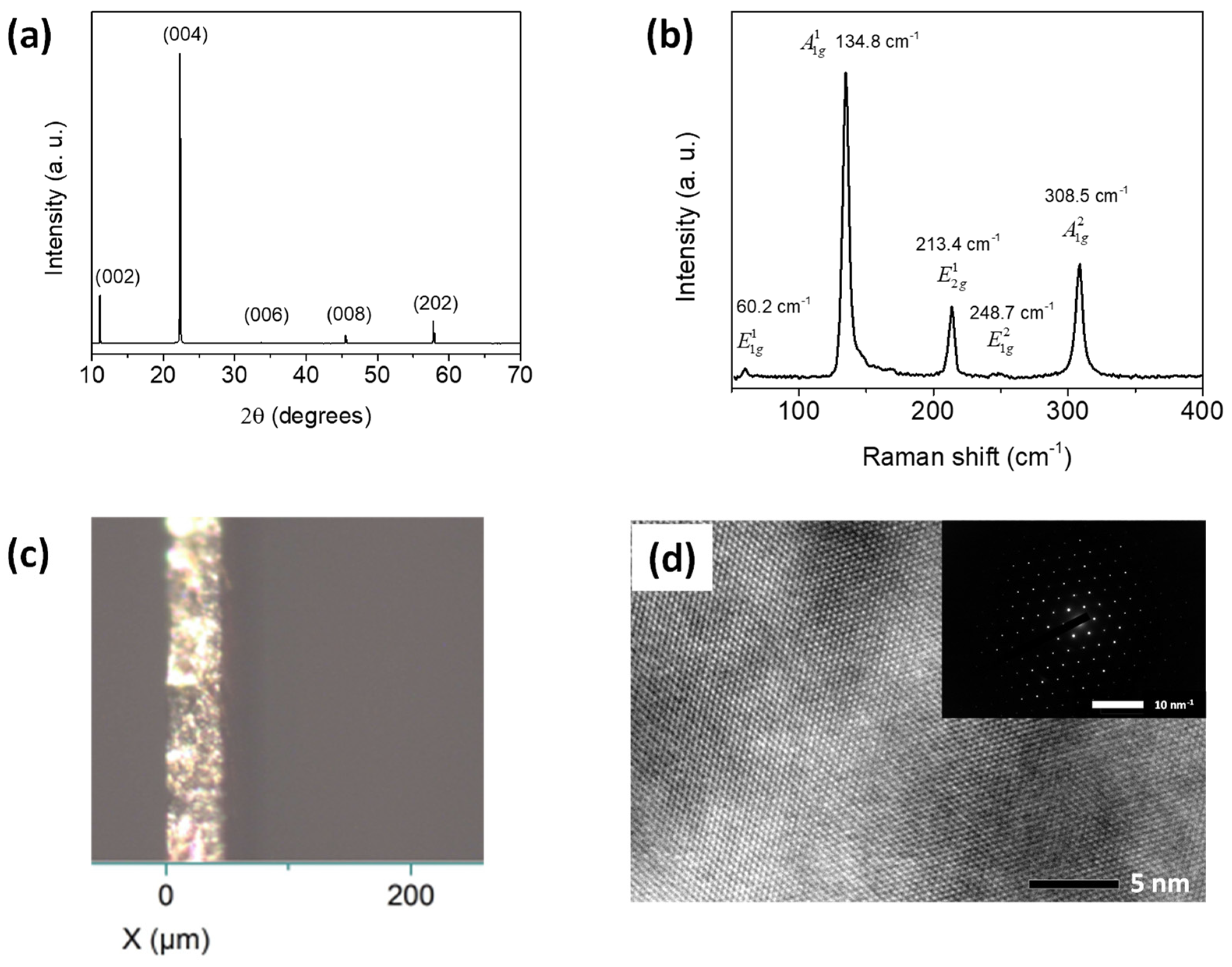
Figure 1. (a) The XRD pattern of the GaSe single crystal aligned along the (001) plane is shown, as well as (b) its Raman spectrum. (c) An optical image of the cross-section of the sample. (d) A HR-TEM image of the sample in the c-plan. The SAED pattern is shown in the inset.
Crystals 14 00539 g001
Figure 2. (a) Temperature dependence of the absorption spectra of the GaSe thin slab from 17 to 300 K is investigated in this study. To improve clarity, we have included several representative spectra. (b) The absorption spectra of the GaSe thin slab are shown for comparison at 17 K and 300 K.
Crystals 14 00539 g002
Figure 3. Temperature-dependent PL spectra of a GaSe thin slab obtained in two temperature ranges: (a) 17 to 150 K and (b) 150 to 300 K.
Crystals 14 00539 g003
Figure 4. PL and absorption spectra of the GaSe thin slab at (a) 17 K and (b) 300 K.
Crystals 14 00539 g004
Figure 5. Energy of the ground excitons (square dots) and excited excitons (circle dots) of GaSe are shown as a function of temperature. The experimental data are represented by dots and the fitted curves are shown as solid lines in both (a) absorption and (b) photoluminescence.
Crystals 14 00539 g005
Figure 6. Luminescence spectra (log scale) of GaSe single crystal measured by excitation into the conduction band with the 355 nm at 17 K and for different excitation intensities.
Crystals 14 00539 g006
Table 1. The best-fit parameters of the temperature dependences of the exciton states of absorption and PL obtained using Equation (1).
Exciton States E 0 (eV) A (meVK) B (meVK) ω p h (meV)
Absorption n = 1 2.111 ± 0.002−0.5 ± 0.7103 ± 1018.6 ± 3.5
n = 2 2.128 ± 0.001−0.6 ± 0.5−111 ± 7820.8 ± 7.8
PL n = 1 2.111 ± 0.001−0.8 ± 0.4−121 ± 720.4 ± 2
n = 2 2.127 ± 0.001−1.1 ± 0.1−217 ± 4029.2 ± 2
Disclaimer/Publisher’s Note: The statements, opinions and data contained in all publications are solely those of the individual author(s) and contributor(s) and not of MDPI and/or the editor(s). MDPI and/or the editor(s) disclaim responsibility for any injury to people or property resulting from any ideas, methods, instructions or products referred to in the content.