Article

Assessment of the Amino Acid L-Histidine as a Corrosion Inhibitor for a 1018 Carbon Steel in Aqueous Sodium Chloride Solution

by
Milena Jacinto da Silva Moura
1,
Roberta Bastos Vasques
1,
Saulo Jose de melo Magalhães
1,
Francisco Wagner de Queiroz Almeida Neto
2,
Pedro de Lima Neto
3,
Luís Paulo Mourão dos Santos
1,
Mauro Andres Cerra Florez
1,*,
Gemma Fargas Ribas
4,
Samuel Lucas Santos Medeiros
1,
Francisco Carlos Carneiro Soares Salomão
5,
Eduardo Bedê Barros
6 and
Walney Silva Araújo
1
1
Graduate Program in Materials Science and Engineering, Federal University of Ceará, Fortaleza 60440-900, Brazil
2
Science and Technology Center of the Inhamuns Region, Ceara State University Education, Taua 60000-000, Brazil
3
Department of Analytical Chemistry and Physical Chemistry, Federal University of Ceará, Fortaleza 60440-900, Brazil
4
CIEFMA, Department of Materials Science and Engineering, Escola d’Enginyeria de Barcelona Est (EEBE), Universitat Politècnica de Catalunya, 08019 Barcelona, Spain
5
Science and Technology Center—Physics Course, Itaperi Campus, Ceará State University, Fortaleza 62930-000, Brazil
6
Department of Physics, Federal University of Ceará, Fortaleza 60455-760, Brazil
*
Author to whom correspondence should be addressed.
Crystals 2024, 14(8), 703; https://doi.org/10.3390/cryst14080703
Submission received: 28 May 2024 / Revised: 20 June 2024 / Accepted: 22 July 2024 / Published: 2 August 2024
(This article belongs to the Special Issue Synthesis, Characterization and Properties of Crystalline Materials)

Abstract

:
The amino acid L-histidine, which has an imidazole ring, was investigated as a corrosion inhibitor for AISI 1018 carbon steel in chloride solution based on the effectiveness of inhibitors containing imidazole in their composition. A neutral environment was chosen for this study due to the scarcity of research on this amino acid in this environment type. Concentrations of 250, 500, and 1000 ppm were evaluated. Various methods were used to determine inhibition effectiveness, including mass loss, open circuit potential, linear potentiodynamic polarization, and electrochemical impedance spectroscopy. For mass loss, the inhibition efficiency varied from 83 to 88% according to the increase in concentration. For the electrochemical tests, the efficiency variation ranged from 62 to 90% with increasing amino acid concentration. Furthermore, a simulation analysis using quantum chemical calculations within the scope of Density Functional Theory (DFT) revealed that histidine’s nucleophilic character is crucial for its corrosion inhibitory capacity in an aqueous medium at pH 7. The inhibition efficiency increased with increasing concentration in a neutral medium, following the Langmuir isotherm for the adsorption of L-histidine. Additional studies were carried out using Fourier transform infrared spectroscopy (FTIR) and thermogravimetry (TGA). Analysis of the substrate surface by scanning electron microscopy (SEM) showed greater preservation with the addition of L-histidine, confirming its adsorption on the steel. Atomic Force Microscopy (AFM) also demonstrated an improvement in surface roughness in the presence of amino acids compared to the medium without an inhibitor.

1. Introduction

Corrosion inhibitors are chemical substances added to a corrosive environment to reduce or prevent corrosion of metallic materials. They function by forming a protective layer on the surface of the metal or changing environmental conditions to make it less corrosive. These compounds are widely used in a variety of industries, including oil, gas, chemical, and infrastructure such as pipelines, tanks, and equipment, where corrosion is a significant concern [1,2,3]. Therefore, several industries use corrosion inhibitors on a large scale to reduce the corrosive process, such as oil companies that apply them to everything from extraction to oil and gas transportation pipelines [4,5,6].
In this context, the adoption of organic corrosion inhibitors, characterized by their non-toxic nature and minimal environmental impact, appears as an attractive alternative to commercial inhibitors, which are normally inorganic inhibitors and contain substances harmful to the environment and humans. The use of green corrosion inhibitors has proven to be a practical approach to protecting iron-based metal alloys immersed in chlorinated media, particularly those with low carbon content, such as AISI 1018 carbon steel [4,5,6,7,8,9].
Organic compounds containing heteroatoms (O, S, N, P), aromatic rings, imidazole derivatives, pyrimidine derivatives, or Schiff bases are being tested as potential corrosion inhibitors [7,8,9,10,11,12,13,14,15,16]. In this context, amino acids are substances that have relevant properties that must be explored, in all corrosive environments, as inhibitory products. They have a low environmental impact, are water-soluble, and non-toxic products that are easy to obtain and handle, and contain heteroatoms in their structure. Thus, these substances have proven effective in protecting against corrosion in metals and their alloys in different corrosive environments [3,17,18,19,20].
In the literature, the amino acid L-histidine has been evaluated as a corrosion inhibitor in acidic environments at different concentrations, where it shows greater efficiency with increasing concentration [21,22]. For this study, L-histidine was evaluated as a corrosion inhibitor in a medium containing 3.5% by weight NaCl, where the pH is in the range of 6.85 ± 0.35. When the amino acid is added, the pH is established in a range of 7.25 ± 0.25; that is, the medium to be analyzed is neutral. Histidine, already studied in a weakly acidic environment, obtained good efficiency. Due to its amphoteric behavior, we will investigate whether it also forms bonds with metal surfaces in neutral environments.

2. Experimental Materials and Methods

The study material was AISI 1018 carbon steel, whose chemical composition was determined by optical emission spectroscopy (% by weight): C 0.197, Mn 0.84, Si 0.19, Cu 0.02, Ni 0.01, Cr 0.09, Mo 0.03, and the remainder Fe. This material was used to manufacture the working electrodes, which were welded with copper wire and immersed in epoxy resin with an exposed area of 0.5 cm2. The working electrodes were polished with different grits: 120, 220, 320, 400, and 600 mesh. They were then washed with distilled water and dried with hot air. The electrochemical cell consists of three electrodes: an AISI 1018 carbon steel working electrode, a 2.45 cm2 platinum plate used as a counter electrode, and a Hastelloy alloy serving as a reference. The conventional reference electrode (Ag/AgCl or saturated calomel), normally used in electrochemical tests, proved unsuitable for this system, as the inhibitor infiltrated the porous tip, contaminating the solution [15,23].
The organic compound used was L-histidine, as shown in Figure 1.
The amino acid used is a product of SIGMA-ALDRICH. Solutions of 3.5 wt.% NaCl were prepared using distilled water without and with three concentrations of L-histidine, namely 250, 500, and 1000 ppm, without additional treatment. The experiments were conducted in triplicate with solutions without and with the inhibitor at room temperature (25 °C).

2.1. Gravimetric and Electrochemical Tests

In the gravimetric test, the variation in mass loss (majúscula W) can be obtained, which is the most direct measure of an inhibitor’s inhibition efficiency.
The test was conducted over 168 h. Cylindrical test specimens with an area of 14.76 cm2 were immersed in a 3.5 wt.% NaCl solution both with and without the addition of inhibitors at three different concentrations. After the immersion test, the test specimens underwent surface treatment. Mass loss tests for L-histidine were performed in triplicate. This experiment was conducted using the ASTM G 31-72 technical standard [24,25]. The samples were weighed before and after immersion without and with the inhibitor in an analytical balance, with precision ± 0.0001 mg. The corrosion rate was calculated using these data [26].
After the mass loss test, the adsorption isotherms were studied. Adsorption is the mass transfer of certain substances present in liquid or gaseous fluids to the surface of certain solids. These substances can be bonded to a solid surface in two ways, physically or chemically, depending on the nature of the forces involved [27,28].
The adsorption of amino acid molecules occurs on the active sites of the material’s surface or deposition of corrosion products on it. This process depends on the nature of the inhibitor molecules and the general condition of the steel surface. Therefore, the degree of surface coverage (θ) is calculated at different concentrations of amino acids to define which interaction occurs between the inhibitor molecules and the substrate surface. The coverage degree values were collected from the mass loss test and used to make adjustments. Inhibitor adsorption occurs on metal surfaces due to its functional groups [29,30,31].
After mass loss tests on the working electrode, in a 3.5% NaCl medium without and with L-histidine at the three concentrations studied, the solutions were used to investigate the formation of complexes between the amino acid and Fe2+ ions from the substrate. This analysis is known as ultraviolet-visible (UV–Vis) spectroscopy. UV–Vis measurements were conducted in absorption mode in the range of 220–700 nm with a Shimadzu® model UV2600 spectrophotometer [32,33].
For the electrochemical tests, conventional three-electrode electrochemical cells were used in a 3.5% weight NaCl solution, both without and with L-histidine, at three concentrations (250, 500, and 1000 ppm).
All electrochemical tests were performed using a potentiostat/galvanostat model PGSTAT 302N Autolab (Metrohm-Eco Chemie, Utrecht, The Netherlands) coupled to a computer with version 2.1.4 of the NOVA® program.
The electrode potential in the 3.5 wt.% NaCl solution was monitored in an open circuit relative to the reference electrode for 1 h before potentiodynamic polarization and electrochemical impedance spectroscopy. In this test, a range of ±0.2 mV relative to the open circuit potential was taken, with a scan rate of 1 mVs−1. The inhibition efficiency (η) was calculated from the polarization parameters [20,34,35].
Impedance measurements were taken 1 h after open circuit potential, as previously stated, with a sinusoidal potential disturbance of 5 mV. The frequency varied between 100 kHz and 6 mHz. From this test, it was possible to calculate the inhibition efficiency (η) with the charge transfer resistance values [36,37].

2.2. Characterization of L-Histidine

L-histidine was analyzed by Fourier transform infrared spectroscopy (FTIR) in the infrared range. Analysis was conducted using a spectrophotometer (Spectrum Frontier; PerkinElmer Corp., Waltham, MA, USA) equipped with an attenuated total reflectance (ATR) accessory and zinc selenide (ZnSe) crystal surface. The spectra were recorded with 32 scans between 4000 and 550 cm−1, with a resolution of 4 cm−1 in transmittance mode [38].
For the thermogravimetric analysis of the L-histidine sample, TGA/SDTA851E equipment from Mettler Toledo, Greifensee, Switzerland was used, operating under an inert nitrogen atmosphere at a flow rate of 50 mL/min. The experiment was carried out with a heating rate of 10 °C/min, varying the temperature from 30 °C to 800 °C, in a semi-open alumina crucible. The equipment consists of a balance that allows continuous weighing of the sample as the temperature increases [39].

2.3. Surface Characterization

Surface characterization occurred after the gravimetric test. The images show the surface morphology of the working electrodes without and with L-histidine, which were carried out using a scanning electron microscope (SEM), Quanta 450-FEG (FEI Company, Hillsboro, OR, USA), and an atomic force microscope (AFM). Atomic Force Microscopy (AFM) measurements were obtained in intermittent contact mode using an Asylum Research (Santa Barbara, CA, USA) MFP-3D BIO system, employing tips with a curvature radius smaller than 20 nm and a resonance frequency of 75 kHz. In this characterization, the average surface roughness of the sample was observed. Three samples were analyzed. One sample was polished with Arotec diamond paste of 6, 3, and 1 µm, and the other two samples were immersed in a NaCl solution without and with L-histidine, at a concentration of 1000 ppm [40].

2.4. Quantum Chemical Calculation

Quantum chemistry calculations were performed within the scope of Density Functional Theory (DFT), using the Gaussian 09 and GaussView 5 programs to visualize the input molecules [41]. A microspecies calculation was performed to determine the precise molecular structure of histidine using Marvin Sketch to investigate corrosion inhibition in a neutral solution. The Molecular Frontier Orbitals (FMO) calculations were calculated at the theoretical level B3LYP/6-311++G(d,p) based on the optimized geometry in the gas phase and the presence of water [42]. The isosurfaces for the Highest Occupied Molecular Orbital (HOMO) and the Lowest Unoccupied Molecular Orbital (LUMO) were rendered using the ChemCraft program, version number of the software is 1.8.

3. Results and Discussion

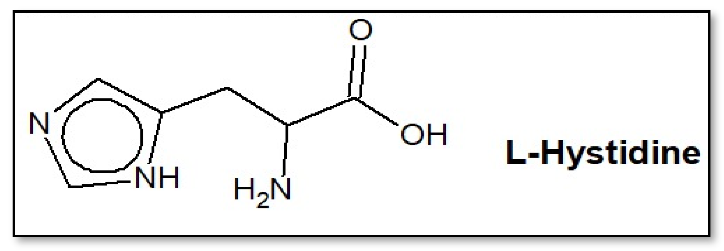
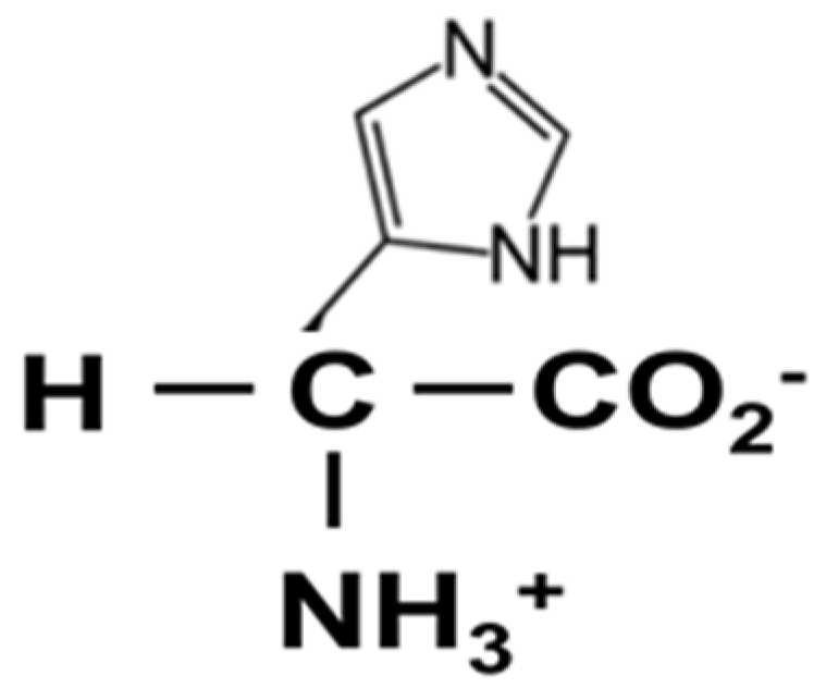
3.1. Amino Acid Characterization: L-Histidine

3.1.1. Fourier Transform Infrared (FTIR) Spectral Analysis

Figure 2 shows an infrared graph of the amino acid L-histidine, where one can identify the characteristic vibrations of the C–H and NH– bonds of the imidazole ring recorded at wavelengths of 3011 cm−1 and 2705 cm−1, respectively. Furthermore, the aliphatic C–H chains are discernible at 2855 cm−1. The peak at 1630 cm−1 is possibly associated with the asymmetric stretching of the carboxylate group and the asymmetric deformation of the NH3 group. The other bands at 1585, 1449, 1341, and 623 cm−1 correspond to the C=C bonds, the symmetric stretching of the NH3 bonds, the symmetric COO group, and the CN bond, respectively [31,38,43].
Analyzing the spectrum, the presence of vibrations characteristic of the NH3+ group at wavelengths of 3011 cm−1, 2855 cm−1, 2705 cm−1, and 1630 cm−1, as well as of the COO group at wavelengths of 1585 cm−1 and 1449 cm−1, is associated with the asymmetric and symmetric stretching of C(=O)2, respectively. These observations reinforce the identification of the amino acid, suggesting its isoelectronic or zwitterionic form, as illustrated in Figure 3. These assignments confirm the presence of several functional groups in the compound [44,45].

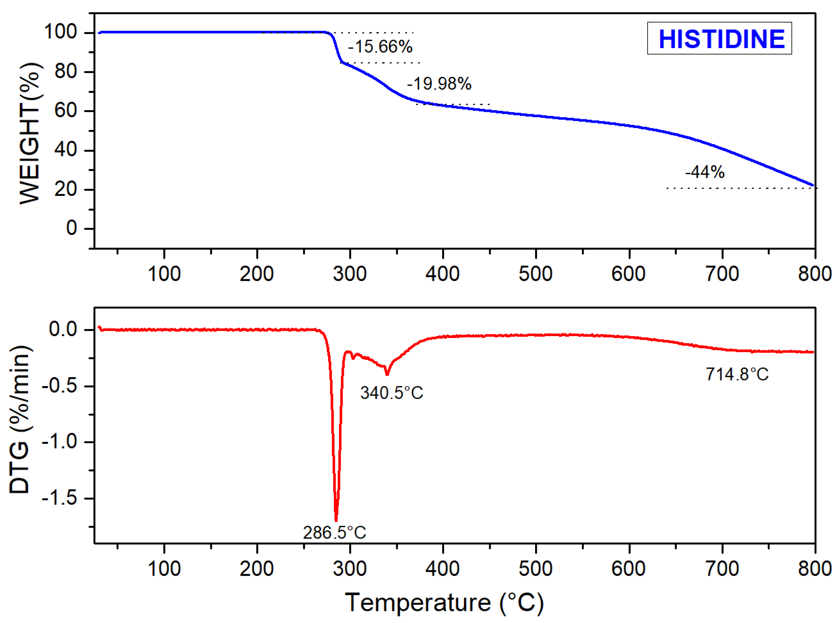
3.1.2. Thermo Gravimetric Analysis (TGA)
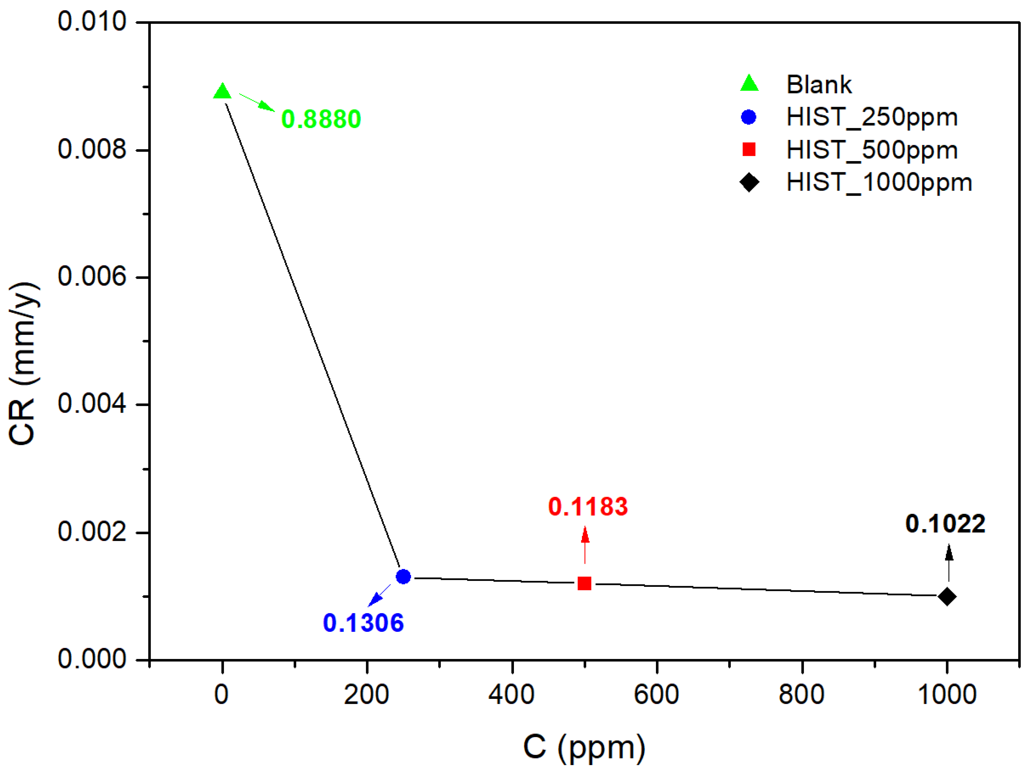
The thermogravimetry test evaluates the thermal stability of the compound and is important for analyzing the degradation of the substance at increasing temperatures. Therefore, this technique investigates variations in the mass of the material in a sample due to physical or chemical transformations over time or temperature. In the test, the dough is continuously monitored, and the data obtained is used to trace the dough degradation curve [43,46].
Two distinct mass loss events can be identified in Figure 4, indicating decomposition in multiple stages. The first event, between temperatures of 270 °C and 400 °C, presents two stages of mass loss, with approximate values of 15.66% and 19.98%. It also presents peak DTG temperatures of 285.5 °C and 340.5 °C, respectively. The second event occurs at temperatures ranging between 400 °C and 800 °C, with a mass loss of around 44% and a peak DTG temperature of around 714.8 °C. Therefore, it is possible to infer that the sample exhibits thermal stability up to approximately 270 °C.

3.2. Weight Loss

Immediately after the 168 h immersion test in the chloride solution of the AISI 1018 carbon steel samples, mass loss values were obtained in the absence and presence of L-histidine (250, 500, and 1000 ppm). The values of inhibition efficiency ( η ), surface coverage ( θ ), and corrosion rate ( C R ) are shown in the table.
W = W 1 W 2 S × t
where W 1 and W 2 are the initial and final mass of the samples in g; S is the total surface area of the samples in cm2; t is the exposure time in h. From the calculated W , the inhibition efficiency ( η ) and surface coverage ( θ ) are obtained [47].
η   % = 1 W c o r r W c o r r 0 × 100
θ = 1 w c o r r w c o r r 0
where w c o r r and w c o r r 0 are the weight loss of the samples in the presence and absence of the inhibitor, respectively.
The corrosion rate ( C R ) uses mass loss data. The constant ( k ) described in the equation depends on the unit adopted for the corrosion rate.
C R = k × Δ w ( g ) t h × A c m 2 × ρ ( g c m 3 )
Table 1 and Figure 5 verify that L-histidine reduces the corrosive process of the medium, with inhibition efficiency increasing with inhibitor concentration. Bobina et al. studied the inhibition behavior of L-histidine in an acidic environment and achieved similar results, such as a decrease in the corrosion rate with the increase in the inhibitor concentration [22].

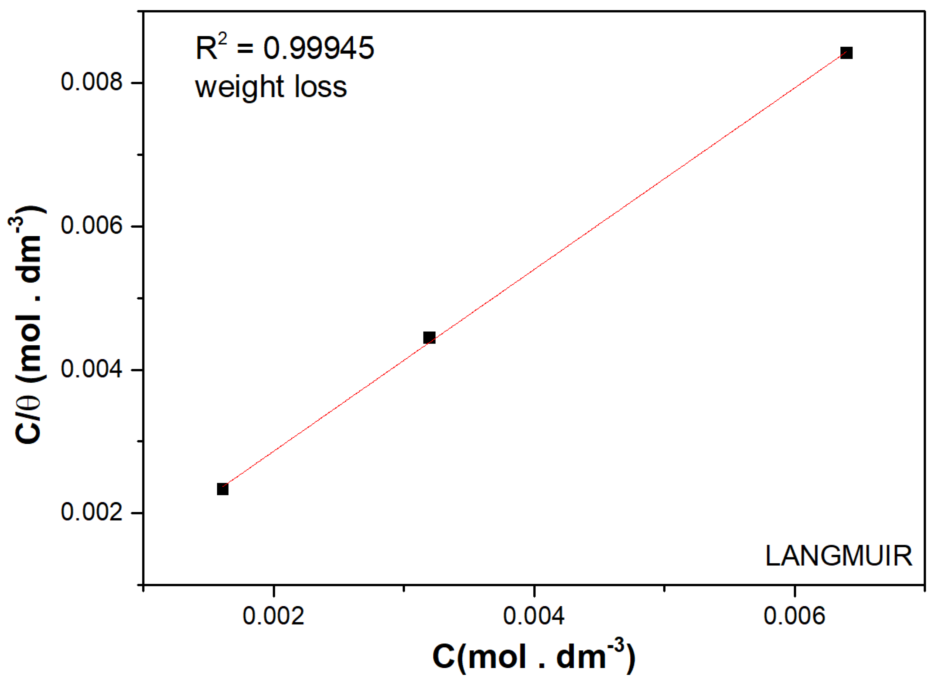
3.3. Adsorption Isotherms

Figure 6 shows that the studied amino acid presents a linear relationship between C / θ and concentration, indicating that substrate coverage increases proportionally with increasing inhibitor concentration. This result indicates that the amino acid follows the Langmuir adsorption model.
The degree of coverage values obtained from the mass loss are calculated using Equation (3) [48,49].
The Langmuir isotherm is based on the theory that all possible active adsorption sites are equivalent and that there is no interaction with the surroundings [3,50]:
K = θ C ( 1 θ )
where K is the adsorption equilibrium constant related to the Gibbs free energy that characterizes the interactivity between the surface of the material and the L-histidine molecules.
Figure 6 shows the plot of C / θ × C . Gibbs free energy was calculated using Equation (6):
Δ G a d s = R T l n ( C S o l v K )
where R is the universal gas constant; T is the temperature in Kelvin; C S o l v represents the molar concentration of the solvent which is 55.5 moles × dm−3 in the case of water. By applying the values obtained from the mass loss of Equation (6), it was possible to verify the value of Δ G a d s = −29 kJ/mol. In this way, L-histidine is physically adsorbed to the steel surface [51,52].

3.4. Electrochemical Tests

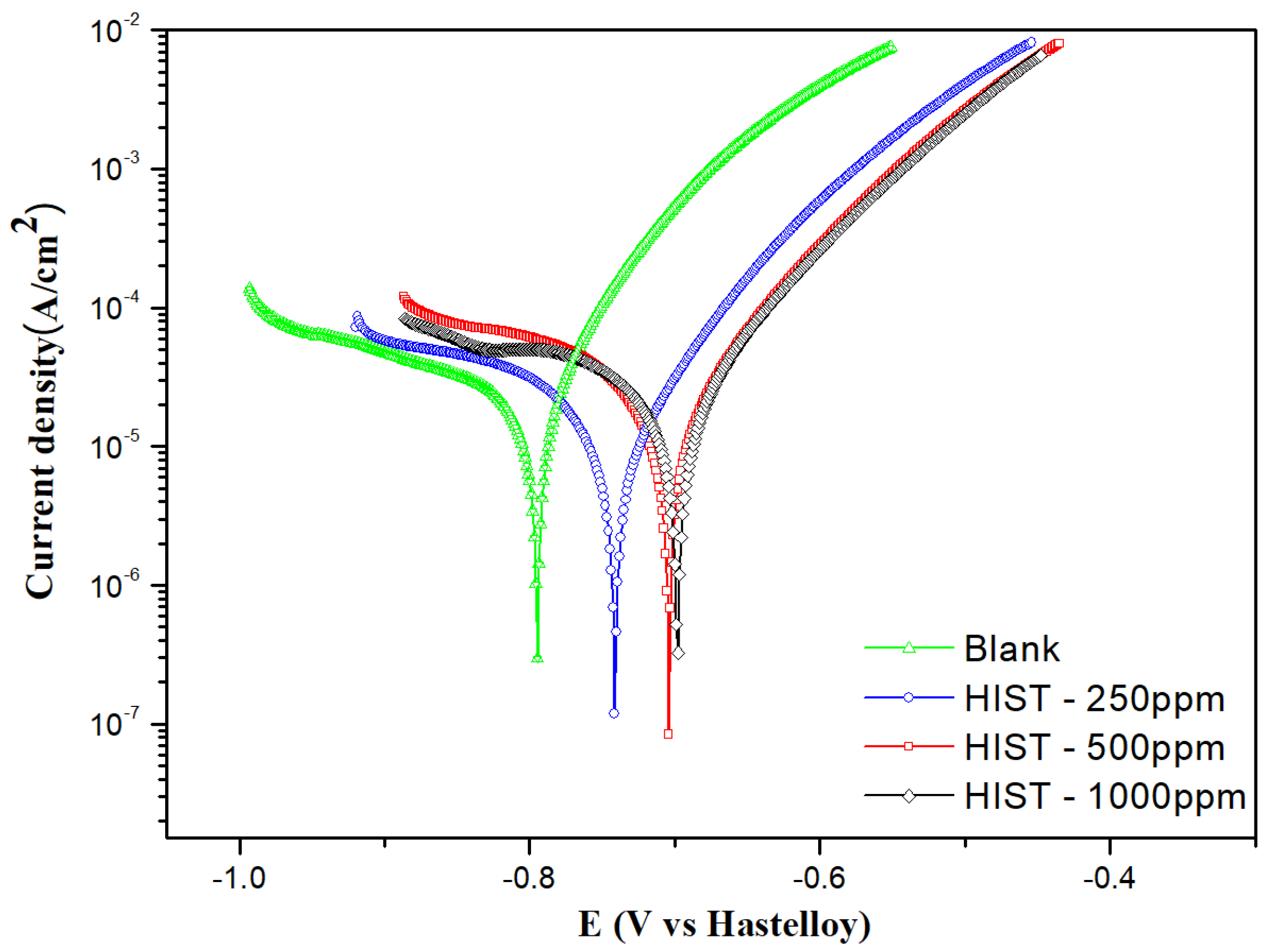
3.4.1. Linear Polarization

Linear polarization is also a technique used to determine the inhibition efficiency of corrosion inhibitors in metals and alloys in corrosive environments. Figure 7 shows the cathodic and anodic polarization curves obtained for carbon steel in a 3.5 wt.% NaCl solution at room temperature, both in the absence and presence of three L-histidine concentrations. The electrochemical parameters associated with this process, such as corrosion potential ( E c o r r ), corrosion current density ( i c o r r ), anodic and cathodic Tafel slopes ( b a e b c ), and polarization resistance ( R p ), were determined by extrapolating potentiodynamic curves using Nova 2.1.4 software. Additionally, inhibition efficiency ( η ) and surface coverage ( θ ) were calculated based on Equations (7) and (8), with values recorded in Table 2 [34,35,53].
η % = i c o r r 0 i c o r r i c o r r 0 × 100
θ = i c o r r 0 i c o r r i c o r r 0
where, i c o r r 0 and i c o r r are the current densities in the absence and presence of L-histidine.
Analyzing the data in Table 2, a decrease in the anodic Tafel slopes is observed, indicating the action of L-histidine as an anodic inhibitor, interfering with metal corrosion. Furthermore, the Tafel slopes do not vary significantly with increasing amino acid concentrations, suggesting that the inhibitor present in the test solution does not affect the process mechanism. It acts as an adsorptive inhibitor that slows down the anodic reaction by blocking active sites. Increasing the inhibitor concentration likely increases the number of inhibiting molecules at the metal–solution interface and reduces corrosion current densities, slightly shifting the corrosion potential to nobler values, as it is possible to observe that the current density decreases with increasing concentration. This inhibition process can be attributed to the adsorption of L-histidine molecules at active corrosion sites and/or the deposition of corrosion products on the surface of carbon steel. In summary, based on the Tafel polarization results, it is concluded that the inhibition mechanism involves blocking the surface of carbon steel by inhibitor molecules through adsorption [22,31,54,55].

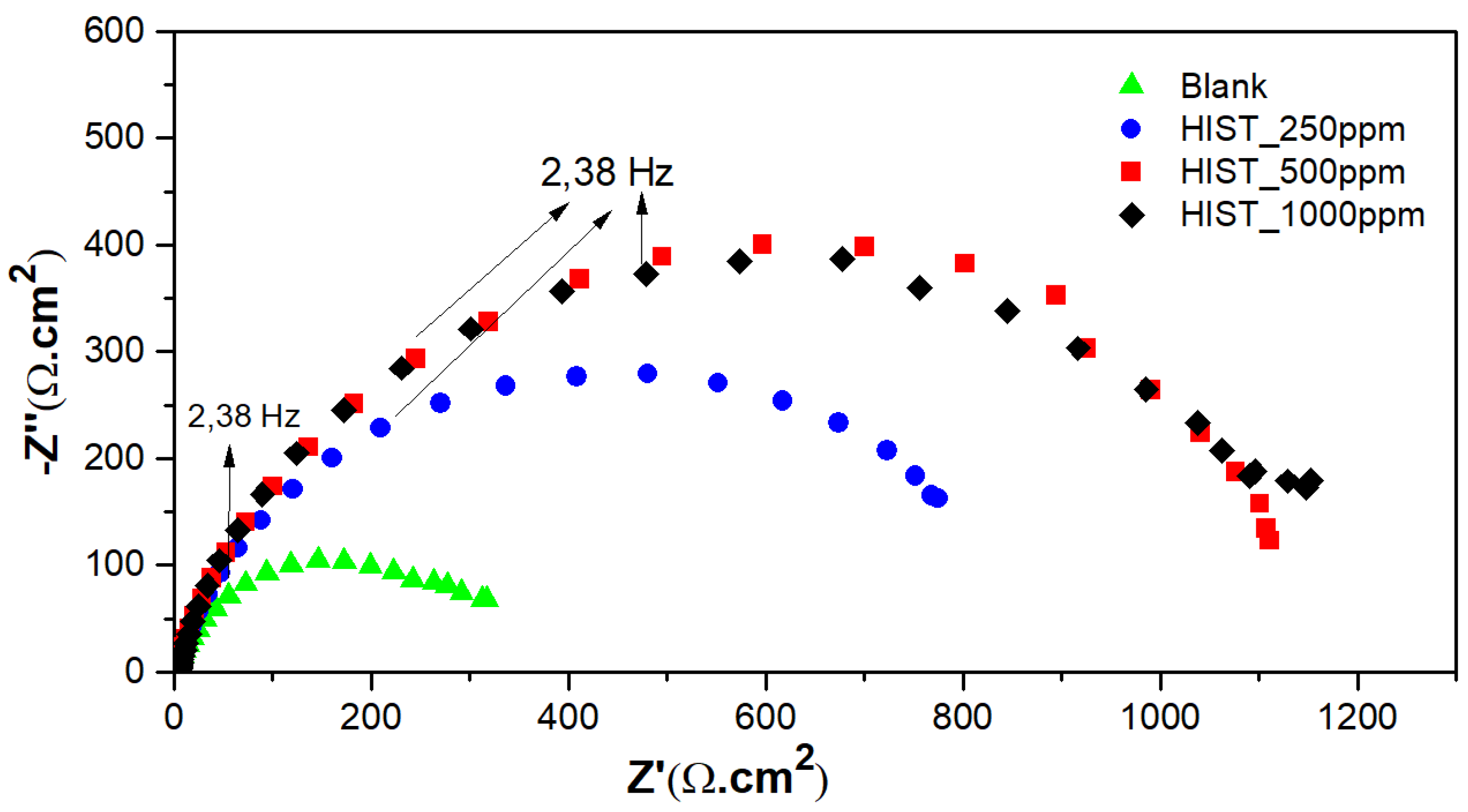
3.4.2. Electrochemical Impedance Spectroscopy (EIE)

From the electrochemical impedance spectroscopy essay, data regarding L-histidine inhibition were obtained and presented through Nyquist and Bode diagrams, as shown in Figure 8 and Figure 9, respectively.
The electrochemical impedance spectroscopy test obtained Nyquist curves, which appeared as capacitive semicircles in a high frequency range attributed to the relaxation of the double layer [55]. By extrapolating these semicircles to the real impedance axis, charge transfer resistance ( R c t ) values are obtained.
In this test, the R c t   value increased with increasing inhibitor concentration. However, the values were not expressive of concentrations from 500 to 1000 ppm. This phenomenon also occurred in potentiodynamic polarization and mass loss tests [22,48].
For the Bode curves presented in Figure 9, it is possible to observe the same behavior in the impedance modulus at high frequencies for the three curves. These impedance modulus values are related to charge transfer resistance. It was noted that with increasing concentrations, the impedance modulus values increase, confirming the data obtained from the Nyquist diagram.
Figure 10 shows a simple equivalent electrical circuit (EEC) is just a model proposal to model the experimental data, analogous to this work. It consists of a series connection between the solution resistance ( R S ) and a parallel connection between the double-layer capacitance and the charge transfer resistance ( R c t ). Typically, the capacitance is replaced by a constant phase element (CPE), which is closer to ideal behavior. The impedance is provided by:
Z C P E = 1 T ( j ω ) n
where T is the electrical constant of the CPE; ω is the angular frequency; j is the imaginary number; and n is the exponent of the CPE, which can vary from 0 to 1.
The inhibition efficiency values were calculated from the charge transfer resistance values, according to Equation (10):
η % = R c t , i n h R c t R c t , i n h × 100
where R c t and R c t , i n h represent the polarization resistance values in the absence and presence of the inhibitor. The inhibition efficiency values obtained presented in Table 3 are in agreement with the values calculated by polarization and weight loss [36,56].

3.5. UV–Vis Absorption Spectroscopy

The UV–Vis absorption spectra were obtained after the substrate immersion test in a medium of 3.5 wt.% NaCl in the presence and absence of the amino acid L-histidine. The spectra were recorded at amino acid concentrations ranging from 250, 500, and 1000 ppm. As shown in Figure 11, three low-intensity absorption bands were detected for a solution in the investigated spectral range containing only Fe2+ ions (lime green curve), centered at approximately 374, 423, and 525 nm. Fontana et al. [32] carried out an extensive investigation into the absorption spectrum of Fe2+ and Fe3+ species from different salts in an aqueous solution. They noted that the Fe2+ system absorption spectrum is independent of the anion. Furthermore, low-intensity spin-forbidden electronic transitions between 300 and 600 nm were observed, which have been rarely explored in the literature. For solutions containing only the amino acid in the corrosive medium (pink, burgundy, and orange curves), there are no electronic transitions in the range of 250–600 nm. In these spectra, there was abrupt absorption in the UV region after 250 nm, possibly due to the electronic transition between the molecular orbitals called HUMO (Highest Occupied Molecular Orbital) and LUMO (Lowest Unoccupied Molecular Orbital).
On the other hand, in solutions containing Fe2+ ions and the amino acid, an absorption band centered at 258 nm indicates the complexation between the Fe2+ ions and the amino acid. Two more absorption bands followed this electronic transition, forming a shoulder at 300 nm and another slightly discrete band at 350 nm, confirming the formation of the metal–amino acid complex. By increasing inhibitor concentrations in the corrosive medium, the metal–amino acid complex species concentration increased, as shown by an increase in band intensity, Figure 11. The formation of a Fe2+ complex with an amino acid has been reported in the literature. According to Li-Kuan et al. [33], the Fe(methionic)2 complex is formed by simultaneously bonding the Fe2+ ion with oxygen atoms in the carbonyl group and nitrogen atoms in the amine group of the amino acid, thus forming a bidentate chelate [38].

3.6. Surface Analyses

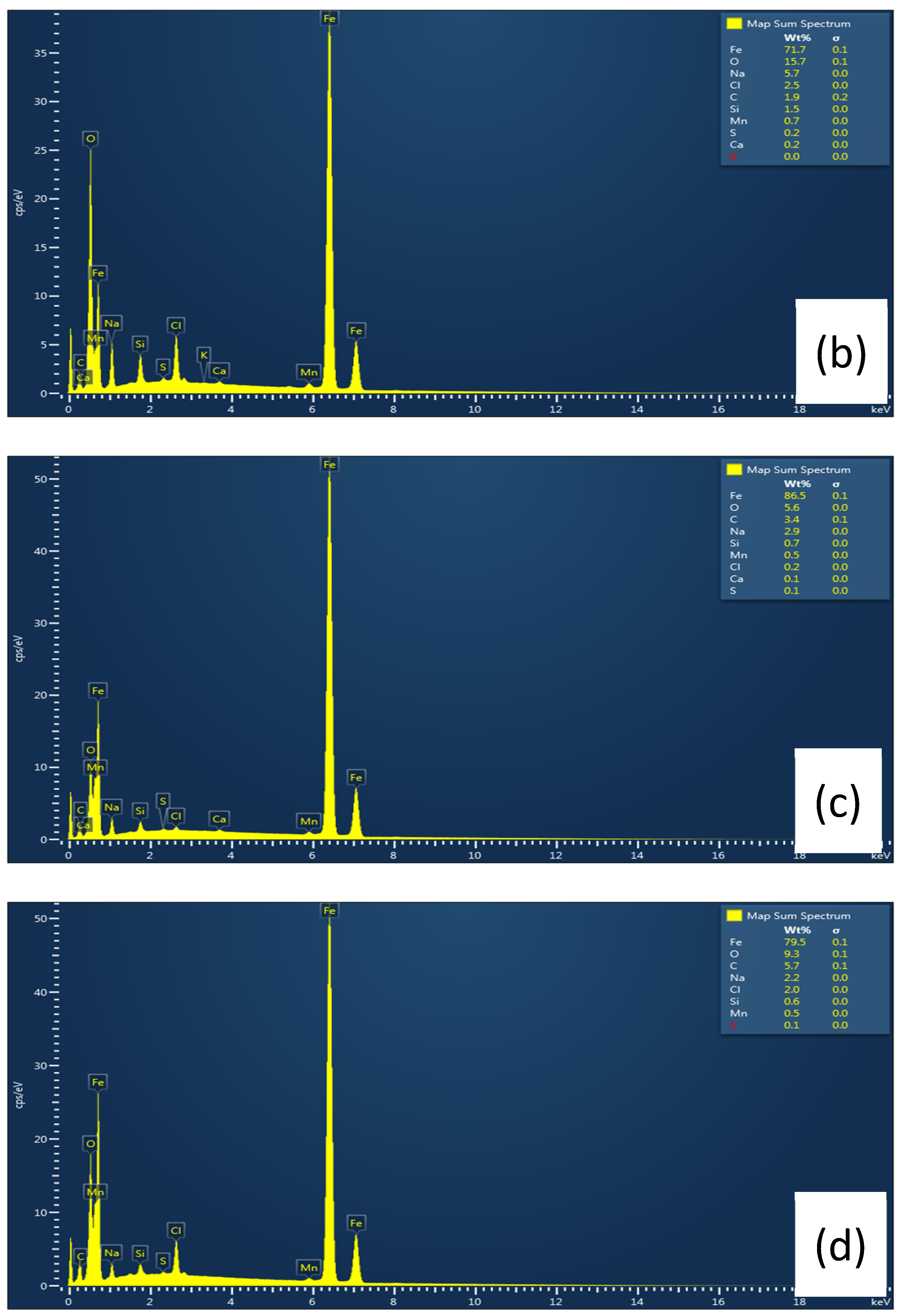
3.6.1. Scanning Electron Microscopy (SEM)

In the gravimetric test, the working electrode was immersed for 168 h in a NaCl solution without and with the addition of L-histidine. Immediately after this test, scanning electron microscopy (SEM) analysis was carried out. Figure 12 shows representative images of the sample submerged in a 3.5 wt.% NaCl solution without the presence of the inhibitor (a) and with the inhibitor at concentrations of 250, 500, and 1000 ppm, as illustrated in (b), (c), and (d), respectively. The sample without an inhibitor (white) had the most corroded surface. However, after the addition of L-histidine, this surface damage decreased with increasing amino acid concentration, which other authors also observed in acidic environments [20,22,31,55].
In Figure 13a, iron (Fe) and oxygen (O) peaks in the blank spectra indicate the formation of iron oxide/hydroxide on the surface of the steel sample. However, in the case of inhibited samples (b, c, and d) in the presence of L-histidine, a lower percentage of oxygen was found, indicating that the amino acid significantly reduced the formation of iron oxide as the concentration increased, thus acting as a corrosion inhibitor for carbon steel [57,58,59].

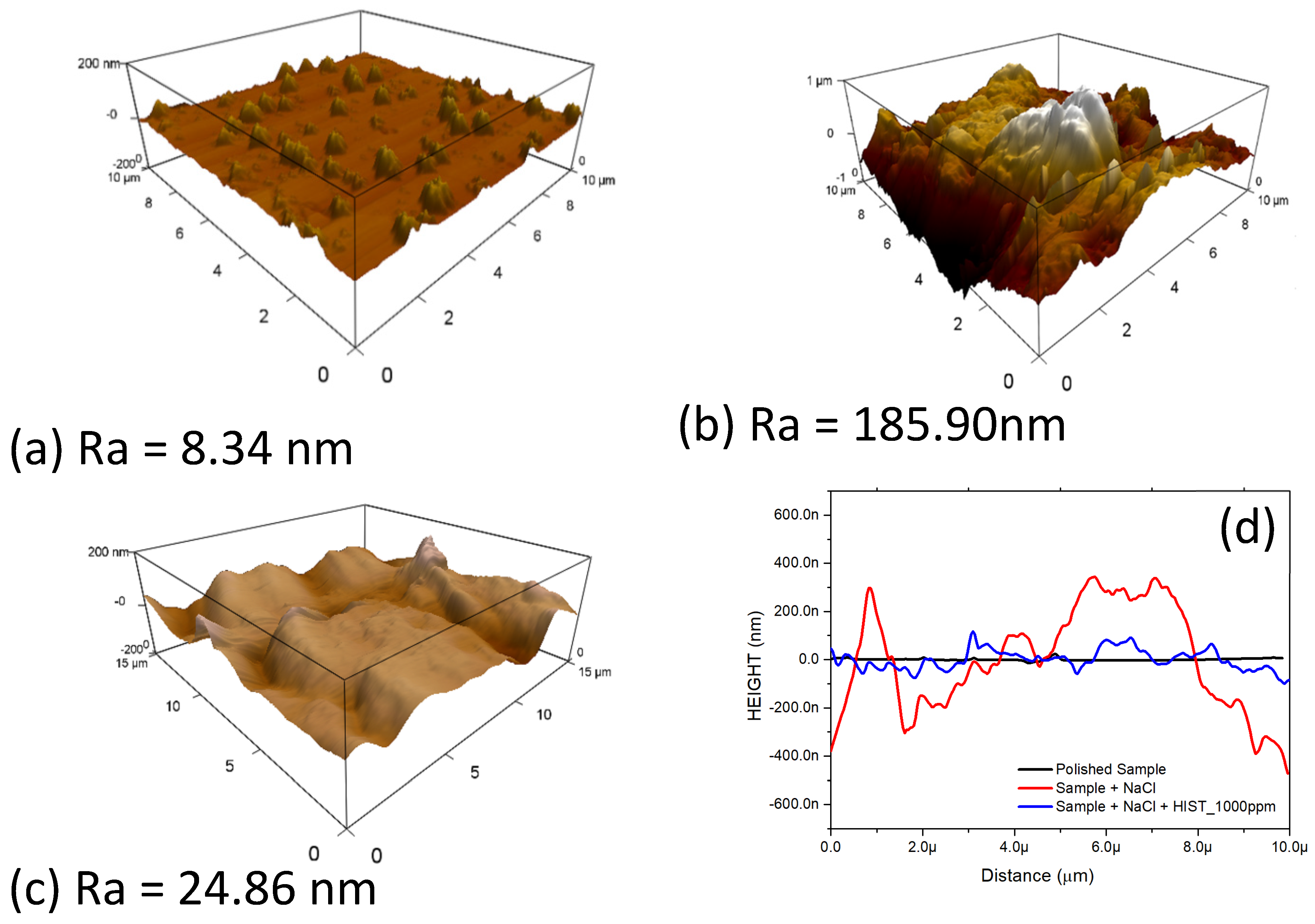
3.6.2. Atomic Force Microscopy (AFM)

Using atomic force microscopy, images of the topography of the sample surface were used to extract average roughness values from the work material, as illustrated in Figure 14, which shows (a) the polished sample; (b) the sample immersed in NaCl solution; (c) the sample immersed in NaCl solution with the addition of L-histidine, and (d) the height profile of each sample tested.
Figure 14a shows the height profile of the polished sample with an average roughness of 8.34 ± 3.38 nm. These values reveal few fluctuations between peaks and valleys on the sample surface. For the image presented in Figure 14b, the height profile of the sample immersed in NaCl solution can be observed without the addition of the inhibitor, with an average roughness of 185.9 ± 12.38 nm. The substrate showed severe corrosion on its surface with an accumulation of corrosion products, resulting in many spikes. Figure 14c shows the height profile of the sample in solution with 1000 ppm of L-histidine. In this condition, the roughness was 24.86 ± 7.06 nm, which may be due to the adsorption of L-histidine on the surface of the sample [60]. Figure 14d shows the Ra values for the three samples, which indicate that the sample in solution with the inhibitor is closer to the value of the polished sample without corrosion. Some researchers observed the same effect for other organic molecules on different substrates in acidic environments [40,61,62].

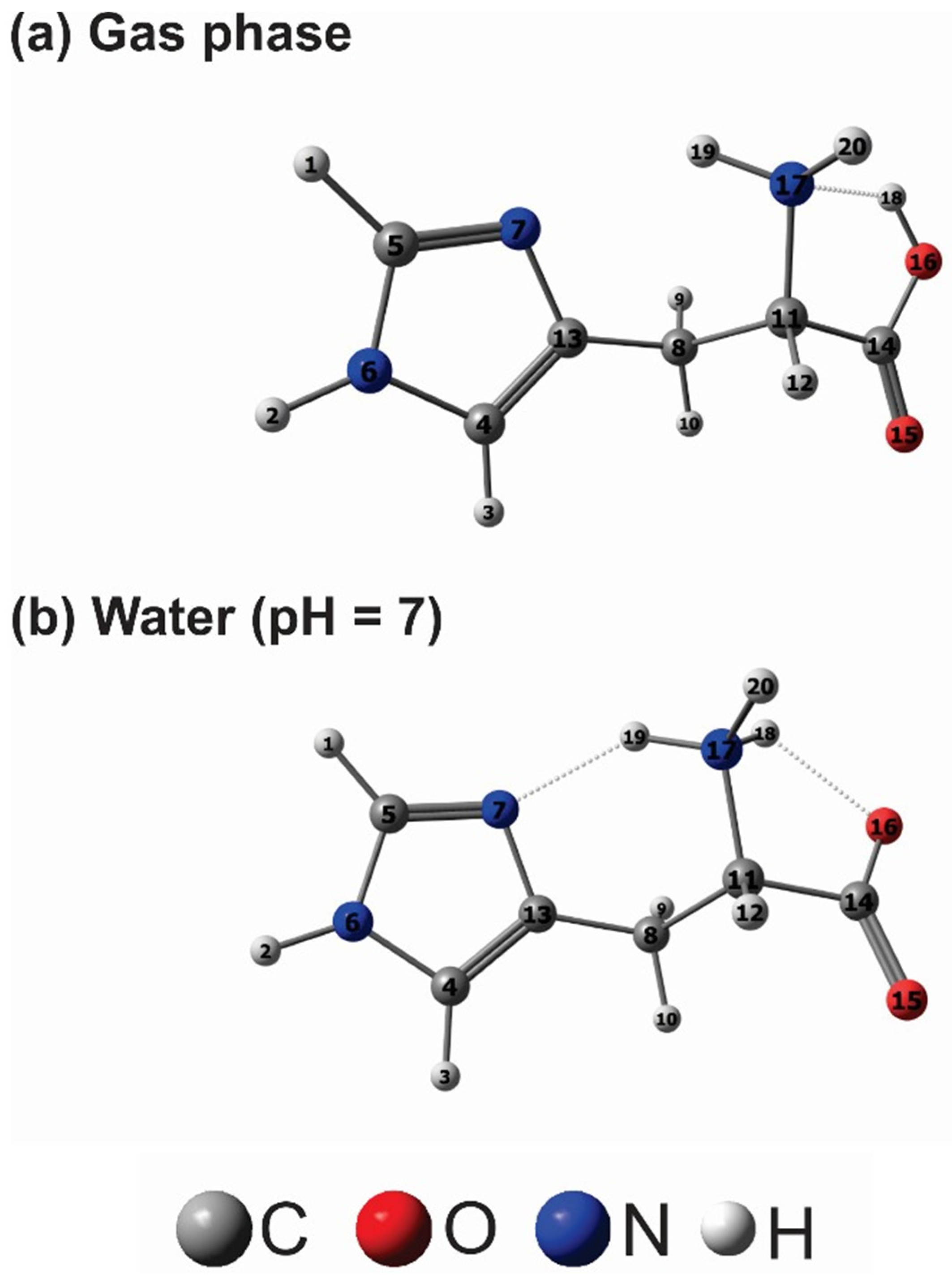
3.7. Theoretical Calculations

Structure 3 in Figure 15 represents the zwitterion structure of L-histidine at neutral pH, simulating the environment where the amino acid will act. The distribution of inhibitor microspecies was analyzed using the DFT method at the B3LYP/6-311++G(d,p) level, where it was possible to detect structural differences between the gas phase and the aqueous medium (Figure 16). Hydrogen bonding patterns varied accordingly, impacting molecule stability and reactivity, with water media exhibiting greater stability due to additional hydrogen bonds. Bond length disparities between gas and water phase structures were attributed to resonance effects in the carboxylate group, affecting molecule stability and reactivity. Figure 16 shows that the angle C8C11N7 changes about 2° from 111.218° (gas phase) to 109.716° (water media).
Frontier Molecular Orbitals (FMO) analysis presents similar electron density distributions over the imidazole ring and the carboxylic group, suggesting comparable interactions with the iron surface. However, the LUMO distribution varied, impacting electron acceptance. The HOMO–LUMO energy gap E G a p reflects a molecule’s tendency to donate or accept electronic density, with higher gaps indicating lower chemical reactivity. Table 4 suggests that the gas-phase histidine molecule is more reactive than its water counterpart, as expected due to the latter’s stabilization via two intramolecular hydrogen bonds. Gas-phase findings align closely with those of Fu et al. [21] and Kaya et al. [63], differing by approximately 0.60 eV from Zhang et al.’s [58] work. Conversely, the histidine molecule in water closely mirrors Kaya et al.’s [63] results, differing only by 0.00821 eV, showcasing the B3LYP method’s accuracy despite varying basis sets. Ionization potential (I), electron affinity (A), and electronegativity (χ) further elucidate molecular behavior, with gas-phase histidine exhibiting a higher electron affinity and electronegativity than its aqueous counterpart. Global hardness (η) and softness (S), influenced by the HOMO–LUMO energy gap, signify the molecule’s reactivity, with water-phase histidine demonstrating higher hardness. Electrophilic (ω) and nucleophilic indices (ε) differ slightly between gas phase and water media, indicating a varied electrophilic character while maintaining similar nucleophilic tendencies. Notably, both gas-phase and water media histidine molecules demonstrate a positive fraction of transferred electrons (∆N > 0) in Table 4, affirming their potential as corrosion inhibitors in neutral environments.

4. Conclusions

Weight loss measurements following immersion tests in chloride solution showed L-histidine’s corrosion inhibition efficiency, with inhibition efficiency increasing with inhibitor concentration. These findings are consistent with previous studies, reinforcing the compound’s potential as an effective corrosion inhibitor. Adsorption isotherms based on Langmuir models provided information about the adsorption behavior of L-histidine on the steel surface. The calculated Gibbs free energy indicated the inhibitor’s physical adsorption, further supporting its protective role against corrosion. The decrease in Tafel anodic inclinations and increase in charge transfer resistance with inhibitor concentration indicated that active corrosion sites on the steel surface were blocked. UV–Vis absorption spectroscopy provided additional evidence of the complexation between L-histidine and Fe2+ ions, revealing its corrosion inhibition mechanism at the molecular level. Surface analyses using scanning electron microscopy (SEM) and atomic force microscopy (AFM) visually confirmed the protective effects of L-histidine, with inhibited samples exhibiting reduced surface corrosion and smoother topographies compared to uninhibited samples. Theoretical calculations complement the experimental findings, providing information about L-histidine’s molecular structure and reactivity in the gas and aqueous phases. The analysis highlighted the compound’s potential as a corrosion inhibitor in neutral environments, supported by its electron transfer properties. In conclusion, this study comprehensively elucidated the corrosion inhibition properties of L-histidine and provided valuable information for its potential application in corrosion control strategies for carbon steel in chloride environments.

Author Contributions

Conceptualization, M.J.d.S.M. and W.S.A.; methodology, M.J.d.S.M., S.J.d.m.M., F.W.d.Q.A.N., L.P.M.d.S., S.L.S.M. and F.C.C.S.S.; software, F.W.d.Q.A.N. and P.d.L.N.; validation, M.A.C.F., G.F.R. and W.S.A.; formal analysis, M.J.d.S.M., G.F.R. and P.d.L.N.; investigation, M.J.d.S.M., F.C.C.S.S., R.B.V. and S.J.d.m.M.; resources, W.S.A., E.B.B., M.A.C.F. and G.F.R.; data curation, M.J.d.S.M., S.J.d.m.M., F.C.C.S.S., F.W.d.Q.A.N., L.P.M.d.S. and S.L.S.M.; writing—original draft preparation, M.J.d.S.M.; writing—review and editing, M.J.d.S.M., G.F.R., F.W.d.Q.A.N. and P.d.L.N.; visualization, M.J.d.S.M., G.F.R., M.A.C.F. and W.S.A.; supervision, W.S.A.; project administration, W.S.A., M.A.C.F. and G.F.R.; funding acquisition, W.S.A., M.A.C.F. and G.F.R. All authors have read and agreed to the published version of the manuscript.

Funding

This research was funded by the Coordenação de Aperfeiçoamento de Pessoal de Nível Superior—Brasil (CAPES)—Finance Code 001.

Data Availability Statement

The original contributions presented in the study are included in the article, further inquiries can be directed to the corresponding author.

Acknowledgments

The authors would like to acknowledge The Federal University of Ceará, to the Corrosion Research Laboratory LPC, the State University of Ceará (UFC), and the UFC Analytical Center.

Conflicts of Interest

The authors declare no conflict of interest. Francisco Carlos Carneiro Soares Salomão.

References

  1. Ma, F.; Zhang, Y.; Wang, H.; Li, W.; Hou, B. Inhibition behavior of chitooligosaccharide derivatives for carbon steel in 3.5% NaCl solution. Int. J. Electrochem. Sci. 2018, 13, 235–249. [Google Scholar] [CrossRef]
  2. Singh, P.; Bhrara, K.; Singh, G. Adsorption and kinetic studies of L-leucine as an inhibitor on mild steel in acidic media. Appl. Surf. Sci. 2008, 254, 5927–5935. [Google Scholar] [CrossRef]
  3. Ashassi-Sorkhabi, H.; Majidi, M.R.; Seyyedi, K. Investigation of inhibition effect of some amino acids against steel corrosion in HCl solution. Appl. Surf. Sci. 2004, 225, 176–185. [Google Scholar] [CrossRef]
  4. Ashassi-Sorkhabi, H.; Ghalebsaz-Jeddi, N. Inhibition effect of polyethylene glycol on the corrosion of carbon steel in sulphuric acid. Mater. Chem. Phys. 2005, 92, 480–486. [Google Scholar] [CrossRef]
  5. Bao, Q.; Zhang, D.; Wan, Y. 2-Mercaptobenzothiazole doped chitosan/11-alkanethiolate acid composite coating: Dual function for copper protection. Appl. Surf. Sci. 2011, 257, 10529–10534. [Google Scholar] [CrossRef]
  6. Cheng, S.; Chen, S.; Liu, T.; Chang, X.; Yin, Y. Carboxymenthylchitosan as an ecofriendly inhibitor for mild steel in 1 M HCl. Mater. Lett. 2007, 61, 3276–3280. [Google Scholar] [CrossRef]
  7. Shamsa, A.; Barker, R.; Hua, Y.; Barmatov, E.; Hughes, T.L.; Neville, A. Impact of corrosion products on performance of imidazoline corrosion inhibitor on X65 carbon steel in CO2 environments. Corros. Sci. 2021, 185, 109423. [Google Scholar] [CrossRef]
  8. Okafor, P.C.; Liu, X.; Zheng, Y.G. Corrosion inhibition of mild steel by ethylamino imidazoline derivative in CO2-saturated solution. Corros. Sci. 2009, 51, 761–768. [Google Scholar] [CrossRef]
  9. Kumar, D.; Jain, V.; Rai, B. Imidazole derivatives as corrosion inhibitors for copper: A DFT and reactive force field study. Corros. Sci. 2020, 171, 108724. [Google Scholar] [CrossRef]
  10. Haque, J.; Ansari, K.R.; Srivastava, V.; Quraishi, M.A.; Obot, I.B. Pyrimidine derivatives as novel acidizing corrosion inhibitors for N80 steel useful for petroleum industry: A combined experimental and theoretical approach. J. Ind. Eng. Chem. 2017, 49, 176–188. [Google Scholar] [CrossRef]
  11. Ashassi-Sorkhabi, H.; Shaabani, B.; Seifzadeh, D. Corrosion inhibition of mild steel by some schiff base compounds in hydrochloric acid. Appl. Surf. Sci. 2005, 239, 154–164. [Google Scholar] [CrossRef]
  12. Barreto, L.S.; Santos, A.M.; de Almeida, T.F.; Silva, D.R.; Cotting, F.; Capelossi, V.R. Application of a Mix of Vegetables Residues as Inhibitor for Carbon Steel. Mater. Res. 2022, 25, 1–10. [Google Scholar] [CrossRef]
  13. Furtado, L.B.; Leoni, G.B.; Nascimento, R.C.; Santos, P.H.C.; Brasil, S.L.D.C.; De Tecnologia, C.; De Janeiro, R.; De Tecnologia, C.; De Janeiro, R.; De Janeiro, R. Experimental and Theoretical Studies of Tailor-made Schiff Bases as Corrosion Inhibitors for Carbon Steel in HCl. Mater. Res. 2023, 26, e20220398. [Google Scholar] [CrossRef]
  14. El Ibrahimi, B.; Jmiai, A.; Bazzi, L.; El Issami, S. Amino acids and their derivatives as corrosion inhibitors for metals and alloys. Arab. J. Chem. 2017, 13, 740–771. [Google Scholar] [CrossRef]
  15. Ismail, K.M. Evaluation of cysteine as environmentally friendly corrosion inhibitor for copper in neutral and acidic chloride solutions. Electrochim. Acta 2007, 52, 7811–7819. [Google Scholar] [CrossRef]
  16. Oguzie, E.E.; Li, Y.; Wang, F.H. Effect of 2-amino-3-mercaptopropanoic acid (cysteine) on the corrosion behaviour of low carbon steel in sulphuric acid. Electrochim. Acta 2007, 53, 909–914. [Google Scholar] [CrossRef]
  17. Jafari, H.; Ameri, E.; Rezaeivala, M.; Berisha, A. Dimethylpropane-1,3-Diyl)Bis(Azanediyl)Bis(Methylene) Bis(2-Methoxyphenol) as New Reduced Form of Schiff Base for Protecting API 5L Grade B in 1 M HCl. Arab. J. Sci. Eng. 2023, 48, 7359–7372. [Google Scholar] [CrossRef]
  18. Özcan, M. AC impedance measurement of cystine adsorption at mild steel/sulfuric acid interface as corrosion inhibitor. J. Solid State Electrochem. 2008, 12, 1653–1661. [Google Scholar] [CrossRef]
  19. Navarrete, J. Electrochemical and XPS studies of decylamides of a-amino acids adsorption on carbon steel in acidic environment. Appl. Surf. Sci. 2006, 252, 2894–2909. [Google Scholar] [CrossRef]
  20. Ashassi-Sorkhabi, H.; Ghasemi, Z.; Seifzadeh, D. The inhibition effect of some amino acids towards the corrosion of aluminum in 1 M HCl + 1 M H2SO4 solution. Appl. Surf. Sci. 2005, 249, 408–418. [Google Scholar] [CrossRef]
  21. Li, J.F.S.; Wang, Y. Computational and electrochemical studies of some amino acid compounds as corrosion inhibitors for mild steel in hydrochloric acid solution. J. Mater. Sci. 2010, 45, 6255–6265. [Google Scholar] [CrossRef]
  22. Bobina, M.; Kellenberger, A.; Millet, J.P.; Muntean, C.; Vaszilcsin, N. Corrosion resistance of carbon steel in weak acid solutions in the presence of l-histidine as corrosion inhibitor. Corros. Sci. 2013, 69, 389–395. [Google Scholar] [CrossRef]
  23. Vasques, R.B.; Levy, M.M.; Wagner, F.; Almeida, D.Q.; Vaz, G.L.; Paes, L. A theoretical and experimental study of phosphate ester inhibitors for AISI 1018 in carbon dioxide—Saturated 3.5 wt% NaCl solution. Mater. Corros. 2021, 72, 1417–1432. [Google Scholar] [CrossRef]
  24. Saxena, A.; Prasad, D.; Haldhar, R. Investigation of Corrosion Inhibition Effect and Adsorption Activities of Achyranthes aspera Extract for Mild Steel in 0.5 M H2SO4. J. Fail. Anal. Prev. 2018, 18, 957–968. [Google Scholar] [CrossRef]
  25. Haldhar, R.; Raorane, C.J.; Mishra, V.K.; Periyasamy, T.; Berisha, A.; Kim, S.C. Development of different chain lengths ionic liquids as green corrosion inhibitors for oil and gas industries: Experimental and theoretical investigations. J. Mol. Liq. 2023, 372, 121168. [Google Scholar] [CrossRef]
  26. Solmaz, R.; Altunba, E.; Karda, G. Adsorption and corrosion inhibition effect of 2-((5-mercapto-1,3,4-thiadiazol-2-ylimino)methyl)phenol Schiff base on mild steel. Mater. Chem. Phys. 2011, 125, 796–801. [Google Scholar] [CrossRef]
  27. Kokalj, A. On the Use of the Langmuir and Other Adsorption Isotherms in Corrosion Inhibition. SSRN Electron. J. 2022, 217, 111112. [Google Scholar] [CrossRef]
  28. Niu, H.; Yang, H.; Zhao, R.; Tong, L.; Cristiani, P. Inhibition of carbonaceous matters adsorbing gold using a green and efficient chlorinating agent TCCA: Inhibition behavior, kinetics and isotherms, inhibition mechanism. Colloids Surf. A Physicochem. Eng. Asp. 2023, 675, 131836. [Google Scholar] [CrossRef]
  29. Chebabe, D.; Chikh, Z.A.; Hajjaji, N.; Srhiri, A.; Zucchi, F. Corrosion inhibition of Armco iron in 1 M, HCl solution by alkyltriazoles. Corros. Sci. 2003, 45, 309–320. [Google Scholar] [CrossRef]
  30. Hegazy, M.A.; Abdallah, M.; Awad, M.K.; Rezk, M. Three novel di-quaternary ammonium salts as corrosion inhibitors for API X65 steel pipeline in acidic solution. Part I: Experimental results. Corros. Sci. 2014, 81, 54–64. [Google Scholar] [CrossRef]
  31. Fawzy, A.; Abdallah, M.; Zaafarany, I.A.; Ahmed, S.A.; Althagafi, I.I. Thermodynamic, kinetic and mechanistic approach to the corrosion inhibition of carbon steel by new synthesized amino acids-based surfactants as green inhibitors in neutral and alkaline aqueous media. J. Mol. Liq. 2018, 265, 276–291. [Google Scholar] [CrossRef]
  32. Fontana, I.; Lauria, A.; Spinolo, G. Optical absorption spectra of Fe2+ and Fe3+ in aqueous solutions and hydrated crystals. Phys. Status Solidi Basic Res. 2007, 244, 4669–4677. [Google Scholar] [CrossRef]
  33. Wang, L.K.; Li, L.; Li, X.M.; Shi, Y.H.; Hu, L.; Le, G.W. Microwave assisted solid state reaction synthesis of methionine complexes of iron (II). Food Chem. 2008, 106, 315–323. [Google Scholar] [CrossRef]
  34. Ghasemi, Z.; Tizpar, A. The inhibition effect of some amino acids towards Pb-Sb-Se-As alloy corrosion in sulfuric acid solution. Appl. Surf. Sci. 2006, 252, 3667–3672. [Google Scholar] [CrossRef]
  35. Şahin, M.; Bilgiç, S.; Yilmaz, H. The inhibition effects of some cyclic nitrogen compounds on the corrosion of the steel in NaCl mediums. Appl. Surf. Sci. 2002, 195, 1–7. [Google Scholar] [CrossRef]
  36. Liang, Y.; Wang, D.; Wu, Y.; Lai, Q.; Xue, L.; Feng, S. Perylene-containing polysiloxane: An effective candidate for corrosion protection of iron surface. Appl. Surf. Sci. 2011, 257, 10576–10580. [Google Scholar] [CrossRef]
  37. Macdonald, J.R.; Barsoukov, E. Theory, Experiment, and Applications, 2nd ed.; John Wiley & Sons, Inc.: Hoboken, NJ, USA, 2005. [Google Scholar]
  38. Dhas, S.A.M.B.; Natarajan, S. Growth and characterization of two new NLO materials from the amino acid family: L-Histidine nitrate and l-Cysteine tartrate monohydrate. Opt. Commun. 2008, 281, 457–462. [Google Scholar] [CrossRef]
  39. Térmica, A.; L-histidina, T.A.D.C.; Medeiros, J.E. Pereira Completion of Course Work: Análise Térmica e Espectroscopia Raman à Temperatura Ambiente de Cristais L-Histidina Departamento de Física Centro de Ciências Universidade Federal do Ceará, (2012). Available online: http://repositorio.ufc.br/handle/riufc/31441 (accessed on 27 May 2024).
  40. Fouda, A.S.; Ismail, M.A.; Elewady, G.Y.; Abousalem, A.S. Evaluation of 4-amidinophenyl-2,2′-bithiophene and its aza-analogue as novel corrosion inhibitors for CS in acidic media: Experimental and theoretical study. J. Mol. Liq. 2017, 240, 372–388. [Google Scholar] [CrossRef]
  41. Becke, A.D. Density functional thermochemistry. I. The effect of the exchange only gradient correction. J. Chem. Phys. 1992, 96, 2155–2160. [Google Scholar] [CrossRef]
  42. Cancès, E.; Mennucci, B.; Tomasi, J. A new integral equation formalism for the polarizable continuum model: Theoretical background and applications to Isotropic and anisotropic dielectrics. J. Chem. Phys. 1997, 107, 3032–3041. [Google Scholar] [CrossRef]
  43. Vinayagamoorthy, R.; Irudayaraj, A.A.; Raj, A.D.; Karthick, S.; Jayakumar, G. Comparative Study of Properties of L-Histidine and L-Histidine Nickel Nitrate Hexahydrate Crystals Grown by Slow Evaporation. Mech. Mater. Sci. Eng. J. 2017, 9, hal-01503639. [Google Scholar] [CrossRef]
  44. Wu, J.; Wu, J.; Lu, L.; Mei, P. Design, characteristics, and theoretical analyses of 8-hydroxyquinoline derivatives with different heteroatoms as effective corrosion inhibitors. Mater. Chem. Phys. 2023, 304, 127929. [Google Scholar] [CrossRef]
  45. Alnajjar, A.O.; El-lateef, H.M.A.; Khalaf, M.M.; Mohamed, I.M.A. Steel protection in acidified 3.5% NaCl by novel hybrid composite of CoCrO3/polyaniline: Chemical fabrication, physicochemical properties, and corrosion inhibition performance. Constr. Build. Mater. 2022, 317, 125918. [Google Scholar] [CrossRef]
  46. Kalantar, M.A.A.M. Thermal Analysis on the Milled AlB2O3 + Si + WO3 System to Synthesize Al2O3-WxSiy-WxBy Powders. Mater. Res. 2020, 23, 1–9. [Google Scholar] [CrossRef]
  47. Behpour, M.; Ghoreishi, S.M.; Soltani, N.; Salavati-Niasari, M.; Hamadanian, M.; Gandomi, A. Electrochemical and theoretical investigation on the corrosion inhibition of mild steel by thiosalicylaldehyde derivatives in hydrochloric acid solution. Corros. Sci. 2008, 50, 2172–2181. [Google Scholar] [CrossRef]
  48. da Silva Hernandes, I.; da Cunha, J.N.; Santana, C.A.; Rodrigues, J.G.A.; D’Elia, E. Application of an aqueous extract of cotton seed as a corrosion inhibitor for mild steel in HCl media. Mater. Res. 2020, 24, 1–10. [Google Scholar] [CrossRef]
  49. Bentiss, F.; Traisnel, M.; Gengembre, L.; Lagrenée, M. New triazole derivative as inhibitor of the acid corrosion of mild steel: Electrochemical studies, weight loss determination, SEM and XPS. Appl. Surf. Sci. 1999, 152, 237–249. [Google Scholar] [CrossRef]
  50. Agarwal, V.K. Langmuir-Blodgett Films. Phys. Today 1988, 41, 40–46. [Google Scholar] [CrossRef]
  51. Silbey, R.J.; Alberty, R.A.; Bawendi, M.G. Physical Chemistry, 4th ed.; John Wiley & Sons, Inc.: Hoboken, NJ, USA, 2004. [Google Scholar]
  52. Paramasivam, S.; Kulanthai, K.; Sadhasivam, G.; Subramani, R. Corrosion Inhibition of Mild Steel in Hydrochloric Acid using4- (pyridin-2yl)-N-p-tolylpiperazine-1-carboxamide. Int. J. Electrochem. Sci. 2016, 11, 3393–3414. [Google Scholar] [CrossRef]
  53. Feng, L.; Zhang, S.; Zhou, Y.; Pan, R.; Du, H.; Liu, F.; Yang, Y. Expired Glucosamine Drugs as Green Corrosion Inhibitors for Carbon Steel in H2SO4 Solution and Synergistic Effect of Glucosamine Molecules with Iodide Ions: Combined experimental and theoretical investigations. Crystals 2023, 13, 205. [Google Scholar] [CrossRef]
  54. da Rocha, J.C. Obtenção de Inibidores de Corrosão a Partir de Extratos de Produtos Naturais. Ph.D. Thesis, Universidade Federal do Rio de Janeiro, Rio de Janeiro, Brazil, 2016; 106p. [Google Scholar]
  55. Zhang, Q.H.; Li, Y.Y.; Lei, Y.; Wang, X.; Liu, H.F.; Zhang, G.A. Comparison of the synergistic inhibition mechanism of two eco-friendly amino acids combined corrosion inhibitors for carbon steel pipelines in oil and gas production. Appl. Surf. Sci. 2022, 583, 152559. [Google Scholar] [CrossRef]
  56. Macdonald, J.R. Impedance Spectroscopy Theory, Experiment, and Applications; John Wiley & Sons, Inc.: Hoboken, NJ, USA, 2005. [Google Scholar]
  57. Zhao, P.; Liang, Q.; Li, Y. Electrochemical, SEM/EDS and quantum chemical study of phthalocyanines as corrosion inhibitors for mild steel in 1 mol/L HCl. Appl. Surf. Sci. 2005, 252, 1596–1607. [Google Scholar] [CrossRef]
  58. Zhang, L.; Zhang, S.; He, Y.; Yang, R.; Ma, L.; Xia, Y.; He, Z. Histidine and histidine dimer as green inhibitors for carbon steel in 3wt% sodium chloride solution; Electrochemical, XPS and quantum chemical calculation studies. Int. J. Electrochem. Sci. 2018, 13, 2136–2153. [Google Scholar] [CrossRef]
  59. Sherif, E.S.M.; Ahmed, A.H. Alleviation of iron corrosion in chloride solution by n,n′-bis[2-methoxynaphthylidene]amino]oxamide as a corrosion inhibitor. Crystals 2021, 11, 1516. [Google Scholar] [CrossRef]
  60. Chauhan, D.S.; Ansari, K.R.; Sorour, A.A.; Quraishi, M.A.; Lgaz, H.; Salghi, R. Thiosemicarbazide and thiocarbohydrazide functionalized chitosan as ecofriendly corrosion inhibitors for carbon steel in hydrochloric acid solution. Int. J. Biol. Macromol. 2018, 107, 1747–1757. [Google Scholar] [CrossRef] [PubMed]
  61. He, X.; Mao, J.; Ma, Q.; Tang, Y. Corrosion inhibition of perimidine derivatives for mild steel in acidic media: Electrochemical and computational studies. J. Mol. Liq. 2018, 269, 260–268. [Google Scholar] [CrossRef]
  62. Rabizadeh, T.; Asl, S.K. Casein as a natural protein to inhibit the corrosion of mild steel in HCl solution. J. Mol. Liq. 2019, 276, 694–704. [Google Scholar] [CrossRef]
  63. Kaya, S.; Tüzün, B.; Kaya, C.; Obot, I.B. Determination of corrosion inhibition effects of amino acids: Quantum chemical and molecular dynamic simulation study. J. Taiwan Inst. Chem. Eng. 2016, 58, 528–535. [Google Scholar] [CrossRef]
Figure 1. Molecular formula of L-histidine (devised by the author).
Crystals 14 00703 g001
Figure 2. FT-IR analysis of L-histidine.
Crystals 14 00703 g002
Figure 3. The structural formula of the amino acid between positive and negative charges (isoelectronic or zwitterionic).
Crystals 14 00703 g003
Figure 4. L-histidine TGA curves.
Crystals 14 00703 g004
Figure 5. Mass loss of AISI 1018 carbon steel without and with L-histidine at concentrations of 250, 500, and 1000 ppm in a medium of 3.5 wt.% NaCl.
Crystals 14 00703 g005
Figure 6. Langmuir adsorption isotherm for L-histidine obtained by weight loss.
Crystals 14 00703 g006
Figure 7. Polarization curves of AISI 1018 carbon steel in a 3.5 wt.% NaCl solution, in the absence and presence of L-histidine in various concentrations.
Crystals 14 00703 g007
Figure 8. Electrochemical impedance spectroscopy of AISI 1018 carbon steel in a 3.5 wt.% NaCl solution without and with L-histidine in various concentrations.
Crystals 14 00703 g008
Figure 9. Bode diagrams—impedance modulus of AISI 1018 carbon steel in a solution of 3.5% by weight of NaCl, in the absence and presence of L-histidine at various concentrations.
Crystals 14 00703 g009
Figure 10. Equivalent electrical circuit (ECC).
Crystals 14 00703 g010
Figure 11. Absorption spectrum in the ultraviolet and visible regions for solutions after the immersion test in the presence and absence of the amino acid L-histidine at various concentrations.
Crystals 14 00703 g011
Figure 12. SEM images obtained for carbon steel samples: immersion in a 3.5 wt.% NaCl solution for 168 h without and with the addition of L-histidine. (a) Without the inhibitor and (bd) with the addition the inhibitor of 250, 500, and 1000 ppm, respectively.
Crystals 14 00703 g012
Figure 13. EDS obtained for carbon steel samples: immersion in a 3.5 wt.% NaCl solution, for 168 h, without and with the addition of L-histidine. (a) Without the inhibitor and (bd) with the addition the inhibito of 250, 500, and 1000 ppm, respectively.
Crystals 14 00703 g013aCrystals 14 00703 g013b
Figure 14. AFM surface topography of the mild steel specimen. (a) The as-polished sample; (b) the sample in a NaCl solution; (c) the sample in NaCl + Lhistidine 1000 ppm; (d) height profiles of the tested samples.
Crystals 14 00703 g014
Figure 15. Microspecies distribution with different values of pH for histidine molecules.
Crystals 14 00703 g015
Figure 16. Optimized molecular geometries at the B3LYP/6-311++G(d,p) computational level for the histidine molecule (a) in the gas phase, and (b) in water as an implicit solvent.
Crystals 14 00703 g016
Table 1. The inhibition efficiency, surface coverage obtained, and corrosion rate by weight loss measurements.
L-Histidine (ppm)η (%)θ (Surface Coverage)CR (mm/y)
0------0.8888 ± 0.091
250830.83 ± 0.090.1306 ± 0.076
500870.87 ± 0.070.1183 ± 0.084
1000880.88 ± 0.040.1022 ± 0.096
Table 2. Parameters of polarization curves, in pH 7 and pH 4 media of 3.5 wt.% NaCl solutions with and without inhibitor.
L-Histidine conc. (ppm) E c o r r (V) i c o r r (A/cm2) b a (mV/dec) b c (mV/dec)Rct
(Ωcm2)
θ η (%)
0−0.794 ± 0.04(3.39 ± 0.83) × 10−568 6239-------
250−0.741 ± 0.06(3.99 ± 0.36) × 10−6425210,1850.882288.22
500−0.704 ± 0.10(3.41 ± 0.19) × 10−6474999630.899589.95
1000−0.698 ± 0.07(3.16 ± 0.18) × 10−6243296680.906890.68
Table 3. Electrochemical impedance spectroscopy parameters in the media of solutions 3.5 wt.% NaCl with and without an inhibitor.
L-Histidine (ppm)Rct (Ωcm2)η%
0336 ± 53
250895 ± 5162
5001119 ± 3869
10001181 ± 4572
Table 4. The histidine molecule’s quantum reactivity descriptors (HOMO and LUMO) in the gas phase, water media, and compared to previous studies.
Quantum Reactivity DescriptorGas PhaseWaterHistidine
[21]
Histidine
[63]
Histidine
[63]
Histidine
[58]
HOMO   energy   ( E H O M O /eV)−6.8268−6.7014−6.367−6.72725−6.71337−6.700
LUMO   energy   ( E L U M O /eV)−0.7606−0.2952−0.272−0.88955−0.31538−0.094
Energy   gap   ( Δ E G a p /eV)6.06636.40626.0955.837706.397996.600
Ionization Potential (I/eV)6.82686.70146.36706.727256.713376.700
Electron Affinity (A/eV)0.76060.29520.27200.889550.315380.094
Electronegativity (χ/eV)3.79373.49833.31953.808403.514383.397
Global Hardness (η/eV)3.03313.20313.04752.918853.199005.200
Global Softness (σ/eV−1)0.32970.31220.32810.342600.312600.303
Electrophilicity index (ε/eV)2.37251.91041.80792.48451.93041.747
Nucleophilicity index (ω/eV−1)0.42150.52350.55310.40250.51800.573
Fraction of electrons transferred (∆N)0.5285440.5466110.57820.5467220.5448000.540
Ref. [21]: B3LYP/6-311G(d,p) in the gas phase. ∆N is related to copper surfaces. Ref. [63]: B3LYP/6-31++G(d,p) in the gas phase and water, respectively. ∆N is related to copper surfaces. Ref. [58]: B3LYP-D3/6-31G(d,p) in the gas phase. ∆N is related to an iron surface.
Disclaimer/Publisher’s Note: The statements, opinions and data contained in all publications are solely those of the individual author(s) and contributor(s) and not of MDPI and/or the editor(s). MDPI and/or the editor(s) disclaim responsibility for any injury to people or property resulting from any ideas, methods, instructions or products referred to in the content.

