Article

Investigations of Some Disordered Quaternary Compounds in the Systems Ag/Pb/Sb/Se and Ag/Pb/Sb/Te

1
Institute for Inorganic Chemistry and Crystallography, Faculty of Chemistry and Mineralogy, University of Leipzig, Johannisallee 29, 04103 Leipzig, Germany
2
Department of Earth System Sciences, Faculty of Mathematics, Informatics and Natural Sciences, Hamburg University, Grindelallee 48, 20146 Hamburg, Germany
*
Author to whom correspondence should be addressed.
Crystals 2024, 14(9), 789; https://doi.org/10.3390/cryst14090789
Submission received: 8 August 2024 / Revised: 30 August 2024 / Accepted: 2 September 2024 / Published: 5 September 2024
(This article belongs to the Section Inorganic Crystalline Materials)

Abstract

:
Electrical and thermal transport measurements on quenched NaCl-type Ag1/3Pb1/3Sb1/3Se reveal an n-type semiconductor with a Seebeck coefficient up to −140 μVK−1 and a thermal conductivity as low as 0.52 WmK−1. Short-range order is indicated by disorder diffuse scattering in electron diffraction patterns. In contrast, 4L-Ag0.61Pb1.79Sb2.61Se6 (space group Cmcm with a = 4.2129(1) Å, b = 13.852(1) Å, and c = 20.866(1) Å, Z = 4) features the first lillianite-type structure in the system Ag/Pb/Sb/Se. It consists of slab-like NaCl-type building blocks that are interconnected via trigonal [PbSe6] prisms. As such structures typically do not form with Te as an anion, the first “sulfosalt-like” compound, Ag0.38Pb0.25Sb2.38Te4, in the system Ag/Pb/Sb/Te forms a layered tetradymite-like structure (space group R 3 - m with a = 4.2887(1) Å, c = 41.544(1) Å, Z = 3). Its slabs, which are separated by van der Waals gaps, are built up from three layers of distorted [MTe6] octahedra. Crystals of Ag0.38Pb0.25Sb2.38Te4 were grown by chemical transport.

1. Introduction

The pseudobinary system AgSbTe2/PbTe features very efficient thermoelectric materials called LAST. Thermoelectric performance is quantified by the dimensionless figure of merit, zT = S2σT/κ, calculated from the Seebeck coefficient S, the electrical conductivity σ, the temperature T, and the thermal conductivity κ. AgPb18SbTe20, for instance, reaches zT values up to 2.2 at 527 °C [1]. This material and similar ones exhibit highly disordered NaCl-type structures and pronounced real structure effects, which involve low intrinsic thermal conductivity [2,3]. In contrast to a plethora of studies focusing on the system Pb/Ag/Sb/Te, much less effort has been devoted to the analogous quaternary system Pb/Ag/Sb/Se. However, it is possible to partially substitute Te with Se, as demonstrated by AgPb18SbTe20−xSex (0 ≤ x ≤ 15) obtained by high-pressure synthesis [4]. Further NaCl-type compounds such as PbSe [5], PbTe [6], and AgSbTe2 [7] also represent well-known materials used for thermoelectric power generation. AgInSe2-doped PbSe, for instance, reaches zT = 1.9 at 600 °C and SrTe-doped PbTe even reaches zT = 2.5 at 650 °C [8,9]. At a rather low temperature of 300 °C, Cd-doped AgSbTe2 reaches a maximal zT value of 2.6, which has been attributed to optimized electronic states and reduced thermal conductivity due to nano-scaled superstructures [10]. NaCl-type PbSe doped with Sb has been observed in isolated nanocrystals of PbmSb2Sem+3 (2 ≤ m ≤ 8) [11]. Defect variants like Pb0.95(Sb0.033□0.017)Se and Pb0.9955Sb0.0045Se reach zT values up to 0.9 at 550 °C and 1.1 at around 650 °C, respectively [12,13]. Related multinary cubic chalcogenides, AgPbBiCh3 (Ch = S, Se, Te), possess low thermal conductivities of <1.3 W/mK [14]. AgSnSbSe1.5Te1.5 and iodine-doped AgPbBiSe3 reach maximum zT values of 1.1 at 450 °C and 0.8 at 541 °C, respectively [15,16]. With mixed anions, n-type AgBiPbSe2S reaches zT = 0.6 at 550 °C, which can be increased to 1.2 by Ag2Se precipitates [17]. The related p-type AgMnSbTe3 reaches zT = 1.5 at 550 °C [18]. CuSnSbS3, CuSnSbSe3, AgPbSbSe3, AgSnSbSe3, CuSnAsSe3 and AgPbAsSe3 have, in addition, been identified as intriguing low-temperature ferroelectric semiconductors [19]. In this line, these compounds have the potential to become high-ohmic resistors with time-dependent resistivity that may be interesting for cryoelectronics [20]. However, compared to the well-investigated cubic phases AgSnSbSe3 and AgPbSbTe3 [2], the Pb/Se variant AgPbSbSe3 [15,21] lacks deeper crystal-chemical and thermoelectric characterization.
In addition to NaCl-type phases, the system Ag/Pb/Sb/Te also features ternary layered compounds with layered structures that crystallize as variants of the tetradymite (Bi2Te2S) structure type [22], such as Pb0.18Sb7.82Te3 [23], Ag0.05Sb1.95Te3 [24], Ag0.24Sb10.76Te4 [25], PbSb2Te4 or PbSb4Te7 [26]. The tetradymite structure type consists of distorted NaCl-type slabs that are stacked along a threefold axis [22]. These slabs are separated by van der Waals gaps between terminating Te atom layers. Such layered tellurides include highly efficient thermoelectric materials, the most prominent example being nanostructured Bi0.5Sb1.5Te3 featuring dislocation arrays, which reaches zT = 1.9 at 47 °C [27]. Furthermore, tetradymite-like compounds are considered a model system for the development of topological insulators with high bulk resistivity and increased electrical conductivity on the surface since pronounced spin–orbit coupling determines the electronic structures of the surface as well as the bulk [28].
In contrast to the NaCl-type and layered structures typical for tellurides, the ternary system Pb/Sb/Se is characterized by several sulfosalt-like structures that contain tilted and interconnected cut-outs of the NaCl-type, which form complex arrangements that define various structure families. Selenides are often isostructural to their sulfide analogs. Pb2Sb2Se5, for instance, is related to the mineral cosalite, Pb2Bi2S5 [29,30]. Further sulfosalt-like compounds include Pb4Sb6Se13 and Pb6Sb6Se17, which are selenium analogs of robinsonite, Pb4Sb6S13, and moëloite, Pb6Sb6S17, respectively [31,32,33,34]. The structure of PbSb2Se4, which has no reported sulfur analog, consists of ribbon-like entities that are interconnected via trigonal prisms occupied by a mixture of Sb and Pb [35]. A homologous series with numerous representatives of various compositions and element combinations—although not Pb/Sb/Se so far—is derived from the mineral lillianite (Pb3Bi2S6) [36]. Its crystal structure can be described by tilted, distorted NaCl-type slabs that are interconnected by cations in bicapped trigonal prisms of S atoms [37,38]. The structures are classified by the maximal number of edge-sharing octahedra across the slab, resulting in symbols like NL or N1,N2L when both slabs have different numbers of octahedra [39,40,41]. Taking into account the abundance of the elements they contain, lillianite-type structures such as 4,5L-Pb7Bi4Se13 exhibit promising thermoelectric properties, with zT reaching a peak value of 0.9 at 502 °C [42]. Ga doping further improved this peak zT value up to 1.35 at 527 °C, which is the highest reported zT value for a lillianite-type compound [43].
This study presents fundamental research on the structural and thermoelectric characterization of Ag1/3Pb1/3Sb1/3Se as well as the structure elucidation of two new compounds in the systems Ag/Pb/Sb/Ch (Ch = Se, Te) by means of synchrotron radiation.

2. Materials and Methods

2.1. Sample Preparation

Ag1/3Pb1/3Sb1/3Se was prepared by sealing stoichiometric amounts of elemental Ag (99.99%, Chempur, Karlsruhe, Germany), Pb (99.99%, Alfa Aesar, Thermo Fisher Scientific, Waltham, MA, USA), Sb (99.9999%, VEB Spurenelemente Freiberg, Freiberg, Germany), and Se (99.999%, Haines & Maassen, Bonn, Germany) in silica glass ampules under vacuum (~0.1 Pa). These samples were heated to 450 °C for 24 h and then to 950 °C for 48 h; they were subsequently quenched at air by removing the ampule from the furnace and allowing it to cool. A few crystals of Ag0.61Pb1.79Sb2.61Se6 were found as a byproduct of this synthesis.
Ag0.38Pb0.25Sb2.38Te4 was obtained in syntheses with a nominal composition of Ag1/3Pb1/3Sb1/3Te starting from elemental Ag, Pb, Sb (see above for sources), and Te (99.999%, Alfa Aesar, Thermo Fisher Scientific, Waltham, MA, USA) sealed in silica glass ampules under vacuum (~10−3 mbar). After heating to 450 °C for 24 h and to 1000 °C for 48 h, the samples were quenched at air. For a chemical transport reaction, 300 mg of the powdered product was loaded into a silica glass ampule, 20 cm in length and 1.5 cm in diameter, together with ~5 wt.-% I2 (99.998%, Ferak, Berlin, Germany, dried over conc. H2SO4). While cooling the end with the starting materials with liquid nitrogen, the ampule was sealed at a pressure of ~10−3 mbar and then placed into a two-zone furnace with a temperature gradient from 650 °C to 580 °C for 7 d. Several crystals up to 300 μm in size formed at the cold side of the ampule.

2.2. Powder X-ray Diffraction and Rietveld Refinements

A Huber G670 diffractometer (Huber Diffraktionstechnik GmbH & Co., KG, Rimsting, Germany) was used to collect powder X-ray diffraction (PXRD) data with Cu-Kα1 radiation (λ = 1.54056 Å, Ge(111) monochromator). The device features Guinier geometry with an image plate detector and an integrated read-out system. The ingots were thoroughly ground in an agate mortar, dispersed in ethanol, and fixed on Mylar foils with hair spray. Rietveld refinements were carried out with TOPAS using a fundamental parameter approach to describe peak profiles [44]. High-temperature (HT) PXRD data were recorded using a Rigaku (Japan) SmartLab powder diffractometer equipped with an Anton Paar HTK 1200 N high-temperature chamber and a HyPix-3000 two-dimensional semiconductor detector using Cu-Kα1 radiation. In order to prevent oxidation, the powder was filled into silica glass capillaries (WJM-Glas Müller GmbH, Berlin, Germany) and sealed under dry argon.

2.3. Single-Crystal X-ray Diffraction

Single-crystal X-ray diffraction (SCXRD) data of Ag1/3Pb1/3Sb1/3Se were collected using an STOE STADIVARI diffractometer (STOE & Cie GmbH, Darmstadt, Germany) utilizing a microsource (AXO, Dresden, Germany) with Ag-Kα radiation (λ = 0.56086 Å) and a Dectris Pilatus 300K detector (Dectris AG, Baden-Dättwil, Switzerland). Data reduction and semiempirical absorption correction based on equivalent intensities were performed with the software package X-Area [45,46]. The data sets of Ag0.38Pb0.25Sb2.38Te4 and Ag0.61Pb1.79Sb2.61Se6 were obtained with synchrotron radiation at the beamline P24 of DESY (Hamburg, Germany), utilizing a Huber four-circle kappa diffractometer (λ = 0.500 Å) equipped with a Pilatus CdTe 1M detector (Dectris AG). Integration was carried out using the CrysAlisPro software package [47] and semiempirical absorption correction was performed with SADABS [48]. Structure solution and refinement were performed using the SHELX software package [49,50]. Crystal structures were visualized with Diamond [51]. Bond-valence sum (BVS) calculations were performed with VaList [52]. Bond length values were taken from SCXRD analyses. Crystal data and refinement results are summed up in Table 1.

2.4. Electron Microscopy and Energy-Dispersive X-ray Spectroscopy

A LEO 1530 Gemini (Carl Zeiss AG, Oberkochen, Germany) scanning electron microscope (SEM) equipped with an energy-dispersive X-ray (EDX) detector (model 7426, Oxford Instruments Ltd., Abingdon, UK) was used to perform chemical analysis and to record backscattered and secondary electron images at an acceleration voltage of 20 kV. EDX spectra of the investigated crystals were evaluated with the INCA software package [53].
A Jeol JEM-2100Plus (JEOL Ltd., Tokyo, Japan) equipped with an LaB6 cathode (200 kV, point resolution of 0.23 nm) was used to obtain high-resolution transmission electron microscopy (HRTEM) images, selected-area electron diffraction (SAED) patterns and EDX spectra using an EDAX TEAM Enhanced system with an Octane T Optima 30 (AMETEK, Inc., Mahwah, NJ, USA). The associated software was used to quantify the EDX spectra of the investigated microcrystals [54]. The jEMS software package was used to calculate simulated HRTEM images (multislice method) and SAED patterns (kinematic approximation) [55,56]. Fourier filtering was applied with DigitalMicrograph [57]. Crystallites of Ag1/3Pb1/3Sb1/3Se were prepared by crushing the material under ethanol and transferring the suspension onto a lacey carbon copper grid (PLANO GmbH, Wetzlar, Germany).

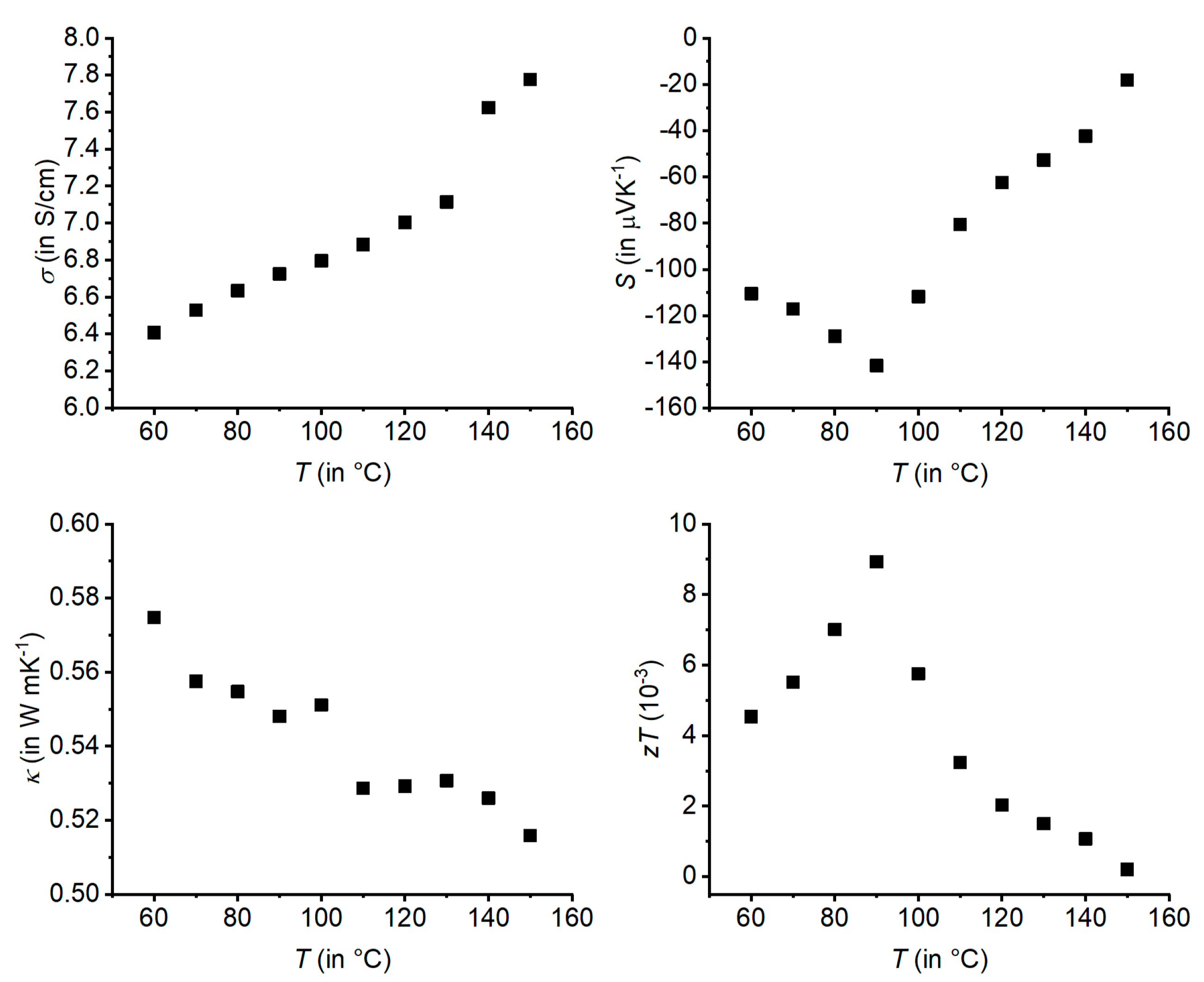
2.5. Transport Properties

Compact bulk samples for the measurement of physical properties were polished into plane parallel shapes using SiC powder with a 6.5 µm grain size and cut into pieces using a diamond wire saw (Well Diamantdrahtsägen GmbH, Mannheim, Germany). The electrical conductivity and the Seebeck coefficient were measured with an LSR-3 (Linseis Messgeräte GmbH, Selb, Germany) equipped with Ni electrodes and NiCr/Ni thermocouples in a bipolar mode, i.e., continuously switching polarity. The relative uncertainties for both values are <10% of the measured values. The thermal diffusivity Dth was measured using a laser flash apparatus (LFA-1000, Linseis Messgeräte GmbH, Selb, Germany) with an InSb detector. Dusza’s model was used for finite-pulse and heat-loss corrections [58]. Archimedes’ method was used for determining the density of Ag1/3Pb1/3Sb1/3Se: ρ = 7.17 g cm−2 corresponds to 99.5% of the X-ray density. The heat capacity Cp = 0.22 J/gK was calculated according to the Dulong–Petit approximation, which seems reasonable, as the measured Cp ≈ 0.27 J/gK of related AgSbSe2 matches the Dulong–Petit value of Cp = 0.26 J/gK [59]. Systematic errors fall within the scope of the precision of measurements, showing uncertainties of <10%. The thermal conductivity was calculated according to κ = CpDthρ and the phononic part of κ according to κph = κtotalLσT. The Lorenz number L can be estimated as L (in 10−8 WΩK−2) ≈ 1.5 + exp(−|S|/116 µVK−1) [60]. Physical measurements were performed under He atmosphere up to 150 °C with a heating rate of 10 K min−1.

3. Results and Discussion

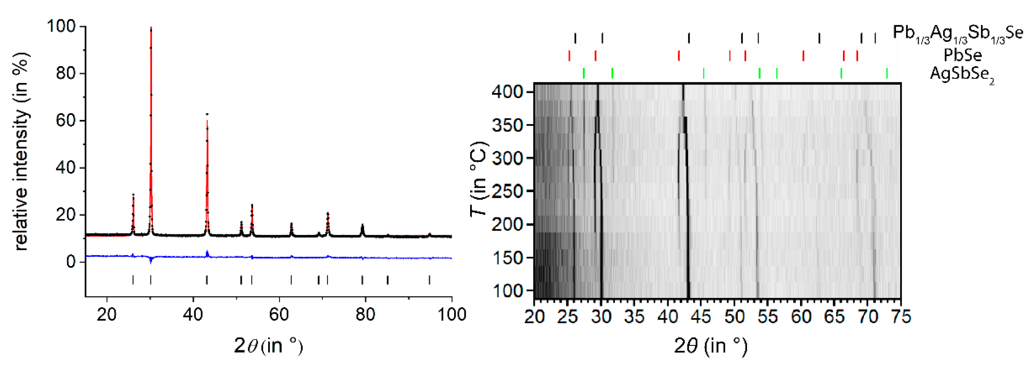
3.1. Ag1/3Pb1/3Sb1/3Se

Originally aiming at the synthesis and characterization of the physical properties of the Se analog of the mineral freieslebenite (AgPbSbS3), which adopts a monoclinic sulfosalt-like structure [61], the analysis of melt-quenched samples with the nominal composition AgPbSbSe3 revealed a cubic phase that corresponds to the disordered NaCl-type model derived from PXRD data in the literature [19]. A Rietveld refinement (Figure 1, left) shows only the cubic phase; further information is given in Tables S1 and S2 (S denotes tables and figures in the Supplementary Materials). The byproduct discussed in Section 3.2. is only present in very small amounts that cannot be detected by PXRD. HTPXRD (Figure 1, right) shows that Ag1/3Pb1/3Sb1/3Se slowly decomposes into PbSe and AgSbSe2 above 150 °C; at an average heating rate of 13 K/min, the decomposition is not complete when the phase apparently forms again at about 375 °C.
The SEM-EDX analysis of the investigated single crystal (Figure S1) yielded the following composition (in at. %, details in Table S3): Ag 18.4(7), Pb 15.9(7), Sb 18.4(6), Se 47.2(7). This result is in good agreement with the nominal composition (Ag 16.7, Pb 16.7, Sb 16.7, Se 50.0), which was, therefore, fixed in the single-crystal structure refinement. Consistent with PXRD, Ag1/3Pb1/3Sb1/3Se adopts a disordered NaCl-type structure with a mixed occupation of the cations Ag, Pb and Sb as shown in Figure 2. Single-crystal data are listed in Table 1, and atomic parameters are given in Table S4. The average cation–anion distance is 2.96 Å. This lies between the interatomic distances in the corresponding binary selenides (Ag2Se: ≥2.65 Å [62]; PbSe: 3.06 Å [63], and Sb2Se3: ≥2.67 Å [64]) but is mainly determined by the largest atom Pb. TEM investigations of Ag1/3Pb1/3Sb1/3Se (crystallite depicted in Figure S2, EDX analysis in Table S5: Ag 14.9(9), Pb 14.9(4), Sb 15.1(5), Se 55.1(9) at. %) show disorder diffuse scattering (DDS) in SAED patterns recorded along zone axes [131] and [212] (Figure 2), indicating a certain degree of short-range order of the cations, possibly associated with tilting of coordination polyhedra and static displacement of the constituent atoms.
Since the detected DDS is very weak, a full interpretation was not attempted. Further SAED patterns, including tilt series as well as experimental and additional simulated HRTEM images, are shown in Figures S3–S5.
Due to the observed decomposition (see above), transport properties were measured only up to 150 °C (Figure 3). The negative values of the Seebeck coefficient S indicate n-type conduction. The electrical conductivity σ increases almost linearly with temperature up to 130 °C, where a slight discontinuity can be observed. The absolute values of σ are rather low but still much higher than the data reported in the literature [19]. This may be due to different synthesis procedures. The discontinuity at 130 °C can also be noticed in the slopes of S and the thermal conductivity κ as functions of temperature. Additional pronounced discontinuities occur in the curves of S and κ at ~90 °C, where the initial decrease in S reverses into an increase, and the declining thermal conductivity shows a sudden drop. These unexpected discontinuities in physical properties probably suggest that the decomposition of Ag1/3Pb1/3Sb1/3Se into PbSe and AgSbSe2 might start at even lower temperatures than 150 °C as indicated by HTPXRD. In total, while exhibiting a reasonable absolute value of S and a low κ, Ag1/3Pb1/3Sb1/3Se features a low zT value of <0.009 due to its low electrical conductivity σ. The bandgap estimated from S by the Goldsmid–Sharp relation is ~0.1 eV [65].

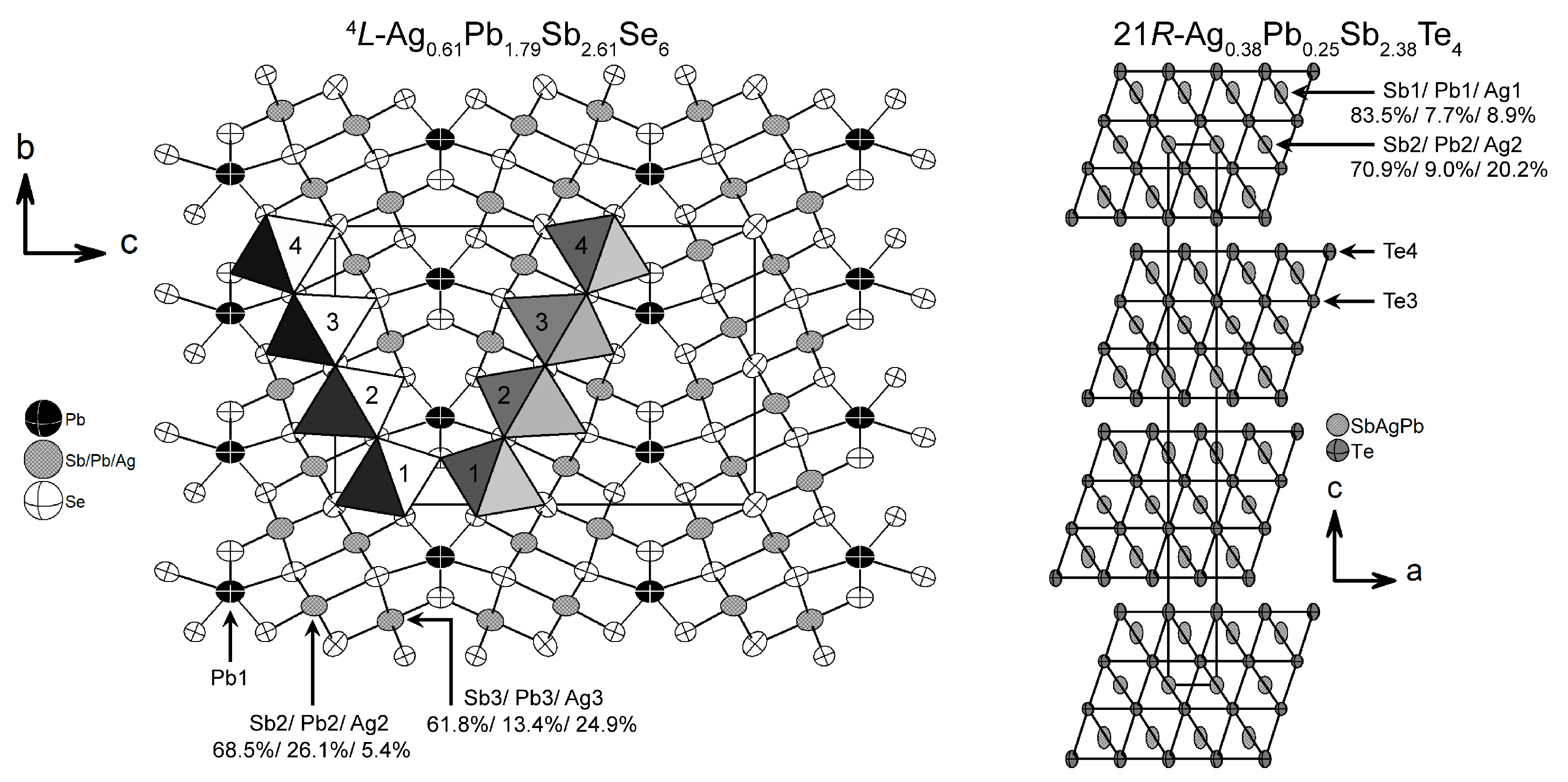
3.2. Ag0.61Pb1.79Sb2.61Se6

In products of syntheses aiming at hypothetical monoclinic Ag1/3Pb1/3Sb1/3Se as described above, SEM-EDX investigations of a single crystal revealed (Figure S6) the following composition (in at. %, details in Table S6): Ag 3.5(4), Pb 14.9(7), Sb 26.1(8), Se 55.5(9). This result is in agreement with the composition derived from SCXRD data: Ag 5.5, Pb 16.3, Sb 23.7, Se 51.5. The crystal structure of Ag0.61Pb1.79Sb2.61Se6 was determined using synchrotron data. The structure solution was straightforward. Atoms sharing one Wyckoff position were refined using equal coordinates and displacement parameters. Electron density and BVS calculations (Table 2) [52,66,67] suggest that the cation site Pb1 is solely occupied by Pb atoms. Although a tentative refinement of the respective site occupancy may indicate a very small amount of vacancies (s.o.f. for Pb: 0.959(2)) or elements with fewer electrons, Pb1 was assumed to be fully occupied in the final refinement. Further cation sites were assumed to be occupied by all three cations, Ag/Pb/Sb, which also improved R values significantly. To avoid insignificant overpopulation, the sums of the s.o.f. values of Ag2/Pb2/Sb2 and Ag3/Pb3/Sb3 were constrained to 1. Without impact on R values or significant changes in site occupancies, the refinement was constrained to yield a charge-balanced sum formula. The result Ag0.61Pb1.79Sb2.61Se6 is in good agreement with the SEM-EDX analysis described above. The crystallographic data for Ag0.61Pb1.79Sb2.61Se6 are listed in Table 1, and the atomic parameters are given in Table S7.
The crystal structure of Ag0.61Pb1.79Sb2.61Se6 can be derived from that of the mineral lillianite (Pb3Bi2Se6) [37]. The space group Cmcm is the typical one. No indications for lower symmetry were found, although variants in Cmc21 have also been reported [68,69,70]. A projection of the crystal structure with highlighted and numbered octahedra, labeled cation sites, and the respective site occupancies is depicted in Figure 4. As outlined in the introduction, it can be classified as a 4,4L type (also known as 4L when centrosymmetric) by counting the edge-sharing octahedra within one slab-like entity. These slabs are interconnected by bicapped trigonal prisms [39] centered by Pb1. The interatomic distances d(Pb–Se) in this prism range from 2.91 to 3.41 Å. This corresponds to typical distances of 2.8 Å ≲ d(Pb–Se) ≲ 3.7 Å in sulfosalt-like compounds that contain at least three of the respective elements [71]. Further mixed cation sites are octahedrally coordinated with little distortion. Such pronounced cation disorder, sometimes including vacancies, is known from related compounds such as 8,8L-Ag5Pb9Bi19Se40 and 5,5L-AgPb3Bi7Se14 [72]. The evaluation of resonant X-ray diffraction data could precisely quantify the amount of vacancies in lillianite-type 7,7L-Sn3.6Bi3.6Se9 and 4,7L-Sn11.49Bi12.39Se30 [69].

3.3. Ag0.38Pb0.25Sb2.38Te4

Exploratory syntheses in the system Ag/Pb/Sb/Te aimed at phases that might not be stable at high temperatures and thus not accessible by melt synthesis. Starting from the nominal composition, Ag1/3Pb1/3Sb1/3Te, the initial product was subjected to a chemical transport reaction using iodine as a transport agent. The detailed reaction conditions are listed in Section 2.5. Crystals formed at the cold side of the ampule. Attempts to obtain homogeneous bulk samples of Ag0.38Pb0.25Sb2.38Te4 by melt synthesis have failed so far. Annealing at temperatures up to 450 °C did not lead to improvement. According to PXRD, such samples consisted of a GeAs2Te4-type phase and a NaCl-type phase.
Diffraction data from a crystal obtained by a transport reaction were collected using synchrotron radiation at beamline P24 at DESY (details cf. Section 2.3). Neglecting a number of weak reflections, integration based on the unit cell of the GeAs2Te4 structure type [73] seemed straightforward using a single-crystal approach. The structure solution confirmed the layered GeAs2Te4 structure type. Anion positions were assumed to be occupied by Te atoms. As Sb and Te exhibit little scattering contrast, antisite disorder could not be taken into account but seems unlikely. Mixed cation positions Ag1/Pb1/Sb1 and Ag2/Pb2/Sb2 were refined using equal coordinates and displacement parameters for each position with an additional constraint that ensures charge neutrality. As the total occupancies of both sites were 1 within standard deviations, full occupancy was assumed in order to reduce the number of refined parameters. The final refinement yielded the formula Ag0.38Pb0.25Sb2.38Te4. Crystallographic data are given in Table 1. Information on Wyckoff positions, atomic coordinates, site occupancy factors, and anisotropic displacement parameters are listed in Table S8 in the Supporting Information.
The crystal structure of Ag0.38Pb0.25Sb2.38Te4 consists of NaCl-type slabs stacked along [001]. The mixed cation positions Ag/Pb/Sb are coordinated in a distorted octahedral fashion by Te atoms. These [MTe6] octahedra form triple layers separated by van der Waals gaps as shown in Figure 4. The 21R stacking sequence is typical for the GeAs2Te4 structure type [73]. The element distribution on the two cation sites is significantly different. SbIII is enriched on the site near the van-der-Waals-like gap as its higher formal charge (vs. PbII or AgI) better compensates the negative charge of terminal Te layers that are coordinated only from one side. This phenomenon is in agreement with the atom distribution in related layered tellurides [25,74,75]. The Ag1/Pb1/Sb1 site next to the terminating Te atoms shows a slightly elongated displacement ellipsoid, which is consistent with both static displacements due to the chemical disorder and with dynamic vibrations in the octahedron, whose distortion is more pronounced than that of the more symmetrical one around Ag2/Pb2/Sb2. The distances d(Te–Te) = 3.59 Å around the van der Waals gap of the title compound agree well with comparable distances in related structures such as Sb2Te3 (3.74 Å) and 42R-Sb4Te3 (3.42 Å) [76,77]. The cation–anion distances of 2.958, 3.075, and 3.301 Å in Ag0.38Pb0.25Sb2.38Te4 agree with those in structurally related Sb2Te3 with d(Sb–Te) values of 2.979 and 3.168 Å [77], and in AgSbTe2 with d(Sb–Te) values of 3.034 and 3.033 Å [2]. The calculated BVS values [52,66,67] of cations in Ag0.38Pb0.25Sb2.38Te4 (Table 2) are based on averaged bond lengths and are thus of limited value; however, they confirm mixed sites with a low Pb content.
The SEM-EDX analysis of the investigated crystal (Figure S7) indicated a composition [in at. %: Ag 2.1(6), Pb 2.0(3), Sb 47.9(7), Te 48.0(4), Table S9] that deviates from the results of the charge-neutral sum formula derived from the single-crystal refinement [calculated for Ag0.38Pb0.25Sb2.38Te4, in at. %: Ag 5.4, Pb 3.5, Sb 34.0, Te 57.1]. This can be explained by epitaxial intergrowth with a crystal of Sb4Te3. The weak reflections in the diffraction pattern, which were not taken into account in the structure determination, correspond to Sb4Te3 [76]. The corresponding unit cell can index these additional reflections, but their intensities were too low to consider them in a multi-crystal approach. Reconstructed reciprocal space sections 0kl can be explained by a superposition of patterns calculated for Ag0.38Pb0.25Sb2.38Te4 and Sb4Te3 (Figure S8, further reciprocal space sections in Figure S9). Both structures crystallize in the space group R 3 - m and share metric and crystal-chemical similarities. The lattice parameters a of both crystal structures are almost equal (4.275 Å). The lattice parameters, c, however, differ: 83.56 Å for Sb4Te3 [76] vs. 41.544 Å for Ag0.38Pb0.25Sb2.38Te4. The intergrown antimony-rich Sb4Te3 explains the large amount of Sb detected by SEM-EDX (Table S9).

4. Conclusions

Cutouts from the NaCl type correspond to building blocks of various sulfosalt-like compounds, which often exhibit complex crystal structures. This study presents a disordered NaCl-type phase Ag1/3Pb1/3Sb1/3Se, as well as Ag0.61Pb1.79Sb2.61Se6, which is built up from corresponding cutouts. TEM investigations on Ag1/3Pb1/3Sb1/3Se revealed weak disorder diffuse scattering that indicated a certain degree of short-range order. Ag1/3Pb1/3Sb1/3Se is an n-type semiconductor with a Seebeck coefficient of up to −140 μVK−1 at 90 °C and a thermal conductivity as low as 0.52 WmK−1 at 150 °C. Its low electrical conductivity does not allow good thermoelectric properties. Doping with Te, for instance, might increase the electrical conductivity without changing the structure and lead to higher zT values. Ag0.61Pb1.79Sb2.61Se6 forms a 4L lillianite-type structure that consists of tilted and distorted NaCl-type slabs interconnected by Pb in bicapped trigonal prisms. 21R-Ag0.38Pb0.25Sb2.38Te4, which is the first layered compound in the system Ag/Pb/Sb/Te, crystallizes in a tetradymite-like structure that can be described as distorted NaCl-type building blocks from Ag1/3Pb1/3Sb1/3Te [2] separated by van der Waals gaps. Mixed-occupied cation positions in both Ag0.61Pb1.79Sb2.61Se6 and Ag0.38Pb0.25Sb2.38Te4 may be beneficial with respect to thermoelectric properties. Future research will aim at the optimization of syntheses to obtain phase-pure samples of these materials needed for the characterization of physical properties and targeted doping experiments.

Supplementary Materials

The following supporting information can be downloaded at https://www.mdpi.com/article/10.3390/cryst14090789/s1. Tables S1 and S2: information on the Rietveld refinement and atomic parameters for Ag1/3Pb1/3Sb1/3Se; Figure S1: SEM images of the Ag1/3Pb1/3Sb1/3Se single crystal; Tables S3 and S4: SEM-EDX and single-crystal data of Ag1/3Pb1/3Sb1/3Se; Figure S2: TEM image of the crystallite of Ag1/3Pb1/3Sb1/3Se used for HRTEM; Table S5: TEM-EDX of Ag1/3Pb1/3Sb1/3Se; Figures S3 and S4: experimental and simulated SAED patterns and respective goniometer settings; Figure S5: HRTEM images of Ag1/3Pb1/3Sb1/3Se and simulations based on the structure model from SCXRD; Figure S6: SEM images of the Ag0.61Pb1.79Sb2.61Se6 single crystal; Tables S6 and S7: SEM-EDX and single crystal data of Ag0.61Pb1.79Sb2.61Se6; Figure S7: SEM images of intergrown crystals of Ag0.38Pb0.25Sb2.38Te4; Table S8: single-crystal data of Ag0.38Pb0.25Sb2.38Te4; Table S9: SEM-EDX of these intergrown crystals compared with the composition Ag0.38Pb0.25Sb2.38Te4; Figures S8 and S9: reconstructed reciprocal space sections of the intergrown crystal and (for 0 kl) superimposed simulations of Ag0.38Pb0.25Sb2.38Te4 and Sb4Te3.

Author Contributions

Conceptualization, M.G. and O.O.; methodology, M.G., V.R., C.B. and C.P.; formal analysis, M.G., V.R. and C.B.; data curation, M.G., C.B. and O.O.; writing—original draft preparation, M.G.; writing—review and editing, O.O., C.B. and C.P.; visualization, M.G.; supervision, O.O.; project administration, O.O.; funding acquisition, O.O. All authors have read and agreed to the published version of the manuscript.

Funding

This research was funded by the Deutsche Forschungsgemeinschaft, grant number OE530/3-2.

Data Availability Statement

CCDC entries: 2226282 (for Ag0.38Pb0.25Sb2.38Te4), 2226283 (for Ag0.61Pb1.79Sb2.61Se6) and 2226284 (for Ag1/3Pb1/3Sb1/3Se) contain supplementary crystallographic data for this paper. Data are provided by the joint Cambridge Crystallographic Data Centre and Fachinformationszentrum Karlsruhe Access Structures service and can be accessed free of charge on www.ccdc.cam.ac.uk/data_request/cif, by e-mail to [email protected], or by contacting The Cambridge Crystallographic Data Centre, 12 Union Road, Cambridge CB2 1EZ, UK; fax: +44 1223 336033.

Acknowledgments

The authors acknowledge support by the Deutsches Elektronen-Synchrotron (DESY), projects I-20190437 and I-20191026. We thank Daniel Günther, Lennart Staab and Tobias Juhlke for help during the synchrotron measurements and the latter also for some EDX measurements. Matthias Jakob is acknowledged for physical measurements. The authors are grateful to Holger Kohlmann for providing the Smartlab diffractometer and to Simon Keilholz for its maintenance.

Conflicts of Interest

The authors declare no conflicts of interest.

References

  1. Hsu, K.F.; Loo, S.; Guo, F.; Chen, W.; Dyck, J.S.; Uher, C.; Hogan, T.; Polychroniadis, E.K.; Kanatzidis, M.G. Cubic AgPbmSbTe2+m: Bulk Thermoelectric Materials with High Figure of Merit. Science 2004, 303, 818–821. [Google Scholar] [CrossRef]
  2. Quarez, E.; Hsu, K.-F.; Pcionek, R.; Frangis, N.; Polychroniadis, E.K.; Kanatzidis, M.G. Nanostructuring, Compositional Fluctuations, and Atomic Ordering in the Thermoelectric Materials AgPbmSbTe2+m. The myth of solid solutions. J. Am. Chem. Soc. 2005, 127, 9177–9190. [Google Scholar] [CrossRef] [PubMed]
  3. Lin, H.; Božin, E.S.; Billinge, S.J.L.; Quarez, E.; Kanatzidis, M.G. Nanoscale clusters in the high performance thermoelectric AgPbmSbTem+2. Phys. Rev. B 2005, 72, 174113. [Google Scholar] [CrossRef]
  4. Su, T.; Zhou, Y.; Li, H.; Li, S.; Li, X.; Deng, L.; Su, Y.; Liu, J.; Sui, Y.; Ma, H.; et al. Enhanced thermoelectric performance of Ag0.8Pb18SbTe20 alloyed with Se. Phys. Status Solidi A 2012, 209, 1124–1127. [Google Scholar] [CrossRef]
  5. Sun, J.; Zhang, Y.; Fan, Y.; Tang, X.; Tan, G. Strategies for boosting thermoelectric performance of PbSe: A review. J. Chem. Eng. 2022, 431, 133699. [Google Scholar] [CrossRef]
  6. Zhong, Y.; Tang, J.; Liu, H.; Chen, Z.; Lin, L.; Ren, D.; Liu, B.; Ang, R. Optimized Strategies for Advancing n-Type PbTe Thermoelectrics: A Review. ACS Appl. Mater. Interfaces 2020, 12, 49323–49334. [Google Scholar] [CrossRef]
  7. Zhang, Y.; Li, Z.; Singh, S.; Nozariasbmarz, A.; Li, W.; Genç, A.; Xia, Y.; Zheng, L.; Lee, S.H.; Karan, S.K.; et al. Defect-Engineering-Stabilized AgSbTe2 with High Thermoelectric Performance. Adv. Mater. 2023, 35, 2208994. [Google Scholar] [CrossRef]
  8. Zhu, Y.; Wang, D.; Hong, T.; Hu, L.; Ina, T.; Zhan, S.; Qin, B.; Shi, H.; Su, L.; Gao, X.; et al. Multiple valence bands convergence and strong phonon scattering lead to high thermoelectric performance in p-type PbSe. Nat. Commun. 2022, 13, 4179. [Google Scholar] [CrossRef] [PubMed]
  9. Tan, G.; Shi, F.; Hao, S.; Zhao, L.-D.; Chi, H.; Zhang, X.; Uher, C.; Wolverton, C.; Dravid, V.P.; Kanatzidis, M.G. Non-equilibrium processing leads to record high thermoelectric figure of merit in PbTe-SrTe. Nat. Commun. 2016, 7, 12167. [Google Scholar] [CrossRef]
  10. Roychowdhury, S.; Ghosh, T.; Arora, R.; Samanta, M.; Xie, L.; Singh, N.K.; Soni, A.; He, J.; Waghmare, U.V.; Biswas, K. Enhanced atomic ordering leads to high thermoelectric performance in AgSbTe2. Science 2021, 371, 722–727. [Google Scholar] [CrossRef]
  11. Soriano, R.B.; Wu, J.; Kanatzidis, M.G. Size as a Parameter to Stabilize New Phases: Rock Salt Phases of PbmSb2nSem+3n. J. Am. Chem. Soc. 2015, 137, 9937–9942. [Google Scholar] [CrossRef]
  12. Zhou, C.; Lee, Y.K.; Cha, J.; Yoo, B.; Cho, S.-P.; Hyeon, T.; Chung, I. Defect Engineering for High-Performance n-Type PbSe Thermoelectrics. J. Am. Chem. Soc. 2018, 140, 9282–9290. [Google Scholar] [CrossRef]
  13. Luo, Z.-Z.; Hao, S.; Zhang, X.; Hua, X.; Cai, S.; Tan, G.; Bailey, T.P.; Ma, R.; Uher, C.; Wolverton, C.; et al. Soft phonon modes from off-center Ge atoms lead to ultralow thermal conductivity and superior thermoelectric performance in n-type PbSe–GeSe. Energy Environ. Sci. 2018, 11, 3220–3230. [Google Scholar] [CrossRef]
  14. Sportouch, S.; Bastea, M.; Brazis, P.; Ireland, J.; Kannewurf, C.R.; Uher, C.; Kanatzidis, M.G. Thermoelectric properties of the cubic family of compounds AgPbBiQ3 (Q = S, Se, Te). Very low thermal conductivity materials. Mat. Res. Soc. Symp. Proc. 1999, 545, 123–130. [Google Scholar] [CrossRef]
  15. Luo, Y.; Hao, S.; Cai, S.; Slade, T.J.; Luo, Z.Z.; Dravid, V.P.; Wolverton, C.; Yan, Q.; Kanatzidis, M.G. High Thermoelectric Performance in the New Cubic Semiconductor AgSnSbSe3 by High-Entropy Engineering. J. Am. Chem. Soc. 2020, 142, 15187–15198. [Google Scholar] [CrossRef] [PubMed]
  16. Dutta, M.; Pal, K.; Waghmare, U.V.; Biswas, K. Bonding heterogeneity and lone pair induced anharmonicity resulted in ultralow thermal conductivity and promising thermoelectric properties in n-type AgPbBiSe3. Chem. Sci. 2019, 10, 4905–4913. [Google Scholar] [CrossRef] [PubMed]
  17. Wei, Y.; Ma, Z.; Li, W.; Li, C.; Yang, B.; Sun, C.; Gang, S.; Zhang, W.; Long, H.; Li, X.; et al. A New n-Type High Entropy Semiconductor AgBiPbSe2S with High Thermoelectric and Mechanical Properties. J. Adv. Funct. Mater. 2024, 34, 2313719. [Google Scholar] [CrossRef]
  18. Luo, Y.; Xu, T.; Ma, Z.; Zhang, D.; Guo, Z.; Jiang, Q.; Yang, J.; Yan, Q.; Kanatzidis, M.G. Cubic AgMnSbTe3 Semiconductor with a High Thermoelectric Performance. J. Am. Chem. Soc. 2021, 143, 13990–13998. [Google Scholar] [CrossRef]
  19. Kheifets, O.L.; Kobelev, L.Y.; Melnikova, N.V.; Nugaeva, L.L. Electrical properties of solid electrolytes with the general formula ABCD3 (A = Ag, Cu; B = Pb, Sn; C = As, Sb; and D = S, Se). Tech. Phys. 2007, 52, 86–92. [Google Scholar] [CrossRef]
  20. Dittrich, H.; Stadler, A.; Topa, D.; Schimper, H.-J.; Basch, A. Progress in sulfosalt research. Phys. Status Solidi A 2009, 206, 1034–1041. [Google Scholar] [CrossRef]
  21. Melnikova, N.V.; Alibekov, A.G.; Saypulaeva, L.A.; Kheifets, O.L.; Babushkin, A.N.; Mollaev, A.Y.; Kallaev, S.N.; Ferzaliev, R.M. Electrical Properties of AgSnSbSe3 under Different External Effects. Phys. Solid State 2011, 53, 2476–2479. [Google Scholar] [CrossRef]
  22. Harker, D. The Crystal Structure of the Mineral Tetradymite, Bi2Te2S. Z. Kristallogr. 1934, 89, 175–181. [Google Scholar] [CrossRef]
  23. Schneider, M.N.; Seibald, M.; Lagally, P.; Oeckler, O. Ambiguities in the structure determination of antimony tellurides arising from almost homometric structure models and stacking disorder. J. Appl. Crystallogr. 2010, 43, 1012–1020. [Google Scholar] [CrossRef]
  24. Navrátil, J.; Klichová, I.; Karamazov, S.; Šrámková, J.; Horák, J. Behavior of Ag Admixtures in Sb2Te3 and Bi2Te3 Single Crystals. J. Solid State Chem. 1998, 140, 29–37. [Google Scholar] [CrossRef]
  25. Schneider, M.N.; Seibald, M.; Oeckler, O. A new series of long-range ordered metastable phases in the system M-Sb-Te (M = Ge, Ag). Dalton Trans. 2009, 2004–2011. [Google Scholar] [CrossRef]
  26. Shelimova, L.E.; Karpinskii, O.G.; Svechnikova, T.E.; Avilov, E.S.; Kretova, M.A.; Zemskov, V.S. Synthesis and structure of layered compounds in the PbTe−Bi2Te3 and PbTe−Sb2Te3 systems. Inorg. Mater. 2004, 40, 1264–1270. [Google Scholar] [CrossRef]
  27. Kim, S.I.; Lee, K.H.; Mun, H.A.; Kim, H.S.; Hwang, S.W.; Roh, J.W.; Yang, D.J.; Shin, W.H.; Li, X.S.; Lee, Y.H.; et al. Dense dislocation arrays embedded in grain boundaries for high-performance bulk thermoelectrics. Science 2015, 348, 109–114. [Google Scholar] [CrossRef]
  28. Heremans, J.; Cava, R.; Samarth, N. Tetradymites as thermoelectrics and topological insulators. Nat. Rev. Mater. 2017, 2, 17049. [Google Scholar] [CrossRef]
  29. Srikrishnan, T.; Nowacki, W. A redetermination of the crystal structure of cosalite, Pb2Bi2S5. Z. Kristallogr.-Cryst. Mater. 1974, 140, 114–136. [Google Scholar] [CrossRef]
  30. Skowron, A.; Brown, I.D. Structure of antimony lead selenide, Pb4Sb4Se10, a selenium analogue of cosalite. Acta Crystallogr. Sect. C 1990, 46, 2287–2291. [Google Scholar] [CrossRef]
  31. Derakhshan, S.; Assoud, A.; Taylor, N.J.; Kleinke, H. Crystal and electronic structures and physical properties of two semiconductors: Pb4Sb6Se13 and Pb6Sb6Se17. Intermetallics 2006, 14, 198–207. [Google Scholar] [CrossRef]
  32. Emirdag-Eanes, M.; Kolis, J.W. Structural Characterization of Pb6Sb6Se17. Z. Anorg. Allg. Chem. 2002, 628, 10–11. [Google Scholar] [CrossRef]
  33. Skowron, A.; Brown, I.D. Refinement of the structure of robinsonite, Pb4Sb6S13. Acta Crystallogr. Sect. C 1990, 46, 527–531. [Google Scholar] [CrossRef]
  34. Orlandi, P.; Meerschaut, A.; Palvadeau, P.; Merlino, S. Lead-antimony sulfosalts from Tuscany (Italy). V. Definition and crystal structure of moeloite, Pb6Sb6S14(S3), a new mineral from the Ceragiola marble quarry. Eur. J. Mineral. 2002, 14, 599–606. [Google Scholar] [CrossRef]
  35. Skowron, A.; Boswell, F.W.; Corbett, J.M.; Taylor, N.J. Structure Determination of PbSb2Se4. J. Solid State Chem. 1994, 112, 251–254. [Google Scholar] [CrossRef]
  36. Moëlo, Y.; Makovicky, E.; Mozgova, N.N.; Jambor, J.L.; Cook, N.; Pring, A.; Paar, W.; Nickel, E.H.; Graeser, S.; Karup-Møller, S.; et al. Sulfosalt systematics: A review. Report of the sulfosalt sub-committee of the IMA Commission on Ore Mineralogy. Eur. J. Mineral. 2008, 20, 7–62. [Google Scholar] [CrossRef]
  37. Takagi, J.; Takéuchi, Y. The crystal structure of lillianite. Acta Crystallogr. Sect. B 1972, 28, 649–651. [Google Scholar] [CrossRef]
  38. Ohsumi, K.; Tsutsui, K.; Takéuchi, Y.; Tokonami, M. Reinvestigation of lillianite structure with synchrotron radiation. Acta Crystallogr. Sect. A 1984, 40, 255–256. [Google Scholar] [CrossRef]
  39. Makovicky, E.; Karup-Møller, S. Chemistry and crystallography of the lillianite homologous series. Part I. General properties and definitions. Neues Jahrb. Mineral. Abh. 1977, 130, 264–287. [Google Scholar]
  40. Makovicky, E.; Karup-Møller, S. Chemistry and crystallography of the lillianite homologous series. Part II. Definition of new minerals: Eskimoite, vikingite, ourayite and treasurite. Redefinition of schirmerite and new data on the lillianite-gustavite solid-solution series. Neues Jahrb. Mineral. Abh. 1977, 131, 56–82. [Google Scholar]
  41. Makovicky, E. Chemistry and crystallography of the lillianite homologous series. Part III. Crystal chemistry of lillianite homologues. Related phases. Neues Jahrb. Mineral. Abh. 1977, 131, 187–207. [Google Scholar]
  42. Olvera, A.; Shi, G.; Djieutedjeu, H.; Page, A.; Uher, C.; Kioupakis, E.; Poudeu, P.F.P. Pb7Bi4Se13: A Lillianite Homologue with Promising Thermoelectric Properties. Inorg. Chem. 2015, 54, 746–755. [Google Scholar] [CrossRef] [PubMed]
  43. Hu, L.; Fang, Y.-W.; Qin, F.; Cao, X.; Zhao, X.; Luo, Y.; Repaka, D.V.M.; Luo, W.; Suwardi, A.; Soldi, T.; et al. High thermoelectric performance enabled by convergence of nested conduction bands in Pb7Bi4Se13 with low thermal conductivity. Nat. Commun. 2021, 12, 4793. [Google Scholar] [CrossRef]
  44. TOPAS, version 5; Bruker AXS GmbH: Karlsruhe, Germany, 2015.
  45. X-Area, version 1.82; Stoe & Cie GmbH: Darmstadt, Germany, 2018.
  46. Blessing, R.H. An empirical correction for absorption anisotropy. Acta Crystallogr. Sect. A 1995, 51, 33–38. [Google Scholar] [CrossRef]
  47. CrysAlisPro Software System, version 1.171.41.113a; Rigaku Oxford Diffraction Ltd.: Yarnton, UK, 2021.
  48. SADABS, version 2.05; Bruker AXS Inc.: Madison, WI, USA, 2016.
  49. Sheldrick, G.M. A short history of SHELX. Acta Crystallogr. Sect. A 2008, 64, 112–122. [Google Scholar] [CrossRef] [PubMed]
  50. Sheldrick, G.M. Crystal structure refinement with SHELXL. Acta Crystallogr. Sect. C 2015, 71, 3–8. [Google Scholar] [CrossRef]
  51. Brandenburg, K. Diamond, version 3.2k; Crystal Impact GbR: Bonn, Germany, 2014.
  52. Wills, A.S. VaList—Bond Valence Calculation and Listing, version 4.0.7; Wills Group: London, UK, 2010.
  53. INCA Suite, version 4.09; Oxford Instruments Analytical Limited: Abingdon, UK, 2007.
  54. EDAX Ametek, version 4.6.2001.0293; AMETEK, Inc.: Cassatt Road Berwyn, PA, USA, 2010.
  55. JEMS, version 3.8326 U2012; CIME-EPFL: Lausanne, Switzerland, 2012.
  56. Stadelmann, P.A. EMS—A software package for electron diffraction analysis and HREM image simulation in materials science. Ultramicroscopy 1987, 21, 131–146. [Google Scholar] [CrossRef]
  57. Digital Micrograph (TM), version 3.6.5; Gatan Inc.: Pleasanton, CA, USA, 1999.
  58. Dusza, L. Combined solution of the simultaneous heat loss and finite pulse time corrections with the laser flash method. High Temp.-High Press. 1995, 27, 467–473. [Google Scholar] [CrossRef]
  59. Liu, Y.; Cadavid, D.; Ibáñez, M.; De Roo, J.; Ortega, S.; Dobrozhan, O.; Kovalenko, M.V.; Cabot, A. Colloidal AgSbSe2 nanocrystals: Surface analysis, electronic doping and processing into thermoelectric nanomaterials. J. Mater. Chem. C 2016, 4, 4756–4762. [Google Scholar] [CrossRef]
  60. Kim, H.-S.; Gibbs, Z.M.; Tang, Y.; Wang, H.; Snyder, G.J. Characterization of Lorenz number with Seebeck coefficient measurement. APL Mater. 2015, 3, 041506. [Google Scholar] [CrossRef]
  61. Ito, T.; Nowacki, W. The crystal structure of freieslebenite, PbAgSbS3. Z. Kristallogr. 1974, 139, 85–102. [Google Scholar] [CrossRef]
  62. Yu, J.; Yun, H. Reinvestigation of the low-temperature form of Ag2Se (naumannite) based on single-crystal data. Acta Crystallogr. Sect. E 2011, 67, 45. [Google Scholar] [CrossRef] [PubMed]
  63. Noda, Y.; Masumoto, K.; Ohba, S.; Saito, Y.; Toriumi, K.; Iwata, Y.; Shibuya, I. Temperature dependence of atomic thermal parameters of lead chalcogenides, PbS, PbSe and PbTe. Acta Crystallogr. Sect. C 1987, 43, 1443–1445. [Google Scholar] [CrossRef]
  64. Kyono, A.; Hayakawa, A.; Horiki, M. Selenium substitution effect on crystal structure of stibnite (Sb2S3). Phys. Chem. Miner. 2015, 42, 475–490. [Google Scholar] [CrossRef]
  65. Goldsmid, H.J.; Sharp, J.W. Estimation of the thermal band gap of a semiconductor from Seebeck measurements. J. Electron. Mater. 1999, 28, 869–872. [Google Scholar] [CrossRef]
  66. Brown, I.D. Chemical and steric constraints in inorganic solids. Acta Crystallogr. Sect. B 1992, 48, 553–572. [Google Scholar] [CrossRef]
  67. Brown, I.D. Influence of Chemical and Spatial Constraints on the Structures of Inorganic Compounds. Acta Crystallogr. Sect. B 1997, 53, 381–393. [Google Scholar] [CrossRef]
  68. Elcoro, L.; Perez-Mato, J.M.; Friese, K.; Petříček, V.; Balić-Žunić, T.; Olsen, L.A. Modular crystals as modulated structures: The case of the lillianite homologous series. Acta Crystallogr. Sect. B 2008, 64, 684–701. [Google Scholar] [CrossRef]
  69. Heinke, F.; Urban, P.; Werwein, A.; Fraunhofer, C.; Rosenthal, T.; Schwarzmüller, S.; Souchay, D.; Fahrnbauer, F.; Dyadkin, V.; Wagner, G.; et al. Cornucopia of Structures in the Pseudobinary System (SnSe)xBi2Se3: A Crystal-Chemical Copycat. Inorg. Chem. 2018, 57, 4427–4440. [Google Scholar] [CrossRef]
  70. Makovicky, E.; Topa, D. Lillianites and andorites: New life for the oldest homologous series of sulfosalts. Mineral. Mag. 2014, 78, 387–414. [Google Scholar] [CrossRef]
  71. Putz, H.; Brandenburg, K. Pearson’s Crystal Data, Crystal Structure Database for Inorganic Compounds, version 2.6; Release 2022/23; Crystal Impact GbR: Bonn, Germany, 2023.
  72. Heinke, F.; Nietschke, F.; Fraunhofer, C.; Dovgaliuk, I.; Schiller, J.; Oeckler, O. Structure and thermoelectric properties of the silver lead bismuth selenides Ag5Pb9Bi19Se40 and AgPb3Bi7Se14. Dalton Trans. 2018, 47, 12431–12438. [Google Scholar] [CrossRef] [PubMed]
  73. Shu, H.W.; Jaulmes, S.; Flahaut, J. Système As–Ge–Te. J. Solid State Chem. 1988, 74, 277–286. [Google Scholar] [CrossRef]
  74. Karpinsky, O.G.; Shelimova, L.E.; Kretova, M.A.; Fleurial, J.-P. An X-ray study of the mixed-layered compounds of (GeTe)n(Sb2Te3)mhomologous series. J. Alloys Compd. 1998, 268, 112–117. [Google Scholar] [CrossRef]
  75. Matsunaga, T.; Yamada, N.; Kubota, Y. Structures of stable and metastable Ge2Sb2Te5, an intermetallic compound in GeTe-Sb2Te3 pseudobinary systems. Acta Crystallogr. Sect. B 2004, 60, 685–691. [Google Scholar] [CrossRef]
  76. Poudeu, P.F.P.; Kanatzidis, M.G. Design in solid state chemistry based on phase homologies. Sb4Te3 and Sb8Te9 as new members of the series (Sb2Te3)m∙(Sb2)n. Chem. Commun. 2005, 2672–2674. [Google Scholar] [CrossRef]
  77. Anderson, T.L.; Krause, H.B. Refinement of the Sb2Te3 and Sb2Te2Se structures and their relationship to nonstoichiometric Sb2Te3−ySeycompounds. Acta Crystallogr. Sect. B 1974, 30, 1307–1310. [Google Scholar] [CrossRef]
Figure 1. (Left): Rietveld refinement for Ag1/3Pb1/3Sb1/3Se; measured (black dots) and calculated profile (red line), difference plot (blue line below), and reflection markers (black lines). (Right): HTPXRD (detection time 45 min per diagram every 25 K, total time of measurement ~10 h) of Ag1/3Pb1/3Sb1/3Se with additional reflection markers for PbSe and AgSbSe2.
Crystals 14 00789 g001
Figure 2. (a) Crystal structure of Ag1/3Pb1/3Sb1/3Se and (b) experimental SAED patterns with simulated ones based on the NaCl structure type; experimental patterns along zone axes [131,212] with weak diffuse scattering.
Crystals 14 00789 g002
Figure 3. The temperature-dependent thermoelectric properties of Ag1/3Pb1/3Sb1/3Se; decomposition sets in at ca. 130 °C (the lower temperature for S is due to the thermal gradient required for Seebeck measurement, part of the sample has higher temperature).
Crystals 14 00789 g003
Figure 4. (Left): crystal structure of 4L-Ag0.61Pb1.79Sb2.61Se6 projected along [100], the highlighted octahedra illustrate nomenclature, cf. text. (Right): crystal structure of Ag0.38Pb0.25Sb2.38Te4 with octahedra outlined in order to show the stacking sequence along [001]. Displacement ellipsoids are drawn at the 95% (Left) and 99% (Right) probability levels, respectively. Site occupations for mixed-occupied cation positions are indicated for both structures; Se and Te sites are fully occupied.
Crystals 14 00789 g004
Table 1. Crystallographic data and structure refinement results (room temperature).
Empirical FormulaAg1/3Pb1/3Sb1/3SeAg0.38Pb0.25Sb2.38Te4Ag0.61Pb1.79Sb2.61Se6
M (in g∙mol−1) 224.56892.191226.96
crystal system/space groupcubic/Fm 3 - m (№ 225)trigonal/R 3 - m (№ 166)orthorhombic/Cmcm (№ 63)
cell parameters (in Å)a = 5.9268(7)a = 4.2887(1)
c = 41.544(1)
a = 4.2118(1)
b = 13.859(1)
c = 20.862(1)
V (in ų)208.19(7)661.74(3)1217.74(11)
X-ray density (in g∙cm−3)7.1656.7166.692
Z (per unit cell)434
F(000)37611022048
radiationAg-Kα1synchrotron
λ (in Å)/E (in keV)0.56086/22.1060.500/24.800
absorption correctionsemiempirical (LANA) [45]semiempirical (SADABS) [48]
μ (in mm−1)27.7759.90219.408
resolution (in Å)0.600.710.65
parameters/constraints4/019/343/3
weighting scheme
P = [Max(0, Fo2) + 2Fc2]/3
w = 1/(σ2(Fo2) + (0.0152 P)2) w = 1/(σ2(Fo2) + (0.0361 P)2 + 3.0996 P) w = 1/(σ2(Fo2) + (0.0313 P)2 + 4.0 P)
ρmax/∆ρmin (in e/Å3)0.494/−0.4342.126/−2.0811.746/−2.632
θmin/θmax4.701/27.6192.069/20.5862.179/22.618
reflections collected134239759589
independent reflections402941325
Rint/Rσ0.0378/0.00750.0278/0.01390.0558/0.0434
R1/wR2 [observed]0.0133/0.02690.0203/0.05990.0360/0.0965
R1/wR2 [all data] 0.0133/0.02690.0208/0.06050.0413/0.0991
GooF [all data]1.2011.1281.348
Table 2. Bond valence sums for Ag, Pb, Sb on the cation positions of Ag0.61Pb1.79Sb2.61Se6 and Ag0.38Pb0.25Sb2.38Te4 [52,66,67]. Note that these values are based on averaged bond lengths.
Ag0.38Pb0.25Sb2.38Te4Ag0.61Pb1.79Sb2.61Se6
Ag1/Pb1/Sb1Ag2/Pb2/Sb2Pb1Ag2/Pb2/Sb2Ag3/Pb3/Sb3
Ag1.2481.302-0.9091.048
Pb3.0453.1802.0582.9023.349
Sb2.5892.706-2.3782.626
Disclaimer/Publisher’s Note: The statements, opinions and data contained in all publications are solely those of the individual author(s) and contributor(s) and not of MDPI and/or the editor(s). MDPI and/or the editor(s) disclaim responsibility for any injury to people or property resulting from any ideas, methods, instructions or products referred to in the content.

