
New Discoveries in the Maijishan Grottoes: Identification of Blue-Green Pigments and Insights into Green Pigment Application Techniques


Abstract
:1. Introduction
2. Materials and Methods
2.1. Painting Samples Investigated in This Study
2.2. Analytical Methodology
3. Results and Discussion
3.1. The Mixed Samples
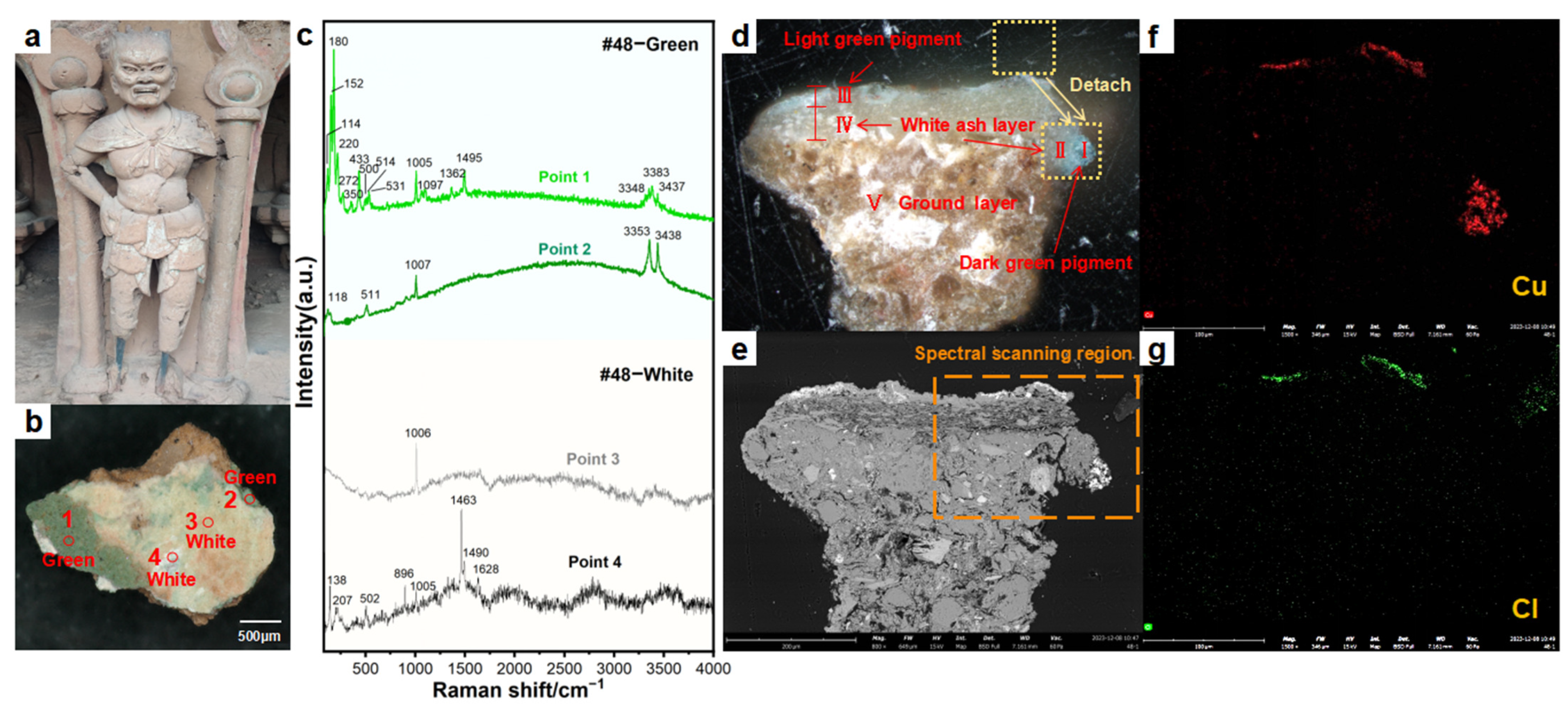
3.1.1. Analysis Results for Cave 48
3.1.2. Analysis Results for Cave 120
3.2. Blue-Green Pigment Samples
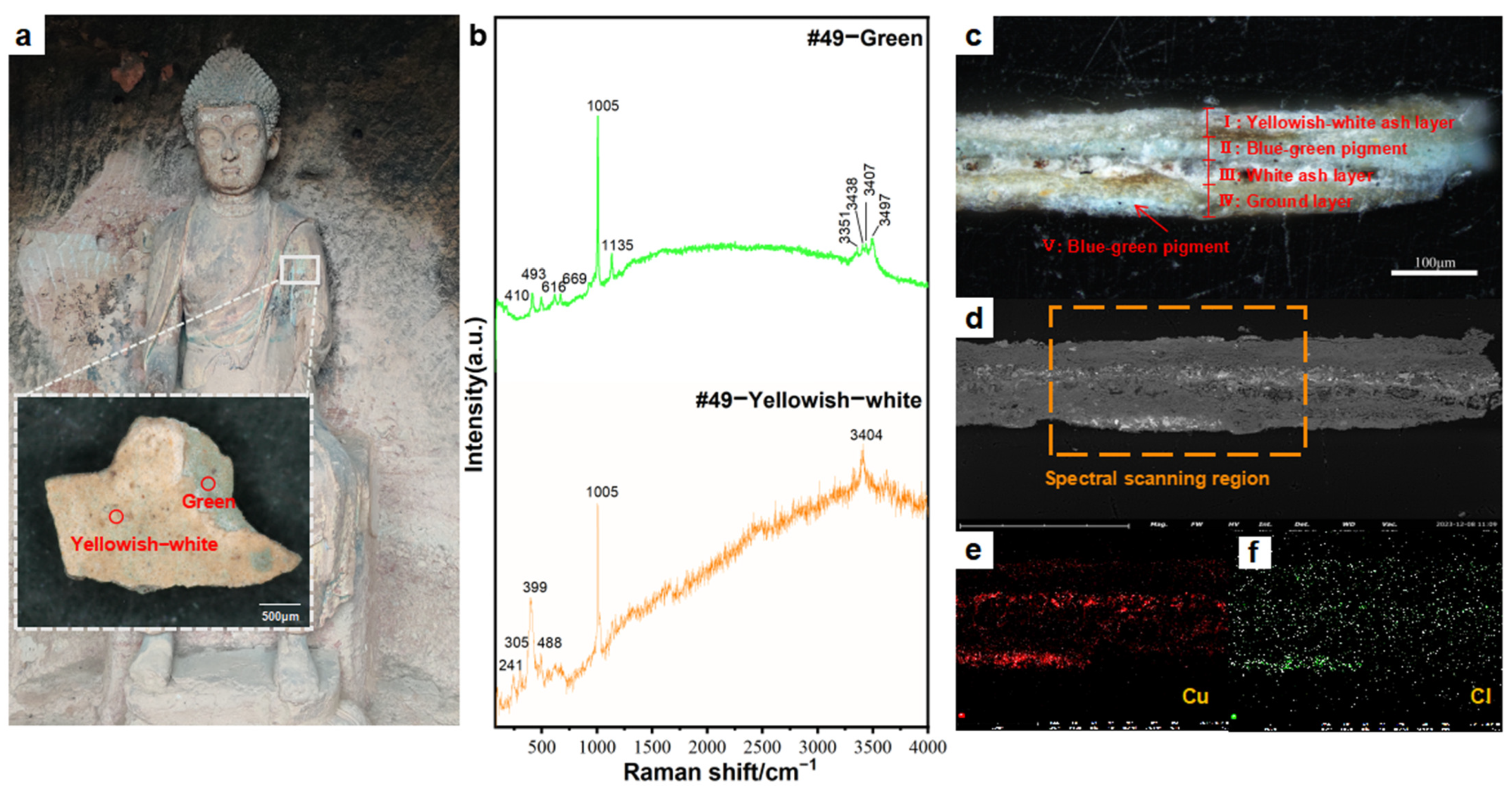
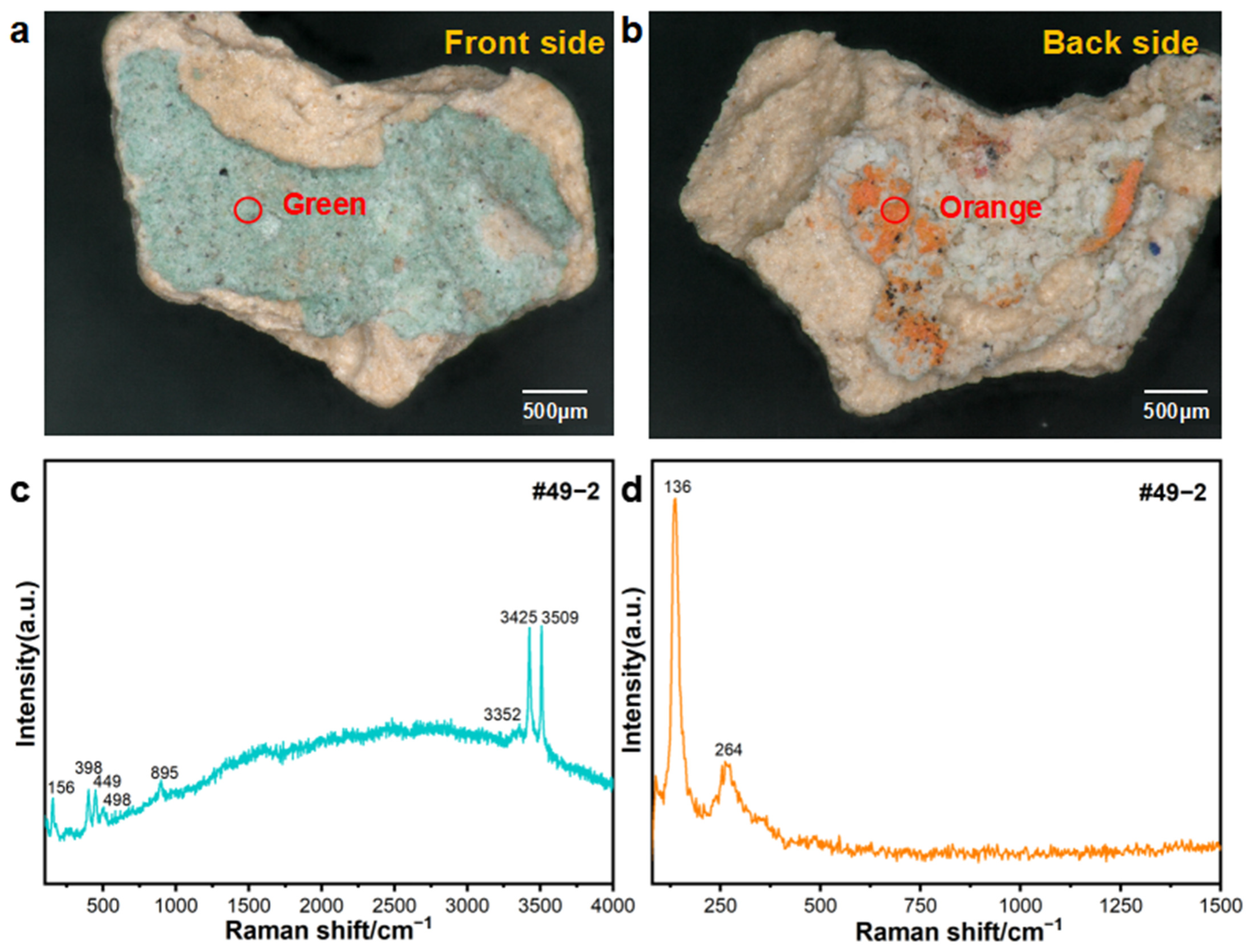
3.2.1. Analysis Results for Cave 49
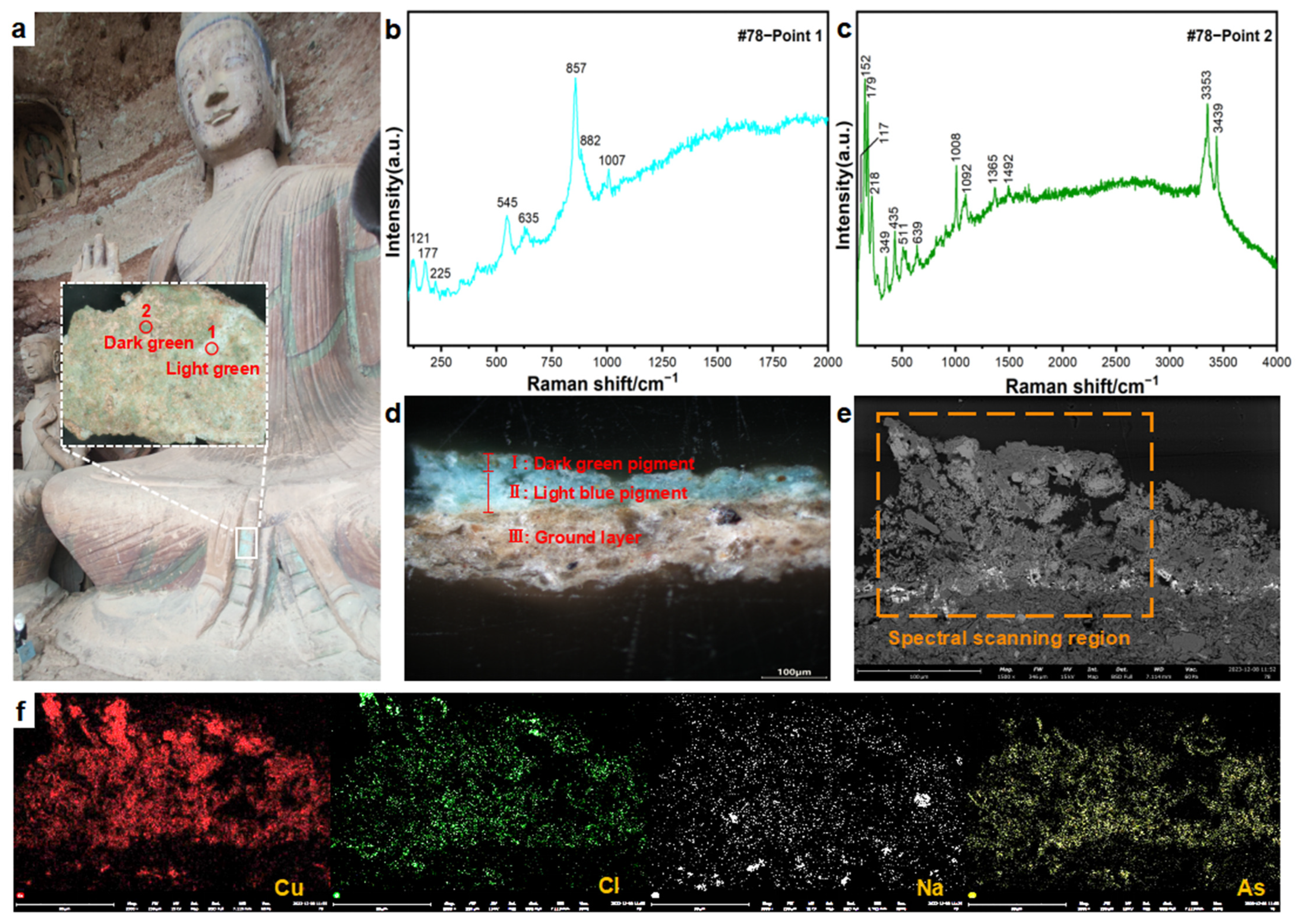
3.2.2. Analysis Results for Cave 78
4. Conclusions
Supplementary Materials
Author Contributions
Funding
Data Availability Statement
Acknowledgments
Conflicts of Interest
References
- Maijishan Grottoes Art Research Institute, Dunhuang Academy. Comprehensive Catalogue of Maijishan Grottoes; Cultural Relics Publishing House Press: Beijing, China, 2023. [Google Scholar]
- Zhang, M. A Study on the Vimalakirti Transformation Art in Cave 133 of the Maijishan Grottoes. Art China 2020, 36–43. [Google Scholar]
- Compilation Committee of Maijishan Grottoes Records; Zhang, J.X. Records of Maijishan Grottoes; Gansu People’s Publishing House Press: Lanzhou, China, 2002. [Google Scholar]
- Yang, W.B. Research on the Statues of Northern Wei Dynasty in Maijishan Grottoes; Lanzhou University Press: Lanzhou, China, 2019. [Google Scholar]
- Zhao, L.Y.; Li, Y.F.; Yu, Z.R.; Wang, X.D.; Su, B.M. Analysis of materials and techniques for making the plaster layers of the Silk Road grottoes murals. Dunhuang Res. 2005, 4, 75–82. [Google Scholar]
- Li, Z.X. Conservation of Mural Paintings and Colored Sculptures in Silk Road Caves; Science Press: Beijing, China, 2005. [Google Scholar]
- Wang, C.Y.; Li, M.; Xia, Y. Study on the manufacturing technology of ancient Chinese grotto murals. Relics Museol. 2014, 4, 74–78. [Google Scholar]
- Wen, J.; Xu, B.K.; Hu, J.J.; Zhang, L.; Wang, H.Y. Study on the production technology and materials for murals in Cave 131 of Maijishan Grottoes. Conserv. Sci. Archaeol. 2023, 35, 104–113. [Google Scholar]
- Zhou, G.X. X-ray Diffraction Analysis of Inorganic Pigments in Maijishan Grottoes Murals and Colored Sculptures. Archaeology 1991, 08, 744–755+776. [Google Scholar]
- Xu, W.Y.; Zhou, G.X.; Li, Y.H. X-ray profiling report of inorganic pigments in Mogao Cave murals and colored sculptures. Dunhuang Res. 1983, 187–197. [Google Scholar]
- Fan, Y.Q.; Chen, G.X.; Li, Z.X.; Wang, J.G.; Su, B.M. Micro diffraction analysis of the rare green pigment botallackite in ancient wall paintings. J. Lanzhou Univ. 2004, 5, 52–55. [Google Scholar]
- Platania, E.; Streeton, L.N.; Lluveras-Tenorio, A.; Kutzke, H.; Colombini, M.P. Identification of green pigments and binders in late medieval painted wings from Norwegian churches. Microchem. J. 2020, 156, 104811. [Google Scholar] [CrossRef]
- Fan, Y.Q.; Chai, B.L.; Yu, Z.R.; Wang, J.G.; Li, Z.X. Multi-spectral non-invasive investigation and studies on the mural technology in early three caves of Mogao Grottoes. Dunhuang Res. 2010, 6, 28–33. [Google Scholar]
- Wang, J.Y.; Wang, J.C. The Study of Verdigris’Application at the Dunhuang Grottoes and the Sources of the Pigment Verdigris. Dunhuang Res. 2002, 4, 23–28. [Google Scholar]
- Yue, Y.Q. Condition Surveys of Deterioration and Research of Wall Paintings in Maijishan Cave-temple. Herit. Conserv. Stud. 2019, 4, 127–131. [Google Scholar]
- Hu, J.J.; Yue, Y.Q. Research on Pigments of Maijishan Grottoes Colored Sculptures and Murals. Silk Road 2015, 10, 71–73. [Google Scholar]
- Yan, H.T.; Sun, K.; Tang, J.; Wang, X.D.; Li, Y.F. Scientific Analysis of the Pigments Used in the Murals of the Tomb of the Prince Yi of the Ming Dynasty. Huaxia Archaeol. 2019, 2, 39–44. [Google Scholar]
- Li, M.; Wang, L.Q.; Xia, Y.; Zhang, J.H.; Chen, W. Research on the Spectral Analysis and Stability of Copper Green. Spectrosc. Spectr. Anal. 2013, 33, 3293–3297. [Google Scholar]
- Li, M.; Xia, Y.; Yun, Q.L.; Wang, T.; Zhang, Y. Analysis of green pigment on Guangyuan Thousand-Buddha Grotto in Sichuan. Sci. Conserv. Archaeol. 2014, 26, 22–27. [Google Scholar]
- Bell, I.M.; Clark, R.J.H.; Gibbs, P.J. Raman spectroscopic library of natural and synthetic pigments (pre-1850AD). Spectrochim. Acta Part A 1997, 53, 2159–2179. [Google Scholar] [CrossRef]
- Liang, J.X.; Wang, T.; Zhang, Y.X.; Chen, L.; Liu, M. Study of the Painted Warrior Figurines Excavated in the Tomb of the Sutong Family (Tang Dynasty) by Spectroscopic Techniques. Spectrosc. Spectr. Anal. 2023, 43, 175–182. [Google Scholar]
- Frost, L.R.; Martens, W.; Kloprogge, T.J.; Williams, P.A.; Leverett, P. Raman spectroscopy of the basic copper chloride minerals atacamite and paratacamite: Implications for the study of copper, brass and bronze objects of archaeological significance. J. Raman Spectrosc. 2002, 33, 801–806. [Google Scholar] [CrossRef]
- Martens, W.N.; Frost, R.L.; Williams, P.A. Raman and infrared spectroscopic study of the basic copper chloride minerals-Implications for the study of the copper and brass corrosion and “bronze disease”. Neues Jahrb. Mineral.-Abh. 2003, 178, 197–215. [Google Scholar]
- Rosi, F.; Miliani, C.; Borgia, I.; Brunetti, B.; Sgamellotti, A. Identification of nineteenth century blue and green pigments by in situ x-ray fluorescence and micro-Raman spectroscopy. J. Raman Spectrosc. 2004, 35, 344–356. [Google Scholar]
- Chen, C.M.; Wen, Y.S.; Shen, Y.Y.; Li, X.H.; Wang, J. Synthesis and Physicochemical Properties of Basic Copper Chloride. Feed Ind. 2004, 5, 38–40. [Google Scholar]
- Yue, Y.Q.; Wang, T.L.; Fu, W.W. Restoration of hollow drum murals in the Maijishan Grottoes. China Cult. Herit. 2016, 2, 75–78. [Google Scholar]
- Wei, H.X. A Literature Review on the Study on Maiji Mountain Grottoes in Tianshui; Northwest Normal University: Lanzhou, China, 2011. [Google Scholar]
- Dong, G.Q. Maijishan Grottoes Pottery Painted Sculpture and Related Issues. Relics Museol. 2009, 6, 147–150. [Google Scholar]
- Mancini, D.; Tournié, A.; Caggiani, M.C.; Vallet, J.-M.; Colombini, M.P. Testing of Raman spectroscopy as a non-invasive tool for the investigation of glass-protected miniature portraits. J. Raman Spectrosc. 2012, 43, 294–302. [Google Scholar] [CrossRef]
- Vasiliev, A.L.; Kovalchuk, M.V.; Yatsishina, E.B. Electron microscopy methods in studies of cultural heritage sites. Crystallogr. Rep. 2016, 61, 873–885. [Google Scholar]
- Antunes, V.; Candeias, A.; Oliveira, M.J.; Teixeira, D.; Gil, M. Characterization of gypsum and anhydrite ground layers in 15th and 16th centuries Portuguese paintings by Raman Spectroscopy and other techniques. J. Raman Spectrosc. 2015, 45, 1026–1033. [Google Scholar] [CrossRef]
- Frost, R.L. Raman spectroscopy of selected copper minerals of significance in corrosion. Spectrochim. Acta Part A Mol. Biomol. Spectrosc. 2003, 59, 1195–1204. [Google Scholar] [CrossRef]
- Cheng, X.L.; Liu, M.; Li, M.; Wang, Y.; Zhang, Q. Identification and interpretation of unusual corrosion products formed on brass coins. Conserv. Sci. Archaeol. 2022, 34, 1–9. [Google Scholar] [CrossRef]
- Sun, Y.Z.; Wang, Z.L.; Wei, S.Y. Analyses of the pigment binding media in the rock painting of the Huashan Mountain, China. Conserv. Sci. Archaeol. 2012, 24, 38–43. [Google Scholar]
- Frost, R.L. Raman spectroscopy of natural oxalates. Anal. Chim. Acta 2004, 517, 207–214. [Google Scholar] [CrossRef]
- Wang, Y.; Liang, P. Application of Microlaser Raman Spectrometer in Corrosion Research of Iron Cultural Relics. Mod. Chem. Res. 2022, 19, 134–136. [Google Scholar]
- Chen, X.L.; Yang, Q. Study of five chloride-containing corrosion products from copper and bronze artifacts using Raman spectroscopy and scanning electron microscopy. Sci. Conserv. Archaeol. 2018, 30, 26–33. [Google Scholar]
- Coccato, A.; Bersani, D.; Coudray, A.; Wouters, J.; Vandenabeele, P. Raman spectroscopy of green minerals and reaction products with an application in Cultural Heritage research. J. Raman Spectrosc. 2016, 47, 1429–1443. [Google Scholar]
- Costantini, I.; Lottici, P.P.; Castro, K.; Madariaga, J.M. Use of Temperature Controlled Stage Confocal Raman Microscopy to Study Phase Transition of Lead Dioxide (Plattnerite). Minerals 2020, 10, 468. [Google Scholar] [CrossRef]
- Lei, Y. Copper trihydroxychlondes as pigments in China. Stud. Conserv. 2012, 57, 106–111. [Google Scholar]
- Wang, L.F.; Yin, H.Y.; Li, Y.; Zhang, X.; Chen, J. Synthesis of three crystalline phases of Cu2(OH)3Cl as Fenton catalysts to mineralize phenol in chloride-free and high chloride solutions. J. Water Process Eng. 2024, 58, 104893. [Google Scholar]
- Li, M.; Xia, Y. Preliminary Exploration of the Properties of Basic Copper Chloride Pigments. Sci. Res. Chin. Cult. Relics 2019, 3, 69–74. [Google Scholar]
- Liu, X.X.; Xu, L.; Huang, Y.L.; Zhang, Q.; Wang, J. Paratacamite phase stability and improved optical properties of Cu2(OH)3Cl crystal via Ni-doping. Mater. Des. 2017, 121, 194–201. [Google Scholar]
- Wang, L.Q.; Ma, Y.N.; Zhang, Y.X.; Li, J.; Chen, W. Pigment Identification of Sleeping Buddha at World Cultural Heritage Dazu Rock Carvings With μ-Raman Spectroscopy and Related Research. Spectrosc. Spectr. Anal. 2020, 40, 3199–3204. [Google Scholar]
- Antonov, A.A.; Nikolaev, A.I. Production of Analogs of the Rare Minerals Sampleite and Lavendulan as Potential Functional Materials. Theor. Found. Chem. Eng. 2022, 56, 541–544. [Google Scholar]
- Chen, B.Z.; Tang, R.H.; Gong, Z.Q.; Li, X.; Wang, H. Thermodynamic analysis of copper arsenate preparation process. Chin. J. Nonferrous Met. 2001, 11, 510–513. [Google Scholar]
- Cui, Q.; Zhang, Y.X.; Shui, B.W.; Li, M.; Wang, T. Study of copper and arsenic-containing green and blue-green pigments of rock carvings at Big Buddha Bay in Dazu. Sci. Conserv. Archaeol. 2020, 32, 87–94. [Google Scholar]
- Liu, L.Y.; Shen, W.; Zhang, B.J.; Wang, J.; Li, X. Microchemical Study of Pigments and Binders in Polychrome Relics from Maijishan Grottoes in Northwestern China. Microsc. Microanal. 2016, 22, 845–856. [Google Scholar] [CrossRef] [PubMed]
- Shen, L.; Yang, J.; Wang, J.; Li, X.; Chen, W. Atacamite discolouration under the influence of arsenates in wall paintings in the Kizil Grottoes, Xinjiang, China. Herit. Sci. 2024, 12, 292. [Google Scholar] [CrossRef]


| Sample Source | Cave Info. | No. | Sampling Location | Sample Type and Color |
|---|---|---|---|---|
| Cave 48 | The Northern Zhou Dynasty (AD 557–581) | 48 | 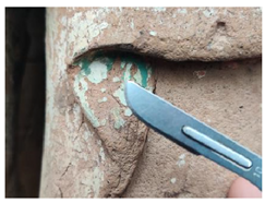 | Dark green and light green pigments |
| Cave 49 | Sui Dynasty (AD 581–618) | 49-1 | 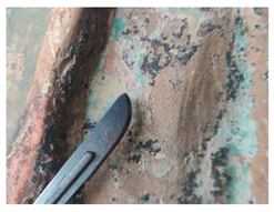 | Blue-green pigment |
| Cave 49 | Sui Dynasty | 49-2 |  | Blue-green pigment |
| Cave 78 | Later Qin Dynast (the Northern Wei Dynasty rebuilt) | 78 | 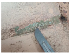 | Blue-green pigment |
| Cave 120 | The West Wei Dynasty | 120 | 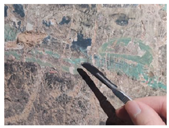 | Dark green and light green pigments |
Disclaimer/Publisher’s Note: The statements, opinions and data contained in all publications are solely those of the individual author(s) and contributor(s) and not of MDPI and/or the editor(s). MDPI and/or the editor(s) disclaim responsibility for any injury to people or property resulting from any ideas, methods, instructions or products referred to in the content. |
© 2025 by the authors. Licensee MDPI, Basel, Switzerland. This article is an open access article distributed under the terms and conditions of the Creative Commons Attribution (CC BY) license (https://creativecommons.org/licenses/by/4.0/).
Share and Cite
Wang, J.; Lv, M.; Song, N.; Zhang, H.; Xu, B.; Zhang, H. New Discoveries in the Maijishan Grottoes: Identification of Blue-Green Pigments and Insights into Green Pigment Application Techniques. Crystals 2025, 15, 339. https://doi.org/10.3390/cryst15040339
Wang J, Lv M, Song N, Zhang H, Xu B, Zhang H. New Discoveries in the Maijishan Grottoes: Identification of Blue-Green Pigments and Insights into Green Pigment Application Techniques. Crystals. 2025; 15(4):339. https://doi.org/10.3390/cryst15040339
Chicago/Turabian StyleWang, Jiakun, Miaoying Lv, Nan Song, Huan Zhang, Bokai Xu, and Hui Zhang. 2025. "New Discoveries in the Maijishan Grottoes: Identification of Blue-Green Pigments and Insights into Green Pigment Application Techniques" Crystals 15, no. 4: 339. https://doi.org/10.3390/cryst15040339
APA StyleWang, J., Lv, M., Song, N., Zhang, H., Xu, B., & Zhang, H. (2025). New Discoveries in the Maijishan Grottoes: Identification of Blue-Green Pigments and Insights into Green Pigment Application Techniques. Crystals, 15(4), 339. https://doi.org/10.3390/cryst15040339