Article

Self-Assembly Motifs of Water in Crystals of Palladium β-Amino Acid Complexes Influenced by Methyl Substitution on the Amino Acid Backbone

Department of Chemistry, Virginia Tech, Blacksburg, VA 24061, USA
*
Author to whom correspondence should be addressed.
Current address: Air Liquide Advanced Materials, Branchburg, NJ 08876, USA.
Crystals 2019, 9(11), 590; https://doi.org/10.3390/cryst9110590
Submission received: 17 October 2019 / Revised: 30 October 2019 / Accepted: 6 November 2019 / Published: 9 November 2019
(This article belongs to the Special Issue Novel Hydrogen-bonded Materials with Significant Physical Properties)

Abstract

:
Amino acid complexes of transition metals show interesting hydrogen-bonding motifs. In this paper, the syntheses and structures of three β-amino acid complexes of palladium that differ only by the substitution on the β-carbon will be discussed. With only hydrogen on the β-carbon, no additional water is incorporated into the crystal lattice and hydrogen-bonding is all complex-to-complex. With the addition of one and two methyl groups on the amino acid β-carbon, water is incorporated into the crystal lattice giving intricate water networks held together by complex-to-water and water-to-water hydrogen-bonding networks.

Graphical Abstract

1. Introduction

Many research teams [1,2,3] including ours [4,5,6,7] have been studying the metal complexes of amino acids, to examine both their catalytic activity as well as their biological activity. Perhaps one of the most influential investigators in this area is Wolfgang Beck whose series “Metal Complexes of Biologically Important Ligands” contains over 175 publications in a research career spanning over 50 years [1]. A great many of those publications deal with alpha-amino acid complexes of a wide variety of metals. Quite appropriately for this paper, one of Beck’s more recent publications concerns Pd and Pt complexes of long chain amino acids [8].
Much less studied, however, are the β-amino acid complexes of metals. This is somewhat surprising since a beta-amino acid should form a stable 6-membered ring chelate with metals, coordinating through the carboxylate O atom and the amino N atom. There are a number of β-amino acid complexes of first-row transition metals such as Co and Cu [9,10,11,12,13,14,15,16] but many fewer examples of heavier metals such as Pd and Pt [17,18,19]. It is not surprising that the hydrogen-bonding (H-bonding) capabilities of amino acids play a large role in the solid-state structures of the their metal complexes. In an iridium valine complex on which we reported, H-bonding sets up a unique helical arrangement of molecules that extends along a 43 screw axis of the space group P43, with L-Valine and D-Valine generating crystals with opposite helical turns in P41 [4,5].
Relevant to the issue of H-bonding between molecules is the bonding of water contained in crystalline lattices. Waters of hydration are a common occurrence in purely inorganic salts [20] but the exact nature of water molecules contained in organic or organometallic crystals is often complex [21,22]. Infantes et al. [23] have mined the examples of water in crystal lattices and have developed a nomenclature for describing these water motifs. It is now possible to search for water motifs in crystals using a feature of the CCDC program, Mercury [24]. Fully understanding all of the factors in directing a particular hydrogen-bonding motif is still an important ongoing topic of study [25,26,27,28,29,30,31,32,33]. Hydrogen-bonding as a tool in crystal engineering is also an increasingly important area [26,34].
Recently, we have been investigating the structures and catalytic properties of amino acid complexes of palladium. We have published a paper on glycine and substituted glycines [35] as well as a paper on palladium complexes of proline and proline homologs [36]. A common and consistent feature of all complexes is their propensity for hydrogen bonding—with the carbonyl oxygen acting as H-bond acceptor and the NH2 hydrogens acting as H-bond donors. In many cases, crystallization readily occurs only with water molecules in the lattice creating some unusual H-bonding networks.
In this paper, we discuss the structures of three Pd β-amino acid compounds and how 0, 1 and 2 methyl groups on the amino acid backbone greatly influence the H-bonding, the inclusion of water and the overall crystal lattice.

2. Results and Discussion

2.1. Complex Syntheses

Three different, readily available β-amino acids were examined: 3-aminopropanoic acid, (S)-3-aminobutanoic acid and 3-amino-3-methylbutanoic acid. Syntheses of the palladium compounds were straightforward and the procedures followed those we have reported on previously [35]. In terms of isomer formation, 3-aminopropanoic acid gave exclusively the trans isomer, trans-bis(3-aminopropanoate) palladium, compound 1. The thermal ellipsoid plot for 1 is shown in Figure 1. The 1H and 13C nuclear magnetic resonance (NMR) spectra also show signals indicative of only one isomer in solution. This complex was the subject of a previous paper by Krylova et al. [19] and a room temperature crystal structure of trans-bis(3-aminopropanoato)palladium was reported. Krylova and co-workers also reported that the cis-isomer could be synthesized by heating the trans isomer to 80° followed by cooling to 0° but no X-ray crystal structure was obtained and we could not reproduce that finding.
(S)-3-aminobutanoic acid gave predominantly the trans isomer of bis-[(S)-3-aminobutanoato] palladium, compound 2 but, in solution, approximately 25% of the cis isomer was observed by NMR spectroscopy. However, despite the 75/25 solution ratio of trans to cis observed in solution, all crystal growing attempts yielded solely the cis isomer. The thermal ellipsoid plot of the cis-isomer of compound 2 is shown in Figure 2.
The reaction between 3-amino-3-methylbutanoic acid and palladium(II) acetate yielded exclusively the trans isomer of [bis-[3-amino-3-methylbutanoato]palladium, compound 3, as evidenced by NMR spectroscopy. The single crystal X-ray structure also shows only the trans isomer, Figure 3.
Gaussian 09 DFT calculations support that the trans isomers are more stable than the cis (see Supplementary Materials) but the results do not explain why some cis isomer was observed in solution for the (S)-3-aminobutanoic acid product and why only the cis product was found in the solid state. Of course, ΔG calculations provide guidance only if the system is at equilibrium. Even if the system were at equilibrium, a lower solubility for the cis isomer of the (S)-3-aminobutanoic acid product could explain why crystallization favors that product. It is also a possibility that the hydrogen-bonding network established in the lattice for the cis (S)-3-aminobutanoic acid product favors crystallization of that isomer even if solubility were not an issue.
Aside from the different isomer possibilities, the inclusion of waters of hydration and the hydrogen-bonding motifs those waters form are very interesting. First, trans-bis-(3-aminopropanoato) palladium crystallizes with no waters of hydration even though the solution from which it was derived was 50% water by volume. The intermolecular interactions in this crystal lattice are derived solely from complex-to-complex hydrogen bonding interactions of the form N−H···O where both hydrogen atoms on the amino nitrogen are involved in donating to oxygens in two different adjacent complexes. Each hydrogen atom is involved in two different forms of H-bonding. The first N-H donates to a carbonyl oxygen of the carboxylate (or, to phrase it differently, to the carboxylate oxygen that is NOT bonded to the palladium) with an H···O distance of 2.09 Å. The second N-H bond appears to be partly H-bonded to both carboxylate oxygen atoms in an adjacent complex with H···O bond distances of 2.37 Å each. We presume that this complex with the simplest β-amino acid has no steric issues with bringing the complex molecules close together enough to satisfy N−H···O bonding without inclusion of waters of hydration.
Addition of a methyl group to the β-carbon atom of the amino acid (S)-3-aminobutanoic acid yields two major differences when the amino acid is used to form a palladium(II) complex. First, the cis, rather than the trans complex is formed. This is somewhat surprising in that the addition of a methyl group would simplistically be expected to favor the trans complex since the methyl group would provide greater steric repulsion in the cis compound. At the moment, the only speculation we would make is that the cis compound is less soluble and thus crystallization favors formation of the cis isomer. Of course, this requires that the cis and trans be in equilibrium in solution and that does not appear to be the case, so solution dynamics and crystallization dynamics need to be studied further.
The second major difference is that the complex molecules are not able to approach each other closely enough to form N−H···O bonds between complex molecules. Instead, several waters of hydration are included in the lattice and the N−H···O bonding is exclusively complex N-H to water oxygen binding as well as water O-H to non-bonded carboxylate oxygen of the complex (as well as to oxygen in other water molecules in the lattice.) Overall, the lattice picture is best described as alternating channels of complex and of water.
The amino acid with 2 methyl groups attached to the β-carbon, 3-amino-3-methylbutanoic acid, now yields solely (by NMR spectroscopy) the trans structure, a result that makes sense based on the increased steric repulsion that placing the 4 methyl groups on the same side of the palladium would involve.
Interestingly, now that the NH2 groups are on opposite sides of the molecule, there is a trajectory along which the N-H hydrogen atom can approach closely enough to hydrogen-bond to the non-bonded carboxylate oxygen atom of an adjacent complex molecule. However, only one of the N-H hydrogen atoms can satisfy its donating ability in this manner. The other donates to waters of hydration in the crystal lattice. The combined features of the line of approach of the complex molecules as well as H-bonding to waters of hydration yields a complex H-bonding motif and an overall picture that, simplistically, can be described as layers of complex interspersed with layers of water.

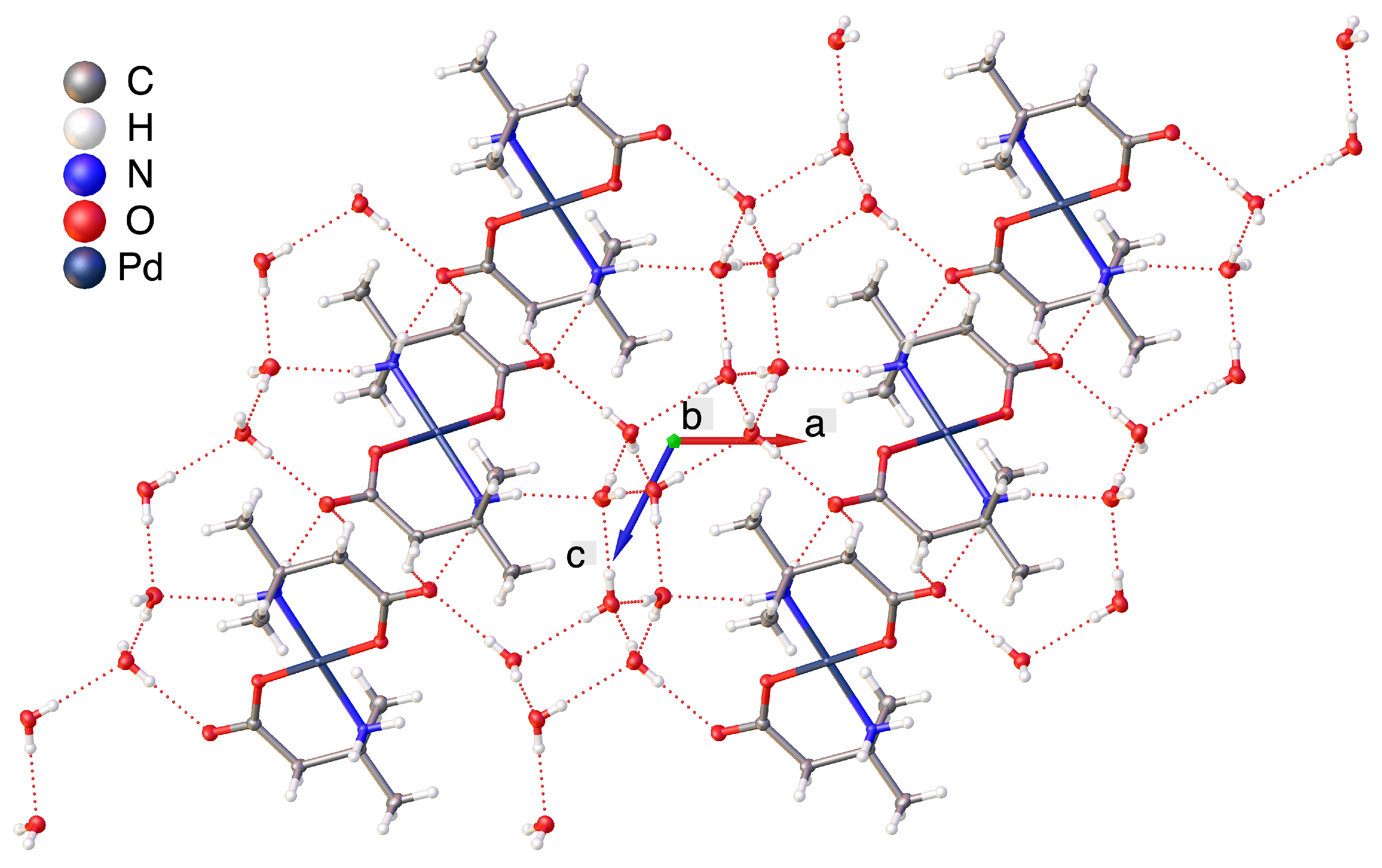
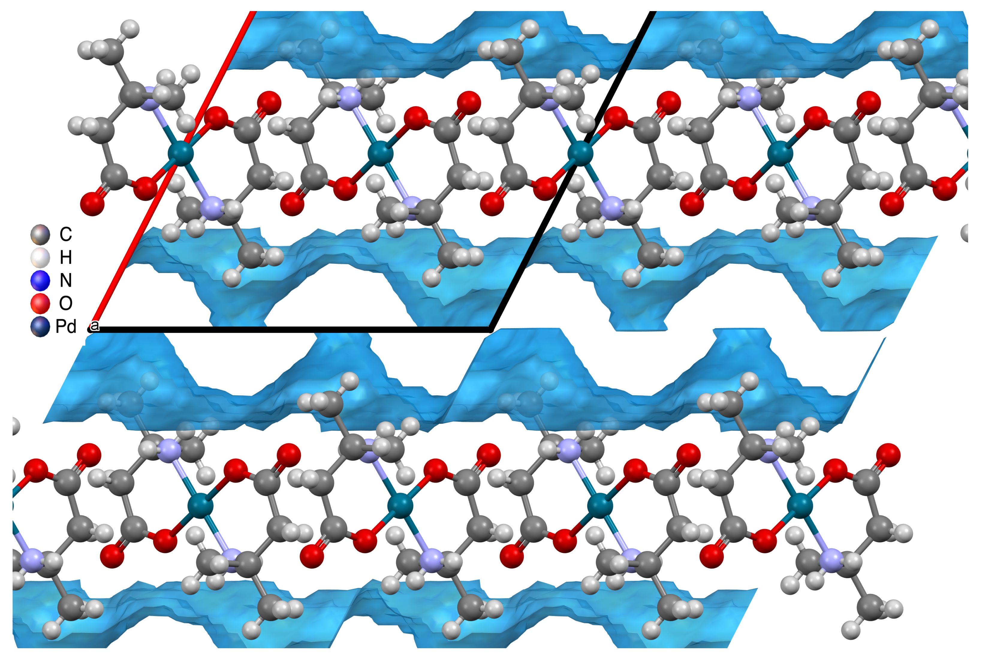
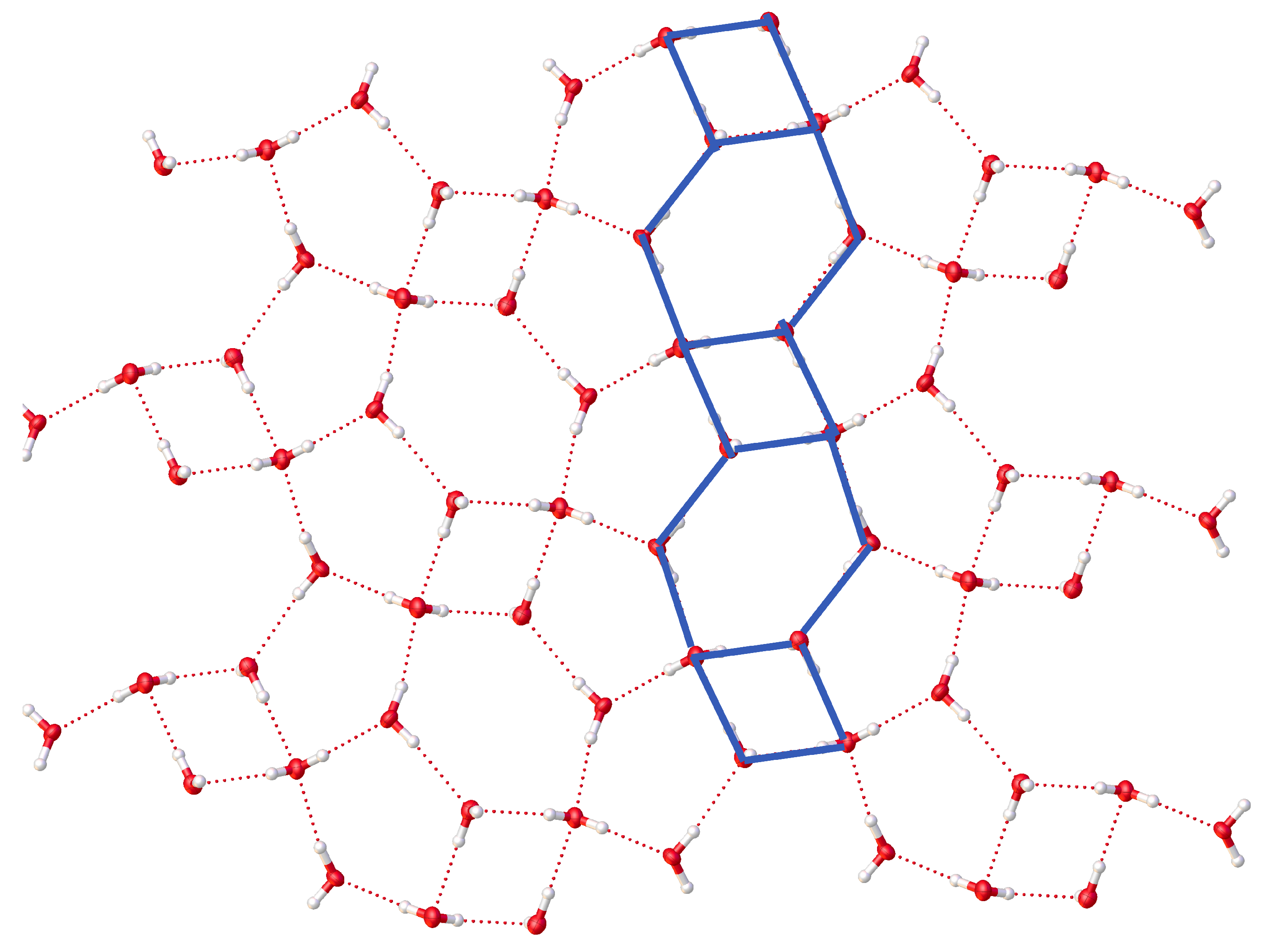
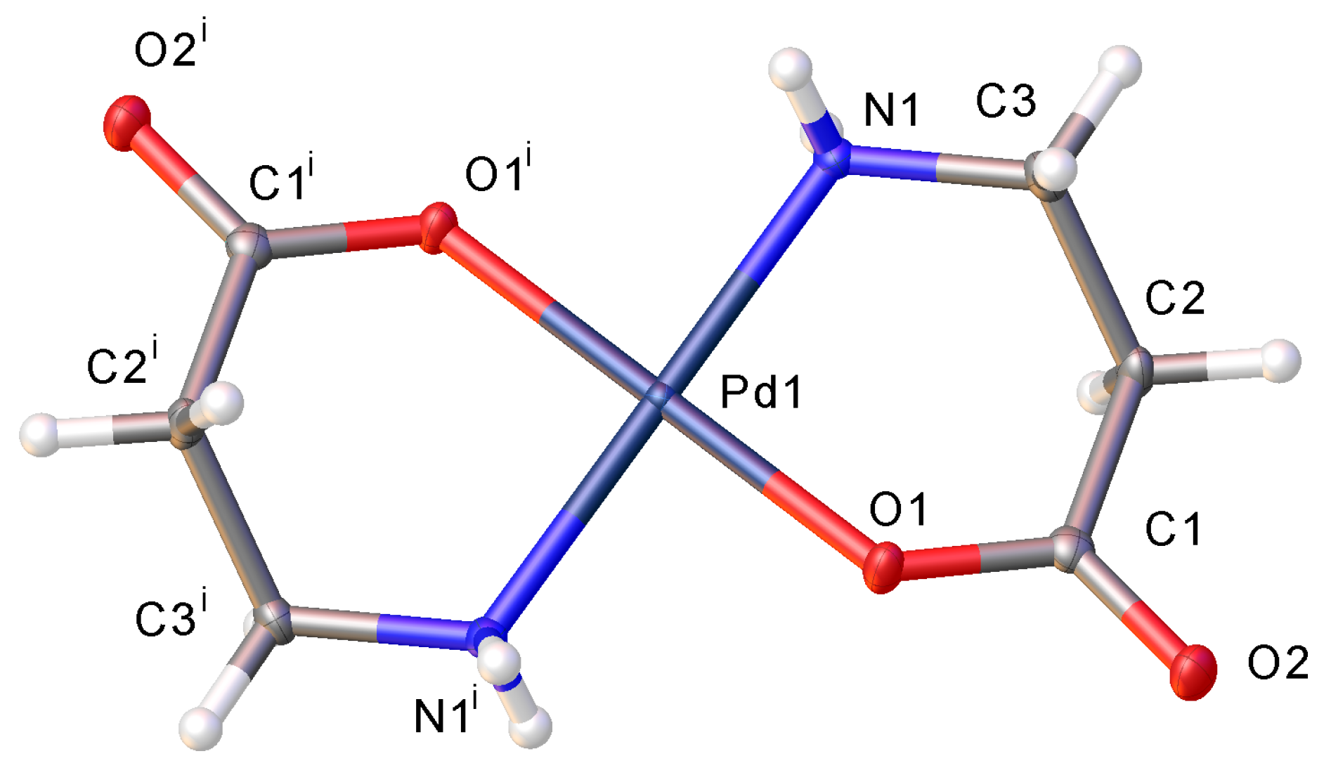
2.2. Hydrogen Bonding Motifs

Compound 1 does not contain any lattice waters as mentioned above. But the hydrogen-bonding network is interesting nonetheless. Figure 4 shows a portion of the packing diagram for compound 1 indicating that there are four channels of one orientation of the molecule surrounding one channel of molecules at approximately right angles to the four with N−H···O bonding holding the complexes together.
Once a methyl group replaces a hydrogen on the β-carbon, the crystal lattice now includes water. Figure 5 shows a thermal ellipsoid plot viewed down the a axis of the crystal lattice highlighting the hydrogen bonding. From this view, it would appear that the crystal lattice may best be described as two-dimensional channels of complex interspersed with two-dimensional channels of water. Using the CCDC program Mercury and its CSD -Materials tools [24] the water space calculation shows that the crystal, by volume, is 20 % water. Also using Mercury, Figure 6 was generated and even more clearly illustrates the channels of water in the crystal lattice.
When both hydrogen atoms on the β-carbon of the parent β-alanine are replaced by methyl groups, even more water is incorporated into the crystal lattice. Figure 7 shows a portion of the packing diagram for compound 3 where it can be seen that layers of complex alternate with layers of water. Mercury analysis of water space more clearly illustrate the alternating layers of water and complex molecules (Figure 8). That same analysis shows that the crystal lattice for compound 3 is 38% water by volume. Lourdes et al. have mined the CCDC database for different motifs of water hydrogen-bonding in crystal structures and have developed different designators for water motifs [23]. For complex 3, the motif would be described as T4(2)6(2). T4(2)6(2) indicates an infinite tape of alternating 4,6-membered rings of oxygen atoms sharing one edge. The “(2)” designator tells that the point of connection between the rings is two oxygen atoms. Figure 9 shows just the water in the crystal water layer with the 4,6-membered ring tape highlighted. The figure also shows that where two of the infinite tapes abut, 5-membered oxygen rings form.

3. Conclusions

In this paper, we have described the synthesis of 3 different palladium complexes of readily available β-amino acids and have shown that the presence of 0, 1 or 2 methyl groups on the β carbon atom greatly affects the crystal lattice. No methyl groups (3-aminopropanoic acid) forms a palladium complex with a trans arrangement of the amino acid ligands with no inclusion of water in the crystal lattice. One methyl group (S-3-aminobutanoic acid) forms complexes with palladium with evidence for both cis and trans isomers. However, only the cis isomer formed suitable crystals for X-ray diffraction and water was included in the crystal lattice. In this case, the water is present in the lattice in one dimensional channels. 3-amino-3-methylbutanoic acid (2 methyl groups on the β-carbon atom) appears to form only the trans isomer and water is included in the crystalline lattice in the form of an infinite layer between layers of complex.
Future work will examine further modifications that can be made to β-amino acids [37,38,39] and the effect that those modifications will have on crystalline lattice formation. For example, N-substitution [18] will not only affect the steric demands of the amino acid ligands but also change the number of hydrogen bond donors. Changing the type of substitution on the β-carbon (phenyl, longer chain alkyl) should also affect how the complexes pack in the lattice and how water can pack as well. This palladium-β-amino acid system affords many possibilities for engineering the crystal lattice and, hopefully, developing consistent principles for hydrogen-bonding motifs.

4. Experimental

4.1. General Synthetic Techniques

All compounds are air-stable and water soluble, so no special handling techniques are needed. The β-amino acids were purchased from commercial sources and used as received. Palladium(II) acetate was purchased from Pressure Chemical (Pittsburgh, PA, USA).

4.2. Synthesis of Complexes

4.2.1. Synthesis of trans-bis-(3-aminopropionato)palladium(II)

A four dram vial was fitted with a magnetic stir bar and charged with palladium(II) acetate (51.1 mg, 0.2276 mmol) and 2.5 mL of 50/50 (v/v) acetone/water. The mixture was stirred until all solids had dissolved. To this was added β-alanine (44.6 mg, 0.5008 mmol) and stirred overnight. The reaction solution turned from a clear red-orange to a clear pale-yellow supernatant with a pale-yellow precipitate. The supernatant was transferred via pipette to a clean vial and allowed to evaporate to give clear yellow rectangular prisms. The pale-yellow precipitate was washed with cold water and dried under vacuum. The combined yield of single crystals and precipitate was 59.6 mg of product (0.2109 mmol, 93% yield). Trans-Pd(C3H6NO2)2 was identified on the basis of the following data: 1H NMR (400 MHz, D2O) δ 2.54–2.44 (m, 2H), 2.42–2.32 (m, 2H). HRMS/ESI+ (m/z): [M + H]+ calcd for Pd(C3H6NO2)2, 282.9910; found, 282.9904.

4.2.2. Synthesis of cis-bis-((S)-3-aminobutanoato)palladium(II)

A four dram vial was fitted with a magnetic stir bar and charged with palladium(II) acetate (53.2 mg, 0.2370 mmol) and 2.5 mL of 50/50 (v/v) acetone/water. The mixture was stirred until all solids had dissolved. To this was added (S)-3-aminobutanoic acid (53.8 mg, 0.5213 mmol) and stirred overnight. The reaction solution turned from a clear red-orange to a clear pale-yellow supernatant with a pale-yellow precipitate. The supernatant was transferred via pipette to a clean vial and allowed to evaporate to give clear yellow rectangular prisms. The pale-yellow precipitate was washed with cold water and dried under vacuum. The combined yield of single crystals and precipitate was 68.6 mg of product (0.2208 mmol, 93% yield). Cis-Pd(C4H8NO2)2 was identified on the basis of the following data: 1H NMR (400 MHz, D2O) δ 3.62 (h, J = 6.7 Hz, 1H), 2.53–2.48 (m, 1H), 2.43–2.36 (m, 1H), 1.32–1.19 (m, 3H). HRMS/ESI+ (m/z): [M + H]+ calcd for Pd(C4H8NO2)2, 311.0223; found, 310.9069.

4.2.3. Synthesis of trans-bis-(3-amino-3-methylbutanoato)palladium(II)

A four dram vial was fitted with a magnetic stir bar and charged with palladium(II) acetate (57.5 mg, 0.2561 mmol) and 3.0 mL of 50/50 (v/v) acetone/water. The mixture was stirred until all solids had dissolved. To this was added of 3-amino-3-methylbutanoic acid (63.1 mg, 0.5386 mmol) and stirred overnight. The reaction solution turned from a clear red-orange to a clear pale-yellow supernatant with a pale-yellow precipitate. The supernatant was transferred via pipette to a clean vial and allowed to evaporate to give clear yellow rhombohedral prisms. The pale-yellow precipitate was washed with cold water and dried under vacuum. The combined yield of single crystals and precipitate was 78.1 mg of product (0.2306 mmol, 90% yield). Trans-Pd(C5H10NO2)2 was identified on the basis of the following data: 1H NMR (400 MHz, Methanol-d4) δ 2.38 (s, 2H), 1.31 (s, 6H). HRMS/ESI+ (m/z): [M + H]+ calcd for Pd(C5H10NO2)2, 339.0536; found, 339.0473.

4.3. X-ray Crystallography

For compounds 1, 2 and 3, crystals suitable for X-ray diffraction formed by allowing the slow evaporation of the reaction solution. Data was collected on an Oxford diffraction Gemini_Mo_Eos diffractometer with crystals maintained at 99.9 K during data collection. Structures were solved using SHELXT [40] and refined using SHELX [41] software and figures and other crystallographic output were made using OLEX2 [42]. Analyses of water in the crystal lattices of complexes 2 and 3 were carried out with the program Mercury [24].
1. Crystal Data for compound 1, C6H12N2O4Pd (M = 446.77 g/mol): monoclinic, space group P21/n (no. 14), a = 5.6938(3) Å, b = 8.8497(4) Å, c = 9.0421(5) Å, β = 104.686(6)°, V = 440.73(4) Å3, Z = 2, T = 97(5) K, µ(MoKα) = 2.090 mm−1, Dcalc = 2.129 g/cm3, 4467 reflections measured (7.68° ≤ 2θ ≤ 64.158°), 1451 unique (Rint = 0.0315, Rsigma = 0.0356) which were used in all calculations. The final R1 was 0.0448 (I > 2σ(I)) and wR2 was 0.0636 (all data).
2. Crystal Data for compound 2, C16H44N4O15Pd2 (M = 745.35 g/mol): monoclinic, space group I2 (no. 5), a = 10.7953(2) Å, b = 7.38120(10) Å, c = 17.8370(3) Å, β = 97.217(2)°, V = 1405.33(4) Å3, Z = 2, T 100.15(5) K, µ(MoKα) = 1.352 mm−1, Dcalc = 1.761 g/cm3, 14648 reflections measured (7.462° ≤ 2θ ≤ 64.882°), 4716 unique (Rint = 0.0381, Rsigma = 0.0421) which were used in all calculations. The final R1 was 0.0292 (I > 2σ(I)) and wR2 was 0.0556 (all data).
3. Crystal Data for compound 3, C10H32N2O10Pd (M = 446.77 g/mol): P21/c (no. 14), a = 12.4228(5) Å, b = 7.0497(2) Å, c = 12.5509(5) Å, β = 117.080(5)°, V = 978.67(7) Å3, Z = 2, T 97(5) K, µ(MoKα) = 0.993 mm−1, Dcalc = 1.516 g/cm3, 9102 reflections measured (6.932° ≤ 2θ ≤ 64.77°), 3258 unique (Rint = 0.0379, Rsigma = 0.0474) which were used in all calculations. The final R1 was 0.0448 (I > 2σ(I)) and wR2 was 0.0636 (all data).

5. Associated Content

Accession Codes

CCDC 1916293–1916295 contain the supplementary crystallographic data for this paper. These data can be obtained free of charge via www.ccdc.cam.ac.uk/data_request/cif, or by emailing [email protected], or by contacting The Cambridge Crystallographic Data Centre, 12 Union Road, Cambridge CB2 1EZ, UK; fax: +44 1223 336033.

Supplementary Materials

The following are available at https://www.mdpi.com/2073-4352/9/11/590/s1, Table of results from DFT calculations, 1H NMR spectra for all 3 compounds, high resolution mass spectra for all 3 compounds and the complete experimental information and results on bond distances and angles from the three X-ray structures.

Author Contributions

Conceptualization, J.S.M.; Formal analysis, D.B.H. and J.F.; Investigation, D.B.H., H.P. and V.G.P.; Project administration, J.S.M.; Resources, J.S.M.; Supervision, D.B.H. and J.S.M.; Writing—original draft, D.B.H. and J.S.M.

Funding

This research received no external funding.

Acknowledgments

The authors thank the Hamilton Corporation for a generous donation of syringes and Christine DuChane for aid in obtaining NMR spectra for compound 2.

Conflicts of Interest

The authors declare no conflict of interest.

References

  1. Severin, K.; Bergs, R.; Beck, W. Bioorganometallic Chemistry ± Transition Metal Complexes with. Angew. Chem. Int. Ed. Engl. 1998, 37, 1634–1654. [Google Scholar] [CrossRef]
  2. Carmona, D.; Pilar Lamata, M.; Viguri, F.; San José, E.; Mendoza, A.; Lahoz, F.J.; García-Orduña, P.; Atencio, R.; Oro, L.A. Half-sandwich organometallic complexes with stereogenic metal centres: Synthesis and characterization of diastereomeric [(η-ring)M(Aa)X] (Aa = α-amino carboxylate) compounds. J. Organomet. Chem. 2012, 717, 152–163. [Google Scholar] [CrossRef]
  3. Biancalana, L.; Abdalghani, I.; Chiellini, F.; Zacchini, S.; Pampaloni, G.; Crucianelli, M.; Marchetti, F. Ruthenium Arene Complexes with α-Aminoacidato Ligands: New Insights into Transfer Hydrogenation Reactions and Cytotoxic Behaviour. Eur. J. Inorg. Chem. 2018, 2018, 3041–3057. [Google Scholar] [CrossRef]
  4. Roy, C.P.; Huff, L.A.; Barker, N.A.; Berg, M.A.G.; Merola, J.S. Iridium(III) hydrido amino acid compounds: Chiral complexes and a helical extended lattice. J. Organomet. Chem. 2006, 691, 2270–2276. [Google Scholar] [CrossRef]
  5. Merola, J.S.; Slebodnick, C.; Berg, M.; Ritchie, M.K. mer-Hydridotris(trimethylphosphane-κP)(d-valinato-κ2 N,O)iridium hexafluoridophosphate dichloromethane 0.675-solvate. Acta Crystallogr. Sect. E Struct. Rep. Online 2014, 70, m82. [Google Scholar] [CrossRef]
  6. Karpin, G.; Merola, J.; Falkinham, J., III. Transition metal-α-amino acid complexes with antibiotic activity against Mycobacterium spp. Antimicrob. Agents Chemother. 2013, 57. [Google Scholar] [CrossRef]
  7. Morris, D.M.; McGeagh, M.; De Peña, D.; Merola, J.S. Extending the range of pentasubstituted cyclopentadienyl compounds: The synthesis of a series of tetramethyl(alkyl or aryl)cyclopentadienes (Cp*R ), their iridium complexes and their catalytic activity for asymmetric transfer hydrogenation. Polyhedron 2014, 84, 120–135. [Google Scholar] [CrossRef]
  8. Wagner-Schuh, B.; Beck, W. Metal Complexes of Biologically Important Ligands, CLXXVII. Dichlorido Platinum(II) and Palladium(II) Complexes with Long Chain Amino Acids and Amino Acid Amides. Z. Anorg. Allg. Chem. 2017, 643, 632–635. [Google Scholar] [CrossRef]
  9. Cai, J.; Hu, X.; Feng, X.; Ji, L.; Bernal, I. Structures of three cis-β1 and three cis-β2 isomers of [Co(trien)(aminoacidato)]2+ complexes. Acta Crystallogr. Sect. B Struct. Sci. 2001, B57, 45–53. [Google Scholar] [CrossRef]
  10. Ng, C.H.; Ong, H.K.A.; Ngai, K.S.; Tan, W.T.; Lim, L.P.; Teoh, S.G.; Chong, T.S. mer-Tris(β-alaninato)cobalt(III): Crystal structure, solution properties and its DNA cleavage in the presence of ascorbic acid. Polyhedron 2005, 24, 1503–1509. [Google Scholar] [CrossRef]
  11. Garg, B.S.; Dwivedi, P. Solution studies on ternary complex formation of bivalent metal ions with cephalexine and amino acids, β-alanine and cysteine. J. Indian Chem. Soc. 2004, 81, 239–241. [Google Scholar]
  12. Gaizer, F.; Gondos, G.; Gera, L. Protonation and complex formation of the cis and trans isomers of alicyclic β-amino acids. Polyhedron 1986, 5, 1149–1156. [Google Scholar] [CrossRef]
  13. Juranic, N.; Prelesnik, B.; Manojlovic-Muir, L.; Andjelkovic, K.; Niketic, S.R.; Celap, M.B. Geometrical isomerism of mixed tris(amino carboxylato)cobalt(III) complexes containing glycinato and β-alaninato ligands. Crystal structure of trans(O5)-(β-alaninato)bis(glycinato)cobalt(III) trihydrate. Inorg. Chem. 2005, 29, 1491–1495. [Google Scholar] [CrossRef]
  14. Gridchin, S.N.; Shekhanov, R.F.; Bychkova, S.A. Stability constants of cobalt (II) complexes of taurine and β-alanine. Izv. Vyss. Uchebnykh Zaved. Khimiya I Khimicheskaya Tekhnol. 2016, 59, 95–96. [Google Scholar] [CrossRef]
  15. Sharma, V.S.; Mathur, H.B. Thermodynamic properties of coordination complexes of transition metal ions with amino acids. Indian J. Chem. 1965, 3, 475–479. [Google Scholar]
  16. Skoulika, S.; Michaelides, A.; Aubry, A. Habit modification of coordination compounds in the presence of additives; a study of the system Ni(β−ala)2·2H2O-glycine. J. Cryst. Growth 1991, 108, 285–289. [Google Scholar] [CrossRef]
  17. Böhm, A.; Seebach, D. Determination of Enantiomer Purity of β- and γ-Amino Acids by NMR Analysis of Diastereoisomeric Palladium Complexes. Helv. Chim. Acta 2000, 83, 3262–3278. [Google Scholar] [CrossRef]
  18. Cui, X.; Qin, T.; Wang, J.R.; Liu, L.; Guo, Q.X. Pd(N, N -Dimethyl β-alaninate) 2 as a High-Turnover-Number, Phosphine-Free Catalyst for the Suzuki Reaction. Synth. (Stuttg.) 2007, 2007, 393–399. [Google Scholar] [CrossRef]
  19. Krylova, L.F.; Kovtunova, L.M.; Romanenko, G.V. Pt(II) and Pd(II) Complexes with β-Alanine. Bioinorg. Chem. Appl. 2008, 2008, 1–10. [Google Scholar] [CrossRef] [Green Version]
  20. Ferraris, G.; Franchini-Angela, M. Survey of the geometry and environment of water molecules in crystalline hydrates studied by neutron diffraction. Acta Crystallogr. Sect. B Struct. Crystallogr. Cryst. Chem. 1972, 28, 3572–3583. [Google Scholar] [CrossRef]
  21. Braga, D.; Grepioni, F.; Desiraju, G.R. Hydrogen bonding in organometallic crystals—A survey. J. Organomet. Chem. 1997, 548, 33–43. [Google Scholar] [CrossRef]
  22. Fucke, K.; Steed, J.W. X-ray and neutron diffraction in the study of organic crystalline hydrates. Water 2010, 2, 333–350. [Google Scholar] [CrossRef]
  23. Infantes, L.; Motherwell, S. Water clusters in organic molecular crystals. CrystEngComm 2002, 4, 454–461. [Google Scholar] [CrossRef]
  24. Macrae, C.F.; Bruno, I.J.; Chisholm, J.A.; Edgington, P.R.; McCabe, P.; Pidcock, E.; Rodriguez-Monge, L.; Taylor, R.; van de Streek, J.; Wood, P.A. Mercury CSD 2.0—New features for the visualization and investigation of crystal structures. J. Appl. Crystallogr. 2008, 41, 466–470. [Google Scholar] [CrossRef]
  25. Braga, D.; Grepioni, F. Intermolecular interactions in nonorganic crystal engineering. Acc. Chem. Res. 2000, 33, 601–608. [Google Scholar] [CrossRef]
  26. Desiraju, G.R. Hydrogen Bridges in Crystal Engineering: Interactions without Borders. Acc. Chem. Res. 2002, 35, 565–573. [Google Scholar] [CrossRef]
  27. Brammer, L.; Mareque Rivas, J.C.; Reinaldo Atencio, J.; Fang, S.; Christopher Pigge, F. Combining hydrogen bonds with coordination chemistry or organometallic π-arene chemistry: Strategies for inorganic crystal engineering. J. Chem. Soc. Dalt. Trans. 2000, 3855–3867. [Google Scholar] [CrossRef]
  28. Pal, S.; Sankaran, N.B.; Samanta, A. Structure of a self-assembled chain of water molecules in a crystal host. Angew. Chem. Int. Ed. 2003, 42, 1741–1743. [Google Scholar] [CrossRef]
  29. Infantes, L.; Chisholma, J.; Motherwell, S. Extended motifs from water and chemical functional groups in organic molecular crystals. CrystEngComm 2003, 5, 480–486. [Google Scholar] [CrossRef]
  30. Mascal, M.; Infantes, L.; Chisholm, J. Water oligomers in crystal hydrates—What’s news and what isn’t? Angew. Chem. Int. Ed. 2005, 45, 32–36. [Google Scholar] [CrossRef]
  31. Galek, P.T.; Fábián, L.; Motherwell, W.D.; Allen, F.H.; Feeder, N. Knowledge-based model of hydrogen-bonding propensity in organic crystals. Acta Crystallogr. Sect. B Struct. Sci. 2007, 63, 768–782. [Google Scholar] [CrossRef] [PubMed]
  32. Galek, P.T.; Fábián, L.; Allen, F.H. Truly prospective prediction: Inter- and intramolecular hydrogen bonding. CrystEngComm 2010, 12, 2091–2099. [Google Scholar] [CrossRef]
  33. Cruz Cabeza, A.J.; Day, G.M.; Motherwell, W.D.; Jones, W. Importance of molecular shape for the overall stability of hydrogen bond motifs in the crystal structures of various carbamazepine-type drug molecules. Cryst. Growth Des. 2007, 7, 100–107. [Google Scholar] [CrossRef]
  34. Desiraju, G.R. Crystal engineering: From molecule to crystal. J. Am. Chem. Soc. 2013, 135, 9952–9967. [Google Scholar] [CrossRef]
  35. Hobart, D.B.; Berg, M.A.G.; Merola, J.S. Bis-glycinato complexes of palladium(II): Synthesis, structural determination, and hydrogen bonding interactions. Inorg. Chim. Acta 2014, 423, 21–30. [Google Scholar] [CrossRef]
  36. Hobart, D.B.; Merola, J.S.; Rogers, H.M.; Saghal, S.; Mitchell, J.; Florio, J.; Merola, J.W. Synthesis, Structure, and Catalytic Reactivity of Pd(II) Complexes of Proline and Proline Homologs. Catalysts 2019, 9, 515. [Google Scholar] [CrossRef]
  37. Dexter, C.S.; Jackson, R.F.W. Efficient synthesis of β- and γ-amino acid derivatives using new functionalized zinc reagents: Enhanced stability and reactivity of β-amido zinc reagents in dimethylformamide. Chem. Commun. 1998, 75–76. [Google Scholar] [CrossRef]
  38. Abdel-Magid, A.F.; Cohen, J.H.; Maryanoff, C.A. Chemical process synthesis of β-amino acids and esters. Curr. Med. Chem. 1999, 6, 955–970. [Google Scholar]
  39. Hoen, R.; Tiemersma-Wegman, T.; Procuranti, B.; Lefort, L.; de Vries, J.G.; Minnaard, A.J.; Feringa, B.L. Enantioselective synthesis of β2-amino acids using rhodium-catalyzed hydrogenation. Org. Biomol. Chem. 2007, 5, 267–275. [Google Scholar] [CrossRef]
  40. Sheldrick, G.M. SHELXT—Integrated space-group and crystal-structure determination. Acta Crystallogr. Sect. A Found. Adv. 2015, 71, 3–8. [Google Scholar] [CrossRef]
  41. Sheldrick, G. Crystal structure refinement with SHELXL. Acta Crystallogr. Sect. C 2015, 71, 3–8. [Google Scholar] [CrossRef] [PubMed]
  42. Dolomanov, O.V.; Bourhis, L.J.; Gildea, R.J.; Howard, J.A.K.; Puschmann, H. OLEX2: A complete structure solution, refinement and analysis program. J. Appl. Crystallogr. 2009, 42, 339–341. [Google Scholar] [CrossRef]
Figure 1. Thermal ellipsoid plot of trans-bis-(3-aminopropanoato)palladium, complex 1. Atoms marked with superscript i are generated by the inversion operation. Ellipsoids are shown at 50% probability.
Crystals 09 00590 g001
Figure 2. Thermal ellipsoid plot of cis-bis-((S)-3-aminobutanoato)palladium, complex 2. Lattice waters are not displayed in this figure. Ellipsoids are shown at 50% probability.
Crystals 09 00590 g002
Figure 3. Thermal ellipsoid plot of trans-bis-(3-amino-3-methylbutanoato)palladium, complex 3. Lattice waters are not displayed in this figure. Ellipsoids are shown at 50% probability.
Crystals 09 00590 g003
Figure 4. Thermal ellipsoid plot of complex 1 showing relative orientations of hydrogen bonding in the lattice (red dotted lines). Ellipsoids are shown at 50% probability.
Crystals 09 00590 g004
Figure 5. Thermal ellipsoid plot of complex 2 viewed down the a axis showing relative orientations of hydrogen bonding in the lattice. Ellipsoids are shown at 50% probability.
Crystals 09 00590 g005
Figure 6. Water space analysis of complex 2 crystal lattice viewed down the a axis showing the water channels in the crystal.
Crystals 09 00590 g006
Figure 7. Thermal ellipsoid plot of a portion of the packing diagram of complex 3 viewed down the b axis showing alternating layers of water and complex molecules.
Crystals 09 00590 g007
Figure 8. Water space calculation plot of complex 3 viewed down the b axis showing alternating layers of water and complex molecules.
Crystals 09 00590 g008
Figure 9. Water motif of complex 3 in the crystal lattice water layer. Red spheres are oxygens, white spheres are hydrogens and dotted red lines show hydrogen bonding. The repeating T4(2)6(2) motif is highlighted in blue.
Crystals 09 00590 g009

