Article

Physically Crosslinked Poly (Vinyl Alcohol)/Kappa-Carrageenan Hydrogels: Structure and Applications

1
Materials Engineering and Welding Department, Transilvania University of Brasov, Eroilor 29 Str, 500036 Brasov, Romania
2
Materials Science Department, Transilvania University of Brasov, Eroilor 29 Str, 500036 Brasov, Romania
3
Product Design Mechatronics and Environment Department, Transilvania University of Brasov, Eroilor 29 Str, 500036 Brasov, Romania
4
Research Institute of renewable energy–ICER, Politehnica University of Timisoara, Piata Victoriei Str., 300006 Timisoara, Romania
*
Authors to whom correspondence should be addressed.
Polymers 2020, 12(3), 560; https://doi.org/10.3390/polym12030560
Submission received: 14 February 2020 / Revised: 26 February 2020 / Accepted: 28 February 2020 / Published: 3 March 2020
(This article belongs to the Special Issue Porous Polymer Micro- and Nano-Structures)

Abstract

:
This paper discusses the structure morphology and the thermal and swelling behavior of physically crosslinked hydrogels, obtained from applying four successive freezing–thawing cycles to poly (vinyl alcohol) blended with various amounts of κ-carrageenan. The addition of carrageenan in a weight ratio of 0.5 determines a twofold increase in the swelling degree and the early diffusion coefficients of the hydrogels when immersed in distilled water, due to a decrease in the crystallinity of the polymer matrix. The diffusion of water into the polymer matrix could be considered as a relaxation-controlled transport (anomalous diffusion). The presence of the sulfate groups determines an increased affinity of the hydrogels towards crystal violet cationic dye. A maximum physisorption capacity of up to 121.4 mg/g for this dye was attained at equilibrium.

1. Introduction

Poly (vinyl alcohol) (PVA) hydrogels are well known for their excellent processability [1], biocompatibility, elasticity, porosity and tunable diffusional properties [2,3], making them of prime importance in various fields, such as medicine (wound dressing, cell growth scaffolds, contact lenses and hygiene products) [4], pharmacy (controlled drug delivery vehicles) [5,6], the food industry [7], environmental applications (sorption of pollutant species and sensors) [8,9] and so forth. These materials can be obtained through various methods, which include chemical (covalent) crosslinking through radiation, the addition of mono- or bifunctional agents, or through thermal crosslinking. Numerous studies in the last three decades have focused on milder and more environmental-friendly physical crosslinking methods, among which the most promising is represented by cryostructuring (i.e., submitting the polymer to alternated freezing and thawing cycles) [10].
Many polysaccharides, in their natural or chemically functionalized state, such as chitosan, starch, dextran, cellulose, xanthan, β-glucans, sodium alginate, carrageenans and so forth, have been previously blended with PVA to obtain cryogels [11,12]. Due to their overall hydrophilic character, the addition of polysaccharides (e.g., starch, hydroxyethyl starch) can increase the swelling degree of the blend cryogels up to 300% compared with neat PVA, submitted to identical physical crosslinking operational parameters [13,14]. Chitosan, starch and various glucans, for example, are reported to improve the hydrophilic character, elasticity and flexibility of the hydrogels for wound dressing applications, while also fine-tuning the diffusion coefficient of various active principles (antibiotics, nutraceuticals and so forth) through the polymeric matrix, from 10−5–10−4 mm2/min with up to one order of magnitude [15,16].
Carrageenans (ι, λ or κ) represent a class of linear sulfated polysaccharides deriving from various species of moss, seaweed and algae. The addition of sulphated polysaccharides to PVA could modulate the release of cationic active principles from the polymer matrix in drug delivery devices [17] or could impart several functional properties to the cryogel, such as antimicrobial character or stimulatory effect for the fibroblast synthesis of collagen for tissue rebuild [18,19]. Also, the presence of the negatively charged sulfate moiety could increase the affinity of the hydrogel for cationic dyes and heavy metal ions, deeming the material useful in environmental applications [20].
This paper aims to obtain hydrogels comprised of PVA and κ-carrageenan (CAR) through cryostructuring (cryogelling) the aqueous solution mixtures of the two polymers by alternate freezing and thawing cycles.
Even if PVA/carrageenan chemically-crosslinked or γ radiation-crosslinked hydrogels are already known and their suitability for controlled delivery devices [21,22,23] is under study, there are few studies regarding the obtaining of PVA/CAR cryogels, such as those by Zhang et al. [24], Hosseinzadeh [25] and Chopra et al. [26], and their use is intended either as composites with various fillers or in lyophilized form. The influence of the quantity of κ-carrageenan on the structural features (intermolecular chemical bonding, crystallinity and morphology) and the sorption/release mechanism in/from the polymer matrix has not been studied extensively up to date for this polymer blend. The sorption kinetic and adsorption mechanism assessing for a widely-employed model cationic pollutant dye, i.e., crystal violet, enables a facile comparison of the results presented in this paper with the reference literature and contributes to the enriching of the international database regarding the potential applications of hydrogel systems.

2. Experimental

2.1. Materials

Poly (vinyl alcohol) (PVA) with Mw~130,000, >99% hydrolyzed, was purchased from Sigma-Aldrich (Munich, Germany). The carrageenan (κ-carrageenan, CAR) with a dynamic viscosity of 15 mPa s, 0.3% in H2O (25 °C) and an ester sulfate group (OSO3) percent of 20.40%, determined experimentally according to the gravimetric method proposed by Dodgson and Price [27], was purchased from Sigma-Aldrich (Munich, Germany). Both polymers were further used without any subsequent purification.
Methylene blue (C16H18ClN3S), crystal violet (C25N3H30Cl), cyclohexane, ethanol and the inorganic reagents used in this study were purchased from the same company.

2.2. Gels Obtaining

The hydrogels were prepared starting from aqueous solutions of the two polymers (10% wt. for PVA, respectively 1.5% wt. for carrageenan). To prepare the aqueous PVA and carrageenan solutions, the corresponding amounts of the polymers were dissolved in distilled water at 80 °C under magnetic stirring (200 rpm) for 2 h until a transparent viscous system was obtained. Several mixtures between the two polymer solutions (10 mL of total volume) were obtained at 80 °C under magnetic stirring, as depicted in Table 1.
The mixtures were cast in 80 mm diameter glass Petri dishes, cooled to room temperature and further submitted to four freezing–thawing cycles. The coding of the samples reflected the gravimetric ratio between carrageenan and PVA.
The parameters employed for the freezing–thawing cycles were the following: freezing temperature −25 °C, freezing duration: 12 h; thawing temperature: 22 °C and thawing duration: 12 h. These parameters were the optimum ones, considering the mechanical properties of the gels (i.e., they should allow easy manipulation without rupturing), and were chosen based on our prior experience with PVA cryogels [28] or with lignocellulose-ionic liquid cryogels [29]. The aspect of the hydrogels could be seen in Figure 1.
The solids content (SC) of the gels were determined as the percentage ratio between the mass of the oven-dried gels (105 °C, 6 h) and the mass of the as-obtained gels and are depicted in Table 1.
The gel content (G) of the materials was determined as follows: the hydrogels were packed in a stainless-steel mesh (25 μm), dried until constant mass and extracted with distilled water in a Soxhlet apparatus for 5 h. After extraction, the gels were re-dried, and the gel content was calculated as the mass of the extracted dried gel, reported to the mass of the initial dried material [30].
The Shore hardness values (00 scale; PCE-OO durometer, PCE Instruments, Palm Beach, FL, USA) for the obtained gels are also illustrated in Table 1. It should be noted that for wound-dressing or drug delivery applications, the specialty literature recommends a minimum hardness value of 60 Shore 00 units [31].
All the obtained hydrogels were processed/analyzed immediately after obtaining, to avoid their structural modification with ageing. Morphology analysis (SEM), IR and XRD structural analyses require the drying of the gels. Swelling kinetic studies are performed on the as-obtained gels and for crystal violet sorption experiments, the gels are swollen (equilibrated) in distilled water, as described in Section 2.3 and Section 2.4. The bulk of the samples are stored in distilled water (in the swollen state).

2.3. Morphological and Structural Characterization

A Quanta FEG 250 scanning electron microscope (FEI, Thermo Fisher Scientific, Hillsboro, OR, USA) was used to determine the morphology of the gels, at a 30 kV acceleration voltage.
The pore volume ratio was determined by immersing the dried gels (mdry) in 10 mL of cyclohexane for 1 h. The cyclohexane uptake into the porous network was measured as the increase in weight of the material (mswollen). The pore volume ratio (Pr) was calculated according to Equation (1) [32]:
P r = m s w o l l e n m d r y m d r y · 100 ( % ) .
For scanning electron micrography (SEM) analysis and porosity-related determinations, the gel samples were chemically dehydrated by successively immersing them in ethanol solutions of 70%, 80%, 90% and 98% vol., at 10 min for each solution, followed by drying in a desiccator for 30 min, according to the procedure described by Braet et al. [33].
The XRD spectra of the dried gel samples were acquired using a Bruker D8 diffractometer (Cu Kα radiation source at 0.1542 nm, Bruker, Billerica, MA, USA), scanning speed of 0.04° s−1 and a Bragg angle interval of 2θ = 10–50°.
Fourier-transform infrared spectroscopy (FTIR) was used to acquire the IR spectra of the dried gels in the 4000–600 cm−1 domain (Bruker Vertex 70 spectrophotometer, 4 cm−1 scanning resolution, ATR mode, 10 scans/spectrum, Bruker, Billerica, MA, USA).
For XRD and IR analysis, the gels were dried in a P2O5-containing desiccator for 7 days.
Differential scanning calorimetry (DSC) analysis of the as-obtained samples was performed in the [−60; +250] °C domain under a nitrogen atmosphere, with a 10 °C/min cooling/heating rate (DSC-200 F3 Maia, Netzsch, Selb, Germany).
Simultaneous thermal analysis (STA) refers to the simultaneous application of Thermogravimetry (TG) and Differential Scanning Calorimetry (DSC) to the same sample in one instrument. Analysis of the samples was performed in the [+20; +450] °C domain under a nitrogen atmosphere, with a 10 °C/min cooling/heating rate (STA 449F3 Jupiter, Netzsch, Selb, Germany).

2.4. Swelling and Sorption Experiments

The swelling degree (SD, Equation (2)) of the hydrogels in distilled water, in aqueous media with the pH between 3.0 and 13.0 (namely 3.0; 5.0; 9.0 and 13.0, adjusted with solutions of 5 N HCl and NaOH, respectively), and in phosphate-buffered saline solution (PBS) (pH 7.4) at room temperature (22 °C) were determined by recurrent immersion of circular pre-weighed samples (initial mass, m0) in Vs = 50 mL of fluid, followed by their extraction at pre-determined time intervals from the aqueous phase, removing excess liquid from their surface with filter paper and weighing (mt), until reaching of the equilibrium sorption value.
S D = m t m 0 m 0 · 100 ( % ) .
The PBS solution was obtained following the Cold Spring Harbor Protocol, by dissolving Na2HPO4 (1.4196 g), KH2PO4 (0.2449 g), NaCl (8.0066 g) and KCl (0.2012 g) in distilled water to make up 1 L of solution.
The amount of κ-carrageenan leached from the gels during swelling was determined spectrophotometrically, based on the complexation reaction between this polymer and methylene blue (MB) dye in aqueous solutions. According to the procedure adapted from Ling and Heng [34], 100 μL of solution were withdrawn from the liquid swelling media (for the CAR gel, this aliquot of solution was prior diluted 100-fold, from which the same volume was extracted) at determined time intervals and mixed with 1000 μL of 10 mg/L MB and 900 μL of distilled water. The absorbance of the resulting solution was measured at λmax = 559 nm (Spekol 11 spectrophotometer, Carl Zeiss, Jena, Germany, glass cuvettes with a 0.996 cm optical path), and the concentration of carrageenan in the swelling media (cCAR) was determined based on the prior-constructed calibration curves (sensibility of 5 mg/L CAR, Lambert–Beer law linearity limit: 60 mg/L). The percent amount of carrageenan leached into the swelling fluid, reported to the initial amount of CAR from the gel (fCAR) was determined with Equation (3):
f C A R = c C A R · V S ( m L ) m 0 · S C · w C A R
where wCAR is the weight fraction of carrageenan from the obtained gels (see Table 1), ccar is the concentration of κ-carrageenan leached into the aqueous storing solutions (μg/mL), vs. is the volume of the swelling medium in contact with the gels (50 mL), m0 is the initial mass of the gel and SC is the solids content of the gels.
The crystal violet (CV) adsorption experiments were performed by immersing water-equilibrated pre-weighed (w0) circular-cut samples in a volume of V = 10 mL of aqueous dye solutions with initial concentrations c0 = (20, 30, 40, 50, 60, 70 and 80) mg/L at 25 °C. The gels were maintained in contact with the CV solutions for 72 h, to ensure the achieving of the sorption equilibrium. The dye uptake was measured based on the decrease in the optical absorbance of the solutions, measured at λmax = 590 nm, taking into consideration the calibration curves for this dye. The equilibrium adsorption capacity of the gels (Qe) was calculated with Equation (4),
Q e = ( c 0 c e q ) · V ( L ) w 0 · S C · 100 ( mg / g )
where ceq is the equilibrium concentration of the dye solutions (mg/L) after sorption equilibrium attainment.
To assess the spontaneity of the adsorption process, several thermodynamic potentials, such as the free energy (ΔG°), enthalpy (ΔH°) and entropy (ΔS°), have been determined from the van’t Hoff plots, by representing the sorption equilibrium constant Ke (Ke = Qe/ce, for c0 = 80 mg/L) as a function of reciprocal temperature (T: 22 °C; 25 °C; 28 °C, 30 °C and 32 °C), according to Equations (5) and (6) (R: universal gas constant, 8.31 J/mol K) [35,36]:
l o g K e = Δ S ° 2.303 R Δ H ° 2.303 · R T
Δ G ° = Δ H ° T Δ S ° .
The sorption kinetics of the CV in the gels was studied by determining the variation in the dye uptake value, Qt (mg/g), through measuring the concentration of the dye at different time intervals (ct) until the reaching of the sorption equilibrium, using the same volume of dye, i.e., 10 mL. The values of Qt were calculated with Equation (4), by replacing ceq with ct. For determining the sorption kinetic, an initial concentration of c0 = 80 mg/L CV was used.
For the swelling and sorption studies, each experiment was performed in triplicate, and the average value was presented in the “Results and Discussion” Section.

3. Results and Discussion

3.1. Gels Structure and Morphology

The cryogelation process of PVA implies phase separation during freezing, which leads to the formation of a water-rich phase and a polymer-rich phase. The poly (vinyl alcohol)/κ-carrageenan/water ternary mixtures could be formed beside the water-rich phase of two polymer rich-phases, one containing a blend of the two polymers and one containing κ-carrageenan, which could separate from the blend at high concentrations [37,38]. The polymer-rich phase is mainly composed of amorphous domains interconnected with several crystalline domains (i.e., crystallites) where the macromolecular chains come in close contact with each other and interact through hydrogen bonding (i.e., cryostructuring). The crystallites represent, in fact the crosslinks responsible for the formation of the cryogels macromolecular network [11].
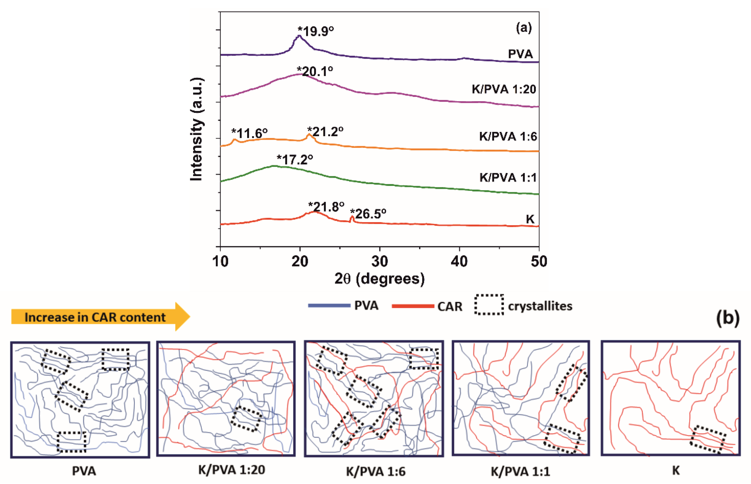
The XRD diffraction pattern of PVA (Figure 2a) is composed of one broad and low-intensity diffraction hallo centered at 2θ~20° (the (101) diffraction plane of a monoclinic unit cell), which indicates a low crystallinity degree (4.2%, Table 2), following other studies regarding PVA hydrogels [39,40]. The diffractogram of pure κ-carrageenan reveals the presence of an amorphous hallo centered at 2θ = 21.8° and a weak diffraction contribution at 26.5°, which was ascribed in previous studies to inorganic trace impurities. The last peak does not appear in the diffractograms of the PVA/CAR mixtures, probably due to the dilution effect [41].
It can be concluded that in contrast to PVA, pure κ-carrageenan does not form crosslinked hydrogels through the application of successive freeze-thaw cycles (almost null gel content: Table 1, and low crystallinity: Table 2). However, its blending with PVA in low amounts determines a cryostructuration of the resulting gel, with the formation of an increased number of highly-ordered domains (crystallites, Figure 2b), through intermolecular cooperative hydrogen bonding between the two polymers (evidenced by FTIR spectroscopy) [42].
An increase in crystallinity comparing to PVA was observed for the K/PVA 1:6 gel (two diffraction peaks at 11.6° and 21.2°, the latter being ascribed to the crystallites formed by PVA). A relatively high value of crystallinity is observed for K/PVA 1:20, while at high CAR: PVA ratios, such as in the K/PVA 1:1 gel, carrageenan determines the destructuration of the crystalline nature of the material.
Shifting of the ~20° maximum to higher diffraction angles for K: PVA 1:6 and K/PVA 1:20 implies a decrease of the crystallite size (through an increase in the macromolecular interactions by hydrogen bonding). In contrast, for K/PVA 1:1, shifting of the diffraction maximum at lower angles implies an increase in the crystallite dimensions (larger crystallites could be formed through the inclusion of CAR macromolecules). The crystallite sizes (D) ascribed to 2θ ~20° (Table 2) are determined with the Debye–Scherrer equation (Equation (7)) [40]:
D = 0.89 · λ B · c o s θ
where λ is the wavelength corresponding to the Cu Kα line (0.40784 nm), B is the full width at half maximum of the diffraction peak and θ is the diffraction angle maximum (in radians).
The number of crystalline domains in the hydrogel network plays an essential role in the performance of these materials. A high crystallinity determines a higher toughness (indicated by the higher Shore 00 hardness values from Table 1 for the PVA and K/PVA 1:6 cryogels), better dimensional stability (through the higher gel content G, depicted in Table 1) and lower diffusion coefficients of the active principles from the polymer network.
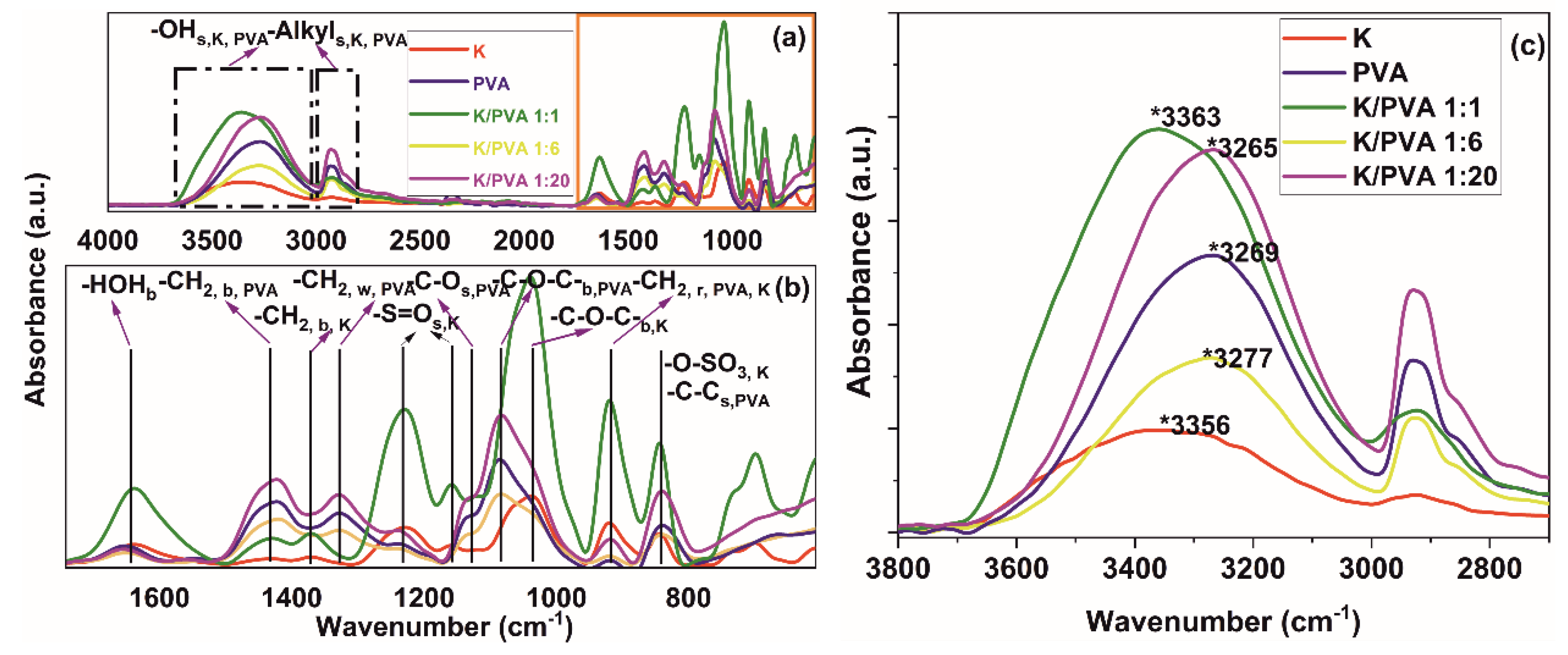
The fingerprint region of the IR absorption spectra (Figure 3a,b) reveals typical absorption bands of PVA, occurring at 839, 917 and 1089 cm−1 ascribed to C–C bending, -CH2 rocking and ester C-O-C bending from the acetyl groups [39,43,44,45]. In κ-carrageenan, the vibrations from 841, 921 and 1035 cm−1 could be assigned to -C-O-SO3- stretching vibrations from the galactose-4-sulphate, -CH2 rocking from the 3,6-anhydro-D-galactose structural unit, glycosidic -C-O-C- linkage (from the 3,6 anhydro-D-galactose) and ester sulfate -S=O stretching (1159, 1235 cm−1) [46,47].
The vibration at 1128 cm−1 from PVA is ascribed to the stretching of C-O from the crystalline region of the macromolecular chain where hydrogen bonds are formed between two neighboring isotactic OH groups. The ratio between the height of the bands centered at 1128 and 1089 cm−1 could be regarded as a crystallinity index (CrI) [43,45], proportional to the crystallinity calculated from the XRD diffractograms (CrXRD) (Table 2).
For both PVA and CAR, strong, broad absorptions are registered at 3269 cm−1 (PVA) and 3356 cm−1 (CAR), attributed to the stretching vibration ν(O-H) from hydrogen-bonded -OH groups and adsorbed water (Figure 3c). Supplementary, bending vibrations due to non-bonded water presence δ(O–H) are registered at ~1640 cm−1. Also, the strong absorptions at ~2932 cm−1 (PVA) and ~2925 cm−1 (CAR) are attributed to C-H stretching [43,44,45,46,47,48,49].
No new absorption bands apart from that characteristic of PVA, respectively CAR, could be noticed in the FTIR spectra of PVA/CAR mixtures, which could be an indication of physical interaction between the two components through hydrogen bonding. The crystallinity-sensible band of PVA (1128 cm−1) shifts to lower wavenumbers for the case of K/PVA 1:6 and K/PVA 1:20, indicating an increase in the intra- and intermolecular associations between the macromolecules through physical bonding (O-H…O), as found in the reference literature for other PVA-(bio)polymer mixtures [42,48]. Similarly, the -S=O stretching band at 1159 cm-1 shifts to lower wavenumbers and increases in width (determined at half maximum) in all PVA/CAR mixtures, possibly indicating SO3H…O hydrogen bonding between CAR and PVA [18].
The O-H…O distances (R, expressed in Å) in hydrogen-bonded crystalline domains could be correlated to the wavenumbers of infrared absorption bands attributed to the O-H stretching vibrations (νOH) with the Pimentel–Sederholm equation (Equation (8)) [50]:
Δ ν ( c m 1 ) = 4430 · ( 2.84 R )
where Δν (cm−1) = νOH- ν0; and ν0 is the wavenumber associated with free -OH groups stretching (3650 cm−1).
The hydrogen bonds energy (EH) could be calculated with Equation (9) [51]:
E H = 1 K · ν 0 ν O H ν 0
where K is a constant equal to 1.6 10−2 kcal [51].
It can be seen from Table 2 that strong interactions through hydrogen bonding occurs for the PVA hydrogel, K/PVA 1:20 and especially K/PVA 1:6 (high values of EH, respectively low values for R). The crystallinity index determined through FTIR band intensity ratios is proportional to the XRD-related crystallinity.
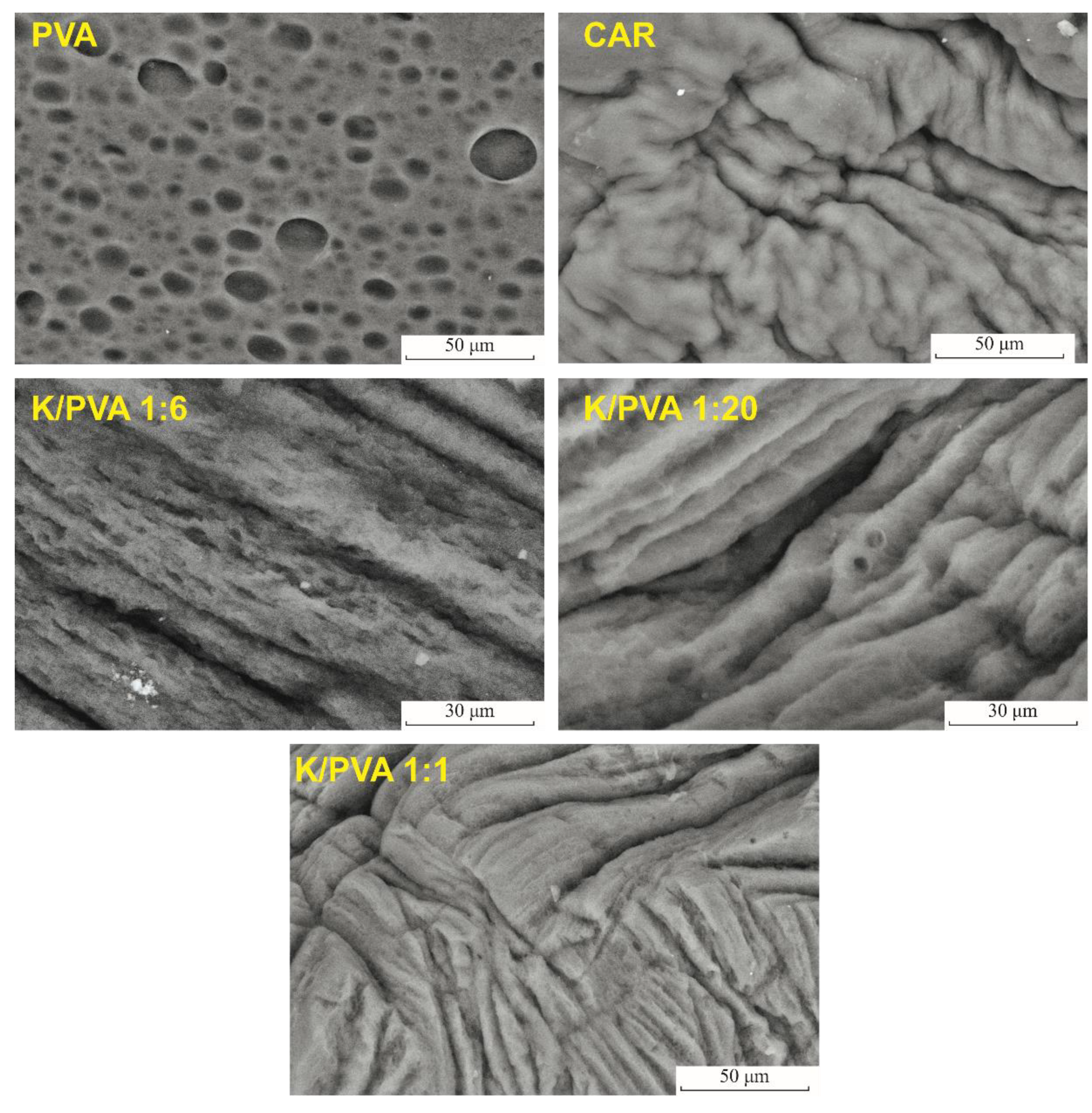
Freezing of the water-rich regions of the PVA aqueous solutions determines the formation of ice crystals, which, upon thawing, generate the characteristic pore network in the structure of the cryogel. Macropores with an average diameter of 9.7 μm were observed on the surface of the dried PVA hydrogel (Figure 4).
The freeze-thawed κ-carrageenan material has a compact surface, with no apparent porosity, which indicates that this polymer’s gel does not form its pore network through the same mechanism as PVA (the water-polymer phase separation occurs in a low amount). Carrageenan addition determines a decrease in the pore size and their relative amount, as can be seen from the SEM micrographs of the dried hydrogels (Figure 4) and the cyclohexane uptake-related pore volume ratio (Table 2). In accordance with the XRD crystallinity and the FTIR crystallinity index of the polymer matrix, the highest porosity for the blend cryogels is recorded for K/PVA 1:6, as, in this case, a maximum increase in polymer-rich zones through phase separation occurs.
Rearrangement of the polymer macromolecules and extensive intermolecular hydrogen bonding determines a compacting of the polymer phase and increase in the pore size, as it was observed in other studies [52,53]. K/PVA 1:20 and K/PVA 1:1 exhibit phase separation to a lower extent.
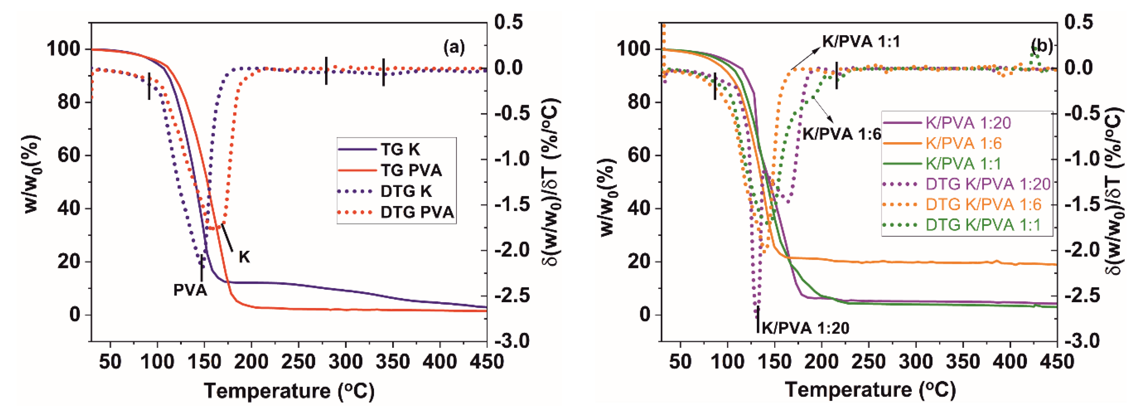
The thermal behavior of PVA, CAR and PVA/CAR mixtures could be observed from the TGA and DTG (first derivative data of the TG curve) thermograms in Figure 5a,b.
The DTG thermograms of the hydrogels reveal the presence of several thermal mass loss/decomposition stages (Table 3). The first mass loss stage is attributed to water evaporation from the materials. It can be observed that the evaporation onsets for the PVA and K materials are generally lower than those for the PVA/CAR mixtures (except for the K/PVA 1:1 hydrogel), which could indicate a higher degree of interaction between the water and polymer(s) through hydrogen bonding [54].
Degradation generally occurs in three distinct steps for all the materials. For PVA, the first degradation step involves the formation of double bonds in the polymer backbone through water elimination, the second degradation step involves the formation of aldehyde and alkene end groups, and the third corresponds to the further degradation of the remaining organic residue [55,56]. For CAR, the first degradation step involves the cleavage of the -CH-O-SO3 bonds, secondly, of the glycosidic bonds between the 3,6 anhydro-D-galactose units, and finally degradation of the bulk polymer backbone [57]. It seems that CAR addition determines an increase in the first degradation step temperature onset, possibly due to an increase in the material’s crystallinity. An indication of the excellent compatibility between PVA and CAR (co-blending) and the mixture’s thermal stability could also be linked with the values of the mass loss due to degradation (∆mdegrad, calculated as the percentual mass difference between the mass of the material at the onset of first degradation stage, and the final mass, determined at 450 °C), respectively with the values of the residue mass (∆mresidue, determined at 450 °C) depicted in Table 3. These values are lower for the PVA/CAR hydrogels, compared to the PVA and K hydrogels. Remarkable stability has been registered for the K/PVA 1:6 hydrogel, similar to other blends of poly (vinyl alcohol) (e.g., PVA/β-cyclodextrin gels [58], PVA-dextran gels [59] or PVA-starch films [60]).
Apart from the material’s crystallinity increase with the addition of CAR (0.047% and 0.139% weight ratios), thermal stability could be enhanced by the decrease in the porosity of the hydrogels (also observed in the SEM micrographs from Figure 4). Therefore, in low amounts, κ-carrageenan acts as a thermal insulator and mass transport barrier for the volatile products generated during thermal decomposition.
Several endothermic peaks could be observed in the DTA thermograms of the PVA and K hydrogels (Figure 6a) and respectively of the PVA/CAR mixtures (Figure 6b). The broad shoulder from 95–105 °C could be ascribed to associated and non-associated water vaporization from the polymer gel. In contrast, the peaks at ~170 °C (PVA), and ~150 °C (K) could be attributed to the melting of PVA [56] and the double-helix conformation destructuration (helix to coil transition) in the case of κ-carrageenan [61]. The exact temperature values for the two transitions are depicted in Table 3. For the K/PVA 1:6 and K/PVA 1:20 hydrogels, only a single peak could be registered by DTA, which could be an indicator of an excellent compatibility between the two polymers and their reciprocal stabilization.
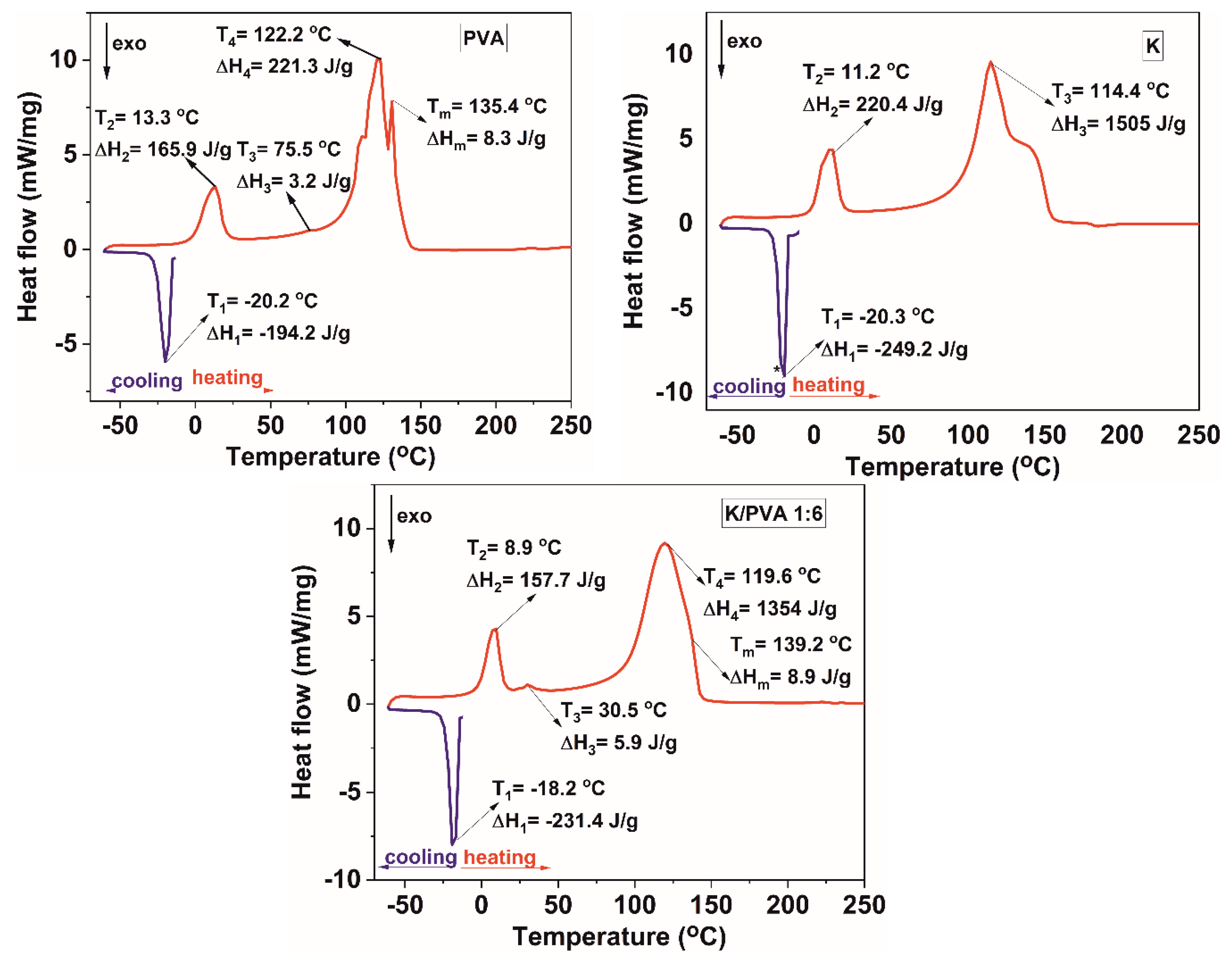
The cooling curve of the thermograms presented in Figure 7 (for PVA, K and the K/PVA 1:6 hydrogel; others not shown here) contains one feature, namely a broad exothermic peak ascribed to the crystallization of water from the hydrogel network [62]. The heating profiles from the DSC thermograms illustrated in Figure 7 reveal several endotherm peaks, indicative of the multiple transitions occurring in this complex system [63].
An endotherm peak at ~13 °C (PVA) and ~11 °C (K) indicates the melting temperature of the non-associated (free) water in the hydrogel [62]. The temperature maxima associated with this transition for the PVA/CAR hydrogels are lower than those corresponding to PVA and K. This decrease in “thawing” temperature is proportional to the κ-carrageenan content from the gels. For the gel with the highest crystallinity degree and highest thermal stability (K/PVA 1:6), this temperature is ~9 °C, as illustrated in Figure 7.
Generally, the presence of a single glass transition peak in the DSC thermograms for a polymeric blend can be related to good impermissibility and interaction between the constituents [42]. In this case, however, a single value for Tg is not correlated to the formation of inter-polymer complexes through hydrogen bonding because κ-carrageenan does not show any presence of this type of transition. For the PVA hydrogel, a peak at ~75 °C could be ascribed to the glass transition of poly (vinyl alcohol), following the values from the reference literature [64].
The crystallinity of the PVA matrix in the gels (CrDSC) was assessed also based on the DSC data, by dividing the value of the melting enthalpy (∆Hm) to the value of the thermodynamic enthalpy corresponding to the melting of a 100% crystalline PVA (150 J/g), as reported in the literature [64]. The results, presented in Table 3 follow the same trend as the XRD-calculated crystallinity and the FTIR crystallinity index, confirming the semi-crystalline nature of the polymer matrix [40]. The highest value being registered for the K/PVA 1:6 cryogel, in which a maximum amount of phase separation and hydrogen bonding occurs.
A decrease in the Tg values was noted for all PVA/CAR mixtures in comparison with the PVA hydrogel (for the K/PVA 1:6 hydrogel, Tg is ~30 °C), this decrease occurs due to the combined plasticizing effect of κ-carrageenan and water.
Complex, broad endotherm peaks appear in the 100–150 °C temperature interval, which could be ascribed to water vaporization from PVA and K, the melting of PVA (which occurs over a range of temperatures, due to the presence of both amorphous and crystalline regions, as well as polymer chains with different stereoisomerism) and destructuration of k-carrageenan [62,63,65].

3.2. Gels Swelling and Adsorption Behavior

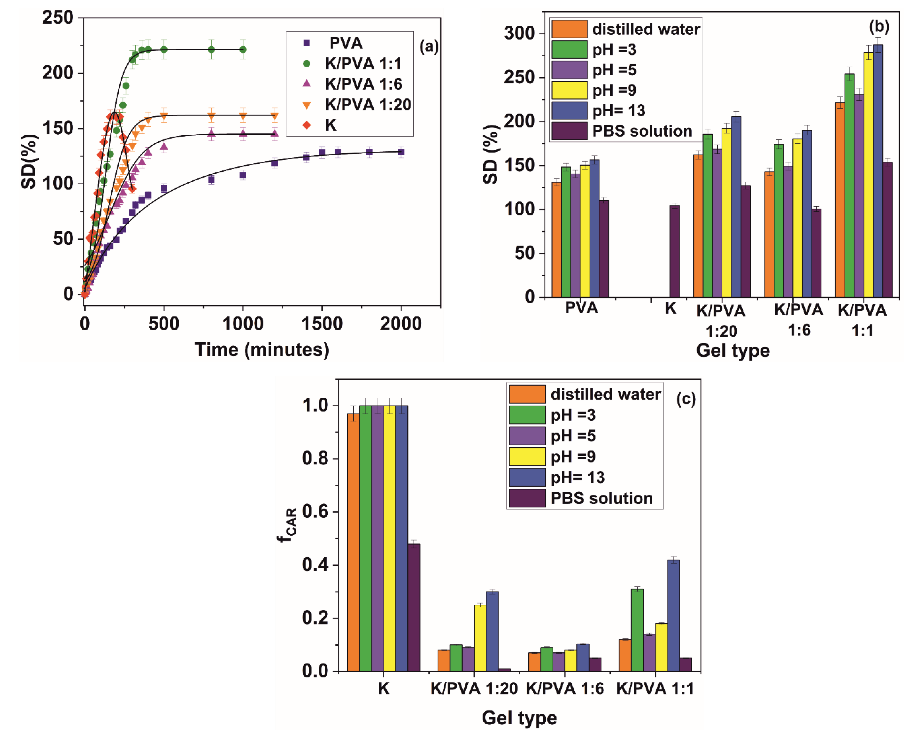
The distilled water swelling kinetic for the obtained hydrogels is presented in Figure 8a, and the values of the swelling degree at equilibrium in different aqueous media is presented in Figure 8b.
The material comprised solely of κ-carrageenan is not stable in distilled water and aqueous media with various acidic, respectively basic pH values. This could be due to the absent crosslinking points in K, also evidenced through XRD and FTIR spectroscopy results and nil gel content value. In the PBS solution, carrageenan is more stable, probably due to the crosslinking effect promoted by the K+ and Na+ cations [66]. Increasing the κ-carrageenan content in the hydrogel determines an increase in the rate of sorption and the equilibrium water content, following the values presented in the reference literature for other types of PVA-hydrophilic biopolymer (starch, chitosan and gelatin) hydrogels [67,68,69].
Modification in the pH values of the solution determines an overall increase in the swelling degree of the hydrogel. Even if PVA is a non-ionic polymer, modifications in the pH could affect the swelling degree of the hydrogel due to the kosmotropic effect of the H+ and OH ions, which determine a restructuration of the hydrogen bonding in PVA and a modification in the association degree of bonded water molecules in the hydrogel [70]. This mechanism is also prevalent in κ-carrageenan, for which the sulfate groups are completely ionized at pH values greater than 2.8 [71]. At pH values between 5.0 and 7.00, the materials containing carrageenan are reasonably stable, as could be deducted from the fractional released amount values presented in Figure 8c. In contrast, at different pH values the carrageenan sol is disrupted, which translates into a decrease in the stability of the materials containing this type of polymer at pH values lower than 5.0 and higher than 7.0. The most stable material in terms of carrageenan leaching from the hydrogel structure is K/PVA 1:6, for which the highest crystallinity values were registered.
Several parameters regarding the water diffusion into the hydrogel matrix were calculated, starting from the experimental swelling curves of the materials and depicted in Table 4.
The rates at which the solvent front advances from the surface to the center of the hydrogel samples were determined by calculating the media penetration velocity (ν), according to Equation (10) [72].
v = 1 2 ρ A · m t .
In Equation (10), ρ represents the density of the swelling medium, A is the area of the hydrogel disc sample, and ∂m/∂t represents the time variation of the hydrogel mass when in contact with the swelling medium, calculated for t < 20 min, for which ∂m/∂t is constant.
For mt/meq ≤ 0.5, Equation (11) permits the determination of the so-called “early-time” diffusion coefficient De, while Equation (12), valid for mt/meq > 0.6, permits the determination of the “late-time” diffusion coefficient, Dl [73]. A complementary modeling method for the early-time approximation is the power-law approach introduced by Peppas and depicted in Equation (13), where k is a constant indicating the swelling rate and n denotes the type of transport mechanism. When n is equal to 0.5 (and according to some researchers ≤ 0.5), the transport rate of the solute into the hydrogel is Fickian, i.e., the solvent penetration rate is higher than the polymer chain relaxation rate (Equation (12) assumes a Fickian diffusion, i.e., n = 0.5 and k = 4 D e / δ π ). When n takes values between 0.5 and 1.0, the solute transport is controlled by polymer relaxation and swelling (anomalous diffusion). Case-II type diffusion occurs when n is equal to 1.0, while super-case II transport occurs when n is greater than 1.0, which both indicate to a great extent a relaxation-controlled transport [74].
m t m e q = 4 [ D e · t π · δ 2 ] 0.5
m t m e q = 1 8 π 2 · e x p [ D l · π · t δ 2 ]
m t m e q = k · t n .
As can be seen from Table 4, the water penetration velocity ν in the hydrogels submerged in distilled water increases with the amount of κ-carrageenan, with one exception: namely K/PVA 1:6, for which the highest crystallinity was registered. In the latter case, the increased number of crosslink points leads to a retarding of the water penetration front into the material. With the modification of pH, all values related to the solvent penetration velocity (and implicitly polymer chain relaxation) increase. In PBS, due to the higher ionic strength of this medium and kosmotropic nature of the ions, a “collapsing” of the hydrogel network could be registered (Figure 8b), which retards the water penetration. The hydrogels exhibiting higher media penetration velocities (i.e., permeability) also presented higher values for the equilibrium swelling degree.
The early-time and late-time diffusion coefficients calculated from Equations (12) and (13) reported in Table 4 range from 5.42 10−5 to 8.97 10−4 mm2/min, respectively 0.0879 to 0.480 mm2/min, increasing in the same manner as the solvent penetration rate ν. The relatively high values for the diffusion coefficients are comparable to other PVA-containing hydrogels [75,76], and imply a high degree of polymer chains relaxation (this could also be deducted from the values of n, which range from 0.73 to 1.00). The hydrogels immersed in PBS represent an exception, in the sense that the water sorption is combined with the PVA “collapsing” and κ-carrageenan crosslinking. This retards the swelling of the polymer matrix. Thus, the diffusion of water in these materials follow a mechanism closer to the ideal Fickian one.
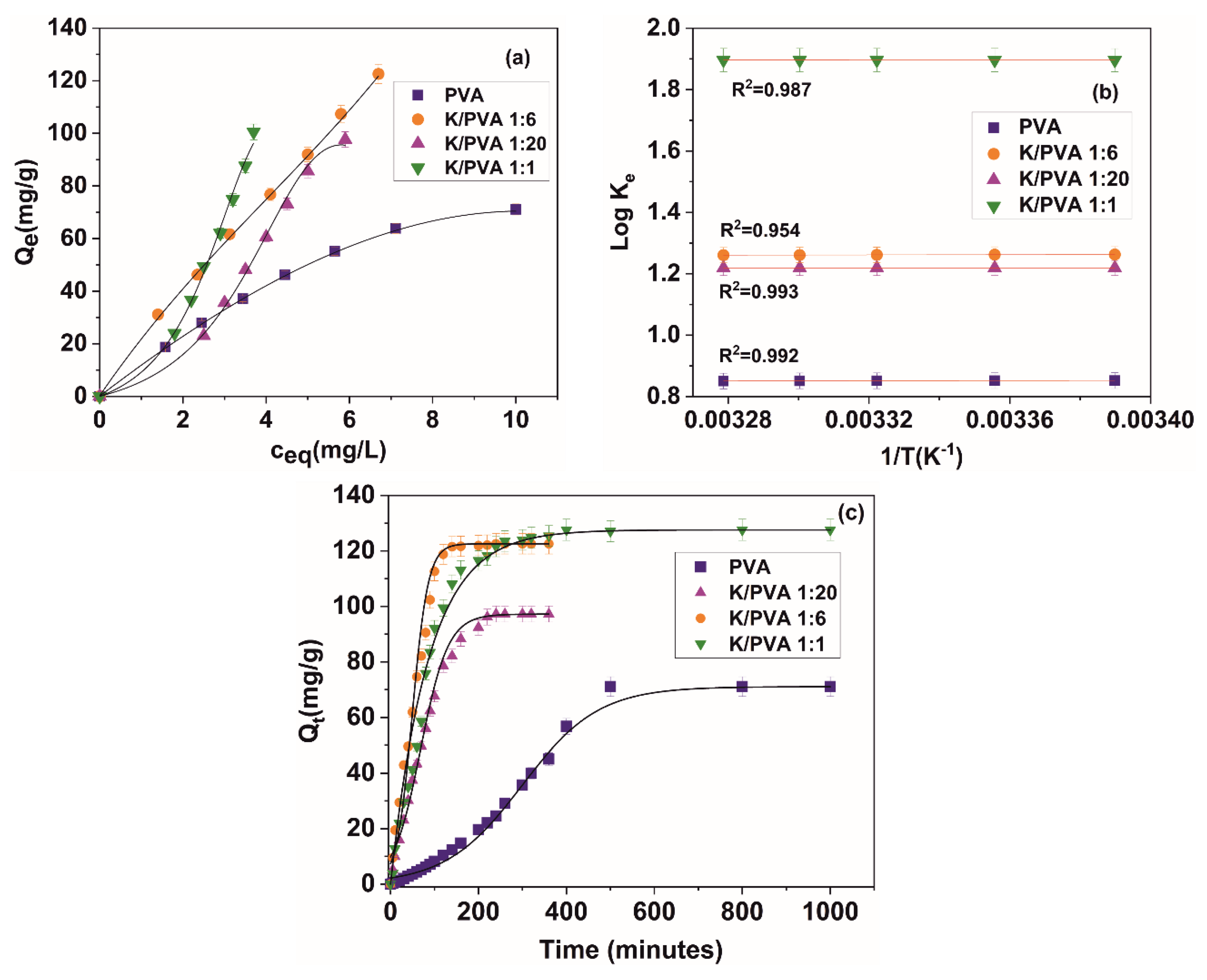
The ability of the hydrogels to function as sorbents for potentially toxic chemical species removal from wastewaters was also studied. The adsorption and kinetic mechanism for crystal violet sorption onto the previously water-equilibrated hydrogels was studied by modelling the adsorption data from Figure 9a with various isotherm models, presented in Table 5. The theory behind each type of isotherm model can be found in the reference literature [77,78,79,80]. Only the physical meaning and measurement units for each parameter were given at the end of the Table 5. Crystal violet was chosen as a model dye for sorption as it does not interact with the hydrogel components through forming complexes that have a maximum absorption at different wavelengths than the original dye and due to the vast available literature for different sorbent types and sorption conditions, allowing for easy comparison of the obtained sorption capacities. The maximum sorption capacities at equilibrium (Qe) for CV range from 63.2 mg/g (in the case of PVA) to 121.4 mg/g (in the case of K/PVA 1:6).
These values are comparable to other results reported in the reference literature, for CV sorption on other types of hydrogels: 49.0 mg/g (poly (acrylic acid) hydrogels) [81]; 105.2 mg/g (carrageenan/multi-walled carbon nanotube hybrid hydrogel) [82] and 16.3 mg/g (κ-carrageenan hydrogel beads) [83].
The results from Table 5 indicate that the Freundlich and the Redlich–Peterson adsorption isotherms best fit the experimental results in the case of PVA, K/PVA 1:20 and K/PVA 1:6. This is indicative of a heterogenous non-ideal adsorption mechanism, not restricted to the formation of a monolayer of adsorbate [77]. For the K/PVA 1:20 and the K/PVA 1:1 hydrogel, the 1/nF values are higher than 1, which indicate a cooperative adsorption mechanism. In the case of PVA and K/PVA 1:6, the adsorption is strongly favored.
From Figure 9b and Table 5 we can see that the adsorption of CV onto the surface of the hydrogel is an exothermic process for PVA and K/PVA 1:6. For these two types of hydrogel, the adsorption is spontaneous and of a physical nature, indicated by the slightly negative values of ∆G [81], and is observed to decrease with the increase in temperature. For all hydrogels, the ∆S values are positive, indicating the increased randomness at the solid/liquid interface in the adsorption process. For K/PVA 1:20 and K/PVA 1:1, the adsorption is not thermodynamically favored (∆G > 0), but at higher temperatures, lower values could be registered for ∆G.
The contact time data from Figure 9c was fitted against various kinetic models, such as pseudo-first order, pseudo-second-order, intra-particle diffusion, liquid film diffusion and Elovich. The significance of each model was not presented here; however, it can be consulted from the reference literature [84,85,86]. The equations of the kinetic models, the parameters of the equations and the determination coefficient indicating the goodness of the fit (R2) are depicted in Table 6.
For PVA, K/PVA 1:20 and K/PVA 1:6, the model that best fits the experimental kinetic data is the pseudo-second order over the entire contact time range, which is consistent with the findings from the reference literature for this type of dye and hydrogel sorbents [85]. The high determination coefficients for the pseudo-second-order model implies that the sharing or exchange of electrons between the polymer substrate and CV (for PVA) and the electrostatic interaction between the ionized chemical groups of κ-carrageenan and the organic cation of CV is the rate-controlling step. For the K/PVA 1:1 hydrogel, the boundary layer effect (i.e., transport of the CV from the boundary liquid film to the surface of the adsorbing material) is the rate-limiting step.
Charge carrier transfer, electrostatic interaction and liquid film diffusion are not the only processes involved in CV adsorption. The Weber and Morris intra-particle diffusion model fits well the first 20% of the sorption kinetic for all types of hydrogel. Based on this model, the sorption of CV on the hydrogels could be described as a three-step process: (1) the transport of CV from the boundary liquid film to the surface of the adsorbent (external film mass transfer); (2) the further transfer of CV from the surface of the adsorbate to the internal active binding sites (intraparticle diffusion); and (3) the interaction of CV with the active binding sites. The appropriateness of the Weber and Morris intra-particle diffusion model to describe the sorption kinetic in the early-time approximation is extensively described by other studies involving hydrogel or biomass-derived materials [85,86]. At longer contact times, the external mass transfer and dynamic equilibrium processes could also play an important role in CV sorption.
From the values of k2 (Table 6), with the increase in the amount of κ-carrageenan, the sorption process occurs with a higher rate and the equilibrium is reached in a shorter period.

4. Conclusions

The addition of κ-carrageenan to PVA determines the obtaining of hydrophilic hydrogels with a high distilled water swelling degree, up to 224%, compared to neat PVA, for which a value of 115% was registered. Carrageenan also determines the decrease with up to 58% in the crystallinity index for the polymer blend matrix, compared to PVA. Also, a higher water diffusion coefficient in/through the polymer matrix (up to 8.24 × 10⁻⁴ mm2/min, compared to 1.82 × 10⁻⁵ mm2/min for PVA) was attained.
The results indicate that several types of interactions occur between the two polymers, implying hydrogen bonding. These interactions are enhanced for the hydrogel containing a κ-carrageenan weight fraction of 0.139, for which the highest stability in aqueous media of various ionic strength and pH was observed. κ-carrageenan also determines an increased affinity of the hydrogels towards crystal violet cationic dye. This physisorption process is generally thermodynamically favored and was well modelled by the Freundlich isotherm mechanism.

Author Contributions

Conceptualization, C.C.; methodology C.C., M.A.P.; formal analysis, C.C., M.A.P., I.C.R.; investigation, C.C., M.A.P., T.B., M.C., I.C.R., I.H.; resources, C.C.; data curation, C.C., I.C.R.; writing—original draft preparation, C.C., M.A.P., I.C.R.; writing—review and editing, C.C., M.A.P., I.C.R.; visualization, C.C., I.C.R.; supervision, C.C.; project administration, C.C.; funding acquisition, C.C. All authors have read and agreed to the published version of the manuscript.

Funding

The authors acknowledge the structural funds project PRO-DD (POS-CCE O.2.2.1, ID 123, SMIS 2637, ctr. No 11/2009) for providing the infrastructure used in this work at the CDI Institute of Transilvania University of Brasov.

Conflicts of Interest

The authors declare no conflict of interest. The funders hfad no role in the design of the study; in the collection, analyses, or interpretation of data; in the writing of the manuscript, or in the decision to publish the results.

References

  1. Wang, X.D.; Liu, X.H.; Yuan, H.Y.; Liu, H.; Liu, C.T.; Li, T.X.; Yan, C.; Yan, X.R.; Shen, C.Y.; Guo, Z.H. Non-covalently functionalized graphene strengthened poly(vinyl alcohol). Mater. Des. 2018, 139, 372–379. [Google Scholar] [CrossRef]
  2. Ahmed, E.M. Hydrogel: Preparation, characterization, and applications: A review. J. Adv. Res. 2015, 6, 105–121. [Google Scholar] [CrossRef] [PubMed] [Green Version]
  3. Wang, Y.J.; Zhang, X.N.; Song, Y.H.; Zhao, Y.P.; Chen, L.; Su, F.M.; Li, L.B.; Wu, Z.L.; Zheng, Q. Ultrastiff and Tough Supramolecular Hydrogels with a Dense and Robust Hydrogen Bond Network. Chem. Mater. 2019, 31, 1430–1440. [Google Scholar] [CrossRef]
  4. Wang, X.; Su, M.; Liu, C.; Shen, C.; Liu, X. Poly (vinyl alcohol)/Graphene Nanocomposite Hydrogel Scaffolds for Control of Cell Adhesion. J. Renew. Mater. 2020, 8, 89–99. [Google Scholar] [CrossRef]
  5. Narayanaswamy, R.; Torchilin, V.P. Hydrogels and Their Applications in Targeted Drug Delivery. Molecules 2019, 24, 603. [Google Scholar] [CrossRef] [Green Version]
  6. Ghanaatian, E.; Entezam, M. Mechanical properties and drug release rate of poly(vinyl alcohol)/poly(ethylene glycol)/clay nanocomposite hydrogels: Correlation with structure and physical properties. J. Appl. Polym. Sci. 2019, 136. [Google Scholar] [CrossRef]
  7. Liu, L.S.; Kost, J.; Yan, F.; Spiro, R.C. Hydrogels from Biopolymer Hybrid for Biomedical, Food, and Functional Food Applications. Polymers (Basel) 2012, 4, 997–1011. [Google Scholar] [CrossRef]
  8. El Halah, A.; Lopez-Carrasquero, F.; Contreras, J. Applications of hydrogels in the adsorption of metallic ions. Cienc. Ing. 2018, 39, 57–70. [Google Scholar]
  9. Guenther, M.; Gerlach, G.; Sorber, J.; Suchaneck, G.; Arndt, K.F.; Richter, A. pH sensors based on polyelectrolytic hydrogels. Smart Struct. Mater. 2005, 5759, 540–548. [Google Scholar] [CrossRef]
  10. Duquette, D.; Dumont, M.J. Comparative studies of chemical crosslinking reactions and applications of bio-based hydrogels. Polym. Bull. 2019, 76, 2683–2710. [Google Scholar] [CrossRef]
  11. Crescenzi, V.; Paradossi, G.; Desideri, P.; Dentini, M.; Cavalieri, F.; Amici, E.; Lisi, R. New hydrogels based on carbohydrate and on carbohydrate-synthetic polymer networks. Polym. Gels. Netw. 1997, 5, 225–239. [Google Scholar] [CrossRef]
  12. Sultankulov, B.; Berillo, D.; Kauanova, S.; Mikhalovsky, S.; Mikhalovska, L.; Saparov, A. Composite Cryogel with Polyelectrolyte Complexes for Growth Factor Delivery. Pharmaceutics 2019, 11, 650. [Google Scholar] [CrossRef] [PubMed] [Green Version]
  13. Bagri, L.P.; Saini, R.K.; Bajpai, A.K.; Choubey, R. Silver hydroxyapatite reinforced poly(vinyl alcohol)-starch cryogel nanocomposites and study of biodegradation, compressive strength and antibacterial activity. Polym. Eng. Sci. 2019, 59, 254–263. [Google Scholar] [CrossRef]
  14. Kamoun, E.A.; Kenawy, E.R.S.; Chen, X. A review on polymeric hydrogel membranes for wound dressing applications: PVA-based hydrogel dressings. J. Adv. Res. 2017, 8, 217–233. [Google Scholar] [CrossRef]
  15. Lotfipour, F.; Alami-Milani, M.; Salatin, S.; Hadavi, A.; Jelyehgari, M. Freeze-thaw-induced cross-linked PVA/chitosan for oxytetracycline-loaded wound dressing: The experimental design and optimization. Res. Pharm. Sci. 2019, 14, 175–189. [Google Scholar] [CrossRef]
  16. Santos, C.; Silva, C.J.; Buttel, Z.; Guimaraes, R.; Pereira, S.B.; Tamagnini, P.; Zille, A. Preparation and characterization of polysaccharides/PVA blend nanofibrous membranes by electrospinning method. Carbohyd. Polym. 2014, 99, 584–592. [Google Scholar] [CrossRef] [Green Version]
  17. Yegappan, R.; Selvaprithiviraj, V.; Amirthalingam, S.; Jayakumar, R. Carrageenan based hydrogels for drug delivery, tissue engineering and wound healing. Carbohyd. Polym. 2018, 198, 385–400. [Google Scholar] [CrossRef]
  18. Bajpai, S.K.; Daheriya, P. Kappa-Carrageenan/PVA Filmswith Antibacterial Properties: Part 1. Optimization of Preparation Conditions and Preliminary Drug Release Studies. J. Macromol. Sci. A 2014, 51, 286–295. [Google Scholar] [CrossRef]
  19. El-Fawal, G.F.; Yassin, A.M.; El-Deeb, N.M. The Novelty in Fabrication of Poly Vinyl Alcohol/kappa-Carrageenan Hydrogel with Lactobacillus bulgaricus Extract as Anti-inflammatory Wound Dressing Agent. AAPS PharmSciTech 2017, 18, 1605–1616. [Google Scholar] [CrossRef]
  20. Soares, S.F.; Simoes, T.R.; Trindade, T.; Daniel-da-Silva, A.L. Highly Efficient Removal of Dye from Water Using Magnetic Carrageenan/Silica Hybrid Nano-adsorbents. Water Air Soil Poll. 2017, 228. [Google Scholar] [CrossRef]
  21. Rasool, A.; Ata, S.; Islam, A.; Khan, R.U. Fabrication of novel carrageenan based stimuli responsive injectable hydrogels for controlled release of cephradine. RSC Adv. 2019, 9, 12282–12290. [Google Scholar] [CrossRef] [Green Version]
  22. Sukhlaaied, W.; Riyajan, S.A. Synthesis and properties of carrageenan grafted copolymer with poly(vinyl alcohol). Carbohyd. Polym. 2013, 98, 677–685. [Google Scholar] [CrossRef] [PubMed]
  23. Dafader, N.C.; Manir, M.S.; Alam, M.F.; Swapna, S.P.; Akter, T.; Huq, D. Effect Of Kappa-Carrageenan On The Properties Of Poly(Vinyl Alcohol) Hydrogel Prepared By The Application Of Gamma Radiation. SOP Trans. Appl. Chem. 2015, 2, 1–12. [Google Scholar] [CrossRef]
  24. Zhang, Y.B.; Ye, L.; Cui, M.; Yang, B.G.; Li, J.J.; Sun, H.; Yao, F.L. Physically crosslinked poly(vinyl alcohol)-carrageenan composite hydrogels: Pore structure stability and cell adhesive ability. RSC Adv. 2015, 5, 78180–78191. [Google Scholar] [CrossRef]
  25. Hosseinzadeh, H. Freezing-Thawing Assisted Synthesis of Novel Carrageenan Nanomagnetic Beads for Controlled Release of Antitumor Drugs. Cell. Chem. Technol. 2017, 51, 529–537. [Google Scholar]
  26. Chopra, P.; Nayak, D.; Nanda, A.; Ashe, S.; Rauta, P.R.; Nayak, B. Fabrication of poly(vinyl alcohol)-Carrageenan scaffolds for cryopreservation: Effect of composition on cell viability. Carbohyd. Polym. 2016, 147, 509–516. [Google Scholar] [CrossRef]
  27. Dodgson, K.S.; Price, R.G. A note on the determination of the ester sulphate content of sulphated polysaccharides. Biochem. J. 1962, 84, 106–110. [Google Scholar] [CrossRef] [Green Version]
  28. Patachia, S.; Croitoru, C. Increasing the adsorption capacity and selectivity of poly(vinyl alcohol) hydrogels by an alternative imprinting technique. J. Appl. Polym. Sci. 2015, 132. [Google Scholar] [CrossRef]
  29. Roata, I.C.; Croitoru, C.; Pascu, A.; Stanciu, E.M. Characterization of Physically Crosslinked Ionic Liquid-lignocellulose Hydrogels. Bioresources 2018, 13, 6110–6121. [Google Scholar]
  30. Varca, G.H.C.; Ferraz, C.C.; Mathor, M.B.; Lopes, P.S.; Rogero, S.O.; Rogero, J.O. Encapsulation and nano-encapsulation of papain active sites to enhance radiolytic stability and decrease toxicity. In Nanoscale Radiation Engineering of Advanced Materials for Potential Biomedical Applications; IAEA RADIATION TECHNOLOGY REPORTS: Vienna, Austria, 2015; pp. 31–46. [Google Scholar]
  31. Kokabi, M.; Sirousazar, M.; Hassan, Z.M. PVA-clay nanocomposite hydrogels for wound dressing. Eur. Polym. J. 2007, 43, 773–781. [Google Scholar] [CrossRef]
  32. Jain, E.; Kumar, A. Designing Supermacroporous Cryogels Based on Polyacrylonitrile and a Polyacrylamide-Chitosan Semi-interpenetrating Network. J. Biomat. Sci. Polym. Ed. 2009, 20, 877–902. [Google Scholar] [CrossRef] [PubMed]
  33. Braet, F.; deZanger, R.; Wisse, E. Drying cells for SEM, AFM and TEM by hexamethyldisilazane: A study on hepatic endothelial cells. J. Microsc. Oxf. 1997, 186, 84–87. [Google Scholar] [CrossRef] [PubMed]
  34. Ling, Y.P.; Heng, L.Y. Complexation between Carrageenan and Methylene Blue for Sensor Design. In Proceedings of the 2013 Ukm Fst Postgraduate Colloquium, Selangor, Malaysian, 3–4 July 2013; Volume 1571, pp. 717–724. [Google Scholar] [CrossRef]
  35. Pehlivan, E.; Arslan, G. Removal of metal ions using lignite in aqueous solution—Low cost biosorbents. Fuel Process. Technol. 2007, 88, 99–106. [Google Scholar] [CrossRef]
  36. Caccavo, D.; Lamberti, G.; Cafaro, M.M.; Barba, A.A.; Kazlauske, J.; Larsson, A. Mathematical modelling of the drug release from an ensemble of coated pellets. Br. J. Pharmacol. 2017, 174, 1797–1809. [Google Scholar] [CrossRef] [PubMed] [Green Version]
  37. Wang, X.L.; Pan, Y.M.; Shen, C.Y.; Liu, C.T.; Liu, X.H. Facile Thermally Impacted Water-Induced Phase Separation Approach for the Fabrication of Skin-Free Thermoplastic Polyurethane Foam and Its Recyclable Counterpart for Oil-Water Separation. Macromol. Rapid Comm. 2018, 39. [Google Scholar] [CrossRef]
  38. Wang, Y.T.; Yang, H.G.; Chen, Z.H.; Chen, N.; Pang, X.C.; Zhang, L.; Minari, T.; Liu, X.Y.; Liu, H.Z.; Chen, J.Z. Recyclable Oil-Absorption Foams via Secondary Phase Separation. ACS Sustain. Chem. Eng. 2018, 6, 13834–13843. [Google Scholar] [CrossRef]
  39. Tretinnikov, O.N.; Zagorskaya, S.A. Determination of the degree of crystallinity of poly(vinyl alcohol) by FTIR spectroscopy. J. Appl. Spectrosc. 2012, 79, 521–526. [Google Scholar] [CrossRef]
  40. Ricciardi, R.; Auriemma, F.; De Rosa, C.; Laupretre, F. X-ray diffraction analysis of poly(vinyl alcohol) hydrogels, obtained by freezing and thawing techniques. Macromolecules 2004, 37, 1921–1927. [Google Scholar] [CrossRef]
  41. Liew, J.W.Y.; Loh, K.S.; Ahmad, A.; Lim, K.L.; Daud, W.R.W. Synthesis and characterization of modified kappa-carrageenan for enhanced proton conductivity as polymer electrolyte membrane. PLoS ONE 2017, 12. [Google Scholar] [CrossRef]
  42. Kantoglu, O.; Caykara, T.; Guven, O. Preparation and characterization of polysaccaride interpolymer complexes: I-PVA/iota-carrageenan. J. Appl. Polym. Sci. 2013, 127, 500–507. [Google Scholar] [CrossRef]
  43. Xu, W.G.; Asai, S.; Sumita, M. Spectroscopic study of ethylene vinyl alcohol copolymer and poly(vinyl alcohol). Sen’i Gakkaishi 1997, 53, 174–182. [Google Scholar] [CrossRef] [Green Version]
  44. Santos, A.M.N.; Moreira, A.P.D.; Carvalho, C.W.P.; Luchese, R.; Ribeiro, E.; McGuinness, G.B.; Mendes, M.F.; Oliveira, R.N. Physically Cross-Linked Gels of PVA with Natural Polymers as Matrices for Manuka Honey Release in Wound-Care Applications. Materials 2019, 12, 559. [Google Scholar] [CrossRef] [Green Version]
  45. Mansur, H.S.; Sadahira, C.M.; Souza, A.N.; Mansur, A.A.P. FTIR spectroscopy characterization of poly (vinyl alcohol) hydrogel with different hydrolysis degree and chemically crosslinked with glutaraldehyde. Mater. Sci. Eng. C Bio. S 2008, 28, 539–548. [Google Scholar] [CrossRef]
  46. Zinoun, M.; Cosson, J.; Deslandes, E. Physicochemical Characterization of Carrageenan from Gigartina-Teedii (Rooth) Lamouroux (Gigartinales, Rhodophyta). J. Appl. Phycol. 1993, 5, 23–28. [Google Scholar] [CrossRef]
  47. Pereira, L.; Amado, A.M.; Ribeiro-Claro, P.J.A.; van de Velde, F. VIBRATIONAL SPECTROSCOPY (FTIR-ATR AND FT-RAMAN) A Rapid and Useful Tool for Phycocolloid Analysis. In Proceedings of the Biodevices 2009: Proceedings of the International Conference on Biomedical Electronics and Devices, Porto, Portugal, 14–17 January 2009; pp. 131–136. [Google Scholar]
  48. Kenawy, E.; Kamoun, E.A.; Eldin, M.S.M.; El-Meligy, M.A. Physically crosslinked poly(vinyl alcohol)-hydroxyethyl starch blend hydrogel membranes: Synthesis and characterization for biomedical applications. Arab. J. Chem. 2014, 7, 372–380. [Google Scholar] [CrossRef] [Green Version]
  49. Gadhave, R.V.; Mahanwar, P.A.; Gadekar, P.T. Effect of glutaraldehyde on thermal and mechanical properties of starch and polyvinyl alcohol blends. Des. Monomers Polym. 2019, 22, 164–170. [Google Scholar] [CrossRef] [PubMed] [Green Version]
  50. Pimentel, G.C.; Sederholm, C.H. Correlation of Infrared Stretching Frequencies and Hydrogen Bond Distances in Crystals. J. Chem. Phys. 1956, 24, 639–641. [Google Scholar] [CrossRef]
  51. Zaltariov, M.F.; Filip, D.; Varganici, C.D.; Macocinschi, D. Atr-Ftir and Thermal Behavior Studies of New Hydrogel Formulations Based on Hydroxypropyl Methylcellulose/Poly(Acrilic Acid) Polymeric Blends. Cell. Chem. Technol. 2018, 52, 619–631. [Google Scholar]
  52. Holloway, J.L.; Lowman, A.M.; Palmese, G.R. The role of crystallization and phase separation in the formation of physically cross-linked PVA hydrogels. Soft Matter 2013, 9, 826–833. [Google Scholar] [CrossRef]
  53. Zhang, C.; Liu, X.H.; Liu, H.; Wang, Y.M.; Guo, Z.H.; Liu, C.T. Multi-walled carbon nanotube in a miscible PEO/PMMA blend: Thermal and rheological behavior. Polym. Test. 2019, 75, 367–372. [Google Scholar] [CrossRef]
  54. Rodriguez-Rodriguez, R.; Garcia-Carvajal, Z.Y.; Jimenez-Palomar, I.; Jimenez-Avalos, J.A.; Espinosa-Andrews, H. Development of gelatin/chitosan/PVA hydrogels: Thermal stability, water state, viscoelasticity, and cytotoxicity assays. J. Appl. Polym. Sci. 2019, 136. [Google Scholar] [CrossRef]
  55. Gomez, I.; Otazo, E.M.; Hernandez, H.; Rubio, E.; Varela, J.; Ramirez, M.; Barajas, I.; Gordillo, A.J. Thermal degradation study of PVA derivative with pendant phenylthionecarbamate groups by DSC/TGA and GC/MS. Polym. Degrad. Stabil. 2015, 112, 132–136. [Google Scholar] [CrossRef]
  56. Holland, B.J.; Hay, J.N. The thermal degradation of poly(vinyl alcohol). Polymer 2001, 42, 6775–6783. [Google Scholar] [CrossRef]
  57. Shahbazi, M.; Rajabzadeh, G.; Ettelaie, R.; Rafe, A. Kinetic study of kappa-carrageenan degradation and its impact on mechanical and structural properties of chitosan/kappa-carrageenan film. Carbohyd. Polym. 2016, 142, 167–176. [Google Scholar] [CrossRef] [PubMed]
  58. Teodorescu, M.; Bercea, M.; Morariu, S. Biomaterials of Poly(vinyl alcohol) and Natural Polymers. Polym. Rev. 2018, 58, 247–287. [Google Scholar] [CrossRef]
  59. Fathi, E.; Atyabi, N.; Imani, M.; Alinejad, Z. Physically crosslinked polyvinyl alcohol-dextran blend xerogels: Morphology and thermal behavior. Carbohyd. Polym. 2011, 84, 145–152. [Google Scholar] [CrossRef]
  60. Salleh, M.S.N.; Nor, N.N.M.; Mohd, N.; Draman, S.F.S. Water Resistance and Thermal Properties of Polyvinyl Alcohol-Starch Fiber Blend Film. In Proceedings of the 6th International Advances in Applied Physics and Materials Science Congress & Exhibition (Apmas 2016), Melville, NY, USA, 1–3 June 2017; Volume 1809. [Google Scholar] [CrossRef] [Green Version]
  61. van de Velde, F.; Antipova, A.S.; Rollema, H.S.; Burova, T.V.; Grinberg, N.V.; Pereira, L.; Gilsenan, P.M.; Tromp, R.H.; Rudolph, B.; Grinberg, V.Y. The structure of kappa/iota-hybrid carrageenans II. Coil-helix transition as a function of chain composition. Carbohyd. Res. 2005, 340, 1113–1129. [Google Scholar] [CrossRef] [Green Version]
  62. Cha, W.I.; Hyon, S.H.; Ikada, Y. Microstructure of Poly(Vinyl Alcohol) Hydrogels Investigated with Differential Scanning Calorimetry. Makromol. Chem. 1993, 194, 2433–2441. [Google Scholar] [CrossRef]
  63. El-Zaher, N.A.; Osiris, W.G. Thermal and structural properties of poly(vinyl alcohol) doped with hydroxypropyl cellulose. J. Appl. Polym. Sci. 2005, 96, 1914–1923. [Google Scholar] [CrossRef]
  64. Stavropoulou, A.; Papadokostaki, K.G.; Sanopoulou, M. Thermal properties of poly(vinyl alcohol)-solute blends studied by TMDSC. J. Appl. Polym. Sci. 2004, 93, 1151–1156. [Google Scholar] [CrossRef]
  65. Braudo, E.E.; Muratalieva, I.R.; Plaschina, I.G.; Tolstoguzov, V.B.; Markovich, I.S. Studies on the Mechanisms of Gelation of Kappa-Carrageenan and Agarose. Colloid. Polym. Sci. 1991, 269, 1148–1156. [Google Scholar] [CrossRef]
  66. Stenner, R.; Matubayasi, N.; Shimizu, S. Gelation of carrageenan: Effects of sugars and polyols. Food Hydrocolloid. 2016, 54, 284–292. [Google Scholar] [CrossRef]
  67. Gunasekaran, S.; Wang, T.; Chai, C.X. Swelling of pH-sensitive chitosan-poly(vinyl alcohol) hydrogels. J. Appl. Polym. Sci. 2006, 102, 4665–4671. [Google Scholar] [CrossRef]
  68. Xiao, C.M.; Yang, M.L. Controlled preparation of physical cross-linked starch-g-PVA hydrogel. Carbohyd. Polym. 2006, 64, 37–40. [Google Scholar] [CrossRef]
  69. Imtiaz, N.; Niazi, M.B.K.; Fasim, F.; Khan, B.A.; Bano, S.A.; Shah, G.M.; Badshah, M.; Menaa, F.; Uzair, B. Fabrication of an Original Transparent PVA/Gelatin Hydrogel: In Vitro Antimicrobial Activity against Skin Pathogens. Int. J. Polym. Sci. 2019. [Google Scholar] [CrossRef] [Green Version]
  70. Zangi, R.; Engberts, J.B.F.N. Physisorption of hydroxide ions from aqueous solution to a hydrophobic surface. J. Am. Chem. Soc. 2005, 127, 2272–2276. [Google Scholar] [CrossRef] [Green Version]
  71. Necas, J.; Bartosikova, L. Carrageenan: A review. Vet. Med. Czech. 2013, 58, 187–205. [Google Scholar] [CrossRef] [Green Version]
  72. Malana, M.A.; Zohra, R. The release behavior and kinetic evaluation of tramadol HCl from chemically cross linked Ter polymeric hydrogels. Daru 2013, 21. [Google Scholar] [CrossRef] [Green Version]
  73. Singh, B.; Vashishtha, M. Development of novel hydrogels by modification of sterculia gum through radiation cross-linking polymerization for use in drug delivery. Nucl. Instrum. Meth. B 2008, 266, 2009–2020. [Google Scholar] [CrossRef]
  74. Brazel, C.S.; Peppas, N.A. Modeling of drug release from swellable polymers. Eur. J. Pharm. Biopharm. 2000, 49, 47–58. [Google Scholar] [CrossRef]
  75. Hill, D.J.T.; Zainuddin Whittaker, A.K. Water diffusion into radiation crosslinked PVA-PVP network hydrogels. Radiat. Phys. Chem. 2011, 80, 213–218. [Google Scholar] [CrossRef]
  76. Gun’ko, V.M.; Savina, I.N.; Mikhalovsky, S.V. Properties of Water Bound in Hydrogels. Gels (Basel) 2017, 3, 37. [Google Scholar] [CrossRef] [PubMed]
  77. Ayawei, N.; Ebelegi, A.N.; Wankasi, D. Modelling and Interpretation of Adsorption Isotherms. J. Chem. N. Y. 2017. [Google Scholar] [CrossRef]
  78. Girods, P.; Dufour, A.; Fierro, V.; Rogaume, Y.; Rogaume, C.; Zoulalian, A.; Celzard, A. Activated carbons prepared from wood particleboard wastes: Characterisation and phenol adsorption capacities. J. Hazard. Mater. 2009, 166, 491–501. [Google Scholar] [CrossRef]
  79. Foo, K.Y.; Hameed, B.H. Insights into the modeling of adsorption isotherm systems. Chem. Eng. J. 2010, 156, 2–10. [Google Scholar] [CrossRef]
  80. Wu, F.C.; Liu, B.L.; Wu, K.T.; Tseng, R.L. A new linear form analysis of Redlich-Peterson isotherm equation for the adsorptions of dyes. Chem. Eng. J. 2010, 162, 21–27. [Google Scholar] [CrossRef]
  81. Yuan, Z.Y.; Wang, J.; Wang, Y.M.; Liu, Q.; Zhong, Y.J.; Wang, Y.; Li, L.; Lincoln, S.F.; Guo, X.H. Preparation of a poly(acrylic acid) based hydrogel with fast adsorption rate and high adsorption capacity for the removal of cationic dyes. RSC Adv. 2019, 9, 21075–21085. [Google Scholar] [CrossRef] [Green Version]
  82. Hosseinzadeh, H. Synthesis of carrageenan/multi-walled carbon nanotube hybrid hydrogel nanocomposite for adsorption of crystal violet from aqueous solution. Pol. J. Chem. Technol. 2015, 17, 70–76. [Google Scholar] [CrossRef] [Green Version]
  83. Mahdavinia, G.R.; Bazmizeynabad, F.; Seyyedi, B. kappa-Carrageenan beads as new adsorbent to remove crystal violet dye from water: Adsorption kinetics and isotherm. Desalin. Water Treat. 2015, 53, 2529–2539. [Google Scholar] [CrossRef]
  84. Kulkarni, M.R.; Anirudh, T.R.; Acharya, A.; Bhat, P. Removal of Crystal Violet dye from aqueous solution using water hyacinth: Equilibrium, kinetics and thermodynamics study. Resour. Effic. Technol. 2017, 3, 71–77. [Google Scholar] [CrossRef]
  85. Miyah, Y.; Lahrichi, A.; Idrissi, M.; Boujraf, S.; Taouda, H.; Zerrouq, F. Assessment of adsorption kinetics for removal potential of Crystal Violet dye from aqueous solutions using Moroccan pyrophyllite. J. Assoc. Arab Univ. Basic Appl. Sci. 2017, 23, 20–28. [Google Scholar] [CrossRef] [Green Version]
  86. Wu, P.; Cai, Z.; Jin, H.; Tang, Y. Adsorption mechanisms of five bisphenol analogues on PVC microplastics. Sci. Total Environ. 2019, 650, 671–678. [Google Scholar] [CrossRef] [PubMed]
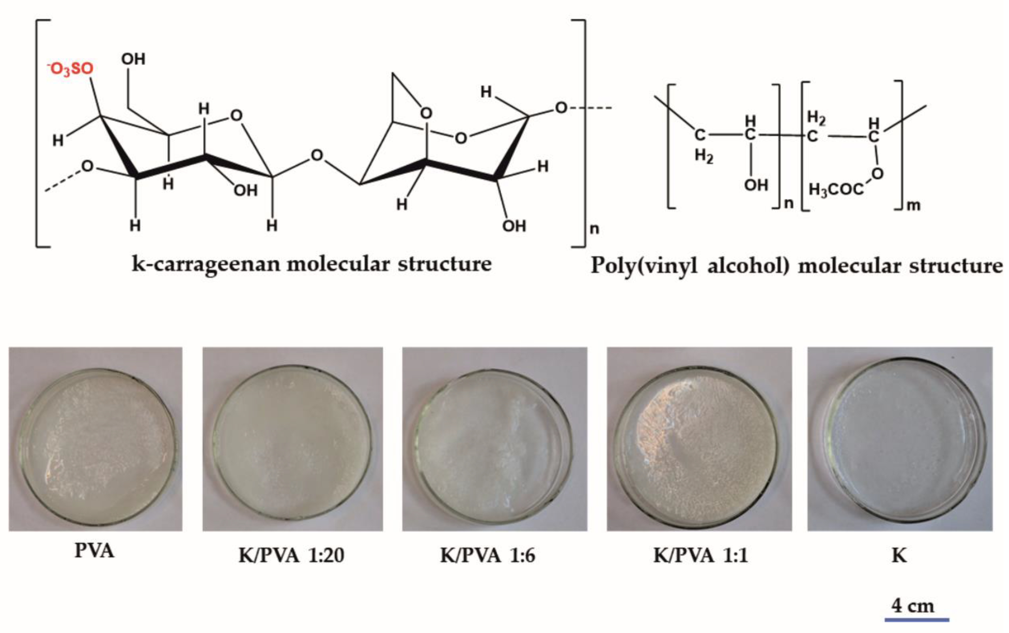
Figure 1. Molecular structure of the polymers and the photographic images of the obtained hydrogels.
Polymers 12 00560 g001
Figure 2. (a): XRD diffractograms of the obtained hydrogels; (b): a tentative mechanism for hydrogels cryostructuring.
Polymers 12 00560 g002
Figure 3. ATR-FTIR spectra of the obtained hydrogels (a); detail of the IR fingerprint region (b); detail on the 3800–2700 cm−1 region (c); spectra detail for -OH and alkyl groups region (s: stretching; b: bending; w: wagging; r: rocking vibrations).
Polymers 12 00560 g003
Figure 4. SEM micrographs of the obtained hydrogels.
Polymers 12 00560 g004
Figure 5. TGA and DTG thermograms of (a) PVA and CAR; (b) PVA/CAR mixtures.
Polymers 12 00560 g005
Figure 6. DTA thermograms of (a) PVA and CAR; (b) PVA/CAR mixtures.
Polymers 12 00560 g006
Figure 7. DSC thermograms of the obtained hydrogels.
Polymers 12 00560 g007
Figure 8. (a): Swelling kinetics in distilled water; (b): swelling degree at equilibrium in different aqueous media and (c): the fractional release of carrageenan at equilibrium for the obtained hydrogels.
Polymers 12 00560 g008
Figure 9. (a): Adsorption isotherms for CV onto the PVA/CAR hydrogels at 25 °C; (b): van’t Hoff plots and (c): kinetic for the CV sorption in the PVA/CAR hydrogels at 25 °C.
Polymers 12 00560 g009
Table 1. Characteristics of the obtained hydrogels. Poly (vinyl alcohol) (PVA), κ-carrageenan (CAR).
Sample CodePVACARwcarGels SC* (%)Shore 00 Hardness *Thickness * δ (mm)G (%)
Vol. (mL)Amount (g)Vol. (mL)Amount (g)
PVA101--09.85682.8582.24
K/PVA 1:207.50.752.50.0370.0477.59642.9476.15
K/PVA 1:64.80.485.20.0780.1395.98693.0283.07
K/PVA 1:11.30.138.70.130.52.62453.1164.80
K--100.1511.54123.160.75
* Maximum relative error: for SC and G: ± 1.5%, for the thickness: ± 0.1%, and for the Shore hardness: ± 0.5%. wcar: weight ratio of carrageenan; SC: solids content (%); G: gel content (%).
Table 2. The characteristics of the obtained hydrogels.
Sample CodeFTIR XRDPore volume Ratio Pr (%)
CrIR (Å)EH (kcal)CrXRD (%)DXRD (nm)
PVA0.1432.9266.5234.25.798.63
K/PVA 1:200.1192.9276.3864.05.056.42
K/PVA 1:60.1882.9206.5924.84.815.53
K/PVA 1:10.0242.9244.9142.36.023.48
K-2.9295.0341.1-2.31
Table 3. The thermal properties of the PVA/CAR hydrogels according to the TGA, DTA and DSC thermogram results.
Sample CodeTGA/DTG ResultsDTA ResultsDSC Results
Twater (°C)Tdegrad (°C)∆mdegrad (%)∆mresidue (%)TDTA (°C)Tg (°C)CrDSC (%)
PVA100.2146.5
273.5
340.3
95.54.0170.775.55.5
K/PVA 1:20109.2133.6
167.1
225.5
91.44.6132.7
169.7
60.55.2
K/PVA 1:6113.1149.1
185.9
214.7
37.819.4145.830.55.9
K/PVA 1:1104.0145.7
206.7
268.8
90.73.5145.529.53.2
K106.8176.8
288.1
333.6
91.91.2150.4 --
Table 4. Parameters regarding the diffusion of water into the hydrogel matrix.
MaterialParameter Swelling Medium
Distilled WaterpH = 3pH = 5pH = 9pH = 13PBS
PVAν (cm/min × 103)1.821.891.841.862.111.67
De (mm2/min × 105)1.82 (0.992)1.94 (0.984)1.90 (0.990)1.84 (0.994)2.01 (0.982)1.43 (0.992)
Dl (mm2/min × 102)6.72(0.977)8.92 (0.971)8.87 (0.989)8.79 (0.977)8.94 (0.975)5.11 (0.976)
k (min−0.5 × 10³)4.10 (0.964)4.89 (0.960)5.16 (0.976)4.63 (0.983)4.75 (0.974)3.46 (0.988)
n0.73 (0.964)0.85 (0.960)0.82 (0.976)0.84 (0.983)0.91 (0.974)0.68 (0.988)
K/PVA 1:20ν (cm/min × 103)1.891.520.930.951.610.72
De (mm2/min × 105)6.31 (0.968)6.58 (0.972)6.40 (0.992)6.61 (0.988)6.72 (0.988)5.62 (0.995)
Dl (mm2/min × 101)2.30 (0.979)2.65 (0.989)2.40 (0.995)2.69 (0.985)2.73 (0.992)1.95 (0.984)
k (min−0.5 × 10³)1.96 (0.967)1.99 (0.973)1.97 (0.989)2.01 (0.9)4.75 (0.974)3.46 (0.988)
n0.99 (0.967)0.85 (0.960)0.82 (0.976)0.84 (0.983)0.91 (0.974)0.68 (0.988)
K/PVA 1:6ν (cm/min × 103)1.651.781.691.711.821.38
De (mm2/min × 105)6.66 (0.986)7.80 (0.989)6.75 (0.992)6.72 (0.994)7.84 (0.992)5.42 (0.991)
Dl (mm2/min × 101)2.97 (0.988)3.18 (0.978)3.08 (0.977)3.11 (0.976)3.24 (0.972)1.47 (0.973)
k (min−0.5 × 10³)4.0 (0.980)4.92 (0.991)4.27 (0.993)4.20 (0.988)5.02 (0.987)3.20 (0.988)
n0.93 (0.980)0.96 (0.991)0.94 (0.993)0.96 (0.988)1.00 (0.987)0.71 (0.988)
K/PVA 1:1ν (cm/min × 103)3.663.903.693.733.981.89
De (mm2/min × 104)8.24 (0.993)8.97 (0.998)8.28 (0.997)8.27 (0.991)8.89 (0.992)5.43 (0.992)
Dl (mm2/min × 101)3.95(0.970)4.72 (0.975)4.12 (0.970)4.06 (0.978)4.80 (0.980)2.99 (0.989)
k (min−0.5 × 10³)5.23 (0.995)5.39 (0.994)5.28 (0.989)5.23 (0.995)5.23 (0.995)5.23 (0.988)
n0.95 (0.995)0.97 (0.994)0.96 (0.989)0.95 (0.995)0.95 (0.995)0.62 (0.988)
Table 5. Parameters of the isotherm models fitted onto the experimental CV adsorption data at 25 °C.
Isotherm ModelIsotherm Equation (Linearized Form)Equation Parameters
PVAK/PVA 1:20K/PVA 1:6K/PVA 1:1
Langmuir c e q Q e = 1 K L · Q m + c e q Q m
R L = 1 ( 1 + K L · c 0 )
KL = 0.092
Qm = 153.84
RL: 0.11 ÷ 0.35
R2 = 0.953
KL = 0.103
Qm = 76.92
RL: 0.11 ÷ 0.32
R2 = 0.731
KL = 0.035
Qm = 625.11
RL: 0.26 ÷ 0.58
R2 = 0.789
KL = 0.176
Qm = 55.55
RL: 0.06 ÷ 0.22
R2 = 0.909
Freundlich ln Q e = ln K F + 1 n F ln c e q KF = 14.270
1/nF = 0.77
R2 = 0.979
KF = 5.371
1/nF = 1.70
R2 = 0.976
KF = 22.510
1/nF = 0.88
R2 = 0.998
KF = 7.882
1/nF = 1.94
R2 = 0.997
Radke–Prausnitz c e q Q e = 1 K R P · Q m + c e q m Q m KRP = 0.146
Qm = 79.42
m = 0.65
R2 = 0.970
KRP = 0.763
Qm = 92.90
m = 1.50
R2 = 0.967
KRP = 0.111
Qm = 123.97
m = 0.90
R2 = 0.998
KRP = 0.940
Qm = 112.59
m = 2.00
R2 = 0.995
Redlich–Peterson ln ( K R · c e q Q e 1 ) = g · ln c e q + ln α R
Q m = K R α R
KR = 8.372
g = 0.74
αR = 0.11
Qm = 76.10
R2 = 0.979
KR = 1.372
g = 0.79
αR = 0.013
Qm = 99.91
R2 = 0.979
KR = 1.619
g = 0.81
αR = 0.013
Qm = 124.59
R2 = 0.998
KR = 1.631
g = 0.86
αR = 0.014
Qm = 116.54
R2 = 0.909
Dubinin–Radushkevich ln Q e = ln Q m β ε 2
ε = R T ln ( 1 + 1 c e q )
E = 1 2 β
Qm = 63.10
β: = 9 10−7
E = 745
R2 = 0.914
Qm = 142.25
β: = 3 10−6
E = 408
R2 = 0.995
Qm = 130.08
β: = 8 10−7
E = 791
R2 = 0.865
Qm = 112.44
β: = 2 10−6
E = 500
R2 = 0.992
Flory–Huggins ln ( θ c 0 ) = ln K F H + n · ln ( 1 θ )
θ = 1 c e q c 0
Δ G ° = R T · ln K F H
KFH = 2.2 10−5
n = 2.85
∆G° = −26.48
R2 = 0.774
KFH = 5.378
n = 2.27
∆G° = 4.16
R2 = 0.951
KFH = 1.8 10−10
n = 1.90
∆G° = −55.44
R2 = 0.879
KFH = 6.529
n = 2.05
∆G° = 4.65
R2 = 0.996
Thermodynamic parameters according to Equations (5) and (6) and Figure 9b for c0 = 80 mg/L (at 25 °C)∆H° = −6.7
∆S° = 15.6
∆G° = −11.3
∆H = 10.5
∆S° = 23.4
∆G° = 11.1
∆H° = −14.5
∆S° = 22.0
∆G° = −21.0
∆H° = 19.7
∆S° = 36.5
∆G° = 20.7
Qm: maximum adsorption capacity at monolayer coverage (mg/g); KL: Langmuir isotherm constant (L/mg); RL: Langmuir separation factor; c0: initial concentration of CV (mg/L); KF: Freundlich isotherm constant ((mg/g) (mg/L)1/n); nF: energetic heterogeneity of the adsorption sites; KRP: Radke–Prausnitz equilibrium constant (L/mg); m: Radke–Prausnitz model exponent; KR: Redlich–Peterson isotherm constant (L/g); g and αR (L/mg) are the Redlich–Peterson constants; β: Dubinin–Radushkevich constant (mol2/J2); ε: Polanyi potential (J/mol); R: universal gas constant (8.31 J/mol K); T: adsorption temperature (298 K); E: mean adsorption energy (J/mol); θ: degree of surface coverage; KFH: Flory–Huggins equilibrium constant (L/mg); n: number of adsorbates occupying the adsorption sites; ∆G°: standard Gibbs free energy change (kJ/mol); ∆HG°: standard enthalpy (kJ/mol); ∆S°: standard entropy (J/mol K); R2: determination coefficient indicating the goodness of the fit with the proposed isotherm model [77,78,79,80].
Table 6. Parameters of the isotherm models fitted onto the experimental CV adsorption data.
Kinetic ModelModel Equation (Linearized Form)Equation Parameters
PVAK/PVA 1:20K/PVA 1:6K/PVA 1:1
Pseudo-first-order log ( Q e Q t ) = log Q e k 1 t 2.303 k1 = 1.8 × 10⁻³
R2 = 0.920
k1 = 6.3 × 10⁻³
R2 = 0.899
k1 = 0.018
R2 = 0.839
k1 = 9.1 × 10⁻³
R2 = 0.849
Pseudo-second-order t Q t = 1 k 2 · Q e 2 + t Q e k2 = 0.4 × 10⁻⁵
R2 = 0.956
k2 = 1.1 × 10⁻⁴
R2 = 0.961
k2 = 7.5 × 10⁻⁴
R2 = 0.974
k2 = 9.4 × 10⁻³
R2 = 0.908
Intraparticle diffusion Q t = k d · t 1 / 2 + C
(20% of the first portion of the kinetic data)
kd = 0.144
C = 3.9 × 10⁻⁸
R2 = 1
kd = 2.314
C = 3.1 × 10⁻¹²
R2 = 1
kd = 2.354
C = 0.254
R2 = 1
kd = 1.739
C = 3.6 × 10⁻¹⁶
R2 = 1
Liquid film diffusion ln ( 1 Q t Q e ) = k f d · t kfd = 2.1 × 10⁻³
R2 = 0.891
kfd = 1.5 × 10⁻²
R2 = 0.901
kfd = 2.6 × 10⁻²
R2 = 0.924
kfd = 1.2 × 10⁻²
R2 = 0.973
Elovich Q t = 1 β ln ( α · β ) + 1 β ln t α = 2.1 × 10⁻¹⁷
β = 2.1 × 10⁻³
R2 = 0.634
α = 1.2 × 10⁻⁸
β = 5.8 × 10⁻²
R2 = 0.325
α = 6 × 10⁻⁷
β = 0.199
R2 = 0.251
α = 9.1 × 10⁻⁴
β = 2.9 × 10⁻²
R2 = 0.929
k1: rate constant of pseudo-first order kinetic (min−1), k2: second-order rate constant (g/mg min), kd: rate constant of intraparticle diffusion (mg/g min1/2), C: intercept of intraparticle diffusion, kfd: liquid film diffusion rate coefficients (min−1), α: (mg/g min) and β: (g/mg) are the initial adsorption rate of the Elovich equation, respectively the desorption constant related to the extent of surface coverage and activation energy constant for sorption [85,86].