
Synthesis of Polyaniline Coated Magnesium and Cobalt Oxide Nanoparticles through Eco-Friendly Approach and Their Application as Antifungal Agents

 ,
,  and
and
Abstract
:
1. Introduction
2. Materials and Methods
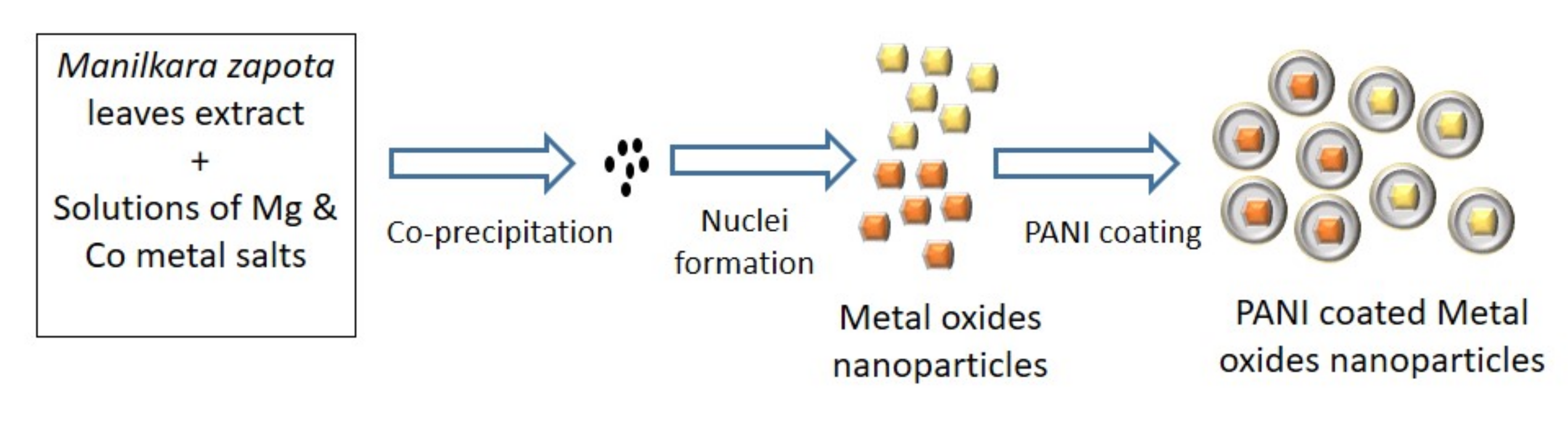
2.1. Green Synthesis of MgO and Co3O4 Nanoparticles
2.2. Coating of Polyaniline (PANI) on MgO and Co3O4 Nanoparticles
2.3. Characterization
2.4. Zone of Inhibition Test
3. Results and Discussion
3.1. FTIR Analysis
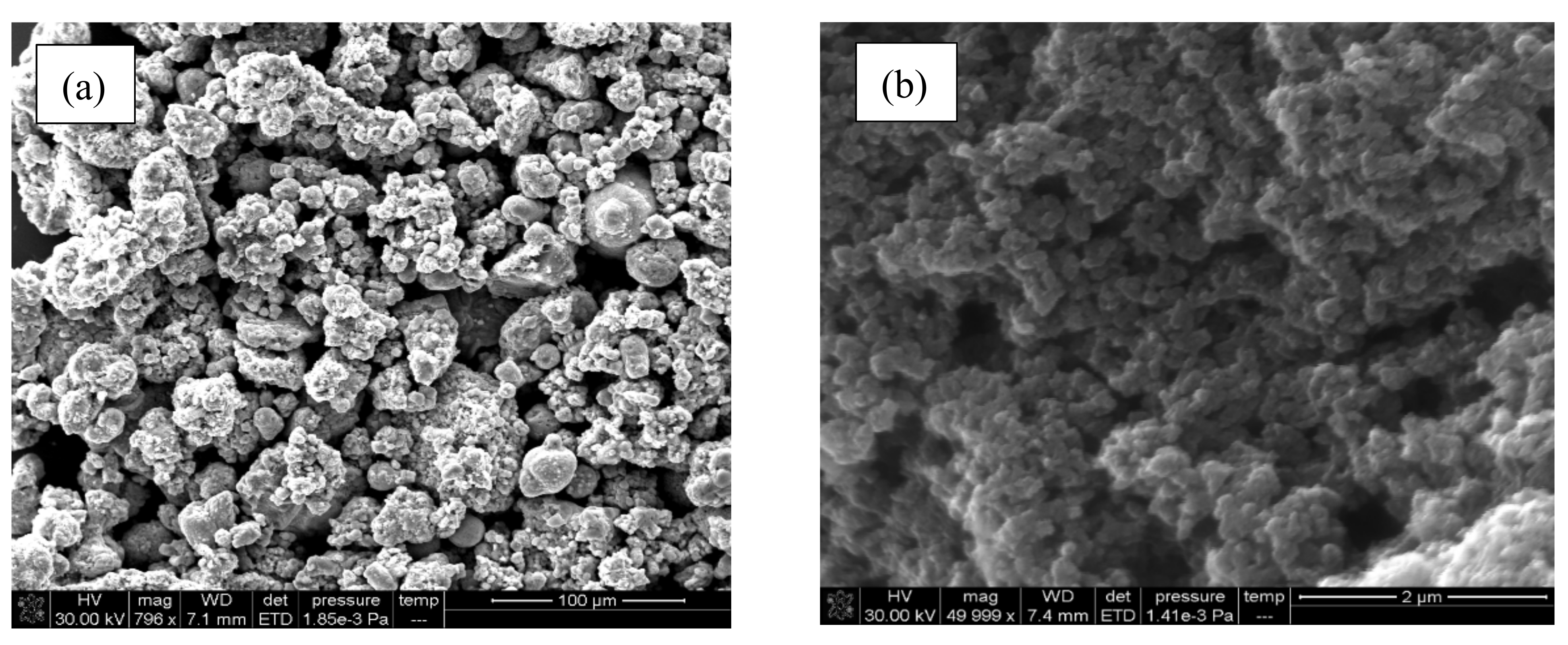
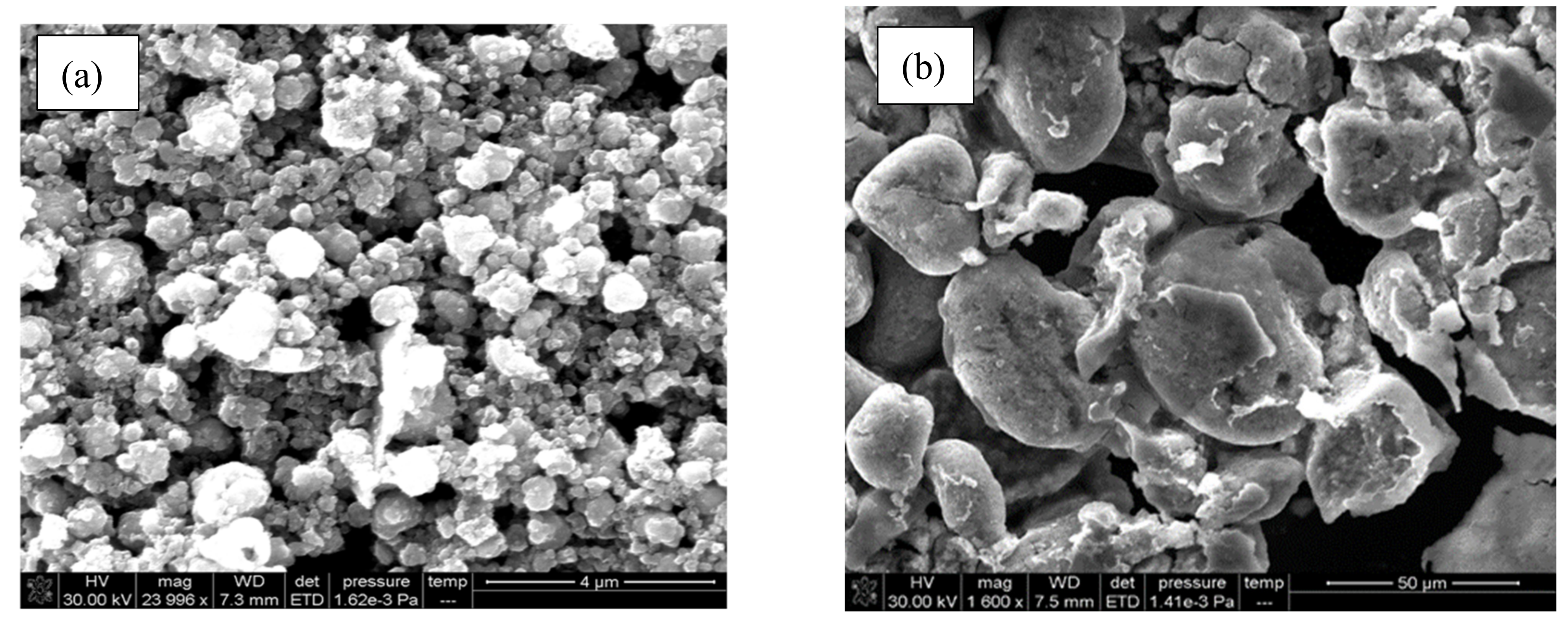
3.2. SEM Analysis
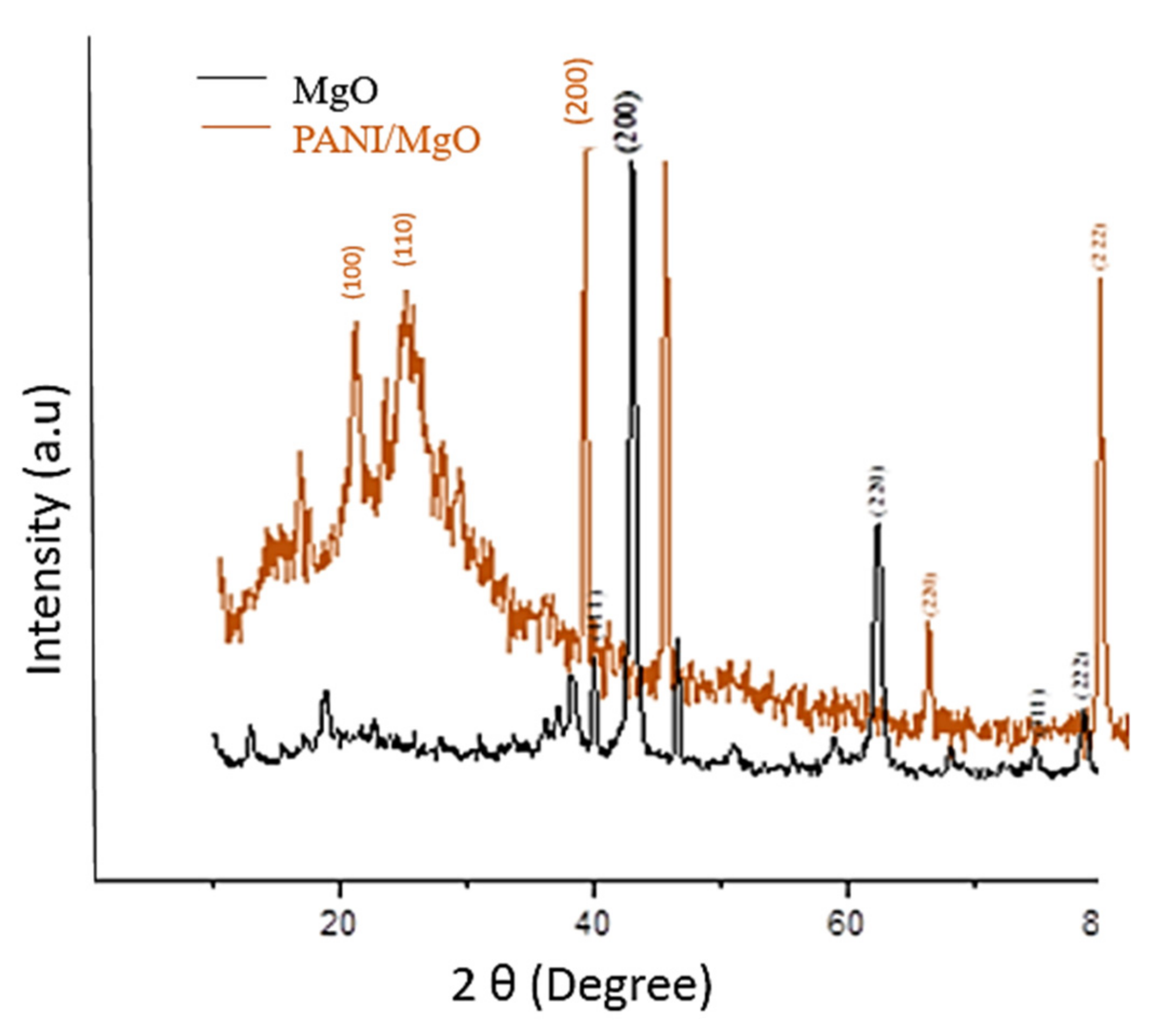
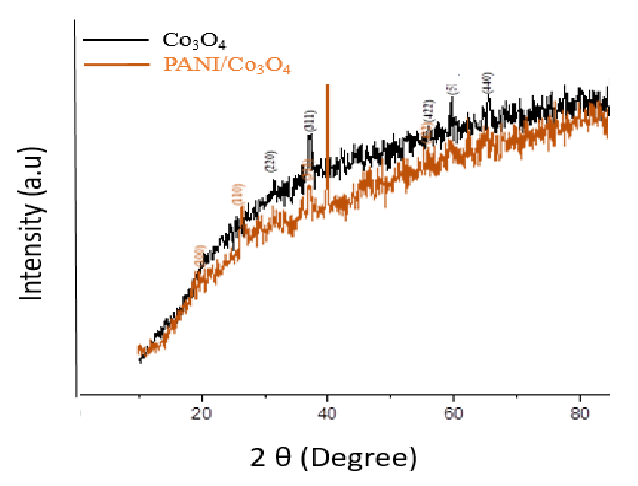
3.3. XRD Analysis
3.4. Zone of Inhibition Test
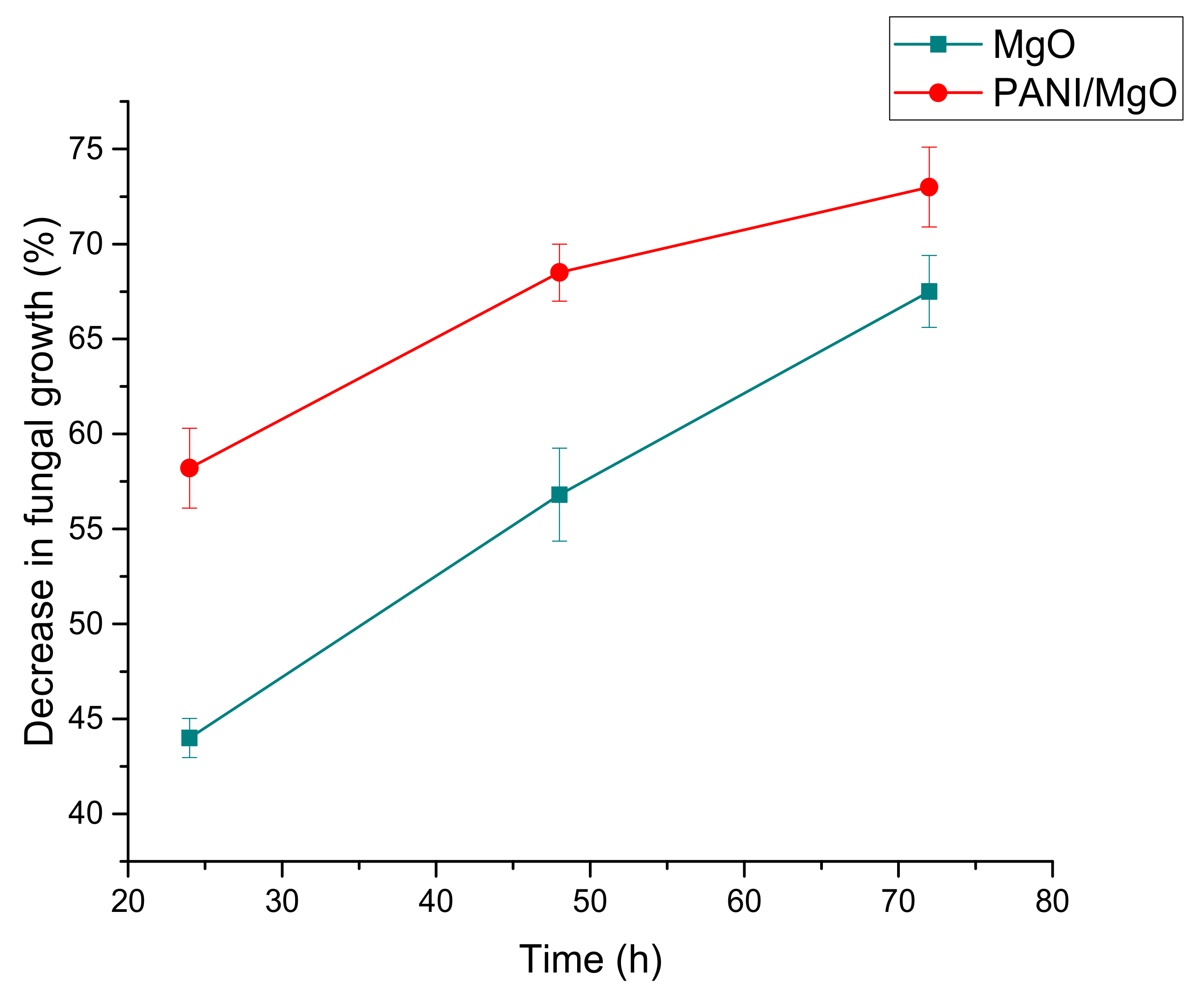
3.5. Concentration and Time Dependent Inhibition
3.6. Performance Evaluation
4. Conclusions
Author Contributions
Funding
Data Availability Statement
Acknowledgments
Conflicts of Interest
References
- Ladaniya, M.S. Commercial fresh citrus cultivars and producing countries. In Citrus Fruit: Biology, Technology and Evaluation; Academic Press: Cambridge, MA, USA, 2008; pp. 13–65. [Google Scholar]
- Tokarzewski, S.; Ziólkowska, G.; Nowakiewicz, A. Susceptibility testing of Aspergillus niger strains isolated from poultry to antifungal drugs-a comparative study of the disk diffusion, broth microdilution (M 38-A) and Etest methods. Pol. J. Vet. Sci. 2012, 15, 125–133. [Google Scholar] [CrossRef] [PubMed] [Green Version]
- Ninkuu, V.; Adetunde, L.; Sackey, I.; Opoku, N.; Diedong, P. Antifungal efficacy of crude extracts of Azadirachta indica and Vernonia amygdalina against pathogenic Aspergillus niger (ATCC 16404). J. Med. Plants Res. 2019, 13, 408–412. [Google Scholar] [CrossRef]
- Wu, Q.; Fan, J.; Chen, X.; Zhu, Z.; Luo, J.; Wan, Y. Sandwich structured membrane adsorber with metal organic frameworks for aflatoxin B1 removal. Sep. Purif. Technol. 2020, 246, 116907. [Google Scholar] [CrossRef]
- Netala, V.R.; Kotakadi, V.S.; Domdi, L.; Gaddam, S.A.; Bobbu, P.; Venkata, S.K.; Ghosh, S.B.; Tartte, V. Biogenic silver nanoparticles: Efficient and effective antifungal agents. Appl. Nanosci. 2016, 6, 475–484. [Google Scholar] [CrossRef] [Green Version]
- Makabenta, J.M.V.; Nabawy, A.; Li, C.-H.; Schmidt-Malan, S.; Patel, R.; Rotello, V.M. Nanomaterial-based therapeutics for antibiotic-resistant bacterial infections. Nat. Rev. Microbiol. 2020, 19, 23–36. [Google Scholar] [CrossRef]
- Sun, Q.; Li, J.; Le, T. Zinc oxide nanoparticle as a novel class of antifungal agents: Current advances and future perspectives. J. Agric. Food Chem. 2018, 66, 11209–11220. [Google Scholar] [CrossRef]
- Arif, R.; Jadoun, S.; Verma, A. Synthesis of Nanomaterials and Their Applications in Textile Industry. In Frontiers of Textile Materials; Scrivener Publishing LLC: Beverly, MA, USA, 2020; pp. 117–133. [Google Scholar]
- Singh, R.P.; Handa, R.; Manchanda, G. Nanoparticles in sustainable agriculture: An emerging opportunity. J. Control. Release 2020, 329, 1234–1248. [Google Scholar] [CrossRef]
- Ahmad, S.; Subhani, K.; Rasheed, A.; Ashraf, M.; Afzal, A.; Ramzan, B.; Sarwar, Z. Development of Conductive Fabrics by Using Silver Nanoparticles for Electronic Applications. J. Electron. Mater. 2020, 49, 1330–1337. [Google Scholar] [CrossRef]
- Utreja, P.; Verma, S.; Rahman, M.; Kumar, L. Use of nanoparticles in medicine. Curr. Biochem. Eng. 2020, 6, 7–24. [Google Scholar] [CrossRef]
- Ziental, D.; Czarczynska-Goslinska, B.; Mlynarczyk, D.T.; Glowacka-Sobotta, A.; Stanisz, B.; Goslinski, T.; Sobotta, L. Titanium dioxide nanoparticles: Prospects and applications in medicine. Nanomaterials 2020, 10, 387. [Google Scholar] [CrossRef] [PubMed] [Green Version]
- Carrouel, F.; Viennot, S.; Ottolenghi, L.; Gaillard, C.; Bourgeois, D. Nanoparticles as anti-microbial, anti-inflammatory, and remineralizing agents in oral care cosmetics: A review of the current situation. Nanomaterials 2020, 10, 140. [Google Scholar] [CrossRef] [Green Version]
- Helan, V.; Prince, J.J.; Al-Dhabi, N.A.; Arasu, M.V.; Ayeshamariam, A.; Madhumitha, G.; Roopan, S.M.; Jayachandran, M. Neem leaves mediated preparation of NiO nanoparticles and its magnetization, coercivity and antibacterial analysis. Results Phys. 2016, 6, 712–718. [Google Scholar] [CrossRef] [Green Version]
- Ates, B.; Koytepe, S.; Ulu, A.; Gurses, C.; Thakur, V.K. Chemistry, structures, and advanced applications of nanocomposites from biorenewable resources. Chem. Rev. 2020, 120, 9304–9362. [Google Scholar] [CrossRef]
- Liang, W.; Wang, F.; Tay, T.E.; Yang, B.; Wang, Z. Experimental and Analytical Investigation of Epoxy/MWCNT Nanocomposites: Electrical, Thermal Properties, and Electric Heating Behavior. Polym. Eng. Sci. 2020, 60, 233–242. [Google Scholar] [CrossRef]
- Sargazi, S.; Hajinezhad, M.R.; Barani, M.; Rahdar, A.; Shahraki, S.; Karimi, P.; Cucchiarini, M.; Khatami, M.; Pandey, S. Synthesis, characterization, toxicity and morphology assessments of newly prepared microemulsion systems for delivery of valproic acid. J. Mol. Liq. 2021, 338, 116625. [Google Scholar] [CrossRef]
- Pandey, S.; De Klerk, C.; Kim, J.; Kang, M.; Fosso-Kankeu, E. Eco friendly approach for synthesis, characterization and biological activities of milk protein stabilized silver nanoparticles. Polymers 2020, 12, 1418. [Google Scholar] [CrossRef]
- Akintelu, S.A.; Folorunso, A.S.; Folorunso, F.A.; Oyebamiji, A.K. Green synthesis of copper oxide nanoparticles for biomedical application and environmental remediation. Heliyon 2020, 6, e04508. [Google Scholar] [CrossRef]
- Chen, J.; Fan, L.; Yang, C.; Wang, S.; Zhang, M.; Xu, J.; Luo, S. Facile synthesis of Ag nanoparticles-loaded chitosan antibacterial nanocomposite and its application in polypropylene. Int. J. Biol. Macromol. 2020, 161, 1286–1295. [Google Scholar] [CrossRef]
- Asiya, S.; Pal, K.; Kralj, S.; El-Sayyad, G.; de Souza, F.; Narayanan, T. Sustainable preparation of gold nanoparticles via green chemistry approach for biogenic applications. Mater. Today Chem. 2020, 17, 100327. [Google Scholar]
- Essien, E.R.; Atasie, V.N.; Okeafor, A.O.; Nwude, D.O. Biogenic synthesis of magnesium oxide nanoparticles using Manihot esculenta (Crantz) leaf extract. Int. Nano Lett. 2020, 10, 43–48. [Google Scholar] [CrossRef] [Green Version]
- Umaralikhan, L.; Jaffar, M.J.M. Green synthesis of MgO nanoparticles and it antibacterial activity. Iran. J. Sci. Technol. Trans. A: Sci. 2018, 42, 477–485. [Google Scholar] [CrossRef]
- Karthik, K.; Dhanuskodi, S.; Kumar, S.P.; Gobinath, C.; Sivaramakrishnan, S. Microwave assisted green synthesis of MgO nanorods and their antibacterial and anti-breast cancer activities. Mater. Lett. 2017, 206, 217–220. [Google Scholar] [CrossRef]
- Kandalkar, S.; Dhawale, D.; Kim, C.-K.; Lokhande, C. Chemical synthesis of cobalt oxide thin film electrode for supercapacitor application. Synth. Met. 2010, 160, 1299–1302. [Google Scholar] [CrossRef]
- Palanisamy, G.; Pazhanivel, T. Green synthesis of MgO nanoparticles for antibacterial activity. Int. Res. J. Eng. Technol. 2017, 4, 137–141. [Google Scholar]
- Nijalingappa, T.; Veeraiah, M.; Basavaraj, R.; Darshan, G.; Sharma, S.; Nagabhushana, H. Antimicrobial properties of green synthesis of MgO micro architectures via Limonia acidissima fruit extract. Biocatal. Agric. Biotechnol. 2019, 18, 100991. [Google Scholar] [CrossRef]
- Abdallah, Y.; Ogunyemi, S.O.; Abdelazez, A.; Zhang, M.; Hong, X.; Ibrahim, E.; Hossain, A.; Fouad, H.; Li, B.; Chen, J. The green synthesis of MgO nano-flowers using Rosmarinus officinalis L.(Rosemary) and the antibacterial activities against Xanthomonas oryzae pv. oryzae. BioMed Res. Int. 2019, 2019, 5620989. [Google Scholar] [CrossRef] [PubMed] [Green Version]
- Moorthy, S.K.; Ashok, C.; Rao, K.V.; Viswanathan, C. Synthesis and characterization of MgO nanoparticles by Neem leaves through green method. Mater. Today Proc. 2015, 2, 4360–4368. [Google Scholar] [CrossRef]
- Bibi, I.; Nazar, N.; Iqbal, M.; Kamal, S.; Nawaz, H.; Nouren, S.; Safa, Y.; Jilani, K.; Sultan, M.; Ata, S.; et al. Green and eco-friendly synthesis of cobalt-oxide nanoparticle: Characterization and photo-catalytic activity. Adv. Powder Technol. 2017, 28, 2035–2043. [Google Scholar] [CrossRef]
- Akhlaghi, N.; Najafpour-Darzi, G.; Younesi, H. Facile and green synthesis of cobalt oxide nanoparticles using ethanolic extract of Trigonella foenumgraceum (Fenugreek) leaves. Adv. Powder Technol. 2020, 31, 3562–3569. [Google Scholar] [CrossRef]
- Siddique, M.; Khan, N.M.; Saeed, M.; Ali, S.; Shah, Z. Green synthesis of cobalt oxide nanoparticles using Citrus medica leaves extract: Characterization and photo-catalytic activity. Z. Phys. Chem. 2021, 235, 663–681. [Google Scholar] [CrossRef]
- Yadi, M.; Mostafavi, E.; Saleh, B.; Davaran, S.; Aliyeva, I.; Khalilov, R.; Nikzamir, M.; Nikzamir, N.; Akbarzadeh, A.; Panahi, Y.; et al. Current developments in green synthesis of metallic nanoparticles using plant extracts: A review. Artif. Cells Nanomed. Biotechnol. 2018, 46, S336–S343. [Google Scholar] [CrossRef] [PubMed] [Green Version]
- Ma, J.; Luo, X.-D.; Protiva, P.; Yang, H.; Ma, C.; Basile, M.J.; Weinstein, I.B.; Kennelly, E.J. Bioactive novel polyphenols from the fruit of Manilkara zapota (Sapodilla). J. Nat. Prod. 2003, 66, 983–986. [Google Scholar] [CrossRef] [PubMed]
- Tamsir, N.M.; Esa, N.M.; Omar, S.N.C.; Shafie, N.H. Manilkara zapota (L.) P. Royen: Potential Source of Natural Antioxidants. Malays. J. Med. Health Sci. 2020, 15, 196–204. [Google Scholar]
- Chen, J.; Wu, L.; Lu, M.; Lu, S.; Li, Z.; Ding, W. Comparative study on the fungicidal activity of metallic MgO nanoparticles and macroscale MgO against soilborne fungal phytopathogens. Front. Microbiol. 2020, 11, 365. [Google Scholar] [CrossRef] [PubMed] [Green Version]
- Zhang, D.; Chen, H.; Hong, R. Preparation and Conductive and Electromagnetic Properties of Fe3O4/PANI Nanocomposite via Reverse In Situ Polymerization. J. Nanomater. 2019, 2019, 7962754. [Google Scholar] [CrossRef] [Green Version]
- Stejskal, J.; Gilbert, R. Polyaniline. Preparation of a conducting polymer (IUPAC technical report). Pure Appl. Chem. 2002, 74, 857–867. [Google Scholar] [CrossRef] [Green Version]
- Bashira, A.; Hanifb, F.; Yasmeena, G.; Maboodc, F.; Hussaind, A.; Abbasa, N.; Yousafe, A.B.; Aamira, M.; Manzoora, S. Polyaniline based magnesium nanoferrite composites as efficient photocatalysts for the photodegradation of Indigo Carmine in aqueous solutions. Desalin. Water Treat. 2019, 164, 368–377. [Google Scholar] [CrossRef]
- Singh, N.; Singh, P.K.; Singh, M.; Tandon, P.; Singh, S.K.; Singh, S. Fabrication and characterization of polyaniline, polyaniline/MgO (30%) and polyaniline/MgO (40%) nanocomposites for their employment in LPG sensing at room temperature. J. Mater. Sci. Mater. Electron. 2019, 30, 4487–4498. [Google Scholar] [CrossRef]
- Shao, W.; Jamal, R.; Xu, F.; Ubul, A.; Abdiryim, T. The effect of a small amount of water on the structure and electrochemical properties of solid-state synthesized polyaniline. Materials 2012, 5, 1811–1825. [Google Scholar] [CrossRef] [Green Version]
- Lázár, I.; Szabó, H.J. Prevention of the aggregation of nanoparticles during the synthesis of nanogold-containing silica aerogels. Gels 2018, 4, 55. [Google Scholar] [CrossRef] [Green Version]
- Bindhu, M.; Umadevi, M.; Micheal, M.K.; Arasu, M.V.; Al-Dhabi, N.A. Structural, morphological and optical properties of MgO nanoparticles for antibacterial applications. Mater. Lett. 2016, 166, 19–22. [Google Scholar] [CrossRef]
- Hafeez, M.; Shaheen, R.; Akram, B.; Haq, S.; Mahsud, S.; Ali, S.; Khan, R.T. Green synthesis of cobalt oxide nanoparticles for potential biological applications. Mater. Res. Express 2020, 7, 025019. [Google Scholar] [CrossRef]
- Pudukudy, M.; Yaakob, Z. Sol-gel synthesis, characterisation, and photocatalytic activity of porous spinel Co3O4 nanosheets. Chem. Pap. 2014, 68, 1087–1096. [Google Scholar] [CrossRef]
- Kumari, M.; Giri, V.P.; Pandey, S.; Kumar, M.; Katiyar, R.; Nautiyal, C.S.; Mishra, A. An insight into the mechanism of antifungal activity of biogenic nanoparticles than their chemical counterparts. Pestic. Biochem. Physiol. 2019, 157, 45–52. [Google Scholar] [CrossRef]
- Shi, N.; Guo, X.; Jing, H.; Gong, J.; Sun, C.; Yang, K. Antibacterial effect of the conducting polyaniline. J. Mater. Sci. Technol. 2006, 22, 289–290. [Google Scholar]
- Robertson, J.; Gizdavic-Nikolaidis, M.; Nieuwoudt, M.K.; Swift, S. The antimicrobial action of polyaniline involves production of oxidative stress while functionalisation of polyaniline introduces additional mechanisms. PeerJ 2018, 6, e5135. [Google Scholar] [CrossRef] [PubMed]
- Ouoba, P.; Ouattara, L.; Bonzi, S.; Yameogo, J.; Somda, I. Evaluation of antifungal activity and phytotoxicity of the essential oil of Zanthoxylum zanthoxyloides fruits. Agric. Sci. Res. J. 2018, 8, 92–99. [Google Scholar]
- Scott, L.J.; Simpson, D. Voriconazole. Drugs 2007, 67, 269–298. [Google Scholar] [CrossRef]
- Alkfaji, F.; Hussaini, I.M.A. Biosynthesis of silver nanoparticles with Mentha spicata against Aspergillus niger. Drug Invent. Today 2020, 14, 806–811. [Google Scholar]
- Kalaiarasan, A. Antibacterial and antifungal activities of Ficus macemosa fruits nanoparticle synthesized extracts it’s from eastern ghats of Tamil nadu state in India. Int. J. Curr. Res. 2016, 8, 43359–43365. [Google Scholar]


| MgO Nanoparticles | PANI/MgO Nanoparticles | ||||||
|---|---|---|---|---|---|---|---|
| Concentration (mM) | Replicates | Time (h) | Time (h) | ||||
| 24 | 48 | 72 | 24 | 48 | 72 | ||
| Zone (cm) | Zone (cm) | ||||||
| 3 | R1 | 0.50 | 1.10 | 1.50 | 0.80 | 1.30 | 1.84 |
| R2 | 0.90 | 1.30 | 1.90 | 1.40 | 1.60 | 1.95 | |
| R3 | 0.90 | 1.50 | 2.00 | 0.80 | 2.30 | 2.34 | |
| Mean | 0.76 ± 0.28 | 1.30 ± 0.20 | 1.80 ± 0.26 | 1.00 ± 0.34 | 1.73 ± 0.51 | 2.04 ± 0.26 | |
| 6 | R1 | 0.50 | 1.10 | 1.90 | 0.90 | 1.30 | 1.90 |
| R2 | 0.70 | 1.30 | 2.00 | 0.90 | 1.90 | 2.10 | |
| R3 | 0.90 | 1.30 | 2.20 | 1.30 | 1.90 | 2.36 | |
| Mean | 0.70 ± 0.20 | 1.23 ± 0.12 | 2.03 ± 0.15 | 1.03 ± 0.23 | 1.70 ± 0.34 | 2.12 ± 0.23 | |
| 12 | R1 | 0.40 | 1.10 | 1.80 | 0.90 | 1.30 | 1.80 |
| R2 | 0.70 | 1.32 | 2.00 | 1.40 | 1.60 | 2.10 | |
| R3 | 1.00 | 1.90 | 2.10 | 1.60 | 1.90 | 2.10 | |
| Mean | 0.70 ± 0.3 | 1.44 ± 0.41 | 1.96 ± 0.15 | 1.30 ± 0.36 | 1.60 ± 0.30 | 2.00 ± 0.17 | |
| 24 | R1 | 0.40 | 1.10 | 1.60 | 1.00 | 1.80 | 2.10 |
| R2 | 0.90 | 1.10 | 2.20 | 1.30 | 1.70 | 2.30 | |
| R3 | 1.40 | 1.60 | 2.40 | 1.90 | 1.30 | 2.10 | |
| Mean | 0.90 ± 0.50 | 1.26 ± 0.29 | 2.06 ± 0.41 | 1.40 ± 0.45 | 1.60 ± 0.26 | 2.16 ± 0.12 | |
| Co3O4 Nanoparticles | PANI/Co3O4 Nanoparticles | ||||||
|---|---|---|---|---|---|---|---|
| Concentration (mM) | Replicates | Time (h) | Time (h) | ||||
| 24 | 48 | 72 | 24 | 48 | 72 | ||
| Zone (cm) | Zone (cm) | ||||||
| 3 | R1 | 0.80 | 1.10 | 1.85 | 0.90 | 1.10 | 1.90 |
| R2 | 1.00 | 1.55 | 1.80 | 0.72 | 1.30 | 2.24 | |
| R3 | 0.90 | 1.60 | 1.98 | 0.60 | 2.00 | 2.05 | |
| Mean | 0.90 ± 0.10 | 1.41 ± 0.27 | 1.87 ± 0.09 | 0.74 ± 0.15 | 1.46 ± 0.47 | 2.06 ± 0.17 | |
| 6 | R1 | 0.80 | 0.95 | 2.10 | 0.80 | 0.95 | 2.10 |
| R2 | 0.90 | 1.30 | 1.94 | 0.70 | 1.46 | 1.95 | |
| R3 | 1.20 | 1.60 | 1.92 | 0.60 | 2.10 | 2.10 | |
| Mean | 0.96 ± 0.20 | 1.28 ± 0.32 | 1.83 ± 0.09 | 0.70 ± 0.10 | 1.50 ± 0.57 | 2.05 ± 0.08 | |
| 12 | R1 | 0.90 | 1.10 | 1.87 | 0.80 | 0.90 | 2.10 |
| R2 | 1.00 | 1.23 | 1.96 | 0.63 | 1.36 | 2.06 | |
| R3 | 0.70 | 1.78 | 2.10 | 0.80 | 1.70 | 2.27 | |
| Mean | 0.86 ± 0.15 | 1.94 ± 0.36 | 1.97 ± 0.11 | 0.74 ± 0.09 | 1.32 ± 0.40 | 2.14 ± 0.11 | |
| 24 | R1 | 0.35 | 1.13 | 1.87 | 0.90 | 1.10 | 2.31 |
| R2 | 1.00 | 1.55 | 1.80 | 0.70 | 1.80 | 1.99 | |
| R3 | 0.90 | 1.90 | 2.10 | 0.80 | 1.70 | 2.30 | |
| Mean | 0.75 ± 0.35 | 1.52 ± 0.38 | 1.92 ± 0.15 | 0.80 ± 0.10 | 1.53 ± 0.37 | 2.20 ± 0.18 | |
| Nanoparticles | Zone of Inhibition (cm) | Reference |
|---|---|---|
| Ag (Green synthesis using Mentha spicata leaves extract) | 0.6 | [51] |
| Ag (Green synthesis using Ficus racemosa fruit extract) | 0.3 | [52] |
| PANI/MgO | 2.16 | Current study |
| PANI/Co3O4 | 2.01 | Current study |
| Voriconazole (Reference fungicide) | 4.8 | Current study |
Publisher’s Note: MDPI stays neutral with regard to jurisdictional claims in published maps and institutional affiliations. |
© 2021 by the authors. Licensee MDPI, Basel, Switzerland. This article is an open access article distributed under the terms and conditions of the Creative Commons Attribution (CC BY) license (https://creativecommons.org/licenses/by/4.0/).
Share and Cite
Manzoor, S.; Yasmin, G.; Raza, N.; Fernandez, J.; Atiq, R.; Chohan, S.; Iqbal, A.; Manzoor, S.; Malik, B.; Winter, F.; et al. Synthesis of Polyaniline Coated Magnesium and Cobalt Oxide Nanoparticles through Eco-Friendly Approach and Their Application as Antifungal Agents. Polymers 2021, 13, 2669. https://doi.org/10.3390/polym13162669
Manzoor S, Yasmin G, Raza N, Fernandez J, Atiq R, Chohan S, Iqbal A, Manzoor S, Malik B, Winter F, et al. Synthesis of Polyaniline Coated Magnesium and Cobalt Oxide Nanoparticles through Eco-Friendly Approach and Their Application as Antifungal Agents. Polymers. 2021; 13(16):2669. https://doi.org/10.3390/polym13162669
Chicago/Turabian StyleManzoor, Suryyia, Ghazala Yasmin, Nadeem Raza, Javier Fernandez, Rashida Atiq, Sobia Chohan, Ayesha Iqbal, Shamaila Manzoor, Barizah Malik, Franz Winter, and et al. 2021. "Synthesis of Polyaniline Coated Magnesium and Cobalt Oxide Nanoparticles through Eco-Friendly Approach and Their Application as Antifungal Agents" Polymers 13, no. 16: 2669. https://doi.org/10.3390/polym13162669
APA StyleManzoor, S., Yasmin, G., Raza, N., Fernandez, J., Atiq, R., Chohan, S., Iqbal, A., Manzoor, S., Malik, B., Winter, F., & Azam, M. (2021). Synthesis of Polyaniline Coated Magnesium and Cobalt Oxide Nanoparticles through Eco-Friendly Approach and Their Application as Antifungal Agents. Polymers, 13(16), 2669. https://doi.org/10.3390/polym13162669