
Novel Polymerization of Dental Composites Using Near-Infrared-Induced Internal Upconversion Blue Luminescence


 ,
,
Abstract
:
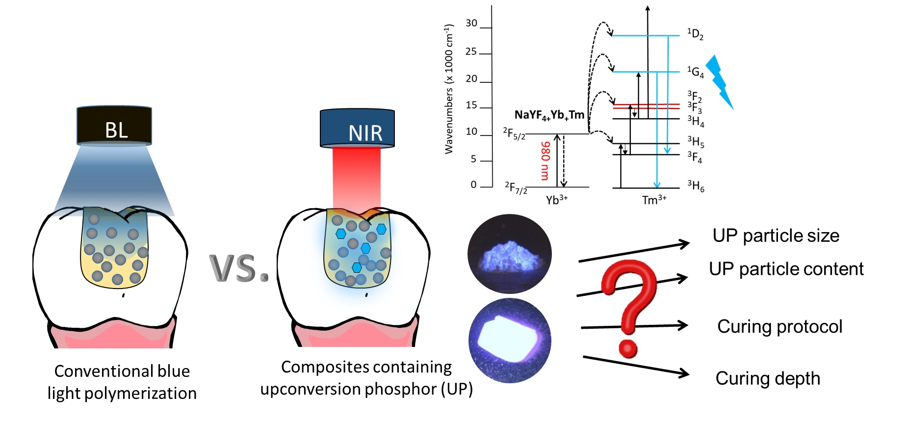
1. Introduction
2. Materials and Methods
2.1. UP Materials’ Preparation and Characterization
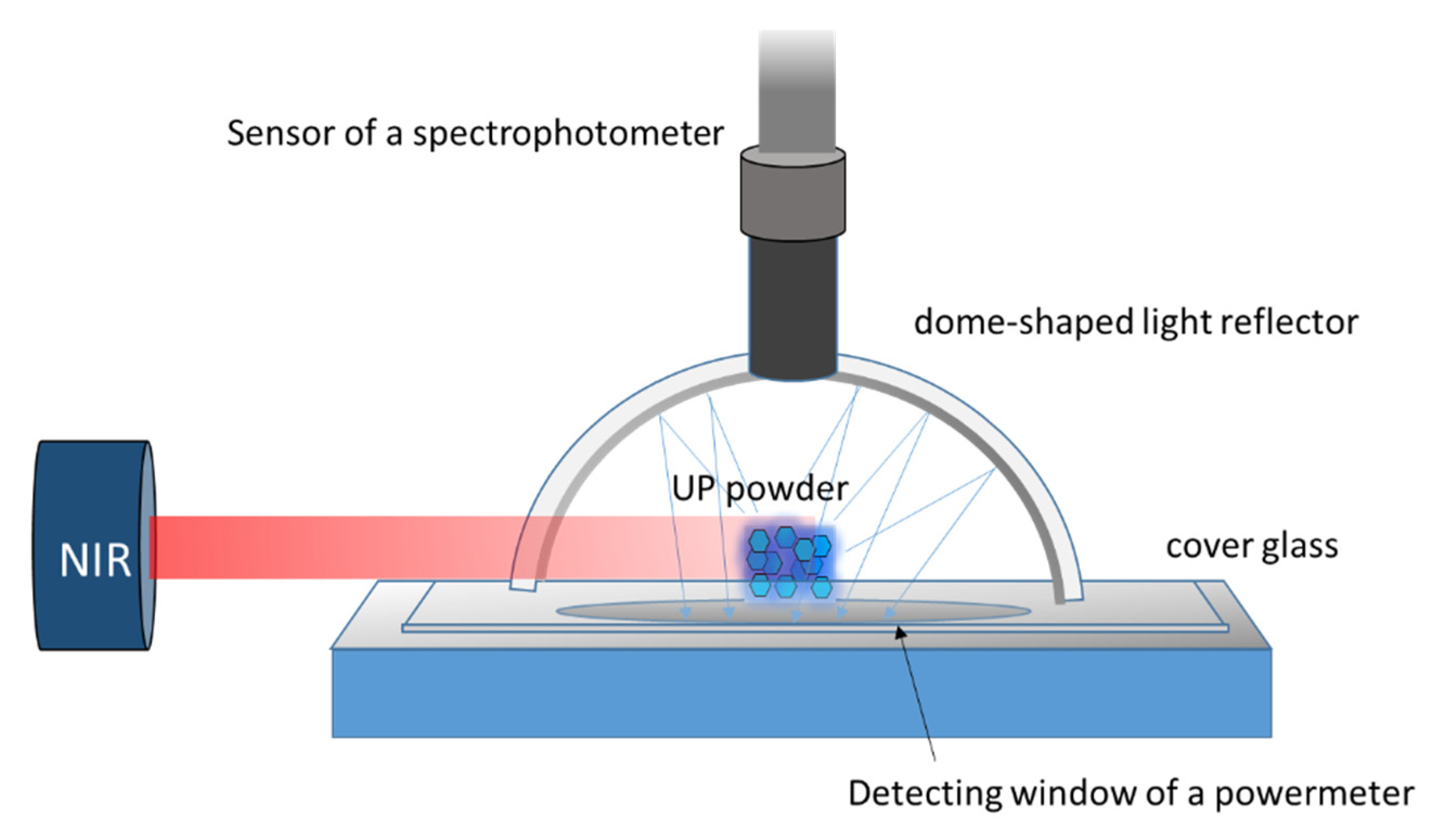
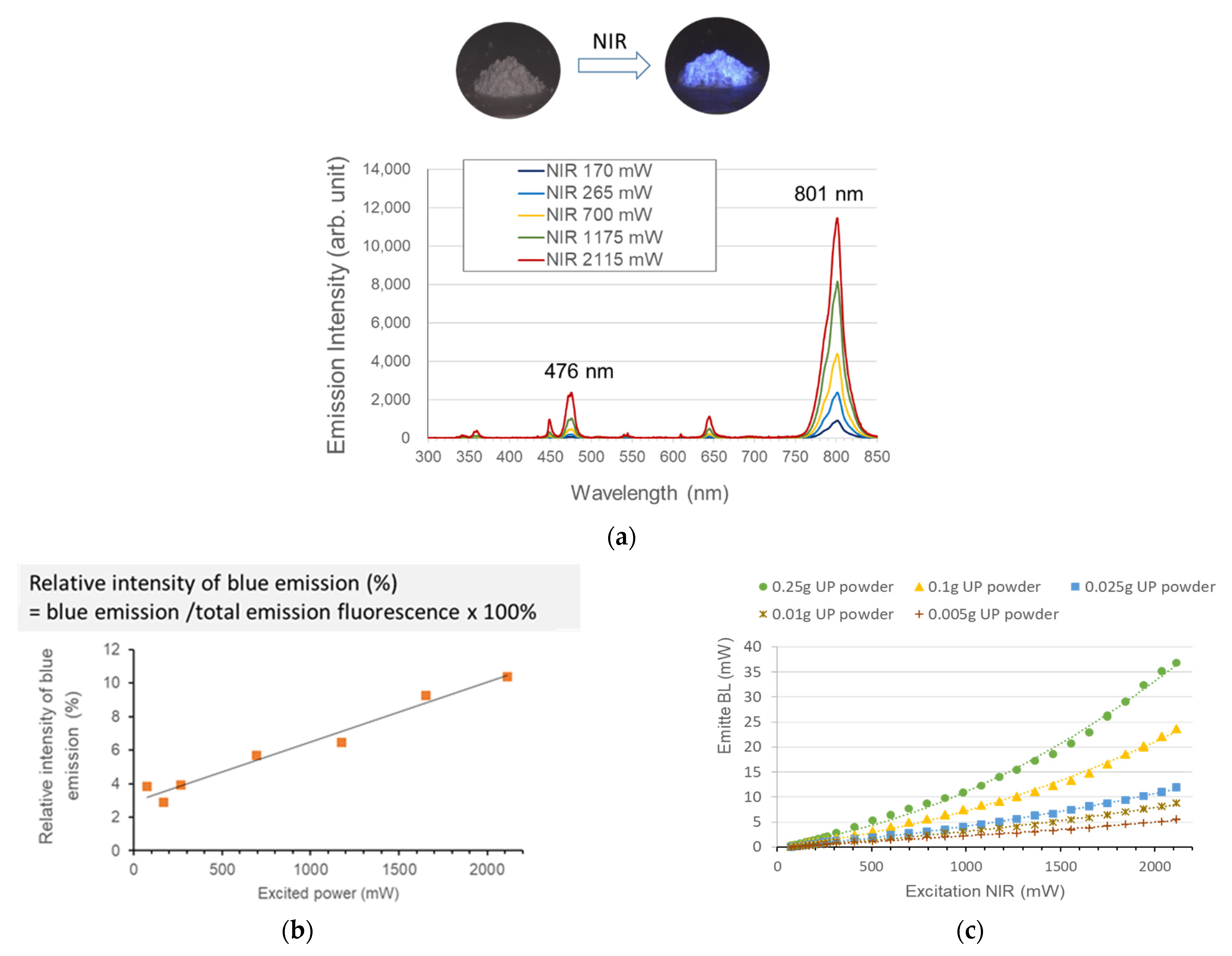
2.2. NIR Irradiation Source and Characterization of Upconversion Emission
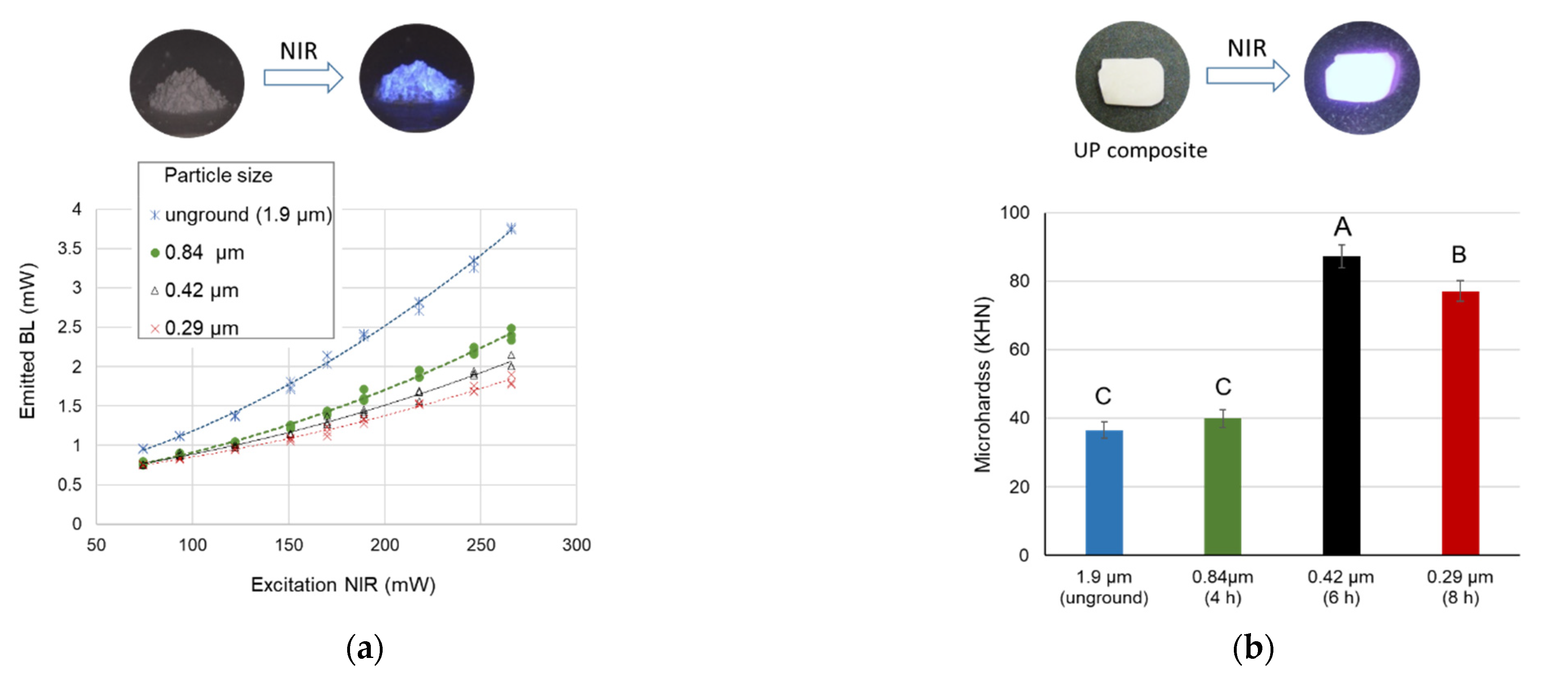
2.3. Effects of UP Particle Size
2.4. Effects of UP Content and Irradiation Protocol on Surface Microhardness
2.5. Depth Profiles of Microhardness
2.6. Statistical Analysis
3. Results
3.1. Characterization of the UP Materials
3.2. Upconversion Luminescence Intensity and Spectrum
3.3. Effect of Particle Size on Luminescence Intensity and Composite Polymerization
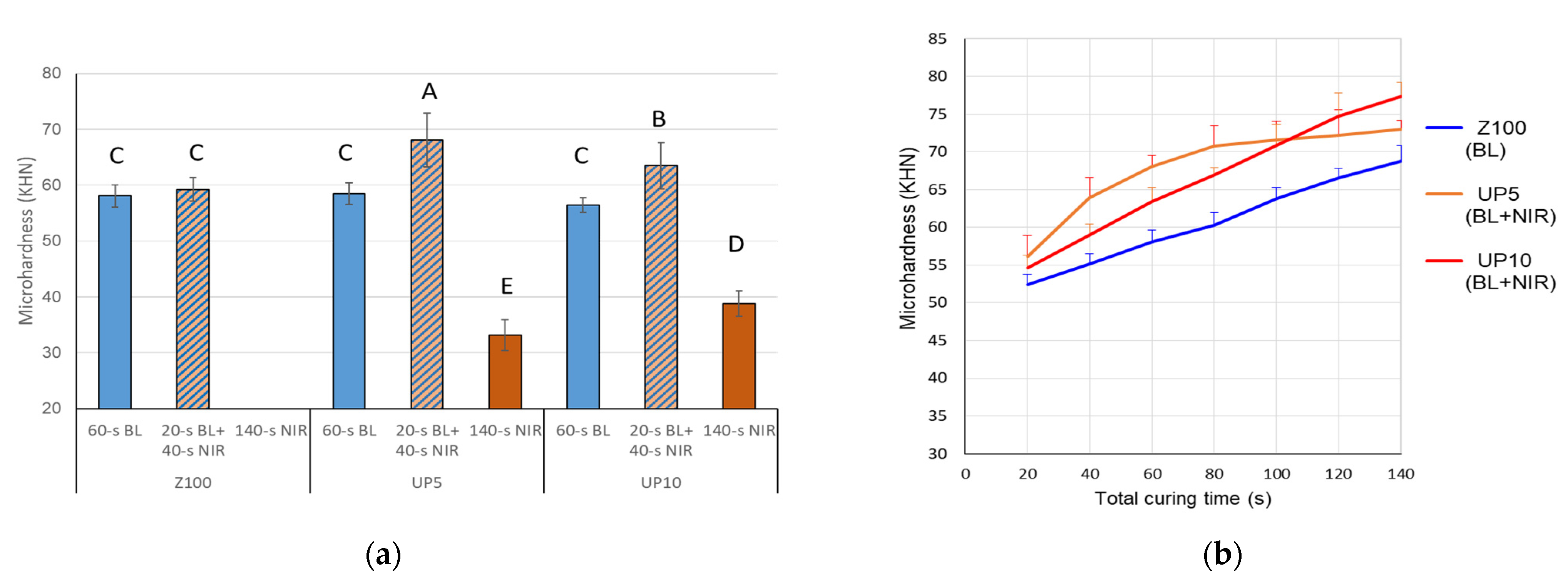
3.4. Effects of UP Content and Irradiation Protocol on Surface Microhardness
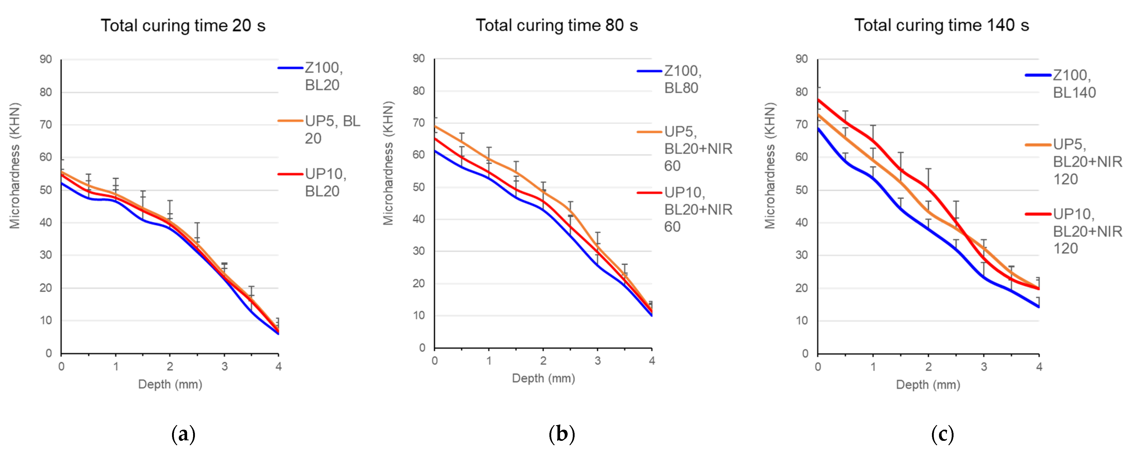
3.5. Depth Profiles of Microhardness
4. Discussion
5. Conclusions
Author Contributions
Funding
Data Availability Statement
Conflicts of Interest
References
- Ruyter, I.E.; Oysaed, H. Conversion in different depths of ultraviolet and visible light activated composite materials. Acta Odontol. Scand. 1982, 40, 179–192. [Google Scholar] [CrossRef] [PubMed]
- Leprince, J.G.; Palin, W.M.; Hadis, M.A.; Devaux, J.; Leloup, G. Progress in dimethacrylate-based dental composite technology and curing efficiency. Dent. Mater. 2013, 29, 139–156. [Google Scholar] [CrossRef]
- Moore, B.K.; Platt, J.A.; Borges, G.; Chu, T.M.; Katsilieri, I. Depth of cure of dental resin composites: ISO 4049 depth and microhardness of types of materials and shades. Oper. Dent. 2008, 33, 408–412. [Google Scholar] [CrossRef] [PubMed] [Green Version]
- Bagis, Y.H.; Baltacioglu, I.H.; Kahyaogullari, S. Comparing microleakage and the layering methods of silorane-based resin composite in wide Class II MOD cavities. Oper. Dent. 2009, 34, 578–585. [Google Scholar] [CrossRef] [PubMed]
- Li, J.; Li, H.; Fok, A.S.; Watts, D.C. Multiple correlations of material parameters of light-cured dental composites. Dent. Mater. 2009, 25, 829–836. [Google Scholar] [CrossRef]
- Hammouda, I.M. Effect of light-curing method on wear and hardness of composite resin. J. Mech. Behav. Biomed. Mater. 2010, 3, 216–222. [Google Scholar] [CrossRef]
- Fonseca, A.S.Q.; Moreira, A.D.L.; de Albuquerque, P.P.A.; de Menezes, L.R.; Pfeifer, C.S.; Schneider, L.F.J. Effect of monomer type on the CC degree of conversion, water sorption and solubility, and color stability of model dental composites. Dent. Mater. 2017, 33, 394–401. [Google Scholar] [CrossRef] [PubMed]
- Moldovan, M.; Balazsi, R.; Soanca, A.; Roman, A.; Sarosi, C.; Prodan, D.; Vlassa, M.; Cojocaru, I.; Saceleanu, V.; Cristescu, I. Evaluation of the degree of conversion, residual monomers and mechanical properties of some light-cured dental resin composites. Materials 2019, 12, 2109. [Google Scholar] [CrossRef] [Green Version]
- Kincses, D.; Böddi, K.; Őri, Z.; Lovász, B.V.; Jeges, S.; Szalma, J.; Kunsági-Máté, S.; Lempel, E. Pre-Heating Effect on Monomer Elution and Degree of Conversion of Contemporary and Thermoviscous Bulk-Fill Resin-Based Dental Composites. Polymers 2021, 13, 3599. [Google Scholar] [CrossRef] [PubMed]
- Chuang, S.F.; Huang, P.S.; Chen, T.Y.F.; Huang, L.H.; Su, K.C.; Chang, C.H. Shrinkage behaviors of dental composite restorations-The experimental-numerical hybrid analysis. Dent. Mater. 2016, 32, E362–E373. [Google Scholar] [CrossRef] [PubMed]
- Chuang, S.F.; Chang, C.H.; Chen, T.Y. Contraction behaviors of dental composite restorations—Finite element investigation with DIC validation. J. Mech. Behav. Biomed. Mater. 2011, 4, 2138–2149. [Google Scholar] [CrossRef]
- Balthazard, R.; Jager, S.; Dahoun, A.; Gerdolle, D.; Engels-Deutsch, M.; Mortier, E. High-resolution tomography study of the porosity of three restorative resin composites. Clin. Oral Investig. 2014, 18, 1613–1618. [Google Scholar] [CrossRef]
- Munchow, E.A.; Meereis, C.T.W.; da Rosa, W.L.D.O.; da Silva, A.F.; Piva, E. Polymerization shrinkage stress of resin-based dental materials: A systematic review and meta-analyses of technique protocol and photo-activation strategies. J. Mech. Behav. Biomed. Mater. 2018, 82, 77–86. [Google Scholar] [CrossRef]
- De Oliveira Correia, A.M.; Tribst, J.P.M.; de Souza Matos, F.; Platt, J.A.; Caneppele, T.M.F.; Borges, A.L.S. Polymerization shrinkage stresses in different restorative techniques for non-carious cervical lesions. J. Dent. 2018, 76, 68–74. [Google Scholar] [CrossRef] [Green Version]
- Uo, M.; Kudo, E.; Okada, A.; Soga, K.; Kogo, Y. Preparation and properties of dental composite resin cured under near Infrared Irradiation. J. Photopolym. Sci. Technol. 2009, 22, 551–554. [Google Scholar] [CrossRef] [Green Version]
- Boyer, J.C.; Johnson, N.J.J.; van Veggel, F.C.J.M. Upconverting lanthanide-doped NaYF4-PMMA polymer composites prepared by in situ polymerization. Chem. Mater. 2009, 21, 2010–2012. [Google Scholar] [CrossRef]
- Darani, M.K.; Bastani, S.; Ghahari, M.; Kardar, P.; Mohajerani, E. NIR induced photopolymerization of acrylate-based composite containing upconversion particles as an internal miniaturized UV sources. Prog. Org. Coat. 2017, 104, 97–103. [Google Scholar] [CrossRef]
- Lavis, L.D.; Raines, R.T. Bright ideas for chemical biology. ACS Chem. Biol. 2008, 3, 142–155. [Google Scholar] [CrossRef] [Green Version]
- Haase, M.; Schafer, H. Upconverting nanoparticles. Angew. Chem. Int. Ed. 2011, 50, 5808–5829. [Google Scholar] [CrossRef]
- Menezes, L.D.; Maciel, G.S.; de Araujo, C.B.; Messaddeq, Y. Phonon-assisted cooperative energy transfer and frequency upconversion in a Yb3+/Tb3+ codoped fluoroindate glass. J. Appl. Phys. 2003, 94, 863–866. [Google Scholar] [CrossRef] [Green Version]
- Sivakumar, R.; van Veggel, F.C.J.M.; Raudsepp, M. Bright white light through up-conversion of a single NIR source from sol-gel-derived thin film made with Ln(3+)-doped LaF3 nanoparticles. J. Am. Chem. Soc. 2005, 127, 12464–12465. [Google Scholar] [CrossRef] [PubMed]
- Wang, F.; Liu, X.G. Recent advances in the chemistry of lanthanide-doped upconversion nanocrystals. Chem. Soc. Rev. 2009, 38, 976–989. [Google Scholar] [CrossRef]
- Stepuk, A.; Mohn, D.; Grass, R.N.; Zehnder, M.; Kramer, K.W.; Pelle, F.; Ferrier, A.; Stark, W.J. Use of NIR light and upconversion phosphors in light-curable polymers. Dent. Mater. 2012, 28, 304–311. [Google Scholar] [CrossRef] [PubMed] [Green Version]
- Ivanova, S.; Pelle, F. Strong 1.53 μm to NIR-VIS-UV upconversion in Er-doped fluoride glass for high-efficiency solar cells. J. Opt. Soc. Am. B 2009, 26, 1930–1938. [Google Scholar] [CrossRef]
- Versluis, A.; Tantbirojn, D.; Douglas, W.H. Do dental composites always shrink toward the light? J. Dent. Res. 1998, 77, 1435–1445. [Google Scholar] [CrossRef]
- Zhou, J.C.; Yang, Z.L.; Dong, W.; Tang, R.J.; Sun, L.D.; Yan, C.H. Bioimaging and toxicity assessments of near-infrared upconversion luminescent NaYF4:Yb,Tm nanocrystals. Biomaterials 2011, 32, 9059–9067. [Google Scholar] [CrossRef]
- Pavani, K.; Kumar, J.S.; Srikanth, K.; Soares, M.; Pereira, E.; Neves, A.; Graça, M. Highly efficient upconversion of Er3+ in Yb3+ codoped non-cytotoxic strontium lanthanum aluminate phosphor for low temperature sensors. Sci. Rep. 2017, 7, 1–15. [Google Scholar] [CrossRef]
- Heer, S.; Kompe, K.; Gudel, H.U.; Haase, M. Highly efficient multicolour upconversion emission in transparent colloids of lanthanide-doped NaYF4 nanocrystals. Adv. Mater. 2004, 16, 2102–2105. [Google Scholar] [CrossRef]
- Vetrone, F.; Mahalingam, V.; Capobianco, J.A. Near-infrared-to-blue upconversion in colloidal BaYF5:Tm3+, Yb3+ nanocrystals. Chem. Mater. 2009, 21, 1847–1851. [Google Scholar] [CrossRef]
- Anderson, R.B.; Smith, S.J.; May, P.S.; Berry, M.T. Revisiting the NIR-to-visible upconversion mechanism in beta-NaYF4:Yb3+,Er3+. J. Phys. Chem. Lett. 2014, 5, 36–42. [Google Scholar] [CrossRef]
- Taylor, D.F.; Kalachandra, S.; Sankarapandian, M.; McGrath, J.E. Relationship between filler and matrix resin characteristics and the properties of uncured composite pastes. Biomaterials 1998, 19, 197–204. [Google Scholar] [CrossRef]
- Del Rosal, B.; Jaque, D. Upconversion nanoparticles for in vivo applications: Limitations and future perspectives. Methods Appl. Fluoresc. 2019, 7, 022001. [Google Scholar] [CrossRef]
- Chen, B.; Wang, F. Emerging frontiers of upconversion nanoparticles. Trends Chem. 2020, 2, 427–439. [Google Scholar] [CrossRef]
- Ilie, N.; Hickel, R. Quality of curing in relation to hardness, degree of cure and polymerization depth measured on a nano-hybrid composite. Am. J. Dent. 2007, 20, 263–268. [Google Scholar]
- Soares, L.E.; Martin, A.A.; Pinheiro, A.L.; Pacheco, M.T. Vicker’s hardness and Raman spectroscopy evaluation of a dental composite cured by an argon laser and a halogen lamp. J. Biomed. Opt. 2004, 9, 601–608. [Google Scholar] [CrossRef] [PubMed]
- Ferracane, J.; Hilton, T.; Stansbury, J.; Watts, D.; Silikas, N.; Ilie, N.; Heintze, S.; Cadenaro, M.; Hickel, R. Academy of Dental Materials guidance—Resin composites: Part II—Technique sensitivity (handling, polymerization, dimensional changes). Dent. Mater. 2017, 33, 1171–1191. [Google Scholar] [CrossRef] [Green Version]
- Ferracane, J.L. Correlation between hardness and degree of conversion during the setting reaction of unfilled dental restorative resins. Dent. Mater. 1985, 1, 11–14. [Google Scholar] [CrossRef]
- Wang, R.; Wang, Y. Depth-dependence of Degree of Conversion and Microhardness for Dual-cure and Light-cure Composites. Oper. Dent. 2020, 45, 396–406. [Google Scholar] [CrossRef]
- Tarle, Z.; Attin, T.; Marovic, D.; Andermatt, L.; Ristic, M.; Tauböck, T. Influence of irradiation time on subsurface degree of conversion and microhardness of high-viscosity bulk-fill resin composites. Clin. Oral Investig. 2015, 19, 831–840. [Google Scholar] [CrossRef] [Green Version]
- Quinn, J.B.; Quinn, G.D. Material properties and fractography of an indirect dental resin composite. Dent. Mater. 2010, 26, 589–599. [Google Scholar] [CrossRef] [Green Version]
- Manhart, J.; Kunzelmann, K.H.; Chen, H.Y.; Hickel, R. Mechanical properties of new composite restorative materials. J. Biomed. Mater. Res. 2000, 53, 353–361. [Google Scholar] [CrossRef]
- Petronijevic Sarcev, B.; Balos, S.; Markovic, D.; Sarcev, I.; Vukcevic, M.; Labus Zlatanovic, D.; Miletic, V. Effect of the Degree of Conversion on Mechanical Properties and Monomer Elution from Self-, Dual-and Light-Cured Core Composites. Materials 2021, 14, 5642. [Google Scholar] [CrossRef] [PubMed]
- Watts, D.C.; Amer, O.M.; Combe, E.C. Surface hardness development in light-cured composites. Dent. Mater. 1987, 3, 265–269. [Google Scholar] [CrossRef]
- Fronza, B.M.; Rueggeberg, F.A.; Braga, R.R.; Mogilevych, B.; Soares, L.E.S.; Martin, A.A.; Ambrosano, G.; Giannini, M. Monomer conversion, microhardness, internal marginal adaptation, and shrinkage stress of bulk-fill resin composites. Dent. Mater. 2015, 31, 1542–1551. [Google Scholar] [CrossRef] [PubMed]
- Kim, K.H.; Ong, J.L.; Okuno, O. The effect of filler loading and morphology on the mechanical properties of contemporary composites. J. Prosthet. Dent. 2002, 87, 642–649. [Google Scholar] [CrossRef] [PubMed]


Publisher’s Note: MDPI stays neutral with regard to jurisdictional claims in published maps and institutional affiliations. |
© 2021 by the authors. Licensee MDPI, Basel, Switzerland. This article is an open access article distributed under the terms and conditions of the Creative Commons Attribution (CC BY) license (https://creativecommons.org/licenses/by/4.0/).
Share and Cite
Chuang, S.-F.; Liao, C.-C.; Lin, J.-C.; Chou, Y.-C.; Lee, T.-L.; Lai, T.-W. Novel Polymerization of Dental Composites Using Near-Infrared-Induced Internal Upconversion Blue Luminescence. Polymers 2021, 13, 4304. https://doi.org/10.3390/polym13244304
Chuang S-F, Liao C-C, Lin J-C, Chou Y-C, Lee T-L, Lai T-W. Novel Polymerization of Dental Composites Using Near-Infrared-Induced Internal Upconversion Blue Luminescence. Polymers. 2021; 13(24):4304. https://doi.org/10.3390/polym13244304
Chicago/Turabian StyleChuang, Shu-Fen, Chu-Chun Liao, Jui-Che Lin, Yu-Cheng Chou, Tsung-Lin Lee, and Ting-Wen Lai. 2021. "Novel Polymerization of Dental Composites Using Near-Infrared-Induced Internal Upconversion Blue Luminescence" Polymers 13, no. 24: 4304. https://doi.org/10.3390/polym13244304