Communication

Heavy Metal Ions Trigger a Fluorescent Quenching in DNA–Organic Semiconductor Hybrid Assemblies

1
National Demonstration Centre for Experimental Chemistry Education, Department of Chemistry, Yanbian University, Yanji 133002, China
2
Jilin Tobacco Industry Co., Ltd., Yanji 133001, China
3
Longjing Municipal Ecological and Environmental Monitoring Station, Longjing 133400, China
4
Department of Chemical Engineering, Inha University, Incheon 22212, Korea
5
Department of Preventive Medicine, Yanbian University, Yanji 133002, China
*
Authors to whom correspondence should be addressed.
Polymers 2022, 14(17), 3591; https://doi.org/10.3390/polym14173591
Submission received: 31 July 2022 / Revised: 24 August 2022 / Accepted: 26 August 2022 / Published: 31 August 2022
(This article belongs to the Special Issue Polymer-Based Materials for Sensors)

Abstract

:
The significance of DNA is no longer limited to its role as a biological information carrier; as a natural polymer, it also become in the field of materials. Single-stranded DNA (ssDNA) molecules with specific sequences can form a G-quadruplex or hairpin-shaped conformation with specific heavy metal ions through coordination bonds. In this study, ssDNA molecules of the four sequences were prepared into hybrid assemblies with one of the famous display materials, the tris-(8-hydroxyquinoline)aluminum (Alq3) semiconductor. Based on these hybrid assemblies, heavy metal ions, namely Pb2+, Hg2+, Cd2+ and As3+, were detected individually at the ppb level. Apart from this, in practical application, many samples containing heavy metal ions are digested with acid. By introducing MES buffer solution, the influence of acidity on the fluorescent signal of Alq3 was excluded. This strategy showed promising results in the practical application of detecting heavy metal ions in shrub branches and leaves.

1. Introduction

With industrialization, urbanization and the frequent use of chemical fertilizers, the situation of soil pollution by heavy metals has become serious. Through food chain accumulation, plants or crops contaminated with heavy metals could have negative effects on human health. The ionic form of these heavy metals can destroy the endocrine and nervous systems, causing motion and cognitive disorders, kidney failure and even DNA damage [1,2]. Hence, quick and sensitive detection methods are of considerable importance for environmental protection and human health. In order to make up for various shortcomings of conventional analytical techniques, such as requiring expensive equipment, time-consuming procedures and highly-trained technicians, numerous methods have been developed to detect these metal ions, including electrochemical techniques [3,4,5,6,7,8,9,10,11,12], spectroscopy [13,14,15,16,17,18] and colorimetry [19,20,21,22,23,24]. Among these, aptamer-based assays for detection of the heavy metal ions have received significant attention owing to their appropriate selectivity, great sensitivity, low cost, facile operation and wide response range [25,26,27,28,29,30]. In general, sequence-designed aptamers and heavy metal ions can form a G-quadruplex or hairpin conformation, and this provides the premise for detection [31,32]. In the previous research of our group, we developed hybrid assemblies consisting of aptamers and organic semiconducting small molecules and applied them to the detection of mercury ions [33,34]. This detection approach was based on a significant “on–off” phenomenon of the fluorescent hybrid assemblies, and their selectivity was also evaluated using other metal ions, such as potassium, sodium, zinc, chromium, cadmium and copper. However, the approach was found to have a fatal shortcoming, a lack of acid resistance which induces the fluorescent quenching of these organic molecules in an acidic environment. Typically, actual samples in which heavy metal ions need to be detected are processed by acid digestion. Therefore, in the detection of heavy metal ions based on organic fluorescent probes, solving the lack of acid resistance is an unavoidable task.
In this research, four single-stranded DNA (ssDNA) sequences corresponding to Pb2+, Hg2+, Cd2+ and As3+ were designed and prepared in hybrid assemblies together with an organic semiconducting molecule, tris-(8-hydroxyquinoline)aluminum (Alq3). In order to solve the problem of acid resistance, we optimized the detection approach and finally achieved acid resistance using MES buffer solution. After that, multiplex detection of the above four heavy metal ions was completed. As a practical application, this optimized approach was also used for the detection of heavy metal ions in shrub branches and leaves. These results will broaden the detection approaches for heavy metal ions based on DNA molecules and provide a meaningful solution to the weak acid resistance of organic fluorescent probes in actual application.

2. Materials and Methods

2.1. Materials

Tris-(8-hydroxyquinoline)aluminum (Alq3, 99.995%), 2-morpholinoethanesulfonic acid (MES, 99%) and 2-[4-(2-hydroxyethyl)piperazin-1-yl]ethanesulfonic acid (HEPES, 99%) were purchased from Sigma-Aldrich. Tetrahydrofuran (THF, 99%) was purchased from Aladdin. ssDNA and Tris-HCl buffer solution were purchased from Shanghai Shenggong Biological, China. Sequences of ssDNA which can specifically bind with four heavy metal ions are as follows:
  • Pb2+: 5′-GGTTGGTGTGGTTGG-3′;
  • Hg2+: 5′-TTCTTTCTTCCCCTTGTTTGTT-3′;
  • Cd2+: 5′-GGGTTCACAGTCCGTT-3′;
  • As3+: 5′-ATGCAAACCCTTAAGAAAGTGGTCGTCCAAAAAACCATTG-3′.
Nitric acid (HNO3, 65.0~68.0%) was purchased from Sinopharm Chemical Reagent. Shrub branches and leaves were obtained from the Institute of Geophysical and Geophysical Exploration, Chinese Academy of Geological Sciences.

2.2. Preparation of Hybrid Assemblies

Commercially available Alq3 powders were dissolved in THF at a concentration of 1 mg/mL to form a stock solution. On the other hand, ssDNA was dissolved in deionized water (DI water) at a concentration of 500 nM. Then, the stock solution (2 mL) was injected into 20 mL of ssDNA solution under vigorous stirring (800 rpm) for 2 min. Subsequently, the mixture was kept at room temperature for 10 h to allow the formation of a visible precipitate.

2.3. Reaction with Heavy Metal Ions

One milliliter of hybrid assembly solution was mixed with various concentrations (50–400 ppb) of heavy metal ions at a volume ratio of 1:1 and kept for at least 30 min.

2.4. Exploration of the Conditions for the Detection of Heavy Metal Ions

One milliliter of hybrid assembly solution was thoroughly mixed with buffer solution (HEPES, Tris-HCl and MES) at a volume ratio of 1:1 and then mixed with 2 mL of Pb2+ (100–400 ppb) with various concentrations and incubated for 30 min.

2.5. Reaction with HNO3

One milliliter of hybrid assembly solution was mixed with various concentrations (0.03–3%) of HNO3 in a volume ratio of 1:1 and incubated for 30 min.
The hybrid assembly solution was thoroughly mixed with MES buffer solution at a volume ratio of 1:1 and then mixed with various concentrations (0.5–4%) of HNO3 at a volume ratio of 1:1 and incubated for 30 min.

2.6. Detection of Heavy Metal Ions in Actual Samples

In the preparation of actual sample, 0.5 g of shrub branches and leaves were weighed and placed in a microwave digestion tank. Then, 5 mL of HNO3, 1 mL of H2O2, 1 mL of HCl and 1 mL of HF were poured into the tank, which was then sealed and placed in a microwave digestion apparatus (Multiwave3000, Anton Paar, Graz, Austria). Digestion was performed at room temperature, 120 °C and 160 °C, for 5 min at each temperature, and then at 200 °C for 30 min. When the temperature dropped below 40 °C, the digestion tank was taken out, and the mixture solution was dried to 1 mL under heating at 130 °C. After cooling to room temperature, the solution was transferred to a volumetric flask and made up to volume using DI water.
For the detection of heavy metal ions, before each test of the actual sample, the standard curve was redrawn. Two milliliters of hybrid assembly solution was thoroughly mixed with MES buffer and then mixed with actual sample solution at a volume ratio of 1:1 and incubated for 30 min. Then, the mixtures were characterized using a fluorescence spectrophotometer with an excitation wavelength of 370 nm.

2.7. Characterization

A scanning electron microscope (Hitachi, SU8010, Tokyo, Japan) was used to observe the morphology of the sample, and a fluorescence spectrophotometer (Jasco, FP-8200, Tokyo, Japan) was used to observe the photoluminescence spectra of the samples.

3. Results

Figure 1 shows the scanning electron microscopy (SEM) images of the hybrid assemblies prepared by a facile re-precipitation method [35]. The ssDNA sequence was 5′-GGTTGGTGTGGTTGG-3′, which could specifically bind with Pb2+. The as-prepared hybrid assemblies showed a hexagonal prism shape with a smooth surface, as shown in Figure 1a. After exposure to the Pb2+ ion solution, cracks began to appear on the smooth surface of the hybrid assemblies. As the concentration of the Pb2+ ion solution increased, the cracks became more obvious.
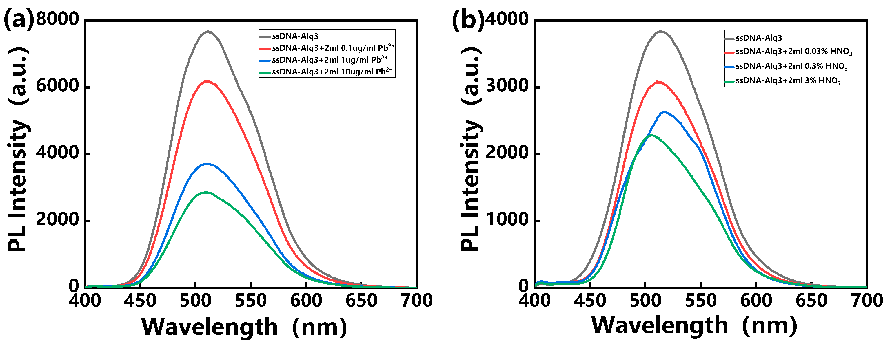
Meanwhile, the photoluminescence (PL) spectra of the hybrid assemblies before and after the interaction with the Pb2+ ion solution were observed, as shown in Figure 2a. At first, a broad PL peak of hybrid assemblies was observed at ~510 nm. The intensity of the PL peak was observed to decrease drastically upon the interaction with different concentrations of the Pb2+ ion solution. Phenomena such as the appearance of cracks and the decrease in PL peak intensity were also observed in previous studies. The reason for the PL quenching is attributed to electron transfer from Alq3 molecules to the mercury ions due to the bridge role of the hairpin conformation of ssDNA [33]. Based on these phenomena, the detection of Pb2+ was realized. It is worth noting that these heavy metal ions were prepared in DI water. However, in practical application, we need to consider the effect of an acidic environment on the PL signal of hybrid assemblies, since many samples are digested by acid. Figure 2b shows the influence of the acidic environment on the PL signal of hybrid assemblies. Obviously, when these hybrid assemblies were exposed to an acidic environment, the PL intensity decreased with increasing acid concentration.
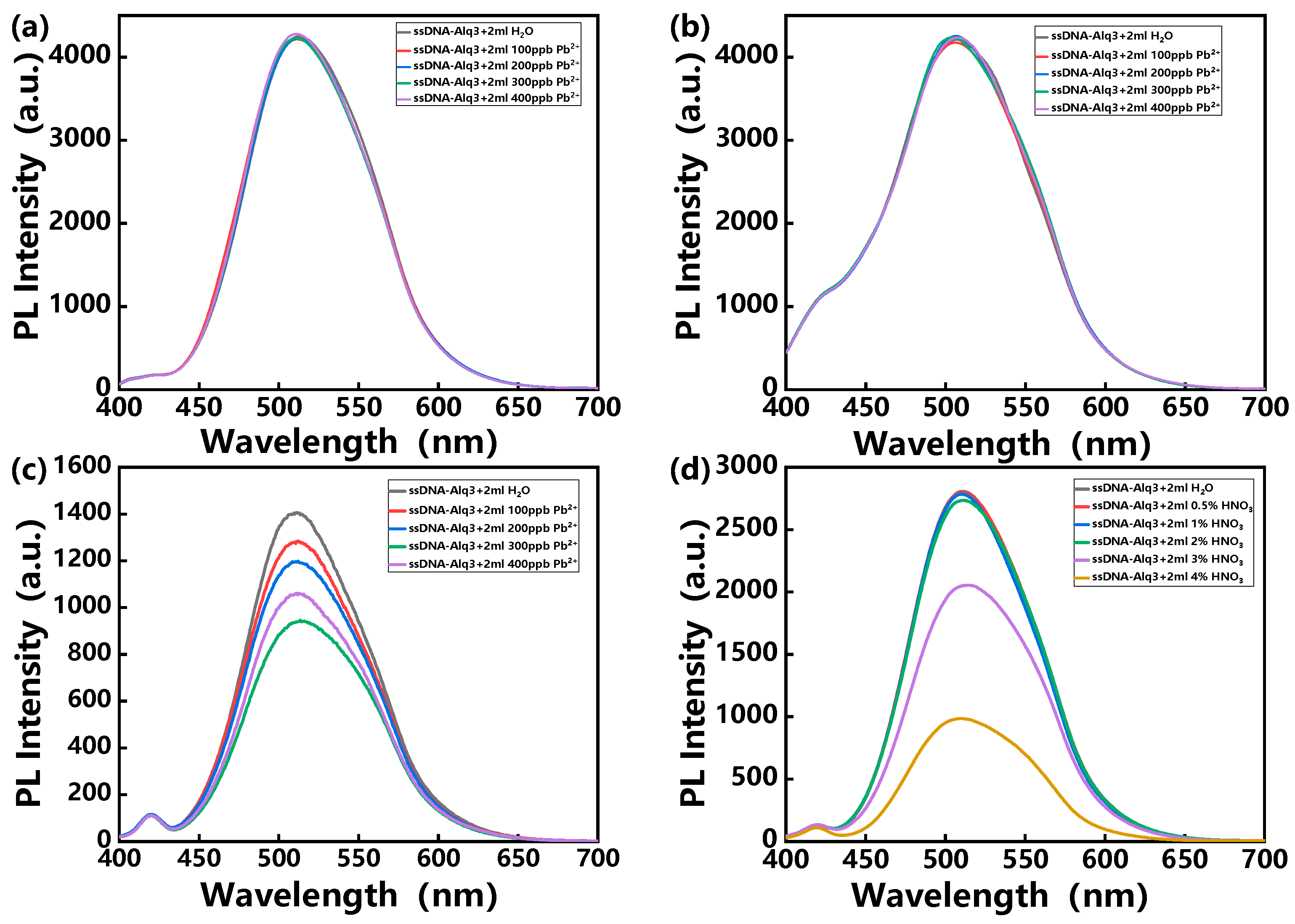
Therefore, in order to exclude the interference of acidity in the detection of heavy metal ions, strategies for using buffer solutions have been introduced. Figure 3a–c show the PL spectra of the hybrid assemblies treated with HEPES, Tris-HCl and MES buffer solution, respectively, and then exposed to various concentrations of Pb2+ ion solutions. Interestingly, the cases of treatment with HEPES and Tris-HCl maintained the original PL intensities after exposure to various concentrations of Pb2+ ion solutions. This may be due to the fact that Pb2+ ions first form complexes with the anionic groups in HEPES or Tris-HCl buffer solution, thus failing to specifically bind to ssDNA molecules of the hybrid assemblies. The PL intensity of the hybrid assemblies decreased only in the case of MES buffer solution. In addition, an acid resistance test of the hybrid assemblies treated with MES buffer was performed, as shown in Figure 3d. It can be seen that the hybrid assemblies treated with MES buffer exhibited resistance to an acidic environment. Hence, we can infer that the PL quenching of the hybrid assemblies shown in Figure 3c is caused by the recognition of lead ions.
Similarly, the other three heavy metal ions, Hg2+, Cd2+ and As3+, were detected by using the hybrid assemblies containing the corresponding ssDNA sequences. As the concentration of heavy metal ions increases, the PL intensities of the hybrid assemblies also decrease sharply, as shown in Figure 4.
Under the optimized conditions, the relationship between the PL intensities of different hybrid assemblies and the concentration of corresponding target ions was investigated. The PL intensities showed a linear relationship with the level of the heavy metal ions in the range from 50 ppb to 350 ppb, as shown in Figure 5. Correlation coefficients (R2) are 0.9945, 0.991, 0.9957 and 0.9917 for Pb2+, Hg2+, Cd2+ and As3+, respectively. The limit of quantification (LOQ) is defined as 10S0/S, where S0 is the standard deviation and S is the slope of the calibration curve [36]. According to this formula, the LOQs of Pb2+, Hg2+, Cd2+ and As3+ were calculated to be 22.92 ppb, 21.74 ppb, 12.68 ppb and 49.29 ppb, respectively.
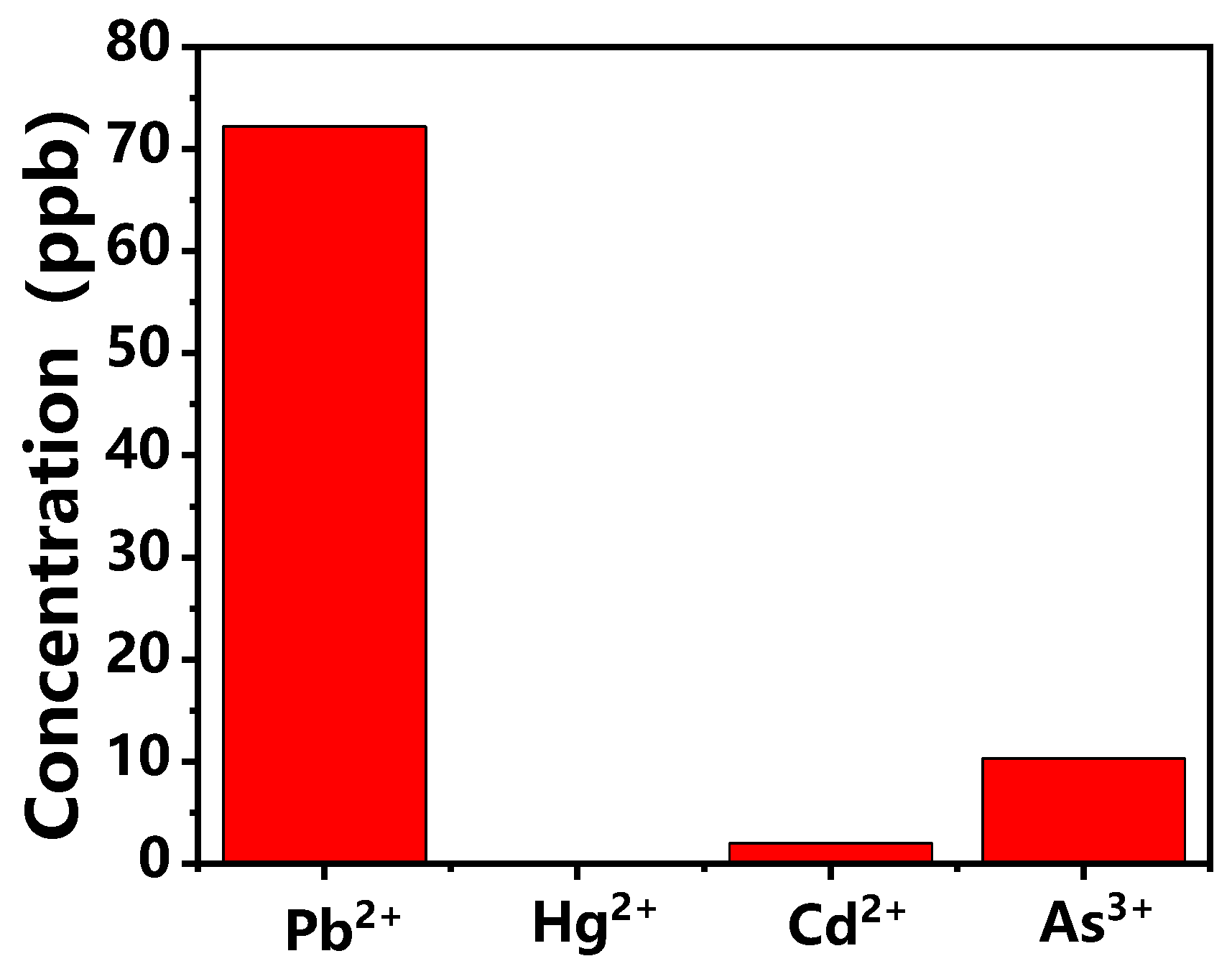
In order to verify the feasibility of the detection method in actual samples, shrub branches and leaves were selected. Their heavy metal ion content was determined using inductively coupled plasma mass spectrometry (ICP-MS). Figure 6 shows the estimated results for the content of heavy metal ions in the sample: 72.21 ppb, 2.00 ppb and 10.27 ppb for Pb2+, Cd2+ and As3+, respectively. The results were all within the reference concentration range. The result that Hg2+ was not detected in the sample was also consistent with the results provided by ICP-MS.

4. Conclusions

In this study, we developed a heavy metal ion probe based on hybrid assemblies of ssDNA molecules and organic semiconducting molecules, and we achieved low-concentration detection of four heavy metal ions, namely Pb2+, Hg2+, Cd2+ and As3+. In addition, by introducing MES buffer solution, the problem of the detection signal of these probes being affected in an acidic environment was solved. Based on this, the practical application purpose of evaluating the content of heavy metal ions in shrub branches and leaves was also achieved. These findings provide meaningful guidance for detection probes based on ssDNA or aptamers, especially for the interference of fluorescent probes in acidic environments.

Author Contributions

Conceptualization, C.C., Y.F. and Y.P.; methodology, X.L.; software, X.L.; validation, T.Y. and L.C.; investigation, D.H.P.; data curation, X.L.; writing—original draft preparation, X.L.; writing—review and editing, C.C.; visualization, Y.F. and Y.P.; supervision, C.C. All authors have read and agreed to the published version of the manuscript.

Funding

This research was funded by National Natural Science Foundation of China grant number [52011540402; 21865037]. And The APC was funded by Jilin Tobacco Industry Co., Ltd.

Institutional Review Board Statement

Not applicable.

Informed Consent Statement

Not applicable.

Data Availability Statement

Not applicable.

Conflicts of Interest

The authors declare no conflict of interest.

References

  1. Li, H.L.; Zhai, J.F.; Tian, J.Q.; Luo, Y.L.; Sun, X.P. Carbon nanoparticle for highly sensitive and selective fluorescent detection of mercury (II) ion in aqueous solution. Biosens. Bioelectron. 2011, 26, 4656–4660. [Google Scholar] [CrossRef]
  2. Xu, H.Y.; Zhang, K.N.; Liu, Q.S.; Xie, M.X. Visual and fluorescent detection of mercury ions by using a dually emissive ratiometric nanohybrid containing carbon dots and CdTe quantum dots. Microchim. Acta 2017, 184, 1199–1206. [Google Scholar] [CrossRef]
  3. Roxby, D.N.; Rivy, H.; Gong, C.; Gong, X.; Yuan, Z.; Chang, G.E.; Chen, Y.C. Microalgae living sensor for metal ion detection with nanocavity-enhanced photoelectrochemistry. Biosens. Bioelectron. 2020, 165, 112420. [Google Scholar] [CrossRef]
  4. Singh, S.; Numan, A.; Zhan, Y.; Singh, V.; Hung, T.V.; Nam, N.D. A novel highly efficient and ultrasensitive electrochemical detection of toxic mercury (II) Ions in canned tuna fish and tap water based on a copper metal-organic framework. J. Hazard. Mater. 2020, 399, 123042. [Google Scholar] [CrossRef]
  5. Hu, J.; Li, Z.; Zhai, C.; Zeng, L.; Zhu, M. Photo-assisted simultaneous electrochemical detection of multiple heavy metal ions with a metal-free carbon black anchored graphitic carbon nitride sensor. Anal. Chim. Acta 2021, 1183, 338951. [Google Scholar] [CrossRef]
  6. Ru, J.; Wang, X.; Cui, X.; Wang, F.; Ji, H.; Du, X.; Lu, X. GaOOH-modified metal-organic frameworks UiO-66-NH2: Selective and sensitive sensing four heavy-metal ions in real wastewater by electrochemical method. Talanta 2021, 234, 122679. [Google Scholar] [CrossRef]
  7. Gupta, P.; Rahm, C.E.; Jiang, D.; Gupta, V.K.; Heineman, W.R.; Justin, G.; Alvarez, N.T. Parts per trillion detection of heavy metals in as-is tap water using carbon nanotube microelectrodes. Anal. Chim. Acta 2021, 1155, 338353. [Google Scholar] [CrossRef]
  8. Kim, E.B.; Imran, M.; Lee, E.H.; Akhtar, M.S.; Ameen, S. Multiple ions detection by field-effect transistor sensors based on ZnO@GO and ZnO@rGO nanomaterials: Application to trace detection of Cr (III) and Cu (II). Chemosphere 2022, 286, 131695. [Google Scholar] [CrossRef]
  9. Chen, X.; Zhao, J.X.; Wang, J.W.; Liu, Y.; Wang, L.C.; Weerasooriya, R.; Wu, Y.C. Doping ZIF-67 with transition metals results in bimetallic centers for electrochemical detection of Hg(II). Electrochim. Acta 2021, 387, 138539. [Google Scholar] [CrossRef]
  10. Li, G.; Belwal, T.; Luo, Z.; Li, Y.; Li, L.; Xu, Y.; Lin, X. Direct detection of Pb2+ and Cd2+ in juice and beverage samples using PDMS modified nanochannels electrochemical sensors. Food Chem. 2021, 386, 129632. [Google Scholar] [CrossRef]
  11. Nguyen, M.B.; Nga, D.T.N.; Thu, V.T.; Piro, B.; Truong, T.N.P.; Yen, P.T.H.; Le, G.H.; Hung, L.Q.; Vu, T.A.; Ha, V.T.T. Novel nanoscale Yb-MOF used as highly efficient electrode for simultaneous detection of heavy metal ions. J. Mater. Sci. 2021, 56, 8172–8185. [Google Scholar] [CrossRef]
  12. Liu, S.; Ni, L.; Chen, W.; Wang, J.; Ma, F. Analysis of lead forms and transition in agricultural soil by nano-fluorescence method. J. Hazard. Mater. 2020, 389, 121469. [Google Scholar] [CrossRef] [PubMed]
  13. Zhao, H.M.; Bao, B.; Sun, T.; Xue, B. Depolymerized phosphorus-doped polymeric carbon nitride: A mercury (II) ion fluorescent probe. Ceram. Int. 2021, 47, 24115–24120. [Google Scholar] [CrossRef]
  14. Li, Y.; Zhang, Y.; Wang, M.; Wang, D.; Chen, K.; Lin, P.; Ge, Y.; Liu, W.; Wu, J. Highly selective fluorescence probe with peptide backbone for imaging mercury ions in living cells based on aggregation-induced emission effect. J. Hazard. Mater. 2021, 415, 125712. [Google Scholar] [CrossRef]
  15. Sharma, R.; Haldar, U.; Turabee, M.H.; Lee, H. Recyclable macromolecular thermogels for Hg(II) detection and separation via sol-gel transition in complex aqueous environments. J. Hazard. Mater. 2021, 410, 124625. [Google Scholar] [CrossRef] [PubMed]
  16. Song, Y.; Ma, Z.; Fang, H.; Zhang, Q.; Zhou, Q.; Chen, Z.; Yang, H.; Wang, F. Au sputtered paper chromatography tandem Raman platform for sensitive detection of heavy metal ions. ACS Sens. 2020, 5, 1455–1464. [Google Scholar] [CrossRef]
  17. Anusuya, T.; Kumar, V.; Kumar, V. Hydrophilic graphene quantum dots as turn-off fluorescent nanoprobes for toxic heavy metal ions detection in aqueous media. Chemosphere 2021, 282, 131019. [Google Scholar] [CrossRef]
  18. Kamal, S.; Yang, C.K. Silver enriched silver phosphate microcubes as an efficient recyclable SERS substrate for the detection of heavy metal ions. J. Colloid Interface Sci. 2022, 605, 173–181. [Google Scholar] [CrossRef]
  19. Liu, J.; Hu, Q.; Qi, L.; Lin, J.M.; Yu, L. Liquid crystal-based sensing platform for detection of Pb2+ assisted by DNAzyme and rolling circle amplification. J. Hazard. Mater. 2020, 400, 123218. [Google Scholar] [CrossRef]
  20. Yu, Y.; Naik, S.S.; Oh, Y.; Theerthagiri, J.; Lee, S.; Choi, M.Y. Lignin-mediated green synthesis of functionalized gold nanoparticles via pulsed laser technique for selective colorimetric detection of lead ions in aqueous media. J. Hazard. Mater. 2021, 420, 126585. [Google Scholar] [CrossRef]
  21. Berlina, A.N.; Komova, N.S.; Zherdev, A.V.; Dzantiev, B.B. Combination of phenylboronic acid and oligocytosine for selective and specific detection of lead(ii) by lateral flow test strip. Anal. Chim. Acta 2021, 1155, 338318. [Google Scholar] [CrossRef] [PubMed]
  22. Zou, Y.; Pang, J.; Zhang, F.; Chai, F. Silver nanoparticles for colorimetric detection and discrimination of mercury ions in lake water. ChemistrySelect 2021, 6, 6077–6082. [Google Scholar] [CrossRef]
  23. Zeng, H.; Tang, Y.; Zou, W.; Wang, C.; Tao, H.; Wu, Y. V6O13 Nanobelts for simultaneous detection of Cd(II) and Pb(II) in water. ACS Appl. Nano Mater. 2021, 4, 4654–4664. [Google Scholar] [CrossRef]
  24. Wang, J.; Wu, J.; Zhang, Y.; Zhou, X.; Hu, Z.; Liao, X.; Sheng, B.; Yuan, K.; Wu, X.; Cai, H.; et al. Colorimetric and SERS dual-mode sensing of mercury (II) based on controllable etching of Au@Ag core/shell nanoparticles. Sens. Actuators B Chem. 2021, 330, 129364. [Google Scholar] [CrossRef]
  25. Khoshbin, Z.; Housaindokht, M.R.; Verdian, A. A low-cost paper-based aptasensor for simultaneous trace-level monitoring of mercury (II) and silver (I) ions. Anal. Biochem. 2020, 597, 113689. [Google Scholar] [CrossRef]
  26. Lu, Z.; Wang, P.; Xiong, W.; Qi, B.; Shi, R.; Xiang, D.; Zhai, K. Simultaneous detection of mercury (II), lead (II) and silver (I) based on fluorescently labelled aptamer probes and graphene oxide. Environ. Technol. 2021, 42, 3065–3072. [Google Scholar] [CrossRef]
  27. Nguyen, D.K.; Jang, C.H. Label-free liquid crystal-based detection of As(III) ions using ssDNA as a recognition probe. Microchem. J. 2020, 156, 104834. [Google Scholar] [CrossRef]
  28. Wang, Y.; Lv, M.; Chen, Z.; Deng, Z.; Liu, N.; Fan, J.; Zhang, W. A fluorescence resonance energy transfer probe based on DNA-modified upconversion and gold nanoparticles for detection of lead ions. Front. Chem. 2020, 8, 238. [Google Scholar] [CrossRef]
  29. Gan, Y.; Liang, T.; Hu, Q.; Zhong, L.; Wang, X.; Wan, H.; Wang, P. In-situ detection of cadmium with aptamer functionalized gold nanoparticles based on smartphone-based colorimetric system. Talanta 2020, 208, 120231. [Google Scholar] [CrossRef]
  30. Chen, T.; Wang, H.; Wang, Z.; Tan, M. Construction of time-resolved luminescence nanoprobe and its application in as (III) detection. Nanomaterials 2020, 10, 551. [Google Scholar] [CrossRef] [Green Version]
  31. Lee, J.; Jun, H.; Kim, J. Mercury biosensors: Polydiacetylene–liposome microarrays for selective and sensitive mercury (II) detection. Adv. Mater. 2009, 21, 367. [Google Scholar]
  32. Li, D.; Wieckowska, A.; Willner, I. Optical analysis of Hg2+ ions by oligonucleotide-gold-nanoparticle hybrids and DNA-based machines. Angew. Chem. Int. Ed. 2008, 47, 3927. [Google Scholar] [CrossRef] [PubMed]
  33. Huang, J.; Park, J.H.; Back, S.H.; Feng, Y.; Cui, C.; Jin, L.Y.; Ahn, D.J. Mercury ion-DNA specificity triggers a distinctive photoluminescence depression in organic semiconductor probes guided with a thymine-rich oligonucleotide sequence. Nanoscale 2018, 10, 17540–17545. [Google Scholar] [CrossRef] [PubMed]
  34. Cui, C.; Park, D.H.; Ahn, D.J. Organic semiconductor-DNA hybrid assemblies. Adv. Mater. 2020, 32, 2002213. [Google Scholar] [CrossRef]
  35. Cui, C.; Park, D.H.; Kim, J.; Joo, J.; Ahn, D.J. Oligonucleotide assisted light-emitting Alq3 microrods: Energy transfer effect with fluorescent dyes. Chem. Commun. 2013, 49, 5360–5362. [Google Scholar] [CrossRef]
  36. Mcnaught, A.D.; Wilkinson, A. IUPAC (1997) Compendium of Chemical Terminology: The Gold Book, 2nd ed.; Blackwell Scientific Publications: Oxford, UK, 1997; p. 839. [Google Scholar]
Figure 1. SEM images of (a) as-prepared hybrid assemblies and hybrid assemblies exposed to (b) 0.1 ug/mL, (c) 1 ug/mL and (d) 10 ug/mL of Pb2+ solution.
Polymers 14 03591 g001
Figure 2. PL spectra of hybrid assemblies exposed to various concentrations of (a) Pb2+ solution and (b) HNO3 solution, with excitation at 370 nm.
Polymers 14 03591 g002
Figure 3. PL spectra of hybrid assemblies treated with (a) HEPES, (b) Tris-HCl and (c) MES buffer solution and exposed to various concentrations of Pb2+ solution, with excitation at 370 nm. (d) PL spectra of hybrid assemblies treated with MES buffer solution treated and exposed to HNO3 solution, with excitation at 370 nm.
Polymers 14 03591 g003
Figure 4. PL spectra of hybrid assemblies treated with MES buffer solution and exposed to various concentrations of (a) Hg2+, (b) Cd2+ and (c) As2+ solution, with excitation at 370 nm.
Polymers 14 03591 g004
Figure 5. The relationships between PL intensities of MES-treated hybrid assemblies and various concentrations of (a) Pb2+, (b) Hg2+, (c) Cd2+ and (d) As3+.
Polymers 14 03591 g005
Figure 6. Evaluation results for four heavy metal ions in shrub branches and leaves.
Polymers 14 03591 g006
Publisher’s Note: MDPI stays neutral with regard to jurisdictional claims in published maps and institutional affiliations.