Article

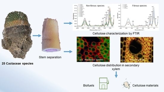
Cellulose in Secondary Xylem of Cactaceae: Crystalline Composition and Anatomical Distribution

by
Agustín Maceda
1,*,
Marcos Soto-Hernández
2 and
Teresa Terrazas
1,*
1
Instituto de Biología, Universidad Nacional Autónoma de México, Mexico City 04510, Mexico
2
Programa de Botánica, Colegio de Postgraduados, Montecillo 56230, Mexico
*
Authors to whom correspondence should be addressed.
Polymers 2022, 14(22), 4840; https://doi.org/10.3390/polym14224840
Submission received: 18 October 2022 / Revised: 6 November 2022 / Accepted: 8 November 2022 / Published: 10 November 2022

Abstract

:
Cellulose is the main polymer that gives strength to the cell wall and is located in the primary and secondary cell walls of plants. In Cactaceae, there are no studies on the composition of cellulose. The objective of this work was to analyze the crystallinity composition and anatomical distribution of cellulose in Cactaceae vascular tissue. Twenty-five species of Cactaceae were collected, dried, and milled. Cellulose was purified and analyzed with Fourier transform infrared spectroscopy, the crystallinity indexes were calculated, and statistical analyzes were performed. Stem sections were fixed, cut, and stained with safranin O/fast green, for observation with epifluorescence microscopy. The crystalline cellulose ratios had statistical differences between Echinocereus pectinatus and Coryphantha pallida. All cacti species presented a higher proportion of crystalline cellulose. The fluorescence emission of the cellulose was red in color and distributed in the primary wall of non-fibrous species; while in the fibrous species, the distribution was in the pits. The high percentages of crystalline cellulose may be related to its distribution in the non-lignified parenchyma and primary walls of tracheary elements with helical or annular thickenings of non-fibrous species, possibly offering structural rigidity and forming part of the defense system against pathogens.

Graphical Abstract

1. Introduction

Plant cell walls give rigidity, delimit the cell space, and function as a physical barrier against pathogens [1]. The main polymers of the cell wall are cellulose, hemicelluloses, and lignin, in addition to other compounds such as pectins, structural proteins, and glycoproteins [2]. The cell wall is divided in two: the primary wall with abundant cellulose microfibers, some hemicelluloses, xyloglucans, and pectins [3]. The primary wall develops during cell growth and maintains some elasticity during the initial stage. The secondary wall has three layers: S1, S2, and S3. A secondary wall develops once cell growth ends, mainly lignin accumulates, and cellulose occurs in a lesser quantity than other polysaccharides such as hemicelluloses [4].
Cellulose is the main component of the cell wall [1] and is a homopolysaccharide composed of repeated glucose residues linked by β-(1→4) bonds that generate long and rigid microfibrils [5] made of 18 cellulose polymers [6]. The presence of cellulose microfibrils in the cell wall confers mechanical and enzymatic resistance [7], structural rigidity in the primary wall [2], and forms the scaffolding for binding with pectins and hemicelluloses [5]. Two types of cellulose are characterized by their orientation and packaging. The cellulose formed by microfibrils is also called crystalline cellulose [8], which is packaged because the microfibrils are linked by hydrogen β-(1→4)-linked D-glucose units, making it more compact, rigid, and ordered [9]. The cellulose matrix that has no order (amorphous) forms a network where the hemicelluloses, pectins, lignin, and phenolic compounds are inserted and joined [3,10].
The importance of studying the crystalline composition of cellulose is due to its use in the paper industry [8] and for the production of biofuels [11]. In addition, Cactaceae species are considered second generation plants for biofuel production because, despite their slow growth, they resist drought conditions and high temperatures, and are not an essential part of human consumption [12]. The species with the highest amount of amorphous cellulose [13] and the lowest presence of lignin or lignin, and with the highest accumulation of syringyl monomers [14], are those that have potential for use in the previously mentioned industries. On the other hand, cellulose studies also focus on anatomical-structural analysis, to identify cell wall interactions with biotic and abiotic factors [15]. At this point, different authors have analyzed the stress effects of the environment on cellulose accumulation [16] and the interaction between cellulose and pathogens [17].
In succulent species, studies are scarce and most have focused on the genus Agave, a member of the Asparagaceae family [18]. In the Cactaceae family, few studies have identified the chemical composition of lignin [19,20,21,22]; furthermore, the crystallinity of cellulose was only analyzed in Opuntia ficus-indica, due to the potential use of its cellulosic compounds in the biofuel and paper industry [23]. However, of the other species of the Opuntioideae, Pereskioideae, and Cactoideae subfamilies, there have been no studies on the composition of cellulose and its distribution in the secondary xylem (wood), which would allow us to understand the structure and functioning of cellulose in the cell wall of the parenchyma, water conductive, and supporting cells. Therefore, this study aimed to analyze the crystalline composition of cellulose and its distribution in wood. The hypothesis was that there would be variation in the crystalline composition of cellulose among the different groups of cacti, due to the type of wood present.

2. Materials and Methods

2.1. Crystalline Analysis of Cellulose

To analyze the crystalline cellulose of Cactaceae species, adult and healthy plants of representative species of Cactaceae (Table 1) were collected. The vascular cylinder of all species was isolated and dried for 72 h. After that, 2 g of each species was weighed, dried, and ground. Subsequently, to obtain free-extractive wood, successive extractions were applied with ethanol: benzene (1:2), ethanol 96%, and hot water at 90 °C, according to the method proposed by Reyes-Rivera and Terrazas [24].
For the free-extractive wood, cellulose purification was performed using the procedure of Maceda et al. [20] based on the Kürschner-Hoffer method. Whereby, 0.5 g of extractive-free wood was weighed and added to 12.5 mL of HNO3/ethanol (1:4 v/v), kept in a reflux system for 1 h, allowed to cool to room temperature, and the sample was decanted. Subsequently, 12.5 mL of HNO3/ethanol was added and the process of reflux, cooling, and decantation was repeated two more times. In the last process, 12.5 mL of 1% aqueous KOH solution was added, kept under reflux for an additional 30 min, and finally filtered through a fine-pore Büchner filter. The residue (cellulose) was dried at 60 °C for 12 h.
Obtainment of the crystalline and amorphous cellulose proportion was performed with attenuated total reflection Fourier transform infrared spectroscopy (ATR-FTIR), because the covalent and non-covalent interactions of cellulose could be identified, and the crystallinity of cellulose could be measured [25]. A small amount of dry cellulose from each species was taken, readings in the spectrum range of 4000–650 cm−1 (30 scans with a resolution of 4 cm−1, 15 s per sample) of each sample were made in an FTIR Spectrometer (Agilent Cary 630 FTIR), and a baseline correction was made with MicroLab PC software (Agilent Technologies, Santa Clara, CA, USA). The raw spectra were converted from transmittance to absorbance, and the average of three spectra was obtained using the Resolution Pro FTIR Software program (Agilent Technologies, Santa Clara, CA, USA).
The total crystallinity index (TCI) proposed by Nelson and O’Connor [26], or also called the proportion of crystallinity [13], was calculated from the ratio between the intensity absorption peaks 1370 cm−1 and 2900 cm−1 [27]. The lateral order index (LOI) [26,27], or the second proportion of crystallinity [13], was calculated from the ratio between the intensity absorption peaks 1430 cm−1 and 893 cm−1. Hydrogen bonding intensity (HBI) was calculated from the ratio between 3350 cm−1 and 1315 cm−1 [28].
The data obtained in triplicate for TCI, LOI, and HBI for each species were analyzed with non-parametric statistics, due to the data having no normality with the Kolmogorov-Smirnov and Shapiro–Wilk tests, even with the square root and log transformations. A non-parametric Kruskal–Wallis test was used to determine if there were differences among the species, followed by Dunn’s post hoc test.

2.2. Epifluorescence Microscopy

To analyze the anatomical distribution of cellulose in Cactaceae species, epifluorescent microscopy was used. Epifluorescence is a technique used to identify the distribution of structural components such as lignin, cellulose, phenolic compounds, starch, and proteins [29]. In the particular case of epifluorescence, it allows the observation of thin sections of plant samples, where the tissues are anisotropic [30]. The method used to make the observations was based on the results obtained by De Micco and Aronne [31] and Maceda et al. [21,22] with epifluorescence and safranin O/fast green staining (SF). However, to confirm that the fluorescence emission by safranin/fast green staining corresponded to the fluorescence of lignified tissues and cellulose, samples of Ferocactus latispinus (HAW.) Britton and Rose were used as a model for comparison with two other typical stains for fluorescence, such as acridine orange–Congo red (AO), in addition to calcofluor (CA), based on the method proposed by Nakaba et al. [32].
Representative samples of wood from the base of the stem of the Cactaceae species were obtained, fixed with a solution of formalin–acetic acid–ethanol (10:5:85) [33], and washed, before dehydrating the samples [30]. The non-fibrous species samples were embedded with paraffin and cut with a rotatory microtome, and the fibrous species were cut with a sliding microtome [20]. All the samples were stained with safranin O/fast green and mounted in synthetic resin according to Loza-Cornejo and Terrazas [34].
Due to the different structural components of the cell wall, presenting autofluorescence and fluorescence emission [29], three different excitation and emission bands were used at the same time [21]. This process allowed obtaining images with a “true color” [29]. Therefore, a wide-field fluorescence microscope (Zeiss Axio Imager Z2) with Apotome 2.0 (Zeiss Apotome.2), an AxioCam MRc 5 (Zeiss), and a microscope metal halide fluorescence light source (Zeiss HXP 120) were used. The multicolor images were obtained with a triple excitation/emission bandwidth: DAPI (blue) with an excitation of 365 nm and emission of 445/50 nm, FITC (green) with an excitation of 470/40 nm and emission of 525/50 nm, and TRITC (red) with an excitation of 546/12 nm and emission of 575–640 nm. Each sample was exposed to fluorescence light with a low power for a maximum of one minute, as proposed by Baldacci-Cresp et al. [35], to avoid overexposure of the samples and cause their photobleaching. Images were obtained with the Zen Blue 2.5 lite (Zeiss, Jena, Germany) program, and brightness adjustments were made to the entire image.

3. Results

3.1. Cellulose Composition

Figure 1 illustrates the cellulose spectra and Table 2 shows the allocation of the main bands. The absence or presence of small bands at 1269 cm−1 corresponding to lignin and hemicelluloses; 1595 cm−1, 1512 cm−1, and 1463 cm−1 assigned to lignin; and 1735 cm−1 corresponding to xylenes and hemicelluloses reflected the effectiveness of the extraction and cellulose purification. The presence of 1640 cm−1 reflected the O-H vibration of absorbed water. The parameters for crystallinity, TCI, LOI, and HBI, had statistically significant differences (p < 0.05) with a non-parametric Kruskal–Wallis analysis (Table 3); and using Dunn’s post hoc test, the species that were statistically different were identified (Table 4 and the website on Data Availability Statement contain the tables for each variable and the comparison between species).
The TCI showed that all species had values above one, because cacti have a higher proportion of crystalline cellulose than amorphous cellulose (Table 4). E. pectinatus had the highest TCI values and had significant differences with C. pallida and C. clavata, which presented the lowest values (Table 4). The lateral order index (LOI) reflected the degree of order in cellulose and the presence of crystalline cellulose II or amorphous cellulose; all species had similar values, except for E. pectinatus, C. pallida, and C. ramillosa, because the last two presented significant differences from the first (Table 4). In HBI, which relates to the crystal system and the bound water, the significant differences were between C. pallida with the highest values, and E. pectinatus, which presented the lowest HBI of the cacti (Table 4). The crystallinity indexes TCI and HBI were high in all Cactaceae species, but had lower values in the LOI index, because the cacti species had a lesser order (disorder) in the crystalline structure, but a higher proportion of crystalline cellulose.

3.2. Staining Methods for Fluorescence

When comparing the bright-field images with the fluorescence images, it was observed that in the bright-field images, the blue tones corresponded to the non-lignified tissue, while the red tones corresponded to the lignified tissue. In the fluorescence images, the green tones reflected the lignified tissue and the red the non-lignified (Figure 2A,B). In the SF, AO, and CA stains (Figure 2) it was observed that the secondary walls of the vessel elements (v) and wide-band tracheids (wbt) showed fluorescence emission in green to bluish tones in all three types of staining. In non-lignified tissues, such as the parenchyma or the primary walls of the vascular tissue, in the SF and AO stains, the cellulose fluoresced in red tones, while, for CA, the tones were bluish to greenish (Figure 2D).

3.3. Cellulose Distribution in Cells of the Secondary Xylem

Figure 3, Figure 4, Figure 5 and Figure 6 show representative species of fibrous (Pereskioideae and Opuntioideae) and non-fibrous (Cactoideae) wood. In the bright-field microscopy images, the lignified cell walls were observed in red tones, with the non-lignified in blue tones. With fluorescence microscopy, the fibrous species reflected bluish-green to yellow tones; while in the non-fibrous species, the tones of the lignified walls were greenish. The presence of cellulose and other components of the non-lignified walls reflected reddish tones in all species. Cylindropuntia leptocaulis (Figure 3A,B) had starch within some parenchyma (p) cells, which were cyan-colored. The distribution of cellulose was different between the fibrous species and non-fibrous species.
In longitudinal sections of Cylindropuntia, Opuntia, and L. lychnidiflora, cellulose was detected in the pits of the VEs (Figure 4A,B,D). In addition, the presence of cellulose was mainly in the simple pits of the fibers (f) of Cylindropuntia and L. lychnidiflora. (Figure 4A,B,D). The unlignified parenchyma of O. stenopetala in longitudinal sections showed primary walls with the presence of cellulose (Figure 4C); whereas, in the lignified parenchyma of Cylindropuntia and Leuenbergeria, lignin was mainly accumulated in the cell wall, and cellulose was exclusively in the pits (Figure 4A,B,D).
In non-fibrous species (Figure 5), cellulose fluorescence emission was detected in the primary wall of p, wbt, and v; unlike in fibrous species, where fluorescence emission was not detected in the primary wall but in the S3 layer of the secondary wall adjacent to the lumen. Furthermore, the non-fibrous species showed secondary walls as helical and annular thickenings in v and wbt, so the primary wall was visible in the longitudinal sections (Figure 6). In all non-fibrous species, the p was abundant and unlignified, in addition to having a greater accumulation of cellulose (Figure 5B,D,F,H and Figure 6B).

4. Discussion

All Cactaceae species had a higher cellulose crystalline proportion, and the distribution of cellulose was in unlignified parenchyma, v, and wbt; all with secondary walls as helical and annular thickenings.

4.1. Crystalline Indexes

The TCI index was based on the proportion between the intensity peaks of 1370 cm−1 and 2900 cm−1, according to Nelson and O’Conner [26]. The TCI is proportional to the degree of crystallinity of cellulose; therefore, the higher the ratio, the higher the percentage of crystalline cellulose, as reported by various authors for different species, fibers, and materials [27,28,38]. In the case of the LOI index, the peak of 1430 cm−1 corresponds to the presence of crystalline cellulose I, while the peak of 893 cm−1 reflects the presence of crystalline cellulose II and amorphous cellulose [39]. LOI reflects the ordered regions perpendicular to the chain direction, which is influenced by the chemical extraction and purification of cellulose [40]. The low values of LOI for Cactaceae species could have been the result of the presence of crystalline cellulose II, which influences the peak 893 cm−1 [41], or the effect of the temperature and concentration of the NaOH solution during the purification of cellulose [39]. In the case of HBI, these values are useful for interpreting qualitative changes in cellulose crystallinity; the lower values indicate the presence of crystalline cellulose, and if the values are higher, this could indicate the presence of cellulose II or amorphous cellulose. However, these values could represent the amount of bound water in the fiber structure and the presence of extractives, hemicelluloses, and lignin that increase HBI values [27].
The results observed in Table 4 reflect that all cacti species had high proportions of crystalline cellulose, because they had high values of TCI and LOI, and similar values in HBI. The species E. pectinatus, C. retusa, and C. ramillosa (except in the LOI value) had the highest crystalline proportions, due to having the highest values of TCI and LOI, and lower values of HBI, which corresponds with the fibers and materials with high crystalline cellulose proportions reported by Colom and Carrillo [42], Carrillo et al. [43], Široký et al. [39], and Poletto et al. [27].
Comparing with the reports in the literature for other cacti species, the high proportions of crystallinity cellulose were similar to most angiosperms reported by Agarwal et al. [44] and to the bark of the cactus Cereus forbesii, which had a percentage of 82% crystalline cellulose [45]. Opuntia ficus-indica is a species with varying crystalline cellulose percentages in different parts of the plant: cladodes 27% [23] and 79% [46], spines 34–70% [47,48], vascular tissue 22–28% [49], fruit epidermis 38% [50], and 60% in seeds [51]. Maceda et al. [46] reported percentages of crystalline cellulose of 76% and 74%, respectively, for Opuntia streptacantha and O. robusta. These values are very similar to those reported in this study for non-fibrous and fibrous species. Therefore, high proportions or percentages of crystalline cellulose is a constant in different Cactaceae species, probably due to the presence of non-lignified parenchyma or non-lignified primary cell walls, as observed in the anatomical distribution. Further studies comparing the purity of cellulose using the Kürschner-Hoffer method with other methods, such as the Seifert [52] method, would allow corroborating the proportions of crystalline cellulose in the Cactaceae species analyzed.

4.2. Staining Methods for Fluorescence

With the three staining methods (SF, AO, and CA; Figure 2), the distribution of the lignified tissue from the non-lignified was identified. The tones in the fluorescence emission for lignin in SF were similar to what was observed in AO and CA, and to what was reported by other authors [22,29,30]. The use of SF for bright-field and fluorescence is advantageous over AO and CA, because images can only be taken using fluorescence microscopy. On the other hand, the AO and CA techniques are semi-permanent, so over time the fluorescence is lost, when the dyes become diluted in the mounting medium used (phosphate buffer:glycerol 1:1 v/v) [30]; while for SF, a permanent mounting medium is used, so slides can be stored without loss of fluorescence.
Finally, the difference in lignin tones was visible in SF and CA, such as v, whose intensity and fluorescence tonalities were different from wbt; while for AO, the tonalities were similar in v and wbt. For the distribution of cellulose in the non-lignified parenchyma and in the vascular tissue, SF was clearly observed in red tones; while in AO and CA, the fluorescence emission of the parenchyma was obscured by the fluorescence of the vascular tissue. Therefore, SF was an efficient method for determining the distribution of lignified tissues and cellulose, as was previously reported in the literature [21,22].

4.3. Cellulose Distribution and Crystalline Composition

The tonalities observed in the fluorescence emission for cellulose and lignin correspond to those reported in other similar studies with a safranin O/fast green staining technique and three excitation bands [21,22,29,30,31]. This technique allowed the detection of differences in the distribution of both structural compounds (cellulose and lignin), between non-fibrous and fibrous species. The safranin O dye allows the detection of lignin autofluorescence and fluorescence in analyzed tissues [30,31], with tonalities of blue to green [29,35]. In the case of celluloses and hemicelluloses that do not have autofluorescence, such as lignin [29], with safranin O/fast green staining, the fluorescence emission of cellulose can be detected at 570–620 nm and its tonalities were reddish, similarly to the results of Maceda et al. [21,22]. However, further studies with immunohistochemistry or specific fluorophores could help confirm the distribution of cellulose [2,29].
When comparing the results with those reported by Maceda et al. [21] for the primary xylem, cellulose accumulated in the primary walls and lignin in the secondary ones of the helical and annular thickenings of the protoxylem and metaxylem of fibrous species, similarly to what was observed in non-fibrous adult plants. Only in the metaxylem of Leuenbergeria lychnidiflora was there a decrease in the accumulation of cellulose in the primary walls and a greater accumulation in the intervascular pits, as obtained in the secondary xylem of the fibrous species.
The presence of high proportions of crystalline cellulose in all studied cacti, mainly in the non-fibrous species (E. pectinatus, C. retusa, and C. ramillosa), could be related to the abundant unlignified parenchyma and the lower accumulation of lignin in the xylem, as seen in Figure 4 and Figure 5B. Even when including fibrous species of Cylindropuntia and Opuntia, there was a high proportion of crystalline cellulose, similarly to non-fibrous species, possibly because the xylem had a greater accumulation of lignin in the cell walls of the v, f, and p [20]. This high proportion of lignin may function as a physical [53] and chemical barrier against the attack of pathogens [54]. Zhao and Dixon [7] and Bacete and Hamann [55] mentioned that the cell wall is a dynamic barrier in conditions of abiotic stress. Therefore, in some cells, the presence of increased lignin accumulation can be observed [56]; while in others, similarly to in the gelatinous layer (G), cellulose is mainly accumulated [8].
In the cells with non-lignified primary cell walls, the accumulation of crystalline cellulose packed by hydrogen chain β-(1→4)-linked D-glucose units [9] makes cellulose more hydrophobic than amorphous cellulose [8], conferring structural support [9] and causing a decrease in the efficiency of cellulase enzymes [57], by not presenting sites for binding with enzymes and making it difficult to degrade [58]. In contrast, amorphous cellulose is slightly hydrophilic [55], susceptible, and degrades rapidly, due to the action of cellulase enzymes [59] and pH changes from pathogens [60].
Infection with some pathogenic fungi occurs when the hyphae invade the roots and subsequently the vascular tissue [16]. In the fibrous species of Cactaceae, the accumulation of lignin works as a physical barrier in Vs [61]. However, in non-fibrous species with little accumulation of lignin in the helical or annular thickenings, the presence of crystalline cellulose in the primary wall of the tracheary elements and the unlignified parenchyma function as a physical barrier, to prevent the spread of the fungus by reducing the effectiveness of cellulase enzymes [55]. For the species analyzed in this work, it has been reported that they have higher percentages of cellulose than lignin in the vascular tissue [20]; thus, possibly, the presence of crystalline cellulose reinforces and protects the vessels with helical and annular secondary thickenings. In subsequent studies using transmission electron microscopy techniques [62], the presence of crystalline cellulose in the primary wall of the tracheary elements could be analyzed and characterized, which will support and confirm this assertion.
The presence of high proportions of crystalline cellulose in fiber species could be due to the succulence of their stems and their distribution in humid environments, such as for Leuenbergeria lychnidiflora, [63] or in the extreme conditions of arid and semi-arid climates with seasons of high humidity. The resistance of plants to stressful conditions is energetically expensive, in addition to the constitutive expression of defense mechanisms, such as the accumulation of callose [1,5], pectins [55], or secondary metabolites [64]. This is not always the best strategy against the colonization of pathogens or diseases, because it can restrict physiological processes and have negative impacts on the plant, such as a reduced seed production and biomass [14]. Therefore, the presence of primary physical barriers, such as lignin [65,66] and crystalline cellulose [9] that inhibit the spread of pathogens, could decrease the expression of the defensive systems (callose, pectins, secondary metabolites) and be energetically expensive [15]. The heterogeneity in the composition of cell walls between species reflects the diversity of defensive mechanisms against the degrading enzymes that pathogens have developed to break down plant cell walls, such as the numerous cell wall-degrading enzymes (CWDEs), polygalacturonases, and xylanases [67].
In these Cactaceae species, as mentioned previously, cellulose may work to confer structural support to the primary wall, without losing flexibility [68], and thus maintaining the cell structure during periods of water stress and rain [69]. Furthermore, the increased amount of crystalline cellulose could function as a defense against pathogens [57], by providing resistance to degradation by glycosyl hydrolase enzymes [9] and enzymes produced by pathogenic fungi [17]. Further analyses in a larger number of dimorphic and non-fibrous cacti, together with other families of succulent plants, may confirm that the presence of crystalline cellulose is due to the presence of non-lignified parenchyma, as was observed in the species analyzed in this work.
In addition, the crystalline cellulose can be used for the production of microfibrillated cellulose nanofibers that could be applied in the production of medical equipment [70,71] or in paper recycling [72]. Amorphous cellulose could be enzymatically degraded for the production of biofuels [23,73]; thus, cacti species have potential for future use [74]. However, it is essential to analyze the profitability in terms of cultivation and plant growth.

5. Conclusions

The non-fibrous species presented a high proportion of crystalline cellulose, reflected in their TCI, LOI, and HBI proportions. The distribution of the cellulose was in the primary cell wall of the tracheary elements and the unlignified parenchyma. In fibrous species, the distribution was in the cell wall near the lumen and the simple and alternate pits of vessel elements and fibers. The high proportion of crystalline cellulose could be related to resistance to pathogens, due to the presence of a non-lignified primary cell wall in all the cacti species.

Author Contributions

Conceptualization, A.M. and T.T.; Formal analysis, A.M., M.S.-H. and T.T.; Funding acquisition, T.T.; Investigation, A.M. and T.T.; Methodology, A.M., M.S.-H. and T.T.; Project administration, T.T.; Resources, M.S.-H.; Writing—original draft, A.M.; Writing—review and editing, M.S.-H. and T.T. All authors have read and agreed to the published version of the manuscript.

Funding

Funding was provided by DGAPA-UNAM postdoctoral fellowship to AM (document number: CJ IC/CTIC I5OO7I2O2I) and by the DGAPA-PAPIIT: UNAM grants IN209012 and IN210115 to TT.

Institutional Review Board Statement

Not applicable.

Informed Consent Statement

Not applicable.

Data Availability Statement

Acknowledgments

Authors thank Pedro Mercado Ruaro from Laboratorio de Morfo-Anatomía y Citogenética (LANABIO, UNAM) for allowing us to use the fluorescence microscope; Rubén San Miguel Chávez for allowing us to use the FTIR, and Julio César Montero-Rojas for artwork.

Conflicts of Interest

The authors declare no conflict of interest.

References

  1. Srivastava, V.; McKee, L.S.; Bulone, V. Plant Cell Walls. In eLS.; John Wiley & Sons, Ltd.: Chichester, UK, 2017; pp. 1–17. [Google Scholar]
  2. Bidhendi, A.J.; Chebli, Y.; Geitmann, A. Fluorescence visualization of cellulose and pectin in the primary plant cell wall. J. Microsc. 2020, 278, 164–181. [Google Scholar] [CrossRef] [PubMed]
  3. Kuki, H.; Yokoyama, R.; Kuroha, T.; Nishitani, K. Xyloglucan is not essential for the formation and integrity of the cellulose network in the primary cell wall regenerated from Arabidopsis protoplasts. Plants 2020, 9, 629. [Google Scholar] [CrossRef] [PubMed]
  4. Harris, P.J. Primary and secondary plant cell walls: A comparative overview. N. Zeal. J. For. Sci. 2006, 36, 36–53. [Google Scholar]
  5. Lampugnani, E.R.; Khan, G.A.; Somssich, M.; Persson, S. Building a plant cell wall at a glance. J. Cell Sci. 2018, 131, jcs207373. [Google Scholar] [CrossRef] [Green Version]
  6. Kubicki, J.D.; Yang, H.; Sawada, D.; O’Neill, H.; Oehme, D.; Cosgrove, D. The shape of native plant cellulose microfibrils. Sci. Rep. 2018, 8, 13983. [Google Scholar] [CrossRef] [Green Version]
  7. Zhao, Q.; Dixon, R.A. Altering the cell wall and its impact on plant disease: From forage to bioenergy. Annu. Rev. Phytopathol. 2014, 52, 69–91. [Google Scholar] [CrossRef] [Green Version]
  8. Festucci-Buselli, R.A.; Otoni, W.C.; Joshi, C.P. Structure, organization, and functions of cellulose synthase complexes in higher plants. Brazilian J. Plant Physiol. 2007, 19, 1–13. [Google Scholar] [CrossRef] [Green Version]
  9. Malinovsky, F.G.; Fangel, J.U.; Willats, W.G.T. The role of the cell wall in plant immunity. Front. Plant Sci. 2014, 5, 178. [Google Scholar] [CrossRef] [Green Version]
  10. Mnich, E.; Bjarnholt, N.; Eudes, A.; Harholt, J.; Holland, C.; Jørgensen, B.; Larsen, F.H.; Liu, M.; Manat, R.; Meyer, A.S.; et al. Phenolic cross-links: Building and de-constructing the plant cell wall. Nat. Prod. Rep. 2020. [Google Scholar] [CrossRef]
  11. Carere, C.R.; Sparling, R.; Cicek, N.; Levin, D.B. Third generation biofuels via direct cellulose fermentation. Int. J. Mol. Sci. 2008, 9, 1342–1360. [Google Scholar] [CrossRef] [Green Version]
  12. Sims, R.E.H.; Mabee, W.; Saddler, J.N.; Taylor, M. An overview of second generation biofuel technologies. Bioresour. Technol. 2010, 101, 1570–1580. [Google Scholar] [CrossRef] [PubMed]
  13. Ciolacu, D.; Ciolacu, F.; Popa, V.I. Amorphous cellulose-structure and characterization. Cellul. Chem. Technol. 2011, 45, 13–21. [Google Scholar]
  14. Alves, A.; Simoes, R.; Stackpole, D.J.; Vaillancourt, R.E.; Potts, B.M.; Schwanninger, M.; Rodrigues, J. Determination of the syringyl/guaiacyl ratio of Eucalyptus globulus wood lignin by near infrared-based partial least squares regression models using analytical pyrolysis as the reference method. J. Near Infrared Spectrosc. 2011, 19, 343–348. [Google Scholar] [CrossRef]
  15. Bacete, L.; Mélida, H.; Miedes, E.; Molina, A. Plant cell wall-mediated immunity: Cell wall changes trigger disease resistance responses. Plant J. 2018, 93, 614–636. [Google Scholar] [CrossRef]
  16. Le Gall, H.; Philippe, F.; Domon, J.-M.; Gillet, F.; Pelloux, J.; Rayon, C. Cell wall metabolism in response to abiotic stress. Plants 2015, 4, 112–166. [Google Scholar] [CrossRef]
  17. Kesten, C.; Gámez-Arjona, F.M.; Menna, A.; Scholl, S.; Dora, S.; Huerta, A.I.; Huang, H.; Tintor, N.; Kinoshita, T.; Rep, M.; et al. Pathogen-induced pH changes regulate the growth-defense balance in plants. EMBO J. 2019, 38, e101822. [Google Scholar] [CrossRef]
  18. Hidalgo-Reyes, M.; Caballero-Caballero, M.; HernáNdez-Gómez, L.H.; Urriolagoitia-Calderón, G. Chemical and morphological characterization of Agave angustifolia bagasse fibers. Bot. Sci. 2015, 93, 807–817. [Google Scholar] [CrossRef] [Green Version]
  19. Reyes-Rivera, J.; Canché-Escamilla, G.; Soto-Hernández, M.; Terrazas, T. Wood chemical composition in species of Cactaceae the relationship between lignification and stem morphology. PLoS ONE 2015, 10, e0123919. [Google Scholar] [CrossRef]
  20. Maceda, A.; Soto-Hernández, M.; Peña-Valdivia, C.B.; Terrazas, T. Chemical composition of cacti wood and comparison with the wood of other taxonomic groups. Chem. Biodivers. 2018, 15, e1700574. [Google Scholar] [CrossRef]
  21. Maceda, A.; Soto-Hernández, M.; Peña-Valdivia, C.B.; Trejo, C.; Terrazas, T. Differences in the structural chemical composition of the primary xylem of Cactaceae: A topochemical perspective. Front. Plant Sci. 2019, 10, 1497. [Google Scholar] [CrossRef] [Green Version]
  22. Maceda, A.; Reyes-Rivera, J.; Soto-Hernández, M.; Terrazas, T. Distribution and chemical composition of lignin in secondary xylem of Cactaceae. Chem. Biodivers. 2021, 18, e2100431. [Google Scholar] [CrossRef] [PubMed]
  23. Yang, L.; Lu, M.; Carl, S.; Mayer, J.A.; Cushman, J.C.; Tian, E.; Lin, H. Biomass characterization of Agave and Opuntia as potential biofuel feedstocks. Biomass Bioenergy 2015, 76, 43–53. [Google Scholar] [CrossRef]
  24. Reyes-Rivera, J.; Terrazas, T. Lignin analysis by HPLC and FTIR. Methods Mol. Biol. 2017, 1544, 193–211. [Google Scholar] [CrossRef]
  25. Kruer-Zerhusen, N.; Cantero-Tubilla, B.; Wilson, D.B. Characterization of cellulose crystallinity after enzymatic treatment using Fourier transform infrared spectroscopy (FTIR). Cellulose 2018, 25, 37–48. [Google Scholar] [CrossRef]
  26. Nelson, M.L.; O’Connor, R.T. Relation of certain infrared bands to cellulose crystallinity and crystal latticed type. Part I. Spectra of lattice types I, II, III and of amorphous cellulose. J. Appl. Polym. Sci. 1964, 8, 1311–1324. [Google Scholar] [CrossRef]
  27. Poletto, M.; Ornaghi, H.L.; Zattera, A.J. Native cellulose: Structure, characterization and thermal properties. Materials 2014, 7, 6105–6119. [Google Scholar] [CrossRef] [Green Version]
  28. Cichosz, S.; Masek, A. IR study on cellulose with the varied moisture contents: Insight into the supramolecular structure. Materials 2020, 13, 4573. [Google Scholar] [CrossRef]
  29. Donaldson, L. Autofluorescence in plants. Molecules 2020, 25, 2393. [Google Scholar] [CrossRef]
  30. Kitin, P.; Nakaba, S.; Hunt, C.G.; Lim, S.; Funada, R. Direct fluorescence imaging of lignocellulosic and suberized cell walls in roots and stems. AoB Plants 2020, 12, plaa032. [Google Scholar] [CrossRef]
  31. De Micco, V.; Aronne, G. Combined histochemistry and autofluorescence for identifying lignin distribution in cell walls. Biotech. Histochem. 2007, 82, 209–216. [Google Scholar] [CrossRef]
  32. Nakaba, S.; Kitin, P.; Yamagishi, Y.; Begum, S.; Kudo, K.; Nugroho, W.D.; Funada, R. Three-dimensional imaging of cambium and secondary xylem cells by confocal laser scanning microscopy. In Plant Microtechniques and Protocols; Springer: Cham, Switzerland, 2015; pp. 431–465. [Google Scholar] [CrossRef]
  33. Ruzin, S.E. Plant Microtechnique and Microscopy; Oxford University Press: Oxford, UK, 1999; ISBN 0195089561. [Google Scholar]
  34. Loza-Cornejo, S.; Terrazas, T. Anatomía del tallo y de la raíz de dos especies de Wilcoxia Britton & Rose (Cactaceae) del noreste de México. Bot. Sci. 1996, 59, 13–23. [Google Scholar] [CrossRef]
  35. Baldacci-Cresp, F.; Spriet, C.; Twyffels, L.; Blervacq, A.; Neutelings, G.; Baucher, M.; Hawkins, S. A rapid and quantitative safranin-based fluorescent microscopy method to evaluate cell wall lignification. Plant J. 2020, 102, 1074–1089. [Google Scholar] [CrossRef] [PubMed]
  36. Lionetto, F.; Del Sole, R.; Cannoletta, D.; Vasapollo, G.; Maffezzoli, A. Monitoring wood degradation during weathering by cellulose crystallinity. Materials 2012, 5, 1910–1922. [Google Scholar] [CrossRef] [Green Version]
  37. Liu, Y.; Kim, H.-J. Fourier Transform Infrared Spectroscopy (FT-IR) and simple algorithm analysis for rapid and non-destructive assessment of developmental cotton fibers. Sensors 2017, 17, 1469. [Google Scholar] [CrossRef] [PubMed]
  38. Poletto, M.; Pistor, V.; Santana, R.M.C.; Zattera, A.J. Materials produced from plant biomass: Part II: Evaluation of crystallinity and degradation kinetics of cellulose. Mater. Res. 2012, 15, 421–427. [Google Scholar] [CrossRef] [Green Version]
  39. Široký, J.; Blackburn, R.S.; Bechtold, T.; Taylor, J.; White, P. Attenuated total reflectance Fourier-transform Infrared spectroscopy analysis of crystallinity changes in lyocell following continuous treatment with sodium hydroxide. Cellulose 2010, 17, 103–115. [Google Scholar] [CrossRef]
  40. Hofmann, D.; Fink, H.P.; Philipp, B. Lateral crystallite size and lattice distortions in cellulose II samples of different origin. Polymer 1989, 30, 237–241. [Google Scholar] [CrossRef]
  41. Kljun, A.; Benians, T.A.S.; Goubet, F.; Meulewaeter, F.; Knox, J.P.; Blackburn, R.S. Comparative analysis of crystallinity changes in cellulose I polymers using ATR-FTIR, X-ray diffraction, and carbohydrate-binding module probes. Biomacromolecules 2011, 12, 4121–4126. [Google Scholar] [CrossRef]
  42. Colom, X.; Carrillo, F. Crystallinity changes in lyocell and viscose-type fibres by caustic treatment. Eur. Polym. J. 2002, 38, 2225–2230. [Google Scholar] [CrossRef]
  43. Carrillo, F.; Colom, X.; Suñol, J.J.; Saurina, J. Structural FTIR analysis and thermal characterisation of lyocell and viscose-type fibres. Eur. Polym. J. 2004, 40, 2229–2234. [Google Scholar] [CrossRef]
  44. Agarwal, U.P.; Reiner, R.R.; Ralph, S.A. Estimation of cellulose crystallinity of lignocelluloses using near-IR FT-Raman spectroscopy and comparison of the Raman and Segal-WAXS methods. J. Agric. Food Chem. 2013, 61, 103–113. [Google Scholar] [CrossRef] [PubMed]
  45. Orrabalis, C.; Rodríguez, D.; Pampillo, L.G.; Londoño-Calderón, C.; Trinidad, M.; Martínez-García, R. Characterization of nanocellulose obtained from Cereus forbesii (a South American cactus). Mater. Res. 2019, 22, 20190243. [Google Scholar] [CrossRef] [Green Version]
  46. Maceda, A.; Soto-Hernández, M.; Peña-Valdivia, C.B.; Trejo, C.; Terrazas, T. Characterization of lignocellulose of Opuntia (Cactaceae) species using FTIR spectroscopy: Possible candidates for renewable raw material. Biomass Convers. Biorefinery 2020, 12, 5165–5174. [Google Scholar] [CrossRef]
  47. Vignon, M.R.; Heux, L.; Malainine, M.-E.; Mahrouz, M. Arabinan-cellulose composite in Opuntia ficus-indica prickly pear spines. Carbohydr. Res. 2004, 339, 123–131. [Google Scholar] [CrossRef] [PubMed]
  48. Marin-Bustamante, M.Q.; Chanona-Pérez, J.J.; Güemes-Vera, N.; Cásarez-Santiago, R.; Pereaflores, M.J.; Arzate-Vázquez, I.; Calderón-Domínguez, G. Production and characterization of cellulose nanoparticles from nopal waste by means of high impact milling. Procedia Eng. 2017, 200, 428–433. [Google Scholar] [CrossRef]
  49. Greco, A.; Maffezzoli, A. Rotational molding of biodegradable composites obtained with PLA reinforced by the wooden backbone of Opuntia ficus-indica cladodes. J. Appl. Polym. Sci. 2015, 132, 42447. [Google Scholar] [CrossRef]
  50. Habibi, Y.; Mahrouz, M.; Vignon, M.R. Microfibrillated cellulose from the peel of prickly pear fruits. Food Chem. 2009, 115, 423–429. [Google Scholar] [CrossRef]
  51. Habibi, Y.; Heux, L.; Mahrouz, M.; Vignon, M.R. Morphological and structural study of seed pericarp of Opuntia ficus-indica prickly pear fruits. Carbohydr. Polym. 2008, 72, 102–112. [Google Scholar] [CrossRef]
  52. Tribulová, T.; Kačík, F.; Evtuguin, D.V.; Čabalová, I.; Ďurkovič, J. The effects of transition metal sulfates on cellulose crystallinity during accelerated ageing of silver fir wood. Cellulose 2019, 26, 2625–2638. [Google Scholar] [CrossRef]
  53. Hamann, T. Plant cell wall integrity maintenance as an essential component of biotic stress response mechanisms. Front. Plant Sci. 2012, 3, 77. [Google Scholar] [CrossRef] [Green Version]
  54. Durkovič, J.; Kačík, F.; Olčák, D.; Kučerová, V.; Krajňáková, J. Host responses and metabolic profiles of wood components in dutch elm hybrids with a contrasting tolerance to dutch elm disease. Ann. Bot. 2014, 114, 47–59. [Google Scholar] [CrossRef] [PubMed]
  55. Bacete, L.; Hamann, T. The role of mechanoperception in plant cell wall integrity maintenance. Plants 2020, 9, 574. [Google Scholar] [CrossRef] [PubMed]
  56. Polo, C.C.; Pereira, L.; Mazzafera, P.; Flores-Borges, D.N.A.; Mayer, J.L.S.; Guizar-Sicairos, M.; Holler, M.; Barsi-Andreeta, M.; Westfahl, H.; Meneau, F. Correlations between lignin content and structural robustness in plants revealed by X-ray ptychography. Sci. Rep. 2020, 10, 6023. [Google Scholar] [CrossRef] [PubMed] [Green Version]
  57. Thomas, L.H.; Trevor Forsyth, V.; Šturcová, A.; Kennedy, C.J.; May, R.P.; Altaner, C.M.; Apperley, D.C.; Wess, T.J.; Jarvis, M.C. Structure of cellulose microfibrils in primary cell walls from collenchyma. Plant Physiol. 2013, 161, 465–476. [Google Scholar] [CrossRef] [PubMed] [Green Version]
  58. Rytioja, J.; Hildén, K.; Yuzon, J.; Hatakka, A.; de Vries, R.P.; Mäkelä, M.R. Plant-polysaccharide-degrading enzymes from Basidiomycetes. Microbiol. Mol. Biol. Rev. 2014, 78, 614–649. [Google Scholar] [CrossRef] [Green Version]
  59. Suchy, M.; Linder, M.B.; Tammelin, T.; Campbell, J.M.; Vuorinen, T.; Kontturi, E. Quantitative assessment of the enzymatic degradation of amorphous cellulose by using a quartz crystal microbalance with dissipation monitoring. Langmuir 2011, 27, 8819–8828. [Google Scholar] [CrossRef]
  60. Kubicek, C.P.; Starr, T.L.; Glass, N.L. Plant cell wall–degrading enzymes and their secretion in plant-pathogenic fungi. Annu. Rev. Phytopathol. 2014, 52, 427–451. [Google Scholar] [CrossRef]
  61. Liu, Q.; Luo, L.; Zheng, L. Lignins: Biosynthesis and biological functions in plants. Int. J. Mol. Sci. 2018, 19, 335. [Google Scholar] [CrossRef] [Green Version]
  62. Ruel, K.; Nishiyama, Y.; Joseleau, J.P. Crystalline and amorphous cellulose in the secondary walls of Arabidopsis. Plant Sci. 2012, 193, 48–61. [Google Scholar] [CrossRef]
  63. Edwards, E.J.; Donoghue, M.J. Pereskia and the origin of the cactus life-form. Am. Nat. 2006, 167, 777–793. [Google Scholar] [CrossRef]
  64. Meraj, T.A.; Fu, J.; Raza, M.A.; Zhu, C.; Shen, Q.; Xu, D.; Wang, Q. Transcriptional factors regulate plant stress responses through mediating secondary metabolism. Genes 2020, 11, 346. [Google Scholar] [CrossRef] [PubMed]
  65. Miedes, E.; Vanholme, R.; Boerjan, W.; Molina, A. The role of the secondary cell wall in plant resistance to pathogens. Front. Plant Sci. 2014, 5, 358. [Google Scholar] [CrossRef] [Green Version]
  66. Maceda, A.; Soto-Hernández, M.; Peña-Valdivia, C.; Trejo, C.; Terrazas, T. Lignina: Composición, síntesis y evolución. Madera Bosques 2021, 27, e2722137. [Google Scholar] [CrossRef]
  67. Annis, S.L.; Goodwin, P.H. Production and regulation of polygalacturonase isozymes in Canadian isolates of Leptosphaeria maculans differing in virulence. Can. J. Plant Pathol. 1997, 19, 358–365. [Google Scholar] [CrossRef]
  68. Moura, J.C.M.S.; Bonine, C.A.V.; de Oliveira Fernandes Viana, J.; Dornelas, M.C.; Mazzafera, P. Abiotic and biotic stresses and changes in the lignin content and composition in plants. J. Integr. Plant Biol. 2010, 52, 360–376. [Google Scholar] [CrossRef] [PubMed]
  69. Garrett, T.Y.; Huynh, C.-V.; North, G.B. Root contraction helps protect the “living rock” cactus Ariocarpus fissuratus from lethal high temperatures when growing in rocky soil. Am. J. Bot. 2010, 97, 1951–1960. [Google Scholar] [CrossRef] [Green Version]
  70. Dlouhá, J.; Suryanegara, L.; Yano, H. The role of cellulose nanofibres in supercritical foaming of polylactic acid and their effect on the foam morphology. Soft Matter 2012, 8, 8704–8713. [Google Scholar] [CrossRef]
  71. Wegner, T.H.; Jones, P.E. Advancing cellulose-based nanotechnology. Cellulose 2006, 13, 115–118. [Google Scholar] [CrossRef]
  72. Robles, N.F.; Saucedo, A.R.; Delgado, E.; Sanjuán, R.; Turrado, J. Effect of cellulose microfibers on paper with high contents of recycled fiber. Rev. Mex. Ciencias For. 2014, 5, 70–78. [Google Scholar]
  73. Zheng, Y.; Pan, Z.; Zhang, R. Overview of biomass pretreatment for cellulosic ethanol production. Int. J. Agric. Biol. Eng. 2009, 2, 51–68. [Google Scholar] [CrossRef]
  74. Khamis, G.; Papenbrock, J. Newly established drought-tolerant plants as renewable primary products as source of bioenergy. Emirates J. Food Agric. 2014, 26, 1067–1080. [Google Scholar] [CrossRef]
Figure 1. FTIR spectra for obtaining cellulose crystalline indexes for the 25 Cactaceae species. (A) Non-fibrous species. (B) Fibrous species.
Polymers 14 04840 g001
Figure 2. Images of the vascular tissue of F. latispinus. (A) Bright-field image of SF. (B) Fluorescence image of SF. (C) Fluorescence image of AO. (D) Fluorescence image of CA. p = parenchyma, v = vessel, wbt =wide-band tracheids. Scale: 100 µm.
Polymers 14 04840 g002
Figure 3. Fluorescence emission of transverse sections of fibrous wood species of Cactaceae. (A,B) Cylindropuntia leptocaulis. (C,D) Opuntia stenopetala. (E,F) Leuenbergeria lychnidiflora. f = fibers, p = parenchyma, v = vessel. Scale: 20 µm.
Polymers 14 04840 g003
Figure 4. Fluorescence emission of longitudinal sections of fibrous wood species of Cactaceae. (A) Cylindropuntia kleiniae. (B) Cylindropuntia leptocaulis. (C) Opuntia stenopetala. (D) Leuenbergeria lychnidiflora. f = fibers, p = parenchyma, v = vessel. Scale: 20 µm: (A,C); 50 µm: (B,D).
Polymers 14 04840 g004
Figure 5. Fluorescence emission of transverse sections of non-fibrous wood species of Cactaceae. (A,B) Coryphantha clavata. (C,D) Echinocereus cinerascens. (E,F) Mammillaria dixathocentron. (G,H) Neolloydia conoidea. p = parenchyma, v = vessel, wbt = wide-band tracheids. Scale: 20 µm: (A,B,E,H); 200 µm: (C,D).
Polymers 14 04840 g005
Figure 6. Fluorescence emission of longitudinal sections of non-fibrous wood species of Cactaceae. (A) Carpathia delaetiana. (B) Neolloydia conoidea. p = parenchyma, v = vessel, wbt = wide-band tracheids. Scale: 20 µm: (A,B).
Polymers 14 04840 g006
Table 1. Morpho-anatomical characteristics of the 25 species of Cactaceae.
SubfamilySpeciesCollection NumberWood TypeStem
CactoideaeCoryphantha clavata (Scheidw.) Backeb.BV2535Non-fibrousCylindrical
C. cornifera (DC.) Lem.BV2534Non-fibrousCylindrical
C. delaetiana (Quehl) A. BergerBV2542Non-fibrousGlobose
C. delicata L. BremerSA1927Non-fibrousCylindrical
C. hintoriorum Dicht & A. LüthyBV2539Non-fibrousCylindrical
C. macromeris (Engelmann) LemaireBV2600Non-fibrousGlobose
C. pallida Britton & RoseSA860Non-fibrousGlobose
C. pseudoechinus Boed.BV2543Non-fibrousCylindrical
C. ramillosa CutakHSM3775Non-fibrousGlobose
C. retusa Britton & RoseSG55Non-fibrousGlobose
Echinocereus cinerascens (DC.) Lem. subsp. tulensisSA1744Non-fibrousCylindrical
E. dasyacanthus Engelm. SA2077Non-fibrousCylindrical
E. pectinatus (Scheidw.) Engelm.SA1918Non-fibrousCylindrical
E. pentalophus (DC.) Lem.SA1740Non-fibrousCylindrical
Mammillaria carnea Zucc. Ex Pfeiff.DA241Non-fibrousCylindrical
M. dixathocentron Backeb. Ex MottramCPNL133Non-fibrousCylindrical
M. magnifica BuchenauUG1411Non-fibrousColumnar
M. mystax Mart.DA238Non-fibrousCylindrical
Neolloydia conoidea (DC.) Britton & RoseBV2595Non-fibrousCylindrical
OpuntioideaeCylindropuntia imbricata (Haw.) F. M. KnuthTT990FibrousTree
C. kleiniae (DC.) F. M. KnuthTT1000FibrousShrub
C. leptocaulis (DC.) F. M. KnuthTT994FibrousShrub
Opuntia stenopetala Lem.TT997FibrousShrub
O. stricta (Haw.) Haw.TT998FibrousShrub
PereskioideaeLeuenbergeria lychnidiflora (DC.) LodéTT967FibrousTree
The vouchers were deposited in the National Herbarium of Mexico (MEXU). Initial collectors were BV, Balbina Vázquez; SA, Salvador Arias; HSM, Hernando Sánchez-Mejorada; UG, Ulises Guzmán; DA, David Aquino; CPNL, Carmen P. Novoa; TT, Teresa Terrazas. SA, verified species identification.
Table 2. Assignment of FTIR absorption bands for the cellulose of Cactaceae species.
Wavenumber (cm−1)Assignments
3000–3600OH stretching [27]
2900CH stretching [13,27]
1430CH2 symmetric bending (crystalline and amorphous cellulose) [13,26,27]
1372C-H and C-O bending vibration bonds [27]
1336C-O-H in-plane bending (amorphous cellulose) [27]
1315CH2 wagging vibration (crystalline cellulose) [26]
1163C-O-C asymmetrical stretching [36]
893Out-of-plane asymmetrical stretching of cellulose ring [13]
670C-O-H out-of-plane stretching [37]
Table 3. Kruskal–Wallis analysis for the crystalline indexes and hydrogen bond index.
Crystalline Indexesχ-SquareDfSignificance
TCI65.81053249.25 × 10−6
LOI52.00702247.81 × 10−4
HBI61.89333243.44 × 10−5
Table 4. Crystallinity indexes with standard deviations, to determine the crystalline cellulose of the 25 Cactaceae species.
SpeciesTCI (A1370/A2900)LOI (A1430/A893)HBI (A3400/A1315)
C. pallida1.118 ± 0.019 a0.503 ± 0.007 a1.365 ± 0.032 a
C. clavata1.126 ± 0.012 ab0.507 ± 0.014 ab1.301 ± 0.048 ab
C. delaetiana1.263 ± 0.020 abcd0.515 ± 0.020 ab1.148 ± 0.018 ab
C. delicata1.227 ± 0.021 abcd0.523 ± 0.010 ab1.231 ± 0.033 ab
C. hintoriorum1.273 ± 0.022 abcd0.561 ± 0.009 ab1.131 ± 0.024 ab
C. macromeris1.291 ± 0.023 abcd0.565 ± 0.009 ab1.131 ± 0.024 ab
C. pseudoechinus1.197 ± 0.030 abcd0.540 ± 0.009 ab1.287 ± 0.030 ab
E. cinerascens1.198 ± 0.026 abcd0.538 ± 0.011 ab1.339 ± 0.037 ab
E. dasyacanthus1.234 ± 0.039 abcd0.535 ± 0.014 ab1.202 ± 0.034 ab
E. pentalophus1.165 ± 0.018 abcd0.540 ± 0.012 ab1.270 ± 0.028 ab
M. dixathocentron1.243 ± 0.021 abcd0.513 ± 0.012 ab1.279 ± 0.026 ab
M. magnifica1.219 ± 0.025 abcd0.532 ± 0.012 ab1.289 ± 0.025 ab
M. mystax1.179 ± 0.028 abcd0.511 ± 0.013 ab1.252 ± 0.017 ab
N. conoidea1.212 ± 0.015 abcd0.513 ± 0.012 ab1.280 ± 0.030 ab
C. imbricata1.141 ± 0.021 abcd0.526 ± 0.013 ab1.277 ± 0.021 ab
C. kleiniae1.192 ± 0.015 abcd0.528 ± 0.006 ab1.293 ± 0.034 ab
C. leptocaulis1.242 ± 0.021 abcd0.567 ± 0.010 ab1.250 ± 0.019 ab
O. stenopetala1.299 ± 0.028 abcd0.535 ± 0.011 ab1.185 ± 0.042 ab
O. stricta1.287 ± 0.023 abcd0.541 ± 0.012 ab1.139 ± 0.035 ab
L. lychnidiflora1.202 ± 0.034 abcd0.510 ± 0.010 ab1.270 ± 0.037 ab
M. carnea1.323 ± 0.024 abcd0.540 ± 0.009 ab1.097 ± 0.028 ab
C. ramillosa1.362 ± 0.027 abcd0.472 ± 0.011 a1.085 ± 0.036 ab
C. retusa1.486 ± 0.039 bcd0.569 ± 0.017 ab1.030 ± 0.019 ab
E. pectinatus1.606 ± 0.042 cd0.647 ± 0.015 b0.516 ± 0.007 b
Different letters in each column indicate significant differences (p < 0.05). Mean ± standard deviation (SD).
Publisher’s Note: MDPI stays neutral with regard to jurisdictional claims in published maps and institutional affiliations.

