
pH-Responsive Nanocarriers in Cancer Therapy


Abstract
:1. Introduction
2. pH-Sensitive Biomaterials and Particles
3. pH-Responsive Nanocarriers for Cancer Therapy
3.1. pH-Responsive Metal-Organic Frameworks (MOFs)
3.2. pH-Responsive Gold Nanoparticles
3.3. pH-Responsive Dendrimers
3.4. pH-Responsive Polymeric Micelles
3.5. pH-Responsive Liposomes
4. Challenges and Opportunities
5. Conclusions
Author Contributions
Funding
Institutional Review Board Statement
Informed Consent Statement
Data Availability Statement
Acknowledgments
Conflicts of Interest
References
- (World Health Organization) WHO. Noncommunicable Diseases. Available online: https://www.who.int/news-room/fact-sheets/detail/noncommunicable-diseases (accessed on 19 April 2021).
- Yildizhan, H.; Barkan, N.P.; Turan, S.K.; Demiralp, Ö.; Demiralp, F.D.Ö.; Uslu, B.; Ōzkan, S.A. Treatment strategies in cancer from past to present. In Drug Targeting and Stimuli Sensitive Drug Delivery Systems; Elsevier: Amsterdam, The Netherlands, 2018; pp. 1–37. [Google Scholar] [CrossRef]
- Couvreur, P. Nanoparticles in drug delivery: Past, present and future. Adv. Drug Deliv. Rev. 2013, 65, 21–23. [Google Scholar] [CrossRef] [PubMed]
- Mudshinge, S.R.; Deore, A.B.; Patil, S.; Bhalgat, C.M. Nanoparticles: Emerging carriers for drug delivery. Saudi Pharm. J. 2011, 19, 129–141. [Google Scholar] [CrossRef] [Green Version]
- Patra, J.K.; Das, G.; Fraceto, L.F.; Campos, E.V.R.; del Pilar Rodriguez-Torres, M.; Acosta-Torres, L.S.; Diaz-Torres, L.A.; Grillo, R.; Swamy, M.K.; Sharma, S.; et al. Nano based drug delivery systems: Recent developments and future prospects 10 Technology 1007 Nanotechnology 03 Chemical Sciences 0306 Physical Chemistry (Incl. Structural) 03 Chemical Sciences 0303 Macromolecular and Materials Chemistry 11 Medical and He. J. Nanobiotechnol. 2018, 16, 71. [Google Scholar] [CrossRef] [PubMed] [Green Version]
- Paul, W.; Sharma, C.P. Inorganic nanoparticles for targeted drug delivery. Biointegration Med. Implant. Mater. Sci. Des. 2010, 204–235. [Google Scholar] [CrossRef]
- Mitchell, M.J.; Billingsley, M.M.; Haley, R.M.; Wechsler, M.E.; Peppas, N.A.; Langer, R. Engineering precision nanoparticles for drug delivery. Nat. Rev. Drug Discov. 2020, 20, 101–124. [Google Scholar] [CrossRef]
- Romero, G.; Moya, S.E. Synthesis of Organic Nanoparticles. Front. Nanosci. 2012, 4, 115–141. [Google Scholar] [CrossRef]
- Chandel, A.K.S.; Nutan, B.; Raval, I.H.; Jewrajka, S.K. Self-Assembly of Partially Alkylated Dextran-graft-poly[(2-dimethylamino)ethyl methacrylate] Copolymer Facilitating Hydrophobic/Hydrophilic Drug Delivery and Improving Conetwork Hydrogel Properties. Biomacromolecules 2018, 19, 1142–1153. [Google Scholar] [CrossRef]
- Cartaxo, A. Nanoparticles Types and Properties—Understanding These Promising Devices in the Biomedical Area. Master’s Thesis, Department of Biomedical Engineering, University of Minho, Braga, Portugal, 2010. [Google Scholar]
- Lamberti, M.; Zappavigna, S.; Sannolo, N.; Porto, S.; Caraglia, M. Advantages and risks of nanotechnologies in cancer patients and occupationally exposed workers. Expert Opin. Drug Deliv. 2014, 11, 1087–1101. [Google Scholar] [CrossRef] [PubMed]
- Lee, J.J.; Yazan, L.S.; Abdullah, C.A.C. A review on current nanomaterials and their drug conjugate for targeted breast cancer treatment. Int. J. Nanomed. 2017, 12, 2373–2384. [Google Scholar] [CrossRef] [PubMed] [Green Version]
- Kahraman, E.; Güngör, S.; Özsoy, Y. Potential enhancement and targeting strategies of polymeric and lipid-based nanocarriers in dermal drug delivery. Ther. Deliv. 2017, 8, 967–985. [Google Scholar] [CrossRef] [PubMed]
- Saleem, K.; Khursheed, Z.; Hano, C.; Anjum, I.; Anjum, S. Applications of Nanomaterials in Leishmaniasis: A Focus on Recent Advances and Challenges. Nanomaterials 2019, 9, 1749. [Google Scholar] [CrossRef] [PubMed] [Green Version]
- Dhakshinamoorthy, A.; Alvaro, M.; Corma, A.; Garcia, H. Delineating similarities and dissimilarities in the use of metal organic frameworks and zeolites as heterogeneous catalysts for organic reactions. Dalton Trans. 2011, 40, 6344–6360. [Google Scholar] [CrossRef] [PubMed]
- Grumezescu, A.M. (Ed.) Nanomaterials for Drug Delivery and Therapy; Elsevier Science: Amsterdam, The Netherlands, 2019. [Google Scholar]
- Ghezzi, M.; Pescina, S.; Padula, C.; Santi, P.; Del Favero, E.; Cantù, L.; Nicoli, S. Polymeric micelles in drug delivery: An insight of the techniques for their characterization and assessment in biorelevant conditions. J. Control. Release 2021, 332, 312–336. [Google Scholar] [CrossRef] [PubMed]
- Noble, G.; Stefanick, J.F.; Ashley, J.D.; Kiziltepe, T.; Bilgicer, B. Ligand-targeted liposome design: Challenges and fundamental considerations. Trends Biotechnol. 2014, 32, 32–45. [Google Scholar] [CrossRef]
- AlSawaftah, N.; Pitt, W.G.; Husseini, G.A. Dual-Targeting and Stimuli-Triggered Liposomal Drug Delivery in Cancer Treatment. ACS Pharmacol. Transl. Sci. 2021, 4, 1028–1049. [Google Scholar] [CrossRef] [PubMed]
- Morales-Cruz, M.; Delgado, Y.; Castillo, B.; Figueroa, C.M.; Molina, A.M.; Torres, A.; Milian, M.; Griebenow, K. Smart Targeting To Improve Cancer Therapeutics. Drug Des. Dev. Ther. 2019, ume 13, 3753–3772. [Google Scholar] [CrossRef] [Green Version]
- Taurin, S.; Nehoff, H.; Van Aswegen, T.; Greish, K. Tumor Vasculature, EPR Effect, and Anticancer Nanomedicine: Connecting the Dots. In Cancer Targeted Drug Delivery: An Elusive Dream; Bae, Y.H., Mrsny, R.J., Park, K., Eds.; Springer: New York, NY, USA, 2013; pp. 207–239. [Google Scholar] [CrossRef]
- Steichen, S.D.; Caldorera-Moore, M.; Peppas, N.A. A review of current nanoparticle and targeting moieties for the delivery of cancer therapeutics. Eur. J. Pharm. Sci. 2013, 48, 416–427. [Google Scholar] [CrossRef] [Green Version]
- Chandel, A.K.S. Devices for Controlled Release Advancements and Effectiveness. In Design and Development of Affordable Healthcare Technologies; IGI Global: Hershey, PA, USA, 2018; pp. 103–134. [Google Scholar]
- Chandel, A.K.S.; Bhingradiya, N. Therapeutic Efficacy of Herbal Formulations Through Novel Drug Delivery Systems. In Enhancing the Therapeutic Efficacy of Herbal Formulations; IGI Global: Hershey, PA, USA, 2021; pp. 1–42. [Google Scholar] [CrossRef]
- Hossen, S.; Hossain, M.K.; Basher, M.K.; Mia, M.N.H.; Rahman, M.T.; Uddin, M.J. Smart nanocarrier-based drug delivery systems for cancer therapy and toxicity studies: A review. J. Adv. Res. 2019, 15, 1–18. [Google Scholar] [CrossRef] [PubMed]
- Raza, A.; Rasheed, T.; Nabeel, F.; Hayat, U.; Bilal, M.; Iqbal, H.M.N. Endogenous and Exogenous Stimuli-Responsive Drug Delivery Systems for Programmed Site-Specific Release. Molecules 2019, 24, 1117. [Google Scholar] [CrossRef] [PubMed] [Green Version]
- Awad, N.S.; Paul, V.; AlSawaftah, N.M.; ter Haar, G.; Allen, T.M.; Pitt, W.G.; Husseini, G.A. Ultrasound-Responsive Nanocarriers in Cancer Treatment: A Review. ACS Pharmacol. Transl. Sci. 2021, 4, 589–612. [Google Scholar] [CrossRef] [PubMed]
- Liu, C.; Chen, H.; Zhou, H.; Yu, S.; Zhao, Y.; Wang, N.; Yao, W.; Lu, A.-H.; Qiao, W. MRI-FI-guided superimposed stimulus-responsive co-assembled liposomes for optimizing transmembrane drug delivery pathways and improving cancer efficacy. Appl. Mater. Today 2022, 26, 101368. [Google Scholar] [CrossRef]
- Sawaftah, N.A.; Paul, V.; Awad, N.; Husseini, G.A. Modeling of Anti-Cancer Drug Release Kinetics From Liposomes and Micelles: A Review. IEEE Trans. NanoBioscience 2021, 20, 565–576. [Google Scholar] [CrossRef] [PubMed]
- Franco, M.S.; Gomes, E.R.; Roque, M.C.; Oliveira, M.C. Triggered Drug Release From Liposomes: Exploiting the Outer and Inner Tumor Environment. Front. Oncol. 2021, 11. [Google Scholar] [CrossRef] [PubMed]
- Sun, Y.; Davis, E. Nanoplatforms for Targeted Stimuli-Responsive Drug Delivery: A Review of Platform Materials and Stimuli-Responsive Release and Targeting Mechanisms. Nanomaterials 2021, 11, 746. [Google Scholar] [CrossRef]
- Liu, G.; Lovell, J.F.; Zhang, L.; Zhang, Y. Stimulus-Responsive Nanomedicines for Disease Diagnosis and Treatment. Int. J. Mol. Sci. 2020, 21, 6380. [Google Scholar] [CrossRef]
- Danhier, F.; Feron, O.; Préat, V. To exploit the tumor microenvironment: Passive and active tumor targeting of nanocarriers for anti-cancer drug delivery. J. Control. Release 2010, 148, 135–146. [Google Scholar] [CrossRef] [PubMed]
- Fernandes, C.; Suares, D.; Yergeri, M.C. Tumor Microenvironment Targeted Nanotherapy. Front. Pharmacol. 2018, 9, 1230. [Google Scholar] [CrossRef]
- Boedtkjer, E.; Pedersen, S.F. The Acidic Tumor Microenvironment as a Driver of Cancer. Annu. Rev. Physiol. 2020, 82, 103–126. [Google Scholar] [CrossRef] [PubMed] [Green Version]
- Liberti, M.V.; Locasale, J.W. The Warburg Effect: How Does it Benefit Cancer Cells? Trends Biochem. Sci. 2016, 41, 211–218. [Google Scholar] [CrossRef] [PubMed] [Green Version]
- Zhuo, S.; Zhang, F.; Yu, J.; Zhang, X.; Yang, G.; Liu, X. pH-Sensitive Biomaterials for Drug Delivery. Molecules 2020, 25, 5649. [Google Scholar] [CrossRef] [PubMed]
- Kanamala, M.; Wilson, W.R.; Yang, M.; Palmer, B.D.; Wu, Z. Mechanisms and biomaterials in pH-responsive tumour targeted drug delivery: A review. Biomaterials 2016, 85, 152–167. [Google Scholar] [CrossRef]
- Yan, Y.; Ding, H. pH-Responsive Nanoparticles for Cancer Immunotherapy: A Brief Review. Nanomaterials 2020, 10, 1613. [Google Scholar] [CrossRef] [PubMed]
- Du, J.-Z.; Lane, L.A.; Nie, S. Stimuli-responsive nanoparticles for targeting the tumor microenvironment. J. Control. Release 2015, 219, 205–214. [Google Scholar] [CrossRef] [PubMed] [Green Version]
- Felber, A.E.; Dufresne, M.-H.; Leroux, J.-C. pH-sensitive vesicles, polymeric micelles, and nanospheres prepared with polycarboxylates. Adv. Drug Deliv. Rev. 2012, 64, 979–992. [Google Scholar] [CrossRef] [PubMed]
- Deirram, N.; Zhang, C.; Kermaniyan, S.S.; Johnston, A.P.R.; Such, G.K. pH-Responsive Polymer Nanoparticles for Drug Delivery. Macromol. Rapid Commun. 2019, 40, e1800917. [Google Scholar] [CrossRef] [PubMed] [Green Version]
- Hafez, I.M.; Ansell, S.; Cullis, P.R. Tunable pH-Sensitive Liposomes Composed of Mixtures of Cationic and Anionic Lipids. Biophys. J. 2000, 79, 1438–1446. [Google Scholar] [CrossRef] [Green Version]
- Fang, Y.; Xue, J.; Gao, S.; Lu, A.; Yang, D.; Jiang, H.; He, Y.; Shi, K. Cleavable PEGylation: A strategy for overcoming the “PEG dilemma” in efficient drug delivery. Drug Deliv. 2017, 24 (Suppl. 1), 22–32. [Google Scholar] [CrossRef] [PubMed] [Green Version]
- Liu, J.; Huang, Y.; Kumar, A.; Tan, A.; Jin, S.; Mozhi, A.; Liang, X.-J. pH-Sensitive nano-systems for drug delivery in cancer therapy. Biotechnol. Adv. 2014, 32, 693–710. [Google Scholar] [CrossRef]
- Wu, M.-X.; Yang, Y.-W. Metal-Organic Framework (MOF)-Based Drug/Cargo Delivery and Cancer Therapy. Adv. Mater. 2017, 29, 1606134. [Google Scholar] [CrossRef]
- Karami, A.; Mohamed, O.; Ahmed, A.; Husseini, G.A.; Sabouni, R. Recent Advances in Metal-Organic Frameworks as Anticancer Drug Delivery Systems: A Review. Anti-Cancer Agents Med. Chem. 2021, 21, 2487–2504. [Google Scholar] [CrossRef]
- Wang, Y.; Yan, J.; Wen, N.; Xiong, H.; Cai, S.; He, Q.; Hu, Y.; Peng, D.; Liu, Z.; Liu, Y. Metal-organic frameworks for stimuli-responsive drug delivery. Biomaterials 2019, 230, 119619. [Google Scholar] [CrossRef] [PubMed]
- Duan, F.; Feng, X.; Yang, X.; Sun, W.; Jin, Y.; Liu, H.; Ge, K.; Li, Z.; Zhang, J. A simple and powerful co-delivery system based on pH-responsive metal-organic frameworks for enhanced cancer immunotherapy. Biomaterials 2017, 122, 23–33. [Google Scholar] [CrossRef] [PubMed]
- Pandeya, A.; Kulkarnia, S.; Vincent, A.P.; Nannuri, S.H.; George, S.D.; Mutalika, S. Hyaluronic acid-drug conjugate modified core-shell MOFs as pH responsive nanoplatform for multimodal therapy of glioblastoma. Int. J. Pharm. 2020, 588, 119735. [Google Scholar] [CrossRef]
- Kumar, K.; Moitra, P.; Bashir, M.; Kondaiah, P.; Bhattacharya, S. Natural tripeptide capped pH-sensitive gold nanoparticles for efficacious doxorubicin delivery both in vitro and in vivo. Nanoscale 2019, 12, 1067–1074. [Google Scholar] [CrossRef] [PubMed] [Green Version]
- Samadian, H.; Mohammad-Rezaei, R.; Jahanban-Esfahlan, R.; Massoumi, B.; Abbasian, M.; Jafarizad, A.; Jaymand, M. A de novo theranostic nanomedicine composed of PEGylated graphene oxide and gold nanoparticles for cancer therapy. J. Mater. Res. 2020, 35, 430–441. [Google Scholar] [CrossRef]
- Khodashenas, B.; Ardjmand, M.; Rad, A.S.; Esfahani, M.R. Gelatin-coated gold nanoparticles as an effective pH-sensitive methotrexate drug delivery system for breast cancer treatment. Mater. Today Chem. 2021, 20, 100474. [Google Scholar] [CrossRef]
- Mukhopadhyay, D.; Sano, C.; AlSawaftah, N.; El-Awady, R.; Husseini, G.A.; Paul, V. Ultrasound-Mediated Cancer Therapeutics Delivery using Micelles and Liposomes: A Review. Recent Pat. Anti-Cancer Drug Discov. 2021, 16, 498–520. [Google Scholar] [CrossRef] [PubMed]
- Karimia, S.; Namaziab, H. Simple preparation of maltose-functionalized dendrimer/graphene quantum dots as a pH-sensitive biocompatible carrier for targeted delivery of doxorubicin. Int. J. Biol. Macromol. 2020, 156, 648–659. [Google Scholar] [CrossRef] [PubMed]
- Zhang, H.-J.; Zhao, X.; Chen, L.-J.; Yang, C.-X.; Yan, X.-P. Dendrimer grafted persistent luminescent nanoplatform for aptamer guided tumor imaging and acid-responsive drug delivery. Talanta 2020, 219, 121209. [Google Scholar] [CrossRef] [PubMed]
- Janas, C.; Mostaphaoui, Z.; Schmiederer, L.; Bauer, J.; Wacker, M.G. Novel polymeric micelles for drug delivery: Material characterization and formulation screening. Int. J. Pharm. 2016, 509, 197–207. [Google Scholar] [CrossRef]
- Sawaftah, N.M.A.; Husseini, G.A. Ultrasound-Mediated Drug Delivery in Cancer Therapy: A Review. J. Nanosci. Nanotechnol. 2020, 20, 7211–7230. [Google Scholar] [CrossRef] [PubMed]
- Domiński, A.; Krawczyk, M.; Konieczny, T.; Kasprów, M.; Foryś, A.; Pastuch-Gawołek, G.; Kurcok, P. Biodegradable pH-responsive micelles loaded with 8-hydroxyquinoline glycoconjugates for Warburg effect based tumor targeting. Eur. J. Pharm. Biopharm. 2020, 154, 317–329. [Google Scholar] [CrossRef]
- Hsu, C.-W.; Hsieh, M.-H.; Xiao, M.-C.; Chou, Y.-H.; Wang, T.-H.; Chiang, W.-H. pH-responsive polymeric micelles self-assembled from benzoic-imine-containing alkyl-modified PEGylated chitosan for delivery of amphiphilic drugs. Int. J. Biol. Macromol. 2020, 163, 1106–1116. [Google Scholar] [CrossRef]
- Son, I.; Lee, Y.; Baek, J.; Park, M.; Han, D.; Min, S.K.; Lee, D.; Kim, B.-S. pH-Responsive Amphiphilic Polyether Micelles with Superior Stability for Smart Drug Delivery. Biomacromolecules 2021, 22, 2043–2056. [Google Scholar] [CrossRef] [PubMed]
- Jiang, Y.; Zhou, Y.; Zhang, C.Y.; Fang, T. Co-Delivery of Paclitaxel and Doxorubicin by pH-Responsive Prodrug Micelles for Cancer Therapy. Int. J. Nanomed. 2020, 15, 3319–3331. [Google Scholar] [CrossRef]
- Zhou, X.X.; Jin, L.; Qi, R.Q.; Ma, T. pH-responsive polymeric micelles self-assembled from amphiphilic copolymer modified with lipid used as doxorubicin delivery carriers. R. Soc. Open Sci. 2018, 5, 171654. [Google Scholar] [CrossRef] [PubMed] [Green Version]
- Nutan, B.; Chandel, A.K.S.; Jewrajka, S.K. Synthesis and Multi-Responsive Self-Assembly of Cationic Poly(caprolactone)-Poly(ethylene glycol) Multiblock Copolymers. Chem. Eur. J. 2017, 23, 8166–8170. [Google Scholar] [CrossRef] [PubMed]
- Kopeckova, K.; Eckschlager, T.; Sirc, J.; Hobzova, R.; Plch, J.; Hrabeta, J.; Michalek, J. Nanodrugs used in cancer therapy. Biomed. Pap. 2019, 163, 122–131. [Google Scholar] [CrossRef] [Green Version]
- Paroha, S.; Chandel, A.K.S.; Dubey, R.D. Nanosystems for drug delivery of coenzyme Q10. Environ. Chem. Lett. 2017, 16, 71–77. [Google Scholar] [CrossRef]
- Zong, W.; Shao, X.; Chai, Y.; Wang, X.; Han, S.; Chu, H.; Zhu, C.; Zhang, X. Liposomes encapsulating artificial cytosol as drug delivery system. Biophys. Chem. 2021, 281, 106728. [Google Scholar] [CrossRef] [PubMed]
- Bulbake, U.; Doppalapudi, S.; Kommineni, N.; Khan, W. Liposomal Formulations in Clinical Use: An Updated Review. Pharmaceutics 2017, 9, 12. [Google Scholar] [CrossRef] [PubMed]
- Abu Lila, A.S.; Ishida, T. Liposomal Delivery Systems: Design Optimization and Current Applications. Biol. Pharm. Bull. 2017, 40, 1–10. [Google Scholar] [CrossRef] [PubMed] [Green Version]
- Yuba, E. Development of functional liposomes by modification of stimuli-responsive materials and their biomedical applications. J. Mater. Chem. B 2020, 8, 1093–1107. [Google Scholar] [CrossRef]
- Zangabad, P.S.; Mirkiani, S.; Shahsavari, S.; Masoudi, B.; Masroor, M.; Hamed, H.; Jafari, Z.; Taghipour, Y.D.; Hashemi, H.; Karimi, M.; et al. Stimulus-responsive liposomes as smart nanoplatforms for drug delivery applications. Nanotechnol. Rev. 2017, 7, 95–122. [Google Scholar] [CrossRef] [PubMed] [Green Version]
- Zhai, L.; Luo, C.; Gao, H.; Du, S.; Shi, J.; Wang, F. A Dual pH-Responsive DOX-Encapsulated Liposome Combined with Glucose Administration Enhanced Therapeutic Efficacy of Chemotherapy for Cancer. Int. J. Nanomed. 2021, 16, 3185–3199. [Google Scholar] [CrossRef] [PubMed]
- Zarrabi, A.; Zarepour, A.; Khosravi, A.; Alimohammadi, Z.; Thakur, V.K.; Zarrabi, A.; Zarepour, A.; Khosravi, A.; Alimohammadi, Z.; Bhattarai, N. Synthesis of Curcumin Loaded Smart pH-Responsive Stealth Liposome as a Novel Nanocarrier for Cancer Treatment. Fibers 2021, 9, 19. [Google Scholar] [CrossRef]
- Wang, D.; Yang, G.; van der Mei, H.C.; Ren, Y.; Busscher, H.J.; Shi, L. Liposomes with Water as a pH-Responsive Functionality for Targeting of Acidic Tumor and Infection Sites. Angew. Chem. 2021, 133, 17855–17860. [Google Scholar] [CrossRef]
- Zhang, N.; Tan, Y.; Yan, L.; Zhang, C.; Xu, M.; Guo, H.; Zhuang, B.; Zhou, L.; Xie, X. Modulation of Tumor Hypoxia by pH-Responsive Liposomes to Inhibit Mitochondrial Respiration for Enhancing Sonodynamic Therapy. Int. J. Nanomed. 2020, 15, 5687–5700. [Google Scholar] [CrossRef]
- Wang, Z.; Lin, X.; Chi, D.; Xu, Z.; Lin, G.; Liu, H.; Sun, J.; He, Z.; Wang, Y. Single-ligand dual-targeting irinotecan liposomes: Control of targeting ligand display by pH-responsive PEG-shedding strategy to enhance tumor-specific therapy and attenuate toxicity. Int. J. Pharm. 2020, 587, 119680. [Google Scholar] [CrossRef]
- Baranei, M.; Taheri, R.A.; Tirgar, M.; Saeidi, A.; Oroojalian, F.; Uzun, L.; Asefnejad, A.; Wurm, F.R.; Goodarzi, V. Anticancer effect of green tea extract (GTE)-Loaded pH-responsive niosome Coated with PEG against different cell lines. Mater. Today Commun. 2020, 26, 101751. [Google Scholar] [CrossRef]
- Xu, H.; Li, C.; Wei, Y.; Zheng, H.; Zheng, H.; Wang, B.; Piao, J.-G.; Li, F. Angiopep-2-modified calcium arsenite-loaded liposomes for targeted and pH-responsive delivery for anti-glioma therapy. Biochem. Biophys. Res. Commun. 2021, 551, 14–20. [Google Scholar] [CrossRef] [PubMed]
- Naziris, N.; Pippa, N.; Sereti, E.; Chrysostomou, V.; Kędzierska, M.; Kajdanek, J.; Ionov, M.; Miłowska, K.; Balcerzak, Ł.; Garofalo, S.; et al. Chimeric Stimuli-Responsive Liposomes as Nanocarriers for the Delivery of the Anti-Glioma Agent TRAM-34. Int. J. Mol. Sci. 2021, 22, 6271. [Google Scholar] [CrossRef]
- Mu, Y.; Gong, L.; Peng, T.; Yao, J.; Lin, Z. Advances in pH-responsive drug delivery systems. OpenNano 2021, 5, 100031. [Google Scholar] [CrossRef]
- Jain, S.; Deore, S.V.; Ghadi, R.; Chaudhari, D.; Kuche, K.; Katiyar, S.S. Tumor microenvironment responsive VEGF-antibody functionalized pH sensitive liposomes of docetaxel for augmented breast cancer therapy. Mater. Sci. Eng. C 2020, 121, 111832. [Google Scholar] [CrossRef] [PubMed]
- Nezhadali, A.; Shapouri, M.R.; Amoli-Diva, M. Anti-cancer combination therapy by co-delivery of hydrophilic and hydrophobic using dual temperature and pH-responsive liposomes. Micro Nano Lett. 2020, 15, 1065–1070. [Google Scholar] [CrossRef]
- Luo, L.; Bian, Y.; Liu, Y.; Zhang, X.; Wang, M.; Xing, S.; Li, L.; Gao, D. Combined Near Infrared Photothermal Therapy and Chemotherapy Using Gold Nanoshells Coated Liposomes to Enhance Antitumor Effect. Small 2016, 12, 4103–4112. [Google Scholar] [CrossRef]
- Choi, J.H.; Yoo, E.; Kim, J.H.; Kim, D. PH-Responsive Nanomedicine for Image-Guided Drug Delivery. In Stimuli-Responsive Nanomedicine; Jenny Stanford Publishing: New York, NY, USA, 2020; pp. 39–68. [Google Scholar] [CrossRef]
- Smith, N.B.; Webb, A. Introduction to Medical Imaging; Cambridge University Press: Cambridge, UK, 2010. [Google Scholar] [CrossRef]
- Kumar, E.K.P.; Um, W.; Park, J.H. Recent Developments in Pathological PH-Responsive Polymeric Nanobiosensors for Cancer Theranostics. Front. Bioeng. Biotechnol. 2020, 8, 1344. [Google Scholar] [CrossRef]
- Huang, X.; Yuan, Y.; Ruan, W.; Liu, L.; Liu, M.; Chen, S.; Zhou, X. PH-Responsive Theranostic Nanocomposites as Synergistically Enhancing Positive and Negative Magnetic Resonance Imaging Contrast Agents. J. Nanobiotechnology 2018, 16, 1–12. [Google Scholar] [CrossRef] [Green Version]
- Qi, T.; Chen, B.; Wang, Z.; Du, H.; Liu, D.; Yin, Q.; Liu, B.; Zhang, Q.; Wang, Y. A pH-Activatable nanoparticle for dual-stage precisely mitochondria-targeted photodynamic anticancer therapy. Biomaterials 2019, 213, 119219. [Google Scholar] [CrossRef] [PubMed]


| NPs | Structure | Advantages | Disadvantages |
|---|---|---|---|
| Liposomes |  |
|
|
| Polymeric micelles |  |
|
|
| Dendrimers |  |
|
|
| Solid lipid nanoparticles |  |
|
|
| Nanoemulsions |  |
|
|
| Hydrogels |  |
|
|
| Nanocarrier | Structure | Advantages | Disadvantages |
|---|---|---|---|
| Magnetic nanoparticles |  |
|
|
| Metal organic frameworks |  |
|
|
| Carbon nanotubes |  |
|
|
| Quantum dots |  |
|
|
| Gold nanoparticles |  |
|
|
| Type | Advantages | Disadvantages |
|---|---|---|
| Visible/near-infrared Light |
|
|
| pH |
|
|
| Magnetic field |
|
|
| Temperature |
|
|
| Redox |
|
|
| Enzymatic level |
|
|
| Ultrasound |
|
|
| Polymer Type | Name | Acronym | Structure | Conformational Changes |
|---|---|---|---|---|
| Anionic | Poly(aspartic acid) | PASP |  | Carboxylate group is deprotonated at pH 7.4 and protonates at pH < 5, which destabilizes the NP. |
| Poly(acrylic acid) | PAA |  | Carboxylate group is protonated at low pH, which destabilizes the NP. | |
| Poly(2-ethylacrylic acid) | PEAA | 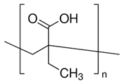 | Carboxylate group is deprotonated at pH 7.4 and protonates at pH < 5, which destabilizes the NP. | |
| Poly (methacrylic acid) | PMAA | 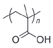 | Carboxylate group is deprotonated at pH 7.4 and protonates at pH < 5, which destabilizes the NP. | |
| Poly-sulfonamides | - | 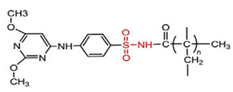 | Picks up a positive charge in response to pH decrease, changing the structure of the NP. | |
| Cationic | Poly(b-amino ester) | - | 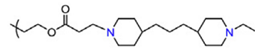 | Neutral and hydrophobic at physiological pH, but is ionized and hydrophilic at pH < 6.5. |
| Poly(N,N-dimethylamino ethyl methacrylate) | PDMAEMA |  | The amine group deprotonates at high pH and protonates/ ionizes at low pH. | |
| poly(L-histidine) | - |  | Imidazole ring deprotonates at physiological pH but is protonated at low pH. |
| Acid-Labile Bond | Structure | Mechanism | Degradation Products |
|---|---|---|---|
| Imine |  | 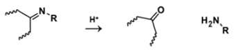 |  |
| Hydrazone | 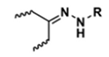 |  | 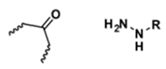 |
| Amides | 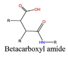 |  |  |
 |  | 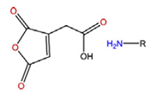 | |
| Phenyl vinyl Ether |  |  | 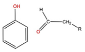 |
| Orthoesters |  |  |  |
| Acetals |  |  | 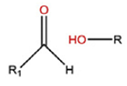 |
| Ketals | 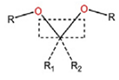 |  |  |
| Oxime |  | 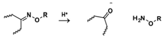 | 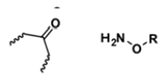 |
| Components | Payload | Cancer Cell Line | pH-Triggered Release | Ref. |
|---|---|---|---|---|
| Poly(ethylene glycol)-b-polycarbonate-b-oligo([R]-3-hydroxybutyrate) (PEG-PKPC-oPHB) | DOX, 8HQ-glu and 8HQ-gla | MCF-7 and HCT-116 cell |
| [59] |
| Amphiphilic chitosan-g-mPEG/DBA | indocyanine green dye (ICG) | MCF-7 |
| [60] |
| Poly(ethylene glycol)-blockpoly(cyclohexyloxy ethyl glycidyl ether)s | Paclitaxel (PTX) and Nile Red dye | SW620 and DU145 cells |
| [61] |
| Poly (ethylene glycol) methyl ether-b-poly (β-amino esters) | PTX and DOX | A549, MDA-MB-231, A2780 and NCL-H460 |
| [62] |
| 1,2-distearoyl-sn-glycero-3-phosphoethanolamine-N-[methoxy(polyethylene glycol)] conjugated poly(β-amino esters) (DSPE-b-PEG-b-PAE-b-PEG-b-DSPE | DOX | B16F10, HepG2 and HeLa cells |
| [63] |
| Poly(caprolactone) (PCL), poly(ethylene glycol) (PEG), and PCL-bPEG-b-PCL | Pyrene, rohdamine-6G and 5-fluorouracil | - |
| [64] |
| Product Name | Active Ingredient | Status | Company |
|---|---|---|---|
| Genexol PM | Paclitaxel | Marketed | Samyang, Seongnam, South Korea |
| NK-911 | Doxorubicin | phase II | Nippon Kayaku Co., Tokyo, Japan |
| NK-105 | Paclitaxel | phase II/III | Nippon Kayaku Co., Tokyo, Japan |
| NC-6004 | Cisplatin | phase III | Nanocarrier Co., Chiba, Japan |
| SP-1049C | Doxorubicin | phase II/III | Supratek Pharma Inc., Quebec, Canada |
| NC-6300 | Epirubicin | phase I/II | Nanocarrier Co., Chiba, Japan |
| Product Name (Year Approved) | Active Agent | Lipid Components | Indication | Company |
|---|---|---|---|---|
| Doxil® (1995) | Doxorubicin | HSPC, cholesterol; PEG 2000-DSPE | Ovarian, breast cancer, Kaposi’s sarcoma | Sequus Pharmaceuticals, California, USA |
| DaunoXome® (1996) | Daunorubicin | DSPC and cholesterol | Kaposi’s sarcoma | NeXstar Pharmaceuticals, Colorado, USA |
| Myocet® (2000) | Mifamurtide | DOPC and POPC | Non-metastatic osteosarcoma | Takeda Pharmaceutical Limited, Tokyo, Japan |
| Marqibo® (2012) | Vincristine | SM and cholesterol | Acute lymphoblastic leukaemia | Talon Therapeutics, Inc., California, USA |
| Onivyde™ (2015) | Irinotecan | DSPC, MPEG-2000 and DSPE | Metastatic adenocarcinoma of the pancreas | Merrimack Pharmaceuticals Inc., Massachusetts, USA |
| Lipoplatin® | Cisplatin | SPC-3, cholesterol and mPEG2000-DSPE | Pancreatic adenocarcinoma, NSCLC, HER2/neu negative metastatic breast cancer and advanced gastric cancer | Regulon Inc., California, USA |
| Lipid Components | pH-Sensitive Component | Payload | Cancer Cell Line | pH-Triggered Release | Ref. |
|---|---|---|---|---|---|
| DOPE, CHEMS, DSPE-PEG2000 | DOPE | DOX | MDA-MB-435S and HeLa cells |
| [72] |
| Citraconic anhydride (CA), DSPC, DSPE-PEG2000 | CA | Curcumin | MCF-7 and L929 |
| [73] |
| DCPA | H2O | Ciprofloxacin, red-fluorescent, rhodamine dye | HepG2 |
| [74] |
| DPPC,DSPE-PEG2000, CHOL, DSPE-PEOz2000 | DSPE-PEOz2000 | Metformin- and IR780 | MDA-MB-231 |
| [75] |
| HSPC, DSPE-PEG2000, C18-AI-PEG5000 and C18-PEG5000 | C18-AI-PEG5000 and C18-PEG5000 | Irinotecan (CPT-11) | MCF-7, BxPC-3 and NIH/3T3 |
| [76] |
| CHEMS, PEG, Nio | pH-sensitive niosomal (Nio) formulation of GTE | Green tea extract (GTE) | MCF-7, HepG2, and HL-60 |
| [77] |
| Egg phosphatidylcholine, CHOL, DSPE-PEG2000-angiopep-2 | DSPE-PEG2000-angiopep-2 | Calcium arsenite | HBMEC and C6 |
| [78] |
| EPC, PDMAEMA-b-PLMA diblock copolymer | PDMAEMA-b-PLMA | TRAM-34 | HEK-293 and GL261 |
| [79] |
Publisher’s Note: MDPI stays neutral with regard to jurisdictional claims in published maps and institutional affiliations. |
© 2022 by the authors. Licensee MDPI, Basel, Switzerland. This article is an open access article distributed under the terms and conditions of the Creative Commons Attribution (CC BY) license (https://creativecommons.org/licenses/by/4.0/).
Share and Cite
AlSawaftah, N.M.; Awad, N.S.; Pitt, W.G.; Husseini, G.A. pH-Responsive Nanocarriers in Cancer Therapy. Polymers 2022, 14, 936. https://doi.org/10.3390/polym14050936
AlSawaftah NM, Awad NS, Pitt WG, Husseini GA. pH-Responsive Nanocarriers in Cancer Therapy. Polymers. 2022; 14(5):936. https://doi.org/10.3390/polym14050936
Chicago/Turabian StyleAlSawaftah, Nour M., Nahid S. Awad, William G. Pitt, and Ghaleb A. Husseini. 2022. "pH-Responsive Nanocarriers in Cancer Therapy" Polymers 14, no. 5: 936. https://doi.org/10.3390/polym14050936