Article

Using Peek as a Framework Material for Maxillofacial Silicone Prosthesis: An In Vitro Study

by
Pinar Cevik
1,2,3,*,
Arzu Zeynep Yildirim
1,
Emine Hulya Demir Sevinc
1,
Aykut Gonder
1 and
Sudarat Kiat-Amnuay
2,3
1
Department of Prosthodontics, Faculty of Dentistry, Gazi University, Ankara 06490, Turkey
2
Department of General Practice and Dental Public Health, School of Dentistry, The University of Texas Health Science Center at Houston, Houston, TX 77054, USA
3
Houston Center of Biomaterials and Biomimetics, Houston, TX 77054, USA
*
Author to whom correspondence should be addressed.
Polymers 2023, 15(12), 2694; https://doi.org/10.3390/polym15122694
Submission received: 17 May 2023 / Revised: 9 June 2023 / Accepted: 12 June 2023 / Published: 15 June 2023
(This article belongs to the Special Issue High-Performance 3D Printing Polymers)

Abstract

:
There are often bonding problems between acrylic resins and silicone. PEEK (polyetheretherketone), which is a high-performance polymer, has great potential for the implant, and fixed or removable prosthodontics. The aim of this study was to evaluate the effect of different surface treatments on PEEK to be bonded to maxillofacial silicone elastomers. A total of 48 specimens were fabricated from either PEEK or PMMA (Polymethylmethacrylate) (n = 8). PMMA specimens acted as a positive control group. PEEK specimens were divided into five study groups as surface treatments as control PEEK, silica-coating, plasma etching, grinding, or nano-second fiber laser. Surface topographies were evaluated by scanning electron microscopy (SEM). A platinum-primer was used on top of all specimens including control groups prior to silicone polymerization. The peel bond strength of the specimens to a platinum-type silicone elastomer was tested at a cross-head speed of 5 mm/min. The data were statistically analyzed (α = 0.05). The control PEEK group showed the highest bond strength (p < 0.05) among the groups. No statistical difference was found between control PEEK, grinding, or plasma etching groups (p > 0.05). The lowest bond strength was seen in the laser group, which was not statistically different from silica-coating (p > 0.05), and statistically different from control PEEK, grinding, or plasma groups (p < 0.05). Positive control PMMA specimens had statistically lower bond strength than either control PEEK or plasma etching groups (p < 0.05). All specimens exhibited adhesive failure after a peel test. The study results indicate that PEEK could serve as a potential alternative substructure for implant-retained silicone prostheses.

1. Introduction

Silicones are frequently used in maxillofacial prosthesis fabrication due to their acceptable physical and mechanical properties [1]. The retention of silicone prostheses is generally provided by implant systems in the maxillofacial region and acrylic resins are mostly used as a substructure between the implant and the silicone [2,3]. However, there are often bonding problems between acrylic resins and silicone [4]. It needs to be renewed due to the breaking of the bonding between silicone and the infrastructure, or the fractures and/or cracks in the acrylic resin [2,5,6]. It is thought that the bonding problems between silicone and acrylic resins used in maxillofacial prostheses are due to the chemical structure difference between the two materials [7,8,9]. Adhesive systems such as primers are applied on acrylic resin in an attempt to eliminate weakness in the bond between them. It has been reported that there is no bonding between the two materials with their different compositions when the primer is not applied [8]. Therefore, adhesive primers are used to enhance the bond between acrylic resins and facial silicone [10].
Primers have been stated to act as an organic solvent agent to react with the silicone and resin materials [11,12], and as a chemical intermediate component of the silicone and the acrylic resin substrates. The hydrophilic and hydrophobic groups of primers react with the functional groups of silica [13]. At the same time, the activation of primers directly on the substrate surface increases the wettability of the substrate and consequently impregnates the surface layer with the polymer content [14]; they act as chemical intermediates of silicone and acrylic resin. Furthermore, it has also been reported that they activate the surfaces via etching or promoting hydrogen bonding and covalent coupling. Some other studies have also suggested the use of primer to increase the bond between silicone and acrylic resin [5,15].
PEEK was introduced as a dental material in 1992 and was initially used for aesthetic abutments due to its superior color compared to metals [16,17]. PEEK is resistant to chewing forces, cost-effective, and anti-allergic. It has several advantages over titanium, such as providing an aesthetic appearance in thin biotype mucosa, good dimensional stability, and an elastic modulus between cortical and cancellous bone [18,19,20,21,22]. Therefore, PEEK is a suitable alternative to traditional implant materials such as titanium and stainless steel and is used in many dental and prosthetic treatments, including fixed restorations, individual abutments, dental implants, removable prostheses, and prosthetic infrastructures [23,24,25].
PEEK needs to be subjected to different surface treatments in order to increase its bonding to polymers [26]. Sandblasting and silica coating [27,28], plasma applications [29,30], different laser systems [29,30,31], sanding and mechanical abrasion [32], and different acid applications [29,30,31,32] are the surface treatments that can be applied to PEEK. To the best of our knowledge, there is no study evaluating the bond strength between silicone and PEEK in the field of maxillofacial prosthodontics. It is important as it is the first study to be done on this subject. The bonding of PEEK with silicone and potential surface treatments are subjects of interest of this study. Conventional applications include the acrylic specimens to be used as a framework material for a maxillofacial silicone prosthesis. Therefore, the aim of this study was to investigate the feasibility of utilizing a new generation high-performance PEEK as a substructure between implant and silicone in maxillofacial prostheses. As a positive control group, polymethyl methacrylate (PMMA) acrylic specimens were included in this study. Additionally, this study aimed to examine the effect of various surface treatments on PEEK specimens to be bonded to maxillofacial silicone, which is a clinically relevant aspect. The null hypotheses of this study are: (1) There would be no differences among different surface treatments on PEEK to be bonded to one type of silicone elastomer with regard to the peel bond test; (2) There would be no difference between untreated PEEK specimens and untreated PMMA specimens.

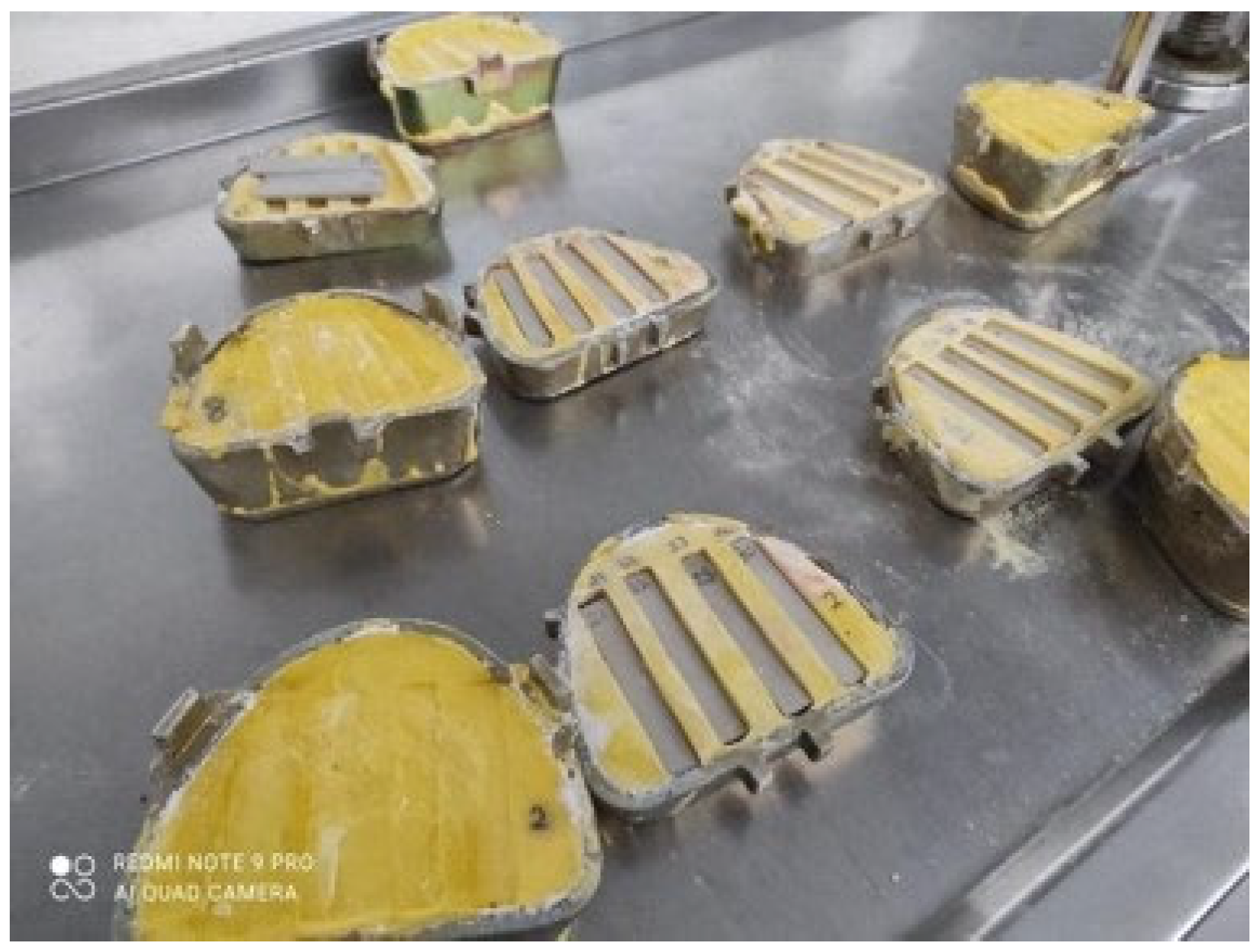
2. Materials and Methods
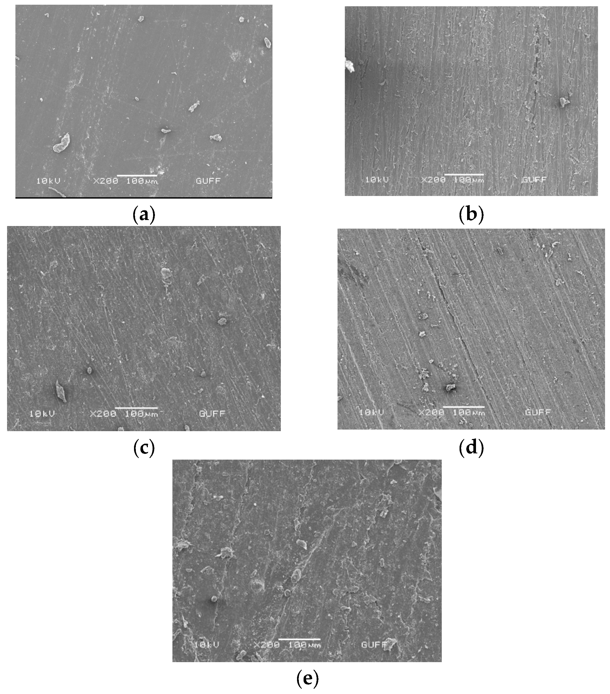
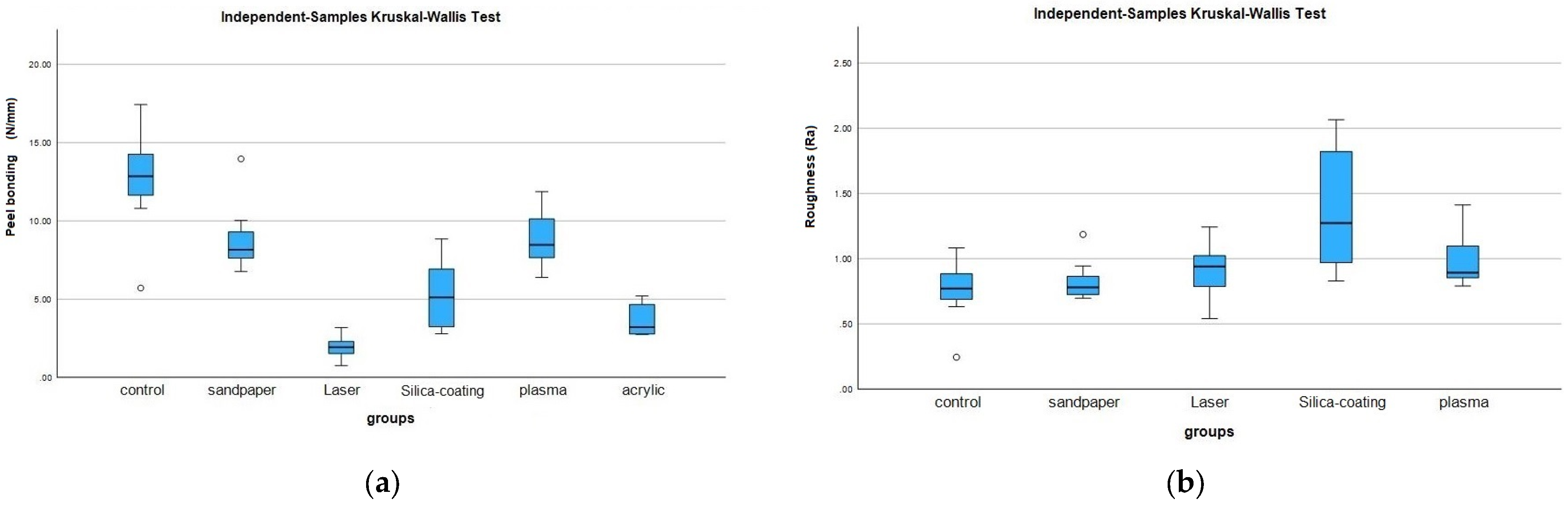
PEEK rectangular specimens with the dimension of 60 mm × 10 mm × 3 mm were fabricated by milling from a high-grade, industrially manufactured PEEK disc in natural color (JUVORA™, JUVORA™ Ltd., Thornton-Cleveleys, UK). A total of 48 specimens (40 specimens were PEEK, 8 specimens were PMMA) prepared and divided into 6 (n = 8) groups according to the surface treatments applied to PEEK or PMMA. The surfaces of all the specimens were cleaned with acetone by using cotton pellets. To ensure the standard, 800-grit sandpaper was first applied to them.
The study groups are as follows:
Group Control (n = 8): It was the control group and a platinum primer (G611) (G611 Platinum Primer, Technovent Ltd., Bridgend, UK) was applied to the top of the PEEK specimens. One-coat primer was applied to the dried specimens with the help of a cotton pellet. The primer was left to dry without any further treatment.
Group Silica-coating (n = 8): Cleaned and dried PEEK specimens’ surfaces were silica coated with 30 µm silica-coated Al2O3 with Cojet Sand (3M ESPE, St. Paul, MN, USA). Sand particles were applied to the specimens for 10 s from a distance of 10 mm at a pressure of 2.5 bar.
Group Plasma (n = 8): Plasma etching was applied to the cleaned and dried PEEK surfaces. Oxygen plasma was applied under 150 m Tor at 100 W power [33]. An apparatus (Vakuum Praha, Prague, Czech Republic) with 13.56 MHz frequency power was attached to oxygen gas reactor. Oxygen gas was released at a volumetric flow rate of 30 sccm (standard cubic centimeters per minute) for 5 min to the top surface of each specimen. The oxygen plasma etching used for 1 min [34]. The specimens were kept under vacuum for gas stabilization.
Group Sandpaper (n = 8): The cleaned and dried specimens underwent sanding using 600 silicon carbide sandpaper following the initial application of 800-grit sandpaper. Subsequently, the specimens were cleaned and dried again.
Group Laser (n = 8): Nano second fiber laser (FiberLAST A.Ş., Ankara, Türkiye) was applied to the cleaned and dried PEEK specimens at a power of 2 W, a speed of 500 mm/s, and a frequency of 40 kHz.
Group PMMA (n = 8): Autopolymerized clear PMMA acrylic specimens (IMICRYL, Konya, Turkiye), with the dimensions of 60 mm × 10 mm × 3 mm were prepared, and used as the positive control group. A platinum primer (G611, Technovent Ltd., Bridgend, UK) was used on the surface prior to the silicone polymerization.
Surface topography of specimens from different groups was analyzed using scanning electron microscopy (SEM). In the control group, the surface topography was analyzed before the primer application, while in the other study groups, it was analyzed immediately after the surface treatment.
The surface roughness values of PEEK specimens were measured with a digital profilometer (Mitotoyo Surf Test SJ 201 P/M; Mitotoyo Corp, Takatsuku, Japan) following the surface treatments, including the control group. A constant force of 0.4 gf at 4 mm was applied to the specimen surface, and the obtained values were digitally generated on the front of the profilometer and recorded as Ra values.
Prior to silicone bonding, all the surfaces were cleaned with acetone, and dried. A high-temperature vulcanized (HTV) platinum-type maxillofacial silicone material (M511 Technovent Ltd., Bridgend, UK) was used in this study. First, PEEK blanks were inserted into the metal gypsum molds (as shown in Figure 1).
The silicone material was prepared by mixing part A and part B in a 10:1 ratio, then packed onto a specified area of PEEK blanks. The silicone was polymerized by heating it in a dry-hot air oven at 135 C for 15 min. After polymerization, a peel test was performed to measure the bond strength between the PEEK and silicone. The 90-degree (90°) peel test was conducted using an Instron machine at a speed of 5 mm/min (Figure 2).
Types of failures were classified as cohesive, adhesive, or mixed. The force needed to cause bond failures was recorded. Peel strength (N/mm) was determined [8]. After the peel strength test, rupture types were evaluated under a fluorescent light, and categorized into adhesive, cohesive, or mixed. Adhesive failure refers to complete separation at the interface between the PEEK and silicone, cohesive failure refers to tearing within the silicone material, and mixed failure refers to both.
The data were statistically analyzed by using a statistical software program (IBM SPSS Statistics, v29.0; IBM Corp., New York, US). Mean values and standard deviations, median, minimum, maximum values, and interquartile ranges (IQRs) for both bond strength and the surface roughness were calculated. Kruskal Wallis test was used to compare differences among groups (α = 0.05). The data for the results are shown in Table 1 and Table 2.

3. Results

Table 1 shows the surface roughness values of the groups in this study. Silica-coated PEEK specimens showed the highest surface roughness values, which was statistically significant compared to those of control PEEK specimens (p < 0.05). There were no statistically significant differences among the groups of silica-coating, laser, sandpaper, or plasma-treated specimens (p > 0.05).
As seen in Table 2, there was statistical difference between the groups regarding the peel test (p ˂ 0.05). The control PEEK group showed the highest peel bond strength, where only primer was used as a surface treatment. However, there was no statistical difference between the control, sandpaper, or plasma etching groups. The nano-fiber laser group demonstrated the lowest bond strength, and this result was not found to be significantly different from the silica-coating and PMMA groups. However, there was a statistically significant difference between the nano-fiber laser group and the control, sandpaper, and plasma groups. The positive control group, consisting of PMMA specimens, had statistically lower bond strength than either the PEEK control, sandpaper or the plasma etching groups.
Figure 3a–e show the results of SEM surface analysis of the different study groups:
In SEM images, plasma-treated specimens exhibited horizontal directional scratches in their surface topography. The SEM images of nano fiber laser group specimens revealed noticeable irregular structures and cracks on the PEEK surface. Specimens in the silica-coating group showed similar surface topography to those in plasma-treated specimens. In contrast, relatively smooth surfaces were observed in the SEM analysis of the control PEEK specimens. Silica-coating specimens showed remarkable point surface changes of the Al2O3 particles remaining on the surface and from sandblasting. These observations provided valuable insights into the effects of different surface treatments on surface topography, which can inform the development of improved material surface properties.
In addition, Figure 4a,b show median values with IQRs for all groups in terms of peel test and surface roughness.
All the failure types were seen as adhesive type in this study.

4. Discussion

The hypotheses of this study were rejected as different surface treatments on PEEK affected the bond strength between PEEK and maxillofacial silicone differently. Furthermore, there was significant difference between study groups regarding the bond strength test.
To evaluate the bonding of maxillofacial silicones with resin materials, various test methods can be employed, such as the tensile test, shear test, and peel test [2,4]. The peel test is particularly suitable for simulating the horizontal component of detaching forces that occur when the patient removes the craniofacial implant-retained prosthesis [35]. For this reason, in our study, we used the 90° peel test to assess the bond strength.
Apart from modified PEEK, also known as BioHPP, with a nano-ceramic content of 20%, it has been reported that the pure PEEK material has a gray-white opaque color and that the PEEK surface should be coated with dental composite resin in order to meet aesthetic expectations in intraoral cavity [36,37,38]. In this regard, various surface treatments have been introduced for PEEK, including sandblasting, silica coating on PEEK surface [27,28,39], 98% sulfuric acid application [32,39], adhesive primer or resin applications [40,41], cold active gas plasma application [29,39], and different laser treatments [28,30].
Despite the emergence of high-power fiber laser systems with advanced features such as air cooling, nanoscale precision, and high-quality beams, their use in dental applications is limited. Nanofiber lasers can modify the material surface mechanically without causing excessive thermal change, unlike conventional laser systems that may create fractures and cracks. These lasers can generate surface topographies at nano- and micron-level scales [42,43]. In our study, SEM photographs showed that nanofiber laser treatment did not produce distinct surface changes visible at 100 μm magnification. Future studies with advanced imaging techniques could investigate the nanoscale changes made by the laser and analyze their homogeneity and effects on PEEK surface properties. Due to the high surface strength of PEEK, only a few surface treatments are effective in providing sufficient surface roughness [26,30,44,45]. Tsuka et al. [30] found that Nd:YVO4 laser treatment increased surface roughness on PEEK, providing optimal bonding between resin and PEEK through micromechanical locking. Laser systems using 2–3 W power have been recommended to avoid overheating defects on material surfaces when applied to resin materials [46,47,48]. The nanofiber laser used in this study had a power of 2 W, based on conventional laser systems in the literature. The surface roughness values generated by the nanofiber laser system were lower than the sandblasting group (p < 0.05), but there was no significant difference between the laser and other groups (p > 0.05). However, the bonding values of the nanofiber laser-treated specimens were significantly lower than the other groups (p < 0.05), suggesting that the nanofiber laser triggered surface degradation on the PEEK surface that disrupted the connection with silicone.
Plasma treatment has been reported to be an effective way to increase the surface energy of the polymer surface [49]. The type of plasma (argon plasma or oxygen plasma) can affect the wettability of the polymer surface. Soft non-thermal plasma, which involves oxygen plasma treatment, has been recognized for its ability to considerably increase hydrophilicity by generating multiple radicals, including excited oxygen molecules, OH radicals, atomic oxygen, superoxide anion radicals, and singlet oxygen without causing thermal damage or altering the surface roughness [50,51]. In our study, plasma treatment did not increase the surface roughness of PEEK specimens as compared with PEEK control group specimens (p > 0.05). In addition, plasma treatment did not show a damaging effect on SEM images of the PEEK specimens. Moreover, the bond strength of the plasma-treated specimens was observed to be lower in comparison to the control PEEK specimens, which demonstrated the highest bond strength within the study. However, it is worth noting that the observed difference in bond strength between the two groups was not found to be statistically significant (p > 0.05). These results suggest that the application of oxygen plasma increases the surface energy of PEEK, resulting in a stronger bond between silicone and PEEK. However, the increase in surface energy in the plasma group is not as substantial as that of the control group since the bond strength values were lower than those of the control group.
Sandblasting alone is not sufficient for bonding PEEK and resin without a primer [52]. Other studies have found that both silica coating and sandblasting improve bond strength through surface modification [26,28]. In our study, silica coating resulted in lower bond strength values than sandpaper, plasma, and control groups (p < 0.05), indicating that it may not enhance bond strength between PEEK and silicone elastomer. On the other hand, the silica-coating group showed higher surface roughness values compared to the plasma group, but this difference was not statistically significant (p > 0.05). In contrast, the bond strength values of the plasma group were higher than those of the silica-coating group (p < 0.05). The observed significant increase in surface roughness of the silica-coated PEEK specimens presented in this statement could be an important indication of the coating’s effectiveness in altering the surface properties of PEEK. However, it is important to consider the influence of other factors such as coating thickness and adhesion to the substrate, which could also affect the coating’s overall performance.
In our study, the control group exhibited the highest bond strength values, which were higher than the laser, sandblasting, and plasma groups (p < 0.05). However, the surface roughness values of the control group were lower than those of the sandblasting and plasma groups (p < 0.05). These results suggest that platinum primers can chemically enhance the bond between silicone and PEEK. It is noteworthy that the bond strength values of the plasma and silica-coating groups, which were expected to increase with the surface roughness generated by mechanical adhesion, were lower than those of the control group with platinum primer. This finding supports the notion that the PEEK–silicone connection is associated with chemical bonding rather than mechanical adhesion.
Maxillofacial silicone elastomers are dimethyl siloxane polymers, while acrylic resins are methyl methacrylate-based polymers [7]. Bonding problems between silicone and acrylic caused by different chemical structures between the two materials can often occur. The bond strength between acrylic substructure and silicone can be improved by using primer on the acrylic structure [3,8,9]. Furthermore, studies have shown that there is a need to apply an adhesive or primer for optimal bonding between the silicone/resin interface [3,4,8,22]. The bond strength between hard acrylic substructure or polyurethane substrate and silicone elastomer may be affected by the type of substrate and primer content. According to this, solvent-based primers are defined as a surface pretreatment method that can modify the polymer surface and result in stronger bonding [53]. The use of a primer to increase surface energy and wettability is a common practice in achieving strong bonding between different materials [54,55]. Organocylane-based primers are often used on silicone surfaces to improve bonding and chemical compatibility [54]. The organocylamine-based primers containing vinylsilan are typically used for bonding platinum-based silicone elastomers. These primers contain solvents such as water, alcohol, or organic solvents such as xylene, toluene, or naptha solution [55]. Studies have demonstrated that primers containing solvents such as ethyl acetate or methylene chloride can result in strong bonding between silicone and polyurethane substrates [3,8,56]. Control PEEK specimens treated with platinum primer tended to show the highest bond strength in this study. The platinum primer used in the study may have improved the wettability and surface energy of PEEK without causing any degradation.
The use of 3D printing technology has revolutionized many industries, including the medical field. This technology, also known as rapid prototyping or additive manufacturing, has the potential to reduce material costs and provide faster solutions for clinical and laboratory applications [57,58,59]. PEEK is a material that has gained interest in the medical and dental fields due to its unique properties. Its lightweight and polishable nature, as well as its color stability and low water absorption, make it an ideal material for maxillary obturators, which are usually made of PMMA resin [60,61,62]. PEEK’s ability to resist polymerization shrinkage during the manufacturing process also makes it a good candidate for maxillofacial prosthetic treatment options, particularly as an infrastructure material [45]. Overall, 3D printing technology combined with PEEK material has great potential for improving and advancing the field of maxillofacial prosthetics [60,61] Future studies should include larger specimen sizes and investigate the combined effects of mechanical and chemical processes on the bond strength between PEEK and silicone.

5. Conclusions

Within the limitations of this study, the following conclusions could be drawn:
  • This study demonstrated that applying a platinum primer to PEEK structures resulted in a favorable bonding between platinum silicone elastomer and PEEK.
  • Surface treatments on PEEK did not affect the bonding between PEEK and silicone. The obtained bond strength values revealed that PEEK may be a preferred supporting material over PMMA frameworks for fabricating implant-supported maxillofacial silicone elastomers. Notably, the control PMMA specimens exhibited significantly lower bond strength compared to the control PEEK specimens.
  • The strong bonding observed between silicone and PEEK has practical implications in the development of maxillofacial silicone prostheses, benefiting both patients and clinicians.
  • Future studies with larger specimen sizes should focus on investigating the bonding of PEEK frameworks with implant-supporting parts in such prostheses, further advancing our understanding in this area.
  • Additionally, future studies could explore a wider range of surface treatment parameters and use larger specimen sizes to provide a more comprehensive understanding of the bonding characteristics between PEEK and silicone in maxillofacial silicone prostheses.

Author Contributions

Conceptualization, P.C. and A.Z.Y.; methodology, P.C., A.Z.Y., E.H.D.S. and A.G.; formal analysis, P.C.; investigation, P.C.; data curation, P.C., E.H.D.S. and A.G.; writing—original draft preparation, P.C.; writing—review and editing, P.C.; visualization, P.C. and A.Z.Y.; supervision, S.K.-A.; project administration, P.C.; funding acquisition, P.C. The work was performed when the authors E.H.D.S. and A.G. were at Gazi University. All authors have read and agreed to the published version of the manuscript.

Funding

This work was supported by Gazi University Projects of Scientific Investigation (BAP) with 6284 Project ID. The first part of the results of this study were presented at ICP-TPID Joint Meeting-25th Scientific Congress of Turkish Prosthodontics and Implantology Association which was held on 4–7 November 2021 Mugla, Turkiye. The second part of this study was presented at the 2023 AADOCR/CADR Annual Meeting & Exhibition which was held on 15–18 March 2023 Portland, OR.

Institutional Review Board Statement

Not applicable.

Data Availability Statement

Not applicable.

Acknowledgments

The authors thank to Aykut ALTINKAYNAK and FiberLAST A.Ş, for their sponsorship of the fiber laser application. Additionally, they would like to thank dental technician Mahmut ECEMIS from Vesson A.Ş and Alyans Digital Dental for the assistance in specimen fabrication.

Conflicts of Interest

The authors declare no conflict of interest.

References

  1. Jindal, S.K.; Sherriff, M.; Waters, M.G.; Smay, J.E.; Coward, T.J. Development of a 3D printable maxillofacial silicone: Part II. Optimization of moderator and thixotropic agent. J. Prosthet. Dent. 2018, 119, 299–304. [Google Scholar] [CrossRef] [PubMed]
  2. Lemon, J.C.; Chambers, M.S. Locking retentive attachment for an implant-retained auricular prosthesis. J. Prosthet. Dent. 2002, 87, 336–338. [Google Scholar] [CrossRef] [PubMed]
  3. Hatamleh, M.M.; Watts, D.C. Bonding of maxillofacial silicone elastomers to an acrylic substrate. Dent. Mater. 2010, 26, 387–395. [Google Scholar] [CrossRef] [PubMed]
  4. McMordie, R.; King, G.E. Evaluation of primers used for bonding silicone to denture base material. J. Prosthet. Dent. 1989, 61, 636–639. [Google Scholar] [CrossRef] [PubMed]
  5. Goiato, M.C.; Garcia-Júnior, I.R.; Magro-Filho, O.; Dos Santos, D.M.; Pellizzer, E.P. Implant-retained thumb prosthesis with anti-rotational attachment for a geriatric patient: Short report. Gerodontology 2010, 27, 243–247. [Google Scholar] [CrossRef]
  6. Hooper, S.M.; Westcott, T.; Evans, P.L.L.; Bocca, A.P.; Jagger, D.C. Implant-supported facial prostheses provided by a maxillofacial unit in a U.K. regional hospital: Longevity and patient opinions. J. Prosthodont. 2005, 14, 32–38. [Google Scholar] [CrossRef]
  7. Qudah, S.; Harrison, A.; Huggett, R. Soft lining materials in prosthetic dentistry: A review. Int. J. Prosthodont. 1990, 3, 477–483. [Google Scholar]
  8. Haddad, M.F.; Goiato, M.C.; dos Santos, D.M.; Crepaldi, N.d.M.; Pesqueira, A.A.; Bannwart, L.C. Bond strength between acrylic resin and maxillofacial silicone. J. Appl. Oral Sci. 2012, 20, 649–654. [Google Scholar] [CrossRef]
  9. Polyzois, G.L.; Frangou, M.I.; Andrcopouioi, A.C. The effect of bonding agents on the bond strengths of facial silicone elastomers to a visible light-activated resin. Int. J. Prosthodont. 1991, 4, 440–444. [Google Scholar]
  10. Sanohkan, S.; Kukiattrakoon, B.; Peampring, C. Tensile bond strength of facial silicone and acrylic resin using different primers. J. Orofac. Sci. 2017, 9, 48–51. [Google Scholar] [CrossRef]
  11. Minami, H.; Suzuki, S.; Ohashi, H.; Kurashige, H.; Tanaka, T. Effect of surface treatment on the bonding of an autopolymerizing soft denture liner to a denture base resin. Int. J. Prosthodont. 2004, 17, 297–301. [Google Scholar]
  12. Polyzois, G.L.; Frangou, M.J. Bonding of Silicone Prosthetic Elastomers to Three Different Denture Resins. Int. J. Prosthodont. 2002, 15, 535–538. [Google Scholar]
  13. Hatamleh, M.M.; Watts, D.C. Effects of bond primers on bending strength and bonding of glass fibers in fiber-embedded maxillofacial silicone prostheses. J. Prosthodont. 2011, 20, 113–119. [Google Scholar] [CrossRef]
  14. Mutluay, M.M.; Ruyter, I.E. Evaluation of bond strength of soft relining materials to denture base polymers. Dent. Mater. 2007, 23, 1373–1381. [Google Scholar] [CrossRef]
  15. Bonatto, L.d.R.; Goiato, M.C.; da Silva, E.V.F.; Oliveira, S.H.P.; Haddad, M.F.; Chaves Neto, A.H.; Brito, V.G.B.; dos Santos, D.M. Biocompatibility of primers and an adhesive used for implant-retained maxillofacial prostheses: An in vitro analysis. J. Prosthet. Dent. 2017, 117, 799–805. [Google Scholar] [CrossRef] [Green Version]
  16. Haleem, A.; Javaid, M. Polyether ether ketone (PEEK) and its manufacturing of customised 3D printed dentistry parts using additive manufacturing. Clin. Epidemiol. Glob. Health 2019, 7, 654–660. [Google Scholar] [CrossRef] [Green Version]
  17. Silthampitag, P.; Chaijareenont, P.; Tattakorn, K.; Banjongprasert, C.; Takahashi, H.; Arksornnukit, M. Effect of surface pretreatments on resin composite bonding to PEEK. Dent. Mater. J. 2016, 35, 668–674. [Google Scholar] [CrossRef] [Green Version]
  18. Najeeb, S.; Khurshid, Z.; Zohaib, S.; Zafar, M.S. Bioactivity and osseointegration of PEEK are inferior to those of titanium: A systematic review. J. Oral Implantol. 2016, 42, 512–516. [Google Scholar] [CrossRef]
  19. Najeeb, S.; Zafar, M.S.; Khurshid, Z.; Siddiqui, F. Applications of polyetheretherketone (PEEK) in oral implantology and prosthodontics. J. Prosthodont. Res. 2016, 60, 12–19. [Google Scholar] [CrossRef]
  20. Skirbutis, G.; Dzingutė, A.; Masiliūnaitė, V.; Šulcaitė, G.; Žilinskas, J. PEEK polymer’s properties and its use in prosthodontics. A review. Stomatologija 2018, 20, 54–58. [Google Scholar]
  21. Fuhrmann, G.; Steiner, M.; Freitag-Wolf, S.; Kern, M. Resin bonding to three types of polyaryletherketones (PAEKs)—Durability and influence of surface conditioning. Dent. Mater. 2014, 30, 357–363. [Google Scholar] [CrossRef] [PubMed]
  22. Taft, R.M.; Cameron, S.M.; Knudson, R.C.; Runyan, D.A. The effect of primers and surface characteristics on the adhesion-in-peel force of silicone elastomers bonded to resin materials. J. Prosthet. Dent. 1996, 76, 515–518. [Google Scholar] [CrossRef] [PubMed]
  23. Kurtz, S.M.; Devine, J.N. PEEK biomaterials in trauma, orthopedic, and spinal implants. Biomaterials 2007, 28, 4845–4869. [Google Scholar] [CrossRef] [PubMed] [Green Version]
  24. Feerick, E.M.; Kennedy, J.; Mullett, H.; FitzPatrick, D.; McGarry, P. Investigation of metallic and carbon fibre PEEK fracture fixation devices for three-part proximal humeral fractures. Med. Eng. Phys. 2013, 35, 712–722. [Google Scholar] [CrossRef] [PubMed]
  25. Yildirim Bicer, A.Z.; Dogan, A.; Keskin, S.; Dogan, O.M. Effect of argon plasma pretreatment on tensile bond strength of a silicone soft liner to denture base polymers. J. Adhes. 2013, 89, 594–610. [Google Scholar] [CrossRef]
  26. Schmidlin, P.R.; Stawarczyk, B.; Wieland, M.; Attin, T.; Hämmerle, C.H.F.; Fischer, J. Effect of different surface pre-treatments and luting materials on shear bond strength to PEEK. Dent. Mater. 2010, 26, 553–559. [Google Scholar] [CrossRef] [Green Version]
  27. Kern, M.; Lehmann, F. Influence of surface conditioning on bonding to polyetheretherketon (PEEK). Dent. Mater. 2012, 28, 1280–1283. [Google Scholar] [CrossRef]
  28. Caglar, I.; Ates, S.M.; Yesil Duymus, Z. An In Vitro Evaluation of the Effect of Various Adhesives and Surface Treatments on Bond Strength of Resin Cement to Polyetheretherketone. J. Prosthodont. 2019, 28, e342–e349. [Google Scholar] [CrossRef] [Green Version]
  29. Stawarczyk, B.; Bähr, N.; Beuer, F.; Wimmer, T.; Eichberger, M.; Gernet, W.; Jahn, D.; Schmidlin, P.R. Influence of plasma pretreatment on shear bond strength of self-adhesive resin cements to polyetheretherketone. Clin. Oral Investig. 2014, 18, 163–170. [Google Scholar] [CrossRef] [Green Version]
  30. Tsuka, H.; Morita, K.; Kato, K.; Kimura, H.; Abekura, H.; Hirata, I.; Kato, K.; Tsuga, K. Effect of laser groove treatment on shear bond strength of resin-based luting agent to polyetheretherketone (PEEK). J. Prosthodont. Res. 2019, 63, 52–57. [Google Scholar] [CrossRef]
  31. Ebnesajjad, S. Surface preparation of thermoplastics, thermosets, and elastomers. In Handbook of Adhesives and Surface Preparation; William Andrew Publishing: Norwich, NY, USA, 2011; pp. 107–134. ISBN 9781437744613. [Google Scholar]
  32. Chaijareenont, P.; Prakhamsai, S.; Silthampitag, P.; Takahashi, H.; Arksornnukit, M. Effects of different sulfuric acid etching concentrations on PEEK surface bonding to resin composite. Dent. Mater. J. 2018, 37, 385–392. [Google Scholar] [CrossRef] [PubMed] [Green Version]
  33. Barani, H.; Calvimontes, A. Effects of oxygen plasma treatment on the physical and chemical properties of wool fiber surface. Plasma Chem. Plasma Process. 2014, 34, 1291–1302. [Google Scholar] [CrossRef]
  34. Yildirim, A.Z.; Unver, S.; Mese, A.; Bayram, C.; Denkbas, E.B.; Cevik, P. Effect of argon plasma and Er:YAG laser on tensile bond strength between denture liner and acrylic resin. J. Prosthet. Dent. 2020, 124, 799.e1–799.e5. [Google Scholar] [CrossRef] [PubMed]
  35. Shetty, U.S.; Guttal, S.S. Evaluation of bonding efficiency between facial silicone and acrylic resin using different bonding agents and surface alterations. J. Adv. Prosthodont. 2012, 4, 121–126. [Google Scholar] [CrossRef] [PubMed] [Green Version]
  36. Georgiev, J.; Vlahova, A.; Kissov, H.; Aleksandrov, S.; Kazakova, R. Possible Application of Biohpp in Prosthetic Dentistry: A Literature Review. J. IMAB—Annu. Proc. (Sci. Pap.) 2018, 24, 1896–1898. [Google Scholar] [CrossRef]
  37. Gama, L.T.; Duque, T.M.; Özcan, M.; Philippi, A.G.; Mezzomo, L.A.M.; Gonçalves, T.M.S.V. Adhesion to high-performance polymers applied in dentistry: A systematic review. Dent. Mater. 2020, 36, e93–e108. [Google Scholar] [CrossRef]
  38. Jin, H.Y.; Teng, M.H.; Wang, Z.J.; Li, X.; Liang, J.Y.; Wang, W.X.; Jiang, S.; Zhao, B.D. Comparative evaluation of BioHPP and titanium as a framework veneered with composite resin for implant-supported fixed dental prostheses. J. Prosthet. Dent. 2019, 122, 383–388. [Google Scholar] [CrossRef]
  39. Zhou, L.; Qian, Y.; Zhu, Y.; Liu, H.; Gan, K.; Guo, J. The effect of different surface treatments on the bond strength of PEEK composite materials (DEMA-D-13-00481). Dent. Mater. 2014, 30, e209–e215. [Google Scholar] [CrossRef]
  40. Uhrenbacher, J.; Schmidlin, P.R.; Keul, C.; Eichberger, M.; Roos, M.; Gernet, W.; Stawarczyk, B. The effect of surface modification on the retention strength of polyetheretherketone crowns adhesively bonded to dentin abutments. J. Prosthet. Dent. 2014, 112, 1489–1497. [Google Scholar] [CrossRef]
  41. Stawarczyk, B.; Taufall, S.; Roos, M.; Schmidlin, P.R.; Lümkemann, N. Bonding of composite resins to PEEK: The influence of adhesive systems and air-abrasion parameters. Clin. Oral Investig. 2018, 22, 763–771. [Google Scholar] [CrossRef]
  42. Hoy, C.L.; Ferhanoǧlu, O.; Yildirim, M.; Kim, K.H.; Karajanagi, S.S.; Chan, K.M.C.; Kobler, J.B.; Zeitels, S.M.; Ben-Yakar, A. Clinical ultrafast laser surgery: Recent advances and future directions. IEEE J. Sel. Top. Quantum Electron. 2014, 20, 242–255. [Google Scholar] [CrossRef]
  43. Kerse, C.; Kalaycloĝ Lu, H.; Elahi, P.; Çetin, B.; Kesim, D.K.; Akçaalan, Ö.; Yavaş, S.; Aşlk, M.D.; Öktem, B.; Hoogland, H.; et al. Ablation-cooled material removal with ultrafast bursts of pulses. Nature 2016, 537, 84–88. [Google Scholar] [CrossRef] [PubMed]
  44. Stawarczyk, B.; Keul, C.; Beuer, F.; Roos, M.; Schmidlin, P.R. Tensile bond strength of veneering resins to PEEK: Impact of different adhesives. Dent. Mater. J. 2013, 32, 441–448. [Google Scholar] [CrossRef] [PubMed] [Green Version]
  45. Stawarczyk, B.; Jordan, P.; Schmidlin, P.R.; Roos, M.; Eichberger, M.; Gernet, W.; Keul, C. PEEK surface treatment effects on tensile bond strength to veneering resins. J. Prosthet. Dent. 2014, 112, 1278–1288. [Google Scholar] [CrossRef] [Green Version]
  46. Korkmaz, F.M.; Bagis, B.; Özcan, M.; Durkan, R.; Turgut, S.; Ates, S.M. Peel strength of denture liner to PMMA and polyamide: Laser versus air-abrasion. J. Adv. Prosthodont. 2013, 5, 287–295. [Google Scholar] [CrossRef] [Green Version]
  47. Ates, S.M.; Korkmaz, F.M.; Caglar, I.S.; Duymus, Z.Y.; Turgut, S.; Bagis, E.A. The effect of ultrafast fiber laser application on the bond strength of resin cement to titanium. Lasers Med. Sci. 2017, 32, 1121–1129. [Google Scholar] [CrossRef]
  48. Korkmaz, F.M.; Aycan, S. Effect of fiber laser irradiation on the shear bond strength between acrylic resin and titanium. Scanning 2019, 2019, 5452919. [Google Scholar] [CrossRef]
  49. Mutel, B.; Grimblot, J.; Dessaux, O.; Goudmand, P. XPS investigations of nitrogen-plasma-treated polypropylene in a reactor coupled to the spectrometer. Surf. Interface Anal. 2000, 30, 401–406. [Google Scholar] [CrossRef]
  50. Lee, J.-H.; Jo, J.-K.; Kim, D.-A.; Patel, K.D.; Kim, H.-W.; Lee, H.-H. Nano-graphene oxide incorporated into PMMA resin to prevent microbial adhesion. Dent. Mater. 2018, 34, e63–e72. [Google Scholar] [CrossRef]
  51. Vesel, A.; Mozetic, M.; Drenik, A.; Milosevic, S.; Krstulovic, N.; Balat-Pichelin, M.; Poberaj, I.; Babic, D. Cleaning of Porous Aluminium Titanate by Oxygen Plasma. Plasma Chem. Plasma Process. 2006, 26, 577–584. [Google Scholar] [CrossRef]
  52. Tsuka, H.; Morita, K.; Kato, K.; Kawano, H.; Abekura, H.; Tsuga, K. Evaluation of shear bond strength between PEEK and resin-based luting material. J. Oral Biosci. 2017, 59, 231–236. [Google Scholar] [CrossRef]
  53. Chen, J.H.; Ruckenstein, E. Solvent-stimulated surface rearrangement of polyurethanes. J. Colloid Interface Sci. 1990, 135, 496–507. [Google Scholar] [CrossRef]
  54. Picard, L.; Phalip, P.; Fleury, E.; Ganachaud, F. Chemical adhesion of silicone elastomers on primed metal surfaces: A comprehensive survey of open and patent literatures. Prog. Org. Coatings 2015, 80, 120–141. [Google Scholar] [CrossRef]
  55. Plueddemann, E.P. Silane Coupling Agents; Plenum Press: New York, NY, USA, 1982. [Google Scholar]
  56. Chang, P.P.; Hansen, N.A.; Phoenix, R.D.; Schneid, T.R. The effects of primers and surface bonding characteristics on the adhesion of polyurethane to two commonly used silicone elastomers. J. Prosthodont. 2009, 18, 23–31. [Google Scholar] [CrossRef]
  57. Cevik, P.; Schimmel, M.; Yilmaz, B. New generation CAD-CAM materials for implant-supported definitive frameworks fabricated by using subtractive technologies. Biomed Res. Int. 2022, 2022, 3074182. [Google Scholar] [CrossRef]
  58. Gutiérrez, T.J. 3D Printing of Biopolymers: Trends and Opportunities for Medical Applications. In Handbook of Biopolymers; Ahmed, S., Kanchi, S., Kumar, G., Eds.; Stanford Publishing: Singapore, 2020; pp. 45–74. ISBN 978-981-4800-17-4. [Google Scholar]
  59. Rekow, E.D. Digital dentistry: The new state of the art—Is it disruptive or destructive? Dent. Mater. 2020, 36, 9–24. [Google Scholar] [CrossRef]
  60. Costa-Palau, S.; Torrents-Nicolas, J.; Brufau-De Barberà, M.; Cabratosa-Termes, J. Use of polyetheretherketone in the fabrication of a maxillary obturator prosthesis: A clinical report. J. Prosthet. Dent. 2014, 112, 680–682. [Google Scholar] [CrossRef]
  61. Tasopoulos, T.; Chatziemmanouil, D.; Kouveliotis, G.; Karaiskou, G.; Wang, J.; Zoidis, P. PEEK Maxillary Obturator Prosthesis Fabrication Using Intraoral Scanning, 3D Printing, and CAD/CAM. Int. J. Prosthodont. 2020, 33, 333–340. [Google Scholar] [CrossRef]
  62. Heimer, S.; Schmidlin, P.R.; Roos, M.; Stawarczyk, B. Surface properties of polyetheretherketone after different laboratory and chairside polishing protocols. J. Prosthet. Dent. 2017, 117, 419–425. [Google Scholar] [CrossRef] [Green Version]
Figure 1. PEEK blanks to be bonded to silicone material.
Polymers 15 02694 g001
Figure 2. Application of 90-degree Peel test with an Instron machine.
Polymers 15 02694 g002
Figure 3. SEM images of control PEEK (a); sandpaper (b); oxygen plasma-treated (c); nano second fiber laser (d); and 30 µm Al2O3 silica-coating (e) specimens.
Polymers 15 02694 g003
Figure 4. (a) Peel test results graphic in N/mm for the study groups. (b) Surface roughness values (Ra) for PEEK study groups.
Polymers 15 02694 g004
Table 1. Surface roughness values (Ra).
Groups Median IQR
Control PEEK0.77 a0.63–0.88
Sandpaper0.78 ab7.10–10.03
Laser0.94 ab0.64–1.04
Silica coating1.27 b0.87–2.01
Plasma 0.89 ab0.81–1.17
p<0.001
Different superscript letters indicate the statistical difference between groups in each column.
Table 2. Peel test results of the groups (N/mm).
Groups Median IQR
Control PEEK12.85 a10.80–14.84
Sandpaper8.15 a7.10–10.03
Laser1.93 b1.12–2.67
Silica coating5.11 b3.08–7.93
Plasma 8.47 a7.53–10.34
PMMA3.21 b2.76–4.79
p<0.001
Different superscript letters indicate the statistical difference between groups in each column.
Disclaimer/Publisher’s Note: The statements, opinions and data contained in all publications are solely those of the individual author(s) and contributor(s) and not of MDPI and/or the editor(s). MDPI and/or the editor(s) disclaim responsibility for any injury to people or property resulting from any ideas, methods, instructions or products referred to in the content.

