Review

Characterization Methods to Determine Interpenetrating Polymer Network (IPN) in Hydrogels

Department of Mechanical, Aerospace, and Biomedical Engineering, University of Tennessee, Knoxville, TN 37996, USA
*
Author to whom correspondence should be addressed.
Polymers 2024, 16(14), 2050; https://doi.org/10.3390/polym16142050
Submission received: 15 May 2024 / Revised: 26 June 2024 / Accepted: 9 July 2024 / Published: 18 July 2024
(This article belongs to the Special Issue Advanced Polymeric Materials: Structure Property Relationships)

Abstract

:
Significant developments have been achieved with the invention of hydrogels. They are effective in many fields such as wastewater treatment, food, agriculture, pharmaceutical applications, and drug delivery. Although hydrogels have been used successfully in these areas, there is a need to make them better for future applications. Interpenetrating polymer networks (IPNs) can be created to make hydrogels more adjustable and suitable for a specific purpose. IPN formation is an innovative approach for polymeric systems. It brings two or more polymer networks together with entanglements. The properties of IPNs are controlled by its chemistry, crosslinking density, and morphology. Therefore, it is necessary to understand characterization methods in order to detect the formation of IPN structure and to develop the properties of hydrogels. In recent studies, IPN structure in hydrogels has been determined via chemical, physical, and mechanical methods such as Fourier transform infrared spectroscopy (FTIR), Raman spectroscopy, scanning electron microscopy (SEM), field emission scanning electron microscopy (FESEM), differential scanning calorimetry (DSC), dynamic mechanical analysis (DMA), X-ray diffraction (XRD), and rheology methods. In this paper, these characterization methods will be explained, recent studies will be scrutinized, and the effectiveness of these methods to confirm IPN formation will be evaluated.

1. Introduction

Hydrogels are defined as three-dimensional and hydrophilic structures that consist of crosslinked networks [1]. Their swelling ability makes them perfect candidates for holding large amounts of water, biological substances, or other liquids. Hydrogels have been used successfully in many fields such as the food industry [2,3,4,5], agriculture [6], wastewater purification processes [7], and medical applications [8,9,10,11,12]. They have been especially impactful in biomedical applications. For instance, they are widely used in wound dressings [13], tissue engineering [13,14,15,16], sensors [17,18], and drug delivery [19,20,21].
Hydrogels can be synthesized from natural and synthetic polymers [22,23,24,25]. Synthetic polymers have a few advantages, such as tunable properties, prolonged stability in different environments, and being formed from various sources. However, they degrade into acidic waste products and may aggregate in the body. This may lead to foreign body response by the immune system and inflammation. In contrast, natural polymers provide suitable in vivo properties since they are known by the body. They can also be easily synthesized from natural sources in abundance [26]. Both synthetic and natural polymers are used in biomedical studies, and they have various benefits to the field. This paper focuses on hydrogels used in biomedical applications.
The network determines the hydrogel’s properties by influencing its softness/stiffness and ability to retain large amounts of liquid. Single network (SN) hydrogels appear to have limited loading capacity [4], and their mechanical properties usually make the hydrogel weak and soft [27,28]. Moreover, low-swelling SN hydrogels need an adjustment to make them more controllable for their applications [29,30]. Therefore, SN hydrogels sometimes do not meet the requirements for specific applications and need to be improved [3,31]. Since it is known that the majority of the properties of a hydrogel are determined by its networks, an advanced network structure may help to provide desired properties for the material [30]. An advanced interpenetrating polymer network (IPN) structure can be shown as a prospective form to upgrade the hydrogel’s properties.
IPNs are polymeric structures that comprise two or more networks (Figure 1). These networks must be at least partially entangled on a molecular scale, but they cannot covalently bond with each other to be considered an IPN [4,32,33]. In most cases, networks entangle with physical bonding, such as electrostatic or hydrophobic interactions, van der Waals forces, and hydrogen bonding [33,34,35,36,37] (Figure 2).
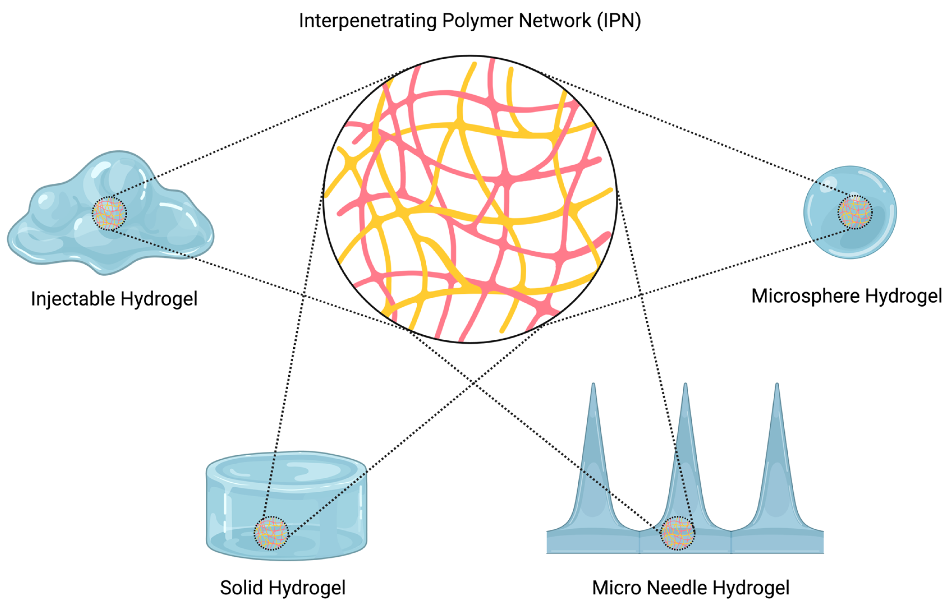
IPN structure in hydrogels can be created in various forms such as films, beads, microspheres, nanoparticles, tablets, and gels [38] (Figure 3). Its structure can be altered as desired for various study fields. From a biomedical point of view, IPNs play an important role because of the gap between their networks. Due to this feature, they can be loaded with different types of drugs, living cells, and liquids [39]. They can even be formed by proteins to create internal reinforcement to structures such as tissue, organs, and systems [35,40]. The mechanical properties of IPNs can be conveniently altered as desired for a particular application. For instance, denser IPN forms may be created for support and reinforcement applications in tissue engineering, material science, and automotive and civil engineering. On the other hand, their structure is immensely practical for loading applications due to their ability to swell. The mechanical properties, such as the stiffness, toughness, and loading capacity, of hydrogels can be ameliorated by forming IPNs. In tissue engineering, wound dressing [39], and drug delivery applications, IPN technology is widely used to enhance treatment methods.
IPNs are usually classified into two main categories based on their formation method [24,40,41,42,43].
Sequential IPN: Formation of polymer network (PN) 1 occurs first. Then, the second PN with/without a crosslinker is polymerized into PN 1.
Simultaneous IPN: Constituent polymers are polymerized at the same time with/without crosslinker to form this kind of IPN.
The most common IPN structures obtained via these formation methods are semi-IPN and full-IPN; however, there are also other types of IPN.
Semi-IPN: Consist of one crosslinked network and additional, uncrosslinked polymer chains.
Full IPN: Consist of two individually crosslinked networks entangled with each other [44].
Pseudo-IPN: Pseudo-IPNs are a type of simultaneous IPN. One polymer has a crosslinked network while the other polymer is linear [44].
Gradient IPN: Gradient IPNs have macroscopically non-homogenous structures in different locations. To synthesize such an IPN, monomer A diffuses into polymer network B. Then, rapid polymerization occurs before diffusion equilibrium happens [45]. Gradient IPNs are collections of layers of insoluble polymer mixtures. Their characteristics can differ gradually in different locations in samples [46].
Thermoplastic IPN: These IPNs show thermoplastic behavior. Physical crosslinking can be used to form these networks. Thermoplastic IPNs behave like thermoplastics because their physical crosslinks break at higher temperatures [44].
IPNs help to bring together polymers that have different properties to form an advanced material. If a polymer or a polymer blend cannot achieve the desired properties, an IPN structure brings a different approach to the application by combining the features of different polymers. Hydrogels with novel properties can be synthesized for different applications with IPN structures. For instance, drug delivery via hydrogels shows great potential to achieve a suitable route to prevent and/or treat diverse diseases. As a novel drug delivery system, IPN hydrogels provide controlled drug release at the target site, minimizing exposure to the drugs at non-target sites so they decrease systemic toxicities and prevent side effects [41,47].
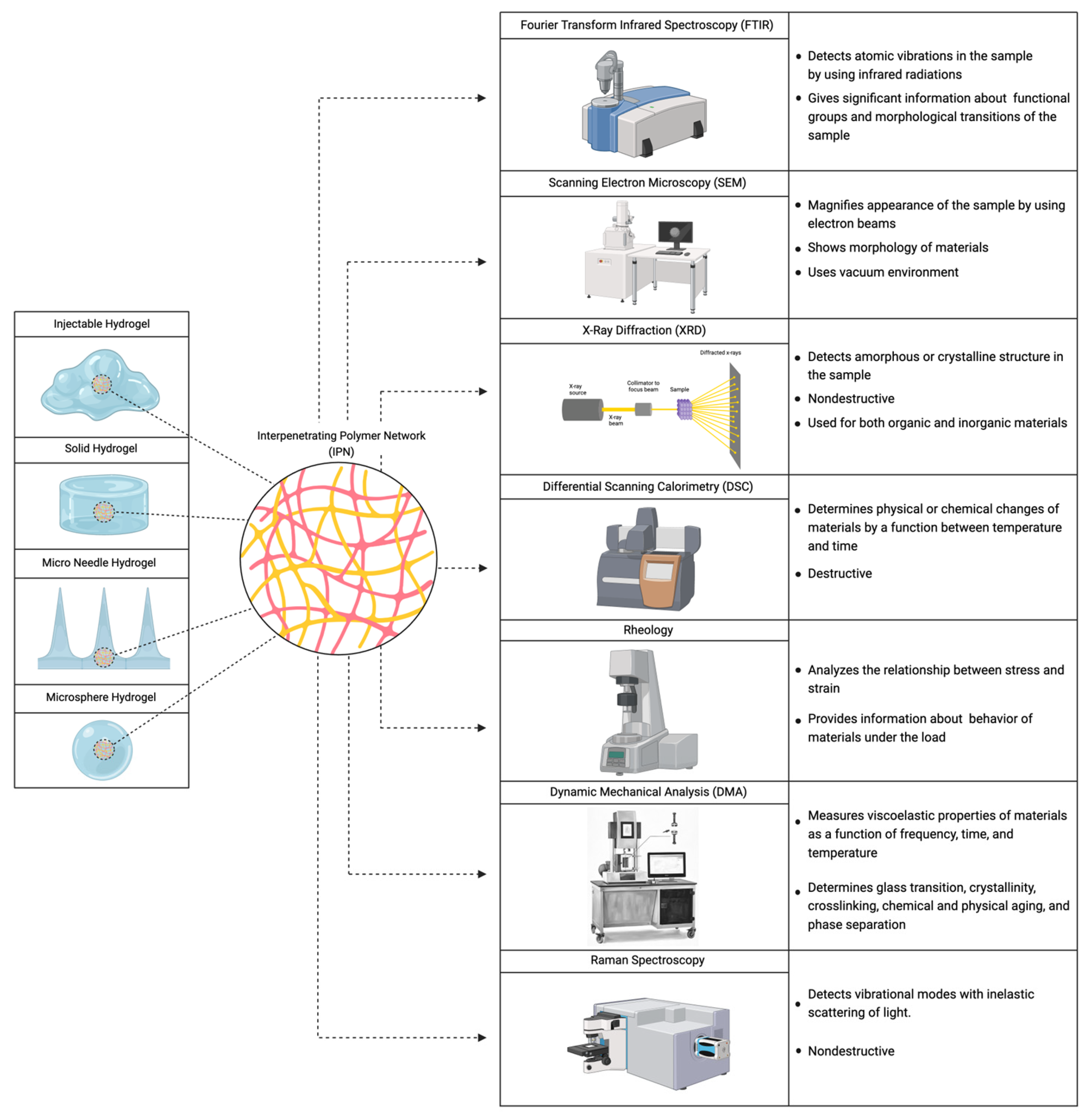
Chemical, physical, and mechanical characterization methods have been conducted to understand the structure of the material and investigate whether it is an IPN or not. The most common characterization methods discussed in the literature are Fourier transform infrared spectroscopy, Raman spectroscopy, scanning electron microscopy, field emission scanning electron microscopy, differential scanning calorimetry, X-ray diffraction, dynamic mechanical analysis, and rheology [48,49]. Researchers use one or more characterization methods to assess the IPN structure of their material. However, these methods have not been brought together and explained in one paper (Figure 4).
In this paper, articles describing the different characterization methods to evaluate IPN structure are reviewed, interpretation of results explained, and an overall recommendation provided for which methods are most useful in proving the presence of IPN structures in hydrogels.

2. Characterization Methods to Determine the Formation of IPNs

First, characterization methods must be understood to analyze the formation of IPNs. A brief introduction to each of these methods is given below.

2.1. Spectroscopy Methods

Fourier transform infrared spectroscopy (FTIR) is used for the characterization of different kinds of materials by infrared radiation (IR). This method is an effective approach for materials/samples that can be present at solid, liquid, gas, film, fiber, and powder forms. It detects molecules’ atomic vibrations in the sample by using IR. FTIR is used in different applications such as drug and medical device development, food analysis, polymer science, industry, and academic research. It provides information about the chemical structure of molecules, functional groups and chemical bonding and chemistry identification/characterization of the sample [50]. Results from the FTIR analysis are understood by reading the FTIR spectrum. This spectrum shows “absorption values versus wavenumbers” or “transmission values versus wavenumbers”. To understand this spectrum, it is necessary to know in which region the analysis is performed. The infrared spectrum is separated into three main regions. These regions are the far-IR spectrum (<400 cm−1), mid-IR spectrum (400–4000 cm−1), and near-IR spectrum (4000–13,000 cm−1). The mid-IR spectrum is used mostly for the samples. Figure 5 shows a general view of a mid-IR spectrum.
According to Kumar et al. (2019), FTIR results provide significant information about materials’ functional groups and morphological transitions [51,52]. Earlier in this article, it was mentioned that physical entanglements between different networks form the IPN structure. Based on this information, the physical bonds between the networks can be found with the FTIR technique. This information may help researchers to interpret whether the IPN structure has occurred in a sample or not. In this article, some examples will be shown to demonstrate how researchers have used FTIR to determine and understand the presence of IPN structure in their studies.
Raman spectroscopy works in a similar manner to FTIR. The main differences between FTIR and Raman spectroscopy are working principle, sensitivity, and sample preparation. Raman spectrometry provides information about vibrational modes by measuring light. As mentioned in the FTIR section, FTIR works by absorbing IR light; however, Raman spectroscopy uses a laser, and its working principle is based on Raman scattering, which is the inelastic scattering of light. One of the most important feature of Raman spectroscopy is being less sensitive to water than FTIR. It is a nondestructive method and requires short preparation times [53]. According to Nigro et al. (2021) and Rajawasam et al. (2024), this method can be used as an identifier of IPN structures as in FTIR [32,54,55].

2.2. Electron Microscopies

Scanning electron microscopy (SEM) is used to examine materials with magnified appearance by using electron beams. This physical method generates 3D-like images that show the morphology of a material by creating a beam of electrons that move on the sample. Electrons do not pass through the sample, but they strike and reflect off a thin metal layer previously coated onto the sample. These electrons are captured by a sensor and evaluated by detectors to create an SEM image [56]. Also, with the help of an electron beam, the sample emits X-rays with unique energy, which are used to identify the material’s composition [57].
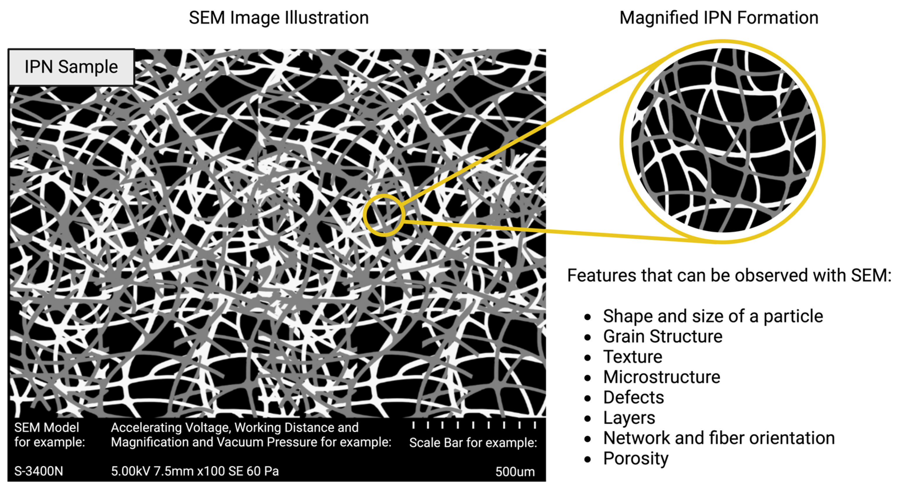
SEM is a nondestructive method that can detect a broad range of materials such as polymers, metals, ceramics, biological samples, and more within a vacuum environment [58,59,60,61,62]. SEM is an important method to determine IPN formation because it shows the arrangements, defects, voids, and structure of the material [63] (Figure 6). Therefore, the IPN structure can be observed and interpreted by using SEM, and these results might be used to prove whether or not an IPN has formed.
Similarly, field emission scanning electron microscopy (FESEM) can be used to observe the material’s morphologic properties and its IPN structure. It is convenient to use in the same applications as SEM, but it creates better quality images with higher sensitivity [64]. However, it also works under vacuum like SEM. If a vacuum environment cannot be formed, the presence of gas in the environment disorganizes the emitted electron beams, leading to inconclusive results [65]. The vacuum environment required for SEM and FESEM constricts their applications with materials such as gels, organisms, cells, and tissues.
Although these two methods seem very suitable for detecting IPNs, they have some drawbacks as mentioned before. SEM and FESEM require vacuuming and coating to examine samples [66], but hydrogels are swollen structures, so if they are vacuumed, their structure may change. For this reason, some researchers freeze-dry their hydrogel before using SEM. Freeze-drying helps keep the hydrogel in its swollen state and allows it to be separated into pieces to see the interior structure. For instance, Che et al. (2018) freeze-dried their hydrogel samples before examining with SEM [67]. This sample preparation method prevents the problems that can be caused by the vacuuming of the hydrogel. However, the freeze-drying method may create some structural changes in samples. Ice crystal formation can be observed with SEM after freeze-drying a hydrogel sample [68]. For these reasons, it is necessary to employ a characterization method other than SEM and FESEM in order to prevent undesirable effects from the vacuum environment and crystal formation.
Environmental scanning electron microscopy (ESEM) is a similar characterization method to see the magnified appearance of materials. It operates under normal/high-vacuum conditions, and pressure can be controlled when working on a sample. While the sample is exposed to atmospheric pressure, the electron column operates in a vacuum [69]. Moreover, it does not require coating of the sample [70]. This method allows researchers to investigate wet, damp, and biological samples without damage or altering their structure [71]. ESEM can be used as a suitable morphology observation method for IPN structure in hydrogels. Previous studies will be shown in the following parts to understand IPN structure observed from SEM, FESEM and ESEM images.

2.3. X-ray Diffraction

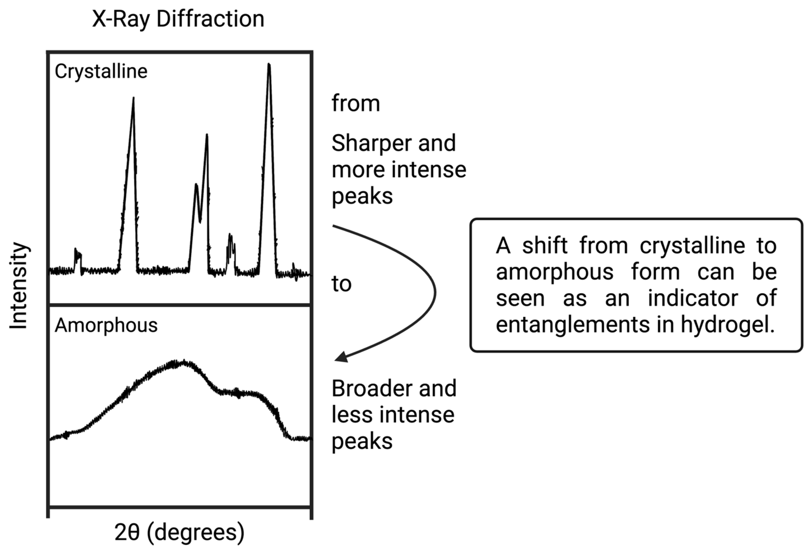
X-ray diffraction (XRD) is a characterization method used to understand the crystal structure of a material [72]. Solid forms, liquids, and powders are examined with this technique. It is a nondestructive method and can be used for both organic and inorganic materials [73]. Briefly, X-ray beams pass into the sample, and they are deflected by the atoms of the sample. Then, diffraction patterns are gathered by a detector to show the sample’s crystallinity [74,75]. Diffraction patterns show the fingerprint of the material, provide information about the amorphous or crystalline structure of the material, and also measure the percent of crystallinity [76]. XRD characterization results can be useful for understanding IPN structure in hydrogels. Crystalline and amorphous structures provide information about the possibility of IPN formation within hydrogel (Figure 7).

2.4. Differential Scanning Calorimetry

Differential scanning calorimetry (DSC) determines physical or chemical changes of materials by a function between temperature and time [77,78]. This method requires small amounts of sample, and it can be used for both solid and liquid samples. However, it is a destructive method for materials due to heat [79]. DSC has two main parts: the thermal computer and a scanning chamber. Two pans are located in the scanning chamber for heating and cooling processes. The sample is settled within the first pan, and the other pan is left empty to use as a reference [75,80]. Temperature is changed gradually to produce a change in the material with the effect of heat quantity. Glass transition temperature (Tg), crystallization heat (Hc), melting point (Tm), crystallization temperature (Tc), and melting heat (Hf) values are determined with DSC [81]. Water loss and thermal degradation studies can be performed for biopolymers. Also, according to Lu and Weiss (1992), Tg value is used to understand biopolymer blends’ miscibility [82,83]. Therefore, DSC can be used to analyze IPN structure in a hydrogel [84].

2.5. Dynamic Mechanical Analysis

Dynamic mechanical analysis (DMA) is a technique to measure the viscoelastic properties of materials as a function of frequency, time, and temperature. This technique is very similar to the rheology method. The differences between the rheology and the DMA methods are kind of the material, testing modes, and data output. Mostly, a rheometer is used for gel–liquid-like soft materials, while DMA is used for solid materials. Also, DMA tests include tension, compression, shear, and bending modes, but a rheometer is used to perform shear and normal stress analyses. DMA determines mechanical properties by the functions of temperature and time. Therefore, this method can be used for detecting morphological structures in a material. Glass transition, crystallinity, crosslinking, chemical and physical aging, and phase separation are used for the characterization of the material by DMA. The glass transition data indicate a relation to the viscoelastic response of the material. Viscoelastic materials, such as gels, show solid-like behavior at lower temperatures while they show gel-like or liquid-like behavior at higher temperatures [32,85,86,87,88,89]. Temperature or frequency can be kept constant while performing a DMA test; however, since it is usually difficult to work with very high and low frequencies, it is useful and fast to collect viscoelastic data with a constant frequency at different temperatures. The viscoelastic data can help to find viscoelastic transitions/relaxations in amorphous materials by the measured glass and secondary transition values. Since secondary transitions cannot be observed with DSC, DMA and DSC are sometimes used together for more accurate results. According to Chartoff et al. (2009), there is a relation between polymer crosslinking, network, and structure integrity with glass transition temperature [85]. This relation is used for characterizing network formation in materials in different applications such as polymer curing, filled polymers, pharmacological and biomedical device optimization studies, polymer composites, and wound healing applications [90,91,92,93,94].

2.6. Rheology

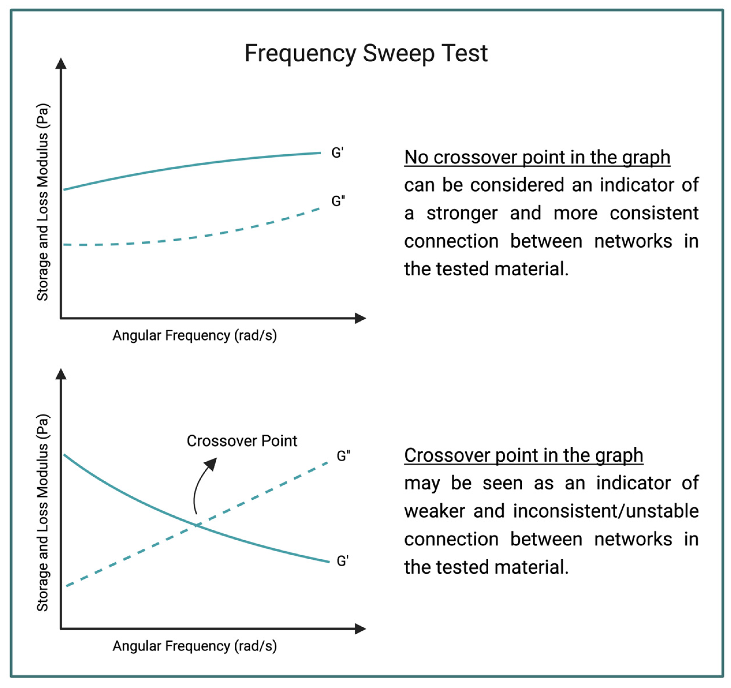
Rheology is a term used for understanding viscoelastic behavior of a material. It analyses the relationship between stress (force) and strain (deformation) when a load is applied [95]. Examining hydrogels with rheology provides information about their behaviors under a load. Hydrogels can be shown as a complex system due to their liquid- and solid-like behaviors. For example, hydrogels used as injectable drug delivery devices display a shear thinning response and their structure can change with these applications. Therefore, it is important to understand the behaviors of injectable hydrogels for in vivo and clinical studies [96]. A rheometer is used to measure the mechanical behaviors of materials, and one of the tests which determine mechanical behavior is the frequency sweep test [97]. Figure 8 shows a generalized illustration of two different frequency sweep test graphs. According to Stojkov et al., crossover point is an indicator of consistent connections in a hydrogel [98]. These rheology tests may help determine IPN structure within a hydrogel [30,99]. Examples of using rheology to characterize IPN hydrogels will be covered in the next section.

3. Studies That Investigate IPN Formation

In this section, a detailed literature review of some IPN hydrogel articles will be discussed. Unfortunately, most of the available IPN studies do not indicate how they confirmed the IPN structure in the hydrogels, mentioning only that their hydrogel has an IPN structure without showing any concrete proof. The articles presented below actually try to prove the IPN structure in their material with different characterization methods.

3.1. Injectable Hydrogels

Wang et al. (2017) synthesized a dynamic covalent hydrogel which is injectable and has IPN structure. The purpose of their study was enhancing mechanical features and improving the structure of the gels for use as a novel approach in different applications such as 3D printing, tissue engineering, and drug delivery. They used reversible addition fragmentation chain transfer polymerization method to create a p(N, N-dimethyl-acrylamide-co-4-acryloyldopamine) p(N, N-dimethylacrylamide-co-3-acrylamidopheynlboronic acid) p(N, N-dimethylacrylamide-co-4-acryloyloxybenzaldehyde) gel. FTIR studies were performed at different pH values, and crosslinking in the gels was determined by peak disappearance within the 1700–1750 cm−1 band range, which was caused by -C=O and -NH-NH2 linkage. The mechanical properties of the gel (at pH 3, 7 and 10) were investigated by using rheometer frequency sweep tests. All the gel samples showed gel-like behavior (G′>G″). According to the authors, IPN hydrogels have higher strength values than single network hydrogels. Therefore, they suggested that the increased density of crosslinking leads to IPN and interlocked structure formation in the gel [100].
Shariatzadeh et al. (2021) synthesized an IPN cryogel which was formed from gelatin methacrylate (GelMA) and hyaluronic acid (HA) to be used for soft tissue engineering. An IPN gel was created by using the sequential method. First, GelMA was chemically crosslinked, then HA was physically crosslinked by using Fe3+ as a crosslinker agent, and ultimately these two networks formed an IPN cyrogel. Mechanical properties were investigated by using frequency sweep tests via rheometer. Samples showed gel-like behavior (G′>G″). FTIR study was performed with GelMA and GelMa-HA to investigate interactions in the material. According to authors’ findings, there was a slight shift in the FTIR bands in comparison with GelMA and GelMA-HA gels. This shift indicated electrostatic/intermolecular interactions or hydrogen bonding between HA and GelMA. Ultimately, it was interpreted as IPN formation in the GelMA-HA cryogel [101].
Cui et al. (2023) used a cellulose nanofiber (CNF) to create IPNs in oxidized alginate (OSA)/gelatin (Gel) hydrogel. The OSA/Gel/CNF hydrogel was formed to be used in bone repair studies because of its injectability, self-healing, and high strength properties. In this study, CNF acted as an IPN reinforcement to the base hydrogel. The effect of varying amounts of CNF (1%, 3% and 5%) to the hydrogel system was investigated by FTIR. Based on the FTIR results, the OSA/Gel had O-H stretching peak at 3521 cm−1, and it shifted to 3521 cm−1 when CNF3 was used to form OSA/Gel/CNF3. According to the authors, this shift at the O-H stretching band indicates the formation of hydrogen bonds. Moreover, this can be interpreted as a hint towards confirmed IPN structure in the OSA/Gel/CNF3 hydrogel [102].
Mu et al. (2020) synthesized an injectable IPN hydrogel which was formed by using injectable platelet-rich fibrin (iPRF) and gelatin nanoparticles (GNPs). The researchers created a hydrogel which had good mechanical strength, self-healing, and degradable properties. The purpose of the iPRF/GNP hydrogel use in bone healing studies as a bioactive material. Scanning electron microscopy was used to understand and opine regarding this double-network hydrogel’s structure. According to the study, the iPRF/GNP hydrogel showed a double-network structure in SEM images. Also, the authors asserted that the iPRF/GNP gel exhibited a porous network which was created from interconnected fibrin fibers and gelatin nanoparticles. They attributed this interconnection to cohesive interactions between these two components. These network formations confirm the IPN structure in the iPRF/GNP hydrogel [103].
Zhao et al. (2014) synthesized an injectable poly(ethylene glycol) methacrylate (PEGMA)/N-isopropylacrylamide (NIPAm)/methacrylated alginate (ALGMA) hydrogel for use in drug and protein release studies. The hydrogel was formed by a copolymerization method [104]. ALGMA was used in this study for its pH-sensitive and network-forming properties. The PEGMA/NIPAm gels with different ALGMA compositions were characterized via rheometer. The storage moduli of PEGMA/NIPAm gels were increased with increased ALGMA amount. According to the authors, this change happened because of the hydrogen bonds which were created by the ALGMA, and these bonds supported IPN structure in the hydrogels. Moreover, they support their rheology findings with FTIR results. FTIR study of the alginate showed a peak at 3440 cm−1 in the hydrogen bond region, and then this peak shifted to 3275 cm−1 for PEGMA/NIPAm/ALGMA hydrogels. Based on these findings, Zhao et al. interpreted the shift as formation of hydrogen bonds between the PEGMA/NIPAm/ALGMA, and this was attributed to IPN formation in the gel [105].

3.2. Drug Delivery Hydrogels

Darge et al. designed a sodium alginate and sulfobetaine methacrylate (SBMA) monomer-based IPN hydrogel to produce microneedles (MN) for drug delivery into the dermal interstitium across the stratum corneum. Lipopolysaccharide (LPS) and doxorubicin (DOX) were loaded into MN arrays for use as an immunochemotherapy agent.
IPN hydrogel was synthesized by using the sequential IPN synthesis method. First, SBMA was chemically crosslinked by photo crosslinking, and it formed a polymer network. Then, alginate was ionically crosslinked by using calcium ions, and it created the second network. These two networks together formed a SBMA/Alginate IPN hydrogel.
FTIR analysis and mechanical testing were utilized for characterization of the IPN hydrogel. In FTIR spectra, the signal at 1720 cm−1 in the SBMA monomer was seen in IPN hydrogel as well. This point indicates that monomers created a network by chemically crosslinking. On the other hand, the COO signal of sodium alginate was seen at 1595 cm−1, then the signal shifted to 1642 cm−1 in the IPN hydrogel. This change indicates the ionic crosslinking for alginate via calcium ions. According to these findings, it was interpreted that there is an IPN hydrogel formation [106].
A universal testing machine was used to obtain mechanical testing results of hydrogels. They examined different synthesis methods on hydrogels. In this article, three synthesis methods were used. These are (i) photo crosslinking, to create SBMA networks, (ii) ionic crosslinking, to get alginate networks, and (iii) photoionic crosslinking, to form IPN hydrogels. By using photo crosslinking, the highest tensile strength was achieved. However, its strain was very low compared to others. Therefore, it was confirmed that the hydrogels that formed by only photo crosslinking method had a brittle and rigid structure. On the other hand, by only using the ionic crosslinking method, the hydrogels had the maximum strain and minimum tensile strength. A soft structure is obtained by this method. Finally, by using the photoionic crosslinking method, desired strength and strain results were achieved. Based on these results, it can be interpreted that the sequentially formed IPN got the properties from both alginate and SBMA networks [106].
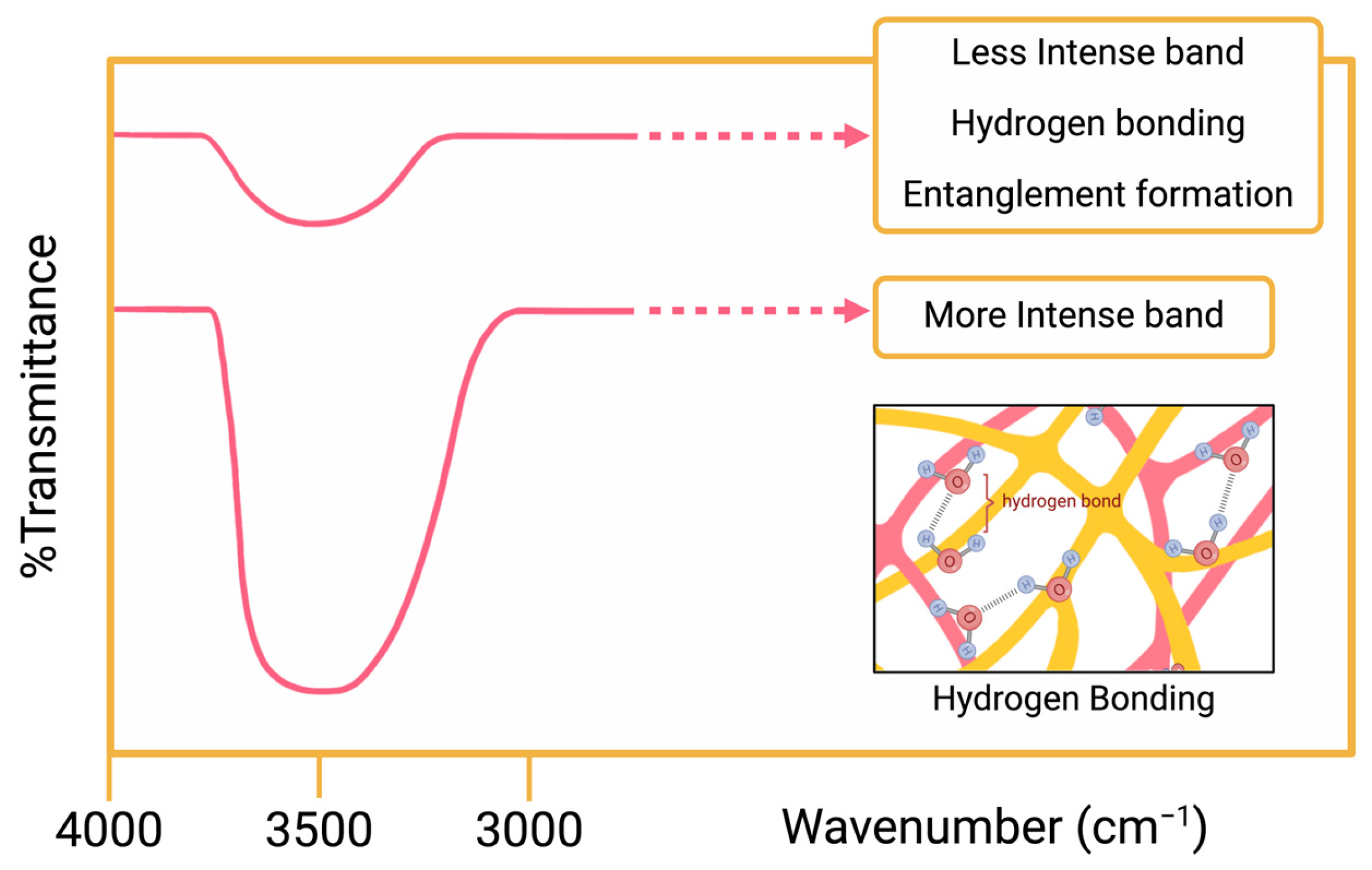
Agnihotri et al. synthesized a novel hydrogel to use in controlled drug delivery. The aims of this study were (i) sustaining a stable blood level, (ii) reducing negative effects of drugs, and (iii) increasing the drug efficacy. To achieve their goals, they formed an IPN microsphere hydrogel from gellan gum (GG) and poly(vinyl alcohol) (PVA) via emulsion crosslinking method. Carvedilol, which is a hypertension treatment drug, was loaded to the microspheres to understand drug release kinetics and glutaraldehyde (GA) crosslinker was used for different crosslinking levels. FTIR, DSC and tensile strength measurements were used to observe interpenetrating network of the microspheres. In the FTIR interpretations, it was observed that GG’s broad band at 3444 cm−1 and PVA’s broad band at 3425 cm−1. Unloaded GG-PVA microspheres showed their broad band at 3487 cm−1. Their values were similar; however, the intensities of the bands showed differences. Figure 9 shows a similar illustration of the intensities of the bands at the hydrogen bond region. Unloaded GG-PVA microspheres had less intensity in contrast to GG and PVA. It was determined that these data were observed because uncrosslinked hydroxyl groups of the glucopyranose ring bonded with hydrogen and created hydrogen bonding [107]. As mentioned at the beginning of this article, physical crosslinking occurs to form IPNs in materials. According to Agnihotri et al., this finding confirms the IPN structure of the microspheres.
Another approach to confirm the hydrogel’s IPN structure is via DSC thermograms. The endothermic peaks of the GG, PVA, and unloaded GG-PVA microspheres were compared. The endothermic peak of the GG was 248 °C. PVA had two endothermic peaks, and they were 210 °C and 290 °C, respectively. The unloaded GG-PVA microspheres showed two peaks. The first one was at 224 °C, which is between the values of GG and PVA, and the second one was at 320 °C, which is a higher value compared to GG and PVA. The interpretation of these findings leads to the conclusion that unloaded GG-PVA microspheres formed a more crystalline polymer structure. As a result, the higher endothermic peaks (which are a sign of high melting temperature) of unloaded GG-PVA indicated that there was an IPN formation because IPN needs higher temperatures to melt compared to single-network structures.
Last but not least, tensile strength measurements were conducted to understand if there was an IPN structure in the microspheres. The increased amount of GA in the synthesis of the microspheres resulted in an increase in strength. When the GG and microspheres were compared, the microspheres showed five to six times higher tensile strength. The researchers interpreted this finding as a confirmation of the IPN structure [107].
Mohamadnia et al. focused their studies on pH-sensitive interpenetrating hydrogel beads to control drug delivery. Some responsive properties, such as pH, temperature, and ionic strength, were used to control the swelling behavior of the beads. Beads were synthesized from two different biopolymers: alginate and carrageenan. Betamethasone acetate, a kind of corticosteroid drug, was loaded into the beads to examine their release behaviors. Also, to enhance the swelling properties, an IPN structure was formed in the hydrogel beads. SEM was used to observe the IPN structure of the hydrogel beads in this study. According to Mohamadnia et al., the interpenetrating network of the beads was the cause of such a unique structure. Loaded beads looked more swollen and orderly due to the encapsulation of the drug into the IPN entanglements [108].
Bhattacharya et al. synthesized sodium carboxymethyl cellulose and sodium carboxymethyl xanthan gum-based (SCMC-SCMXG) IPN hydrogel beads via an emulsion gelation process. They loaded the beads with diclofenac sodium (DS), which is an anti-inflammatory drug, to observe the drug release mechanism. XRD analysis of the beads was carried out to investigate their IPN structure. According to Bhattacharya et al., the drug’s crystalline form shifted to an amorphous form in the beads [109] because the drug had been distributed into the entanglements of the IPN structure. They associated the crystallinity change with the IPN structure and found proof to confirm IPN structure in the hydrogel beads.
Pan et al. formed an IPN hydrogel that contained sugar bagasse cellulose (SBC), carboxymethylcellulose (CMC), and poly(N-isopropylacrylamide) (PNIPAm). Epichlorohydrin was added as a crosslinker. The hydrogel was intended to be used as a smart drug delivery system. SEM figures of the SBC-CMC hydrogel and the IPN hydrogel can be found in the paper by Pan et al. In their SEM figures, the SBC-CMC hydrogel displayed a porous form. However, the IPN hydrogel showed a net-like structure around the first porous form. According to Pan, this confirms the IPN structure of the hydrogel. Additionally, FTIR analyses of the hydrogels provided results to prove that its structure formed an IPN. In the FTIR studies, there was no significant peak shifting between the crosslinked SBC-CMC hydrogel and IPN hydrogel. Their peaks were reasonably similar to each other, so it was inferred as there was no chemical bonding between them and they were just entangled [110].
Kim et al. (2018) worked on a temperature- and pH-sensitive IPN hydrogel which consisted of hyaluronic acid (HA) and poly (N-isopropylacrylamide) (PNIPAM). The injectable hydrogel was created for transdermal delivery of luteolin as psoriasis skin relief. PNIPAM and HA were used for temperature sensitivity and pH sensitivity, respectively. Kim et al. created the first network by radical polymerization of PNIPAM and with the HA the gel structure formed a semi-IPN. The second network was formed by Michael addition for HA. As a result, a full IPN structure was achieved in the study. According to swelling ratio studies, the full IPN hydrogel had a denser structure than the semi-IPN hydrogel. Also, studies on the swelling rate change by pH showed that the acidic setting supported intramolecular hydrogen bond formation and reduced swelling of hydrogen, which can be attributed to IPN formation in the hydrogel [111].

3.3. Hydrogels for Other Biomedical Applications

Wahid et al. aimed to prepare a hydrogel with advanced mechanical and antibacterial features to be used in the biomedical field. Bacterial cellulose (BC), chitosan (CS), and glutaraldehyde crosslinker were used to synthesize semi-IPN hydrogels. In this manner, the BC’s properties, such as a great amount of water retention, biocompatibility, biodegradability, high porosity, and nontoxicity, were combined with chitosan’s antibacterial properties to obtain a biomedically applicable material. Characterization of the hydrogels was carried out with FTIR. In the FTIR analysis, new peaks occurred at 1705 cm−1 only for CS-containing hydrogels. This peak was explained by glutaraldehyde’s C=O of the aldehyde group. Moreover, imide bond formation was observed for CS at 1558 cm−1 in the presence of glutaraldehyde. These peaks indicate that the CS interacted with glutaraldehyde. On the other hand, OH and NH2 shifting were detected for CS, while H-O-H shifting was detected for BC [34]. These lower wavenumbers are interpretable as caused by hydrogen bond formation between BC and CS. As a result, it was understood that CS chains linked with other CS polymer chains with glutaraldehyde while BC polymer chains entangle to them via hydrogen bonding. In conclusion, it was confirmed that the BC-CS hydrogels had a semi-IPN structure [34].
The presence of hydrogen bonding was also examined with XRD analysis. In these studies, CS hydrogel showed an amorphous structure at ~20°. Normally, BC has a crystalline structure because of the hydrogen bonding between its layers. However, it showed a wide and less intense peak at ~20°, in the form of BC-CS hydrogel. This was observed because BC’s hydrogen bonds were entangled with the CS polymer chains instead of its own layers, which caused it to alter its structure from crystalline to amorphous. Therefore, this situation indicates that a semi-IPN structure was formed in the hydrogel.
Another characterization method to reveal the IPN structure was FESEM analysis. Only CS-containing hydrogel and BC-CS-containing hydrogels (with different ratios of BC) were examined under FESEM. CS hydrogel showed big pores. However, BC-CS hydrogels’ pore size was reduced when the amount of BC increased in the hydrogel (BC-20 and BC-40). Increased network structure occurred due to entanglements, so this confirms that the entanglement of the BC to the CS and semi-IPN structure [34].
Sampath et al. prepared a cellulose nanocrystal (CNC) reinforced chitosan (CS) hydrogel with the support of glutaraldehyde crosslinker. This hydrogel was defined as a semi-IPN hydrogel due to FTIR analysis [112].
FTIR analyses were conducted for CNC, CS, CS, and CNC-CS hydrogels to confirm the semi-IPN structure of the hydrogel. Both CNC and CS hydrogel peaks were observed in the CNC-CS hydrogels. There was not an extreme amount of shifting in the peak of the CNC-CS hydrogels. According to Sampath, this means that CNCs were physically bonded to the CS hydrogel, leading to the determination of the semi-IPN structure [112].
Tığlı and Gümüşderelioğlu developed a cartilage scaffold from alginate (Alg) and chitosan (CS). They synthesized this Alg-CS semi-IPN scaffold by freeze-drying. Calcium chloride (CaCl2) crosslinker was used to synthesize the alginate scaffold. SEM and fluorescence microscope images were analyzed to see the scaffold’s semi-IPN structure. The Alg-CS (70:30) % (v/v) scaffold shows that the chitosan settled into the alginate scaffold. On the other hand, to see this network formation clearly, fluorescence microscope images were used.
According to figures in Tığlı and Gümüşderelioğlu’s studies, chitosan is marked with fluorescein isothiocyanate. The bright sections were chitosan, which collapsed in the alginate scaffold. Distribution of the chitosan was shown in its exact location with the help of the fluorescence microscope [113].

3.4. Hydrogels for Non-Biomedical Applications

Due to the toxic behavior of azo dyes in the environment, Ngwabebhoh et al. developed a superabsorbent material to enhance the wastewater treatment process. They synthesized chitosan/starch semi-IPN hydrogels to achieve improved adsorptive removal of the dye Direct Red (DR80) from an aqueous medium. Glutaraldehyde was used as a crosslinking agent. FTIR analyses were conducted to characterize the structure of their material. The band observed at 1644 cm−1 for hydrogel indicates that the crosslinking process via glutaraldehyde was achieved with only chitosan. Also, hemiacetal and acetal bond formations between starch and glutaraldehyde made unsteady interactions with each other. Thus, it was concluded that starch has physical interactions with crosslinked chitosan in the hydrogel. As an inference, the hydrogels were classified as semi-IPN [9] because it is known that if one polymer crosslinks and the second one entangles the first polymer, it is called a “semi-IPN”. According to study, chitosan chains were crosslinked with each other. However, starch polymers were seen to be entangled with them.

4. Discussion

In this review article, FTIR, Raman, SEM, FESEM, DSC, XRD, DMA and rheology methods were examined for confirming IPNs in hydrogels.
While gathering the literature from different articles, it was observed that the great majority of the authors did not give concrete results about how they determined the IPN structure in their material. However, it is important to provide confirming data to say if the material is IPN or not because IPNs’ definition can be easily confused with polymer blends. It is useful to conduct different characterization methods to obtain information about a hydrogel’s internal structure.
The articles that were chosen from the literature mentioned how they confirmed IPN structure with characterization methods in their studies (Table 1). There is not only one characterization method to confirm networks in a hydrogel, and there is not only one type of evidence to confirm the structure. All the characterization processes of IPN depend upon the experiment method, the material’s nature, and the researchers’ experience with the literature and truly interpreting these results.
It is seen that the most used detection method was FTIR. This method gives the researcher the most relatable results regarding hydrogel IPN structure. The chemical bonds and physical interactions between the networks are identified with this technique. Also, it is not an invasive method for materials and does not alter the sample’s structure.
XRD and DSC give information about a material’s amorphous and crystalline structure. DSC can be used in samples that have different structures from each other. If a material consists of these two different arrangement types (amorphous and crystalline), DSC can be used. However, DSC may not be suitable for more than two networks due to the complexity of the identification process. Also, it is a destructive method.
Mechanical testing methods indicate a material’s mechanical properties. Stress–strain curves of the base materials and the IPN hydrogel are compared with each other to determine IPN structure. Rheology method is a mechanical testing method. The storage moduli of the base and IPN samples are compared to each other to find a relatable connection between them. Rheology does not give a chemical viewpoint to the material, so the entanglement theory cannot be completely confirmed by using only this method.
SEM and FESEM are used to obtain an image of the material’s morphology. These methods require a vacuum environment, so hydrogels must be prepared for this condition. The preparation process can cause structural changes within the hydrogel. These methods give the clearest structural identification for the samples, but the most important point here is the accurate interpretation and experience of the researcher within the field. In some cases, entanglements may not be seen with these techniques.

5. Conclusions

As a result, FTIR was found to be the most promising method for determining IPN structures in hydrogels. Other characterization methods are also useful for confirming the structure; however, they need much more effort, experience, and clearness for their interpretations. These methods can be used together to confirm the IPN structure, and a combination of these methods would give even more accurate results. It is feasible to use more than one method and gain experience to correctly interpret the results. In the future, as studies on IPN hydrogel increase, they will provide more information and data about the accuracy of these determining methods.

Author Contributions

Conceptualization, C.C. and E.B.; Writing—original draft preparation, C.C.; Writing—review and editing, K.B.; visualization, C.C.; supervision, E.B. All authors have read and agreed to the published version of the manuscript.

Funding

This research received no external funding.

Data Availability Statement

No new data were created.

Conflicts of Interest

The authors declare no conflicts of interest.

References

  1. Patrickios, C.S. Polymer networks: Recent developments. In Macromolecular Symposia; Wiley Online Library: Hoboken, NJ, USA, 2010. [Google Scholar]
  2. Niu, Y.; Xia, Q.; Gu, M.; Yu, L.L. Interpenetrating network gels composed of gelatin and soluble dietary fibers from tomato peels. Food Hydrocoll. 2019, 89, 95–99. [Google Scholar] [CrossRef]
  3. Yin, Y.; Gu, Q.; Liu, X.; Liu, F.; McClements, D.J. Double-network hydrogels: Design, fabrication, and application in foods and biomedicines. Adv. Colloid Interface Sci. 2023, 320, 102999. [Google Scholar] [CrossRef] [PubMed]
  4. Du, M.; Lu, W.; Zhang, Y.; Mata, A.; Fang, Y. Natural polymer-sourced interpenetrating network hydrogels: Fabrication, properties, mechanism and food applications. Trends Food Sci. Technol. 2021, 116, 342–356. [Google Scholar] [CrossRef]
  5. Lin, Q.; Hu, Y.; Qiu, C.; Li, X.; Sang, S.; McClements, D.J.; Chen, L.; Long, J.; Xu, X.; Wang, J.; et al. Peanut protein-polysaccharide hydrogels based on semi-interpenetrating networks used for 3D/4D printing. Food Hydrocoll. 2023, 137, 108332. [Google Scholar] [CrossRef]
  6. Jia, C.; Zhang, X.; Li, Y.; Jiang, Y.; Zhang, M.; Lu, P.; Chen, H. Synthesis and characterization of bio-based PA/EP interpenetrating network polymer as coating material for controlled release fertilizers. J. Appl. Polym. Sci. 2018, 135, 46052. [Google Scholar] [CrossRef]
  7. Chen, R.; Yang, S.; Liu, B.; Liao, Y. Eco-Friendly Semi-Interpenetrating Polymer Network Hydrogels of Sodium Carboxymethyl Cellulose/Gelatin for Methylene Blue Removal. Materials 2023, 16, 3385. [Google Scholar] [CrossRef] [PubMed]
  8. Cavalcante, J.; Oldal, D.G.; Peskov, M.V.; Beke, A.K.; Hardian, R.; Schwingenschlogl, U.; Szekely, G. Biobased Interpenetrating Polymer Network Membranes for Sustainable Molecular Sieving. ACS Nano 2024, 18, 7433–7443. [Google Scholar] [CrossRef] [PubMed]
  9. Ngwabebhoh, F.A.; Gazi, M.; Oladipo, A.A. Adsorptive removal of multi-azo dye from aqueous phase using a semi-IPN superabsorbent chitosan-starch hydrogel. Chem. Eng. Res. Des. 2016, 112, 274–288. [Google Scholar] [CrossRef]
  10. Cui, L.; Xiong, Z.; Guo, Y.; Liu, Y.; Zhao, J.; Zhang, C.; Zhu, P. Fabrication of interpenetrating polymer network chitosan/gelatin porous materials and study on dye adsorption properties. Carbohydr. Polym. 2015, 132, 330–337. [Google Scholar] [CrossRef] [PubMed]
  11. Zhu, H.; Yang, T.; Chen, S.; Wang, X.; He, J.; Luo, Y. Construction and characterization of chitosan/poly (acrylamide-[2-(methacryloyloxy) ethyl] trimethylammonium chloride) double-network hydrogel with enhanced antibacterial activity. Adv. Compos. Hybrid Mater. 2023, 6, 192. [Google Scholar] [CrossRef]
  12. Chen, Q.; Tian, X.; Fan, J.; Tong, H.; Ao, Q.; Wang, X. An interpenetrating alginate/gelatin network for three-dimensional (3D) cell cultures and organ bioprinting. Molecules 2020, 25, 756. [Google Scholar] [CrossRef]
  13. Huang, Y.; Zhao, X.; Zhang, Z.; Liang, Y.; Yin, Z.; Chen, B.; Bai, L.; Han, Y.; Guo, B. Degradable gelatin-based IPN cryogel hemostat for rapidly stopping deep noncompressible hemorrhage and simultaneously improving wound healing. Chem. Mater. 2020, 32, 6595–6610. [Google Scholar] [CrossRef]
  14. Wang, L.; Zhang, X.; Yang, K.; Fu, Y.V.; Xu, T.; Li, S.; Zhang, D.; Wang, L.N.; Lee, C.S. A novel double-crosslinking-double-network design for injectable hydrogels with enhanced tissue adhesion and antibacterial capability for wound treatment. Adv. Funct. Mater. 2020, 30, 1904156. [Google Scholar] [CrossRef]
  15. Cassimjee, H.; Kumar, P.; Ubanako, P.; Choonara, Y.E. Genipin-crosslinked, proteosaccharide scaffolds for potential neural tissue engineering applications. Pharmaceutics 2022, 14, 441. [Google Scholar] [CrossRef]
  16. Yang, Q.; Peng, J.; Xiao, H.; Xu, X.; Qian, Z. Polysaccharide hydrogels: Functionalization, construction and served as scaffold for tissue engineering. Carbohydr. Polym. 2022, 278, 118952. [Google Scholar] [CrossRef]
  17. Sun, X.; Wang, H.; Ding, Y.; Yao, Y.; Liu, Y.; Tang, J. Fe3+-Coordination mediated synergistic dual-network conductive hydrogel as a sensitive and highly-stretchable strain sensor with adjustable mechanical properties. J. Mater. Chem. B 2022, 10, 1442–1452. [Google Scholar] [CrossRef]
  18. Shi, X.; Deng, Z.; Zhang, P.; Wang, Y.; Zhou, G.; de Haan, L.T. Wearable optical sensing of strain and humidity: A patterned dual-responsive semi-interpenetrating network of a cholesteric main-chain polymer and a poly (ampholyte). Adv. Funct. Mater. 2021, 31, 2104641. [Google Scholar] [CrossRef]
  19. Song, F.; Gong, J.; Tao, Y.; Cheng, Y.; Lu, J.; Wang, H. A robust regenerated cellulose-based dual stimuli-responsive hydrogel as an intelligent switch for controlled drug delivery. Int. J. Biol. Macromol. 2021, 176, 448–458. [Google Scholar] [CrossRef]
  20. Chen, S.; Matsumoto, H.; Moro-Oka, Y.; Tanaka, M.; Miyahara, Y.; Suganami, T.; Matsumoto, A. Smart microneedle fabricated with silk fibroin combined semi-interpenetrating network hydrogel for glucose-responsive insulin delivery. ACS Biomater. Sci. Eng. 2019, 5, 5781–5789. [Google Scholar] [CrossRef]
  21. Sellamuthu, K.; Angappan, S. Formulation and Characterization of Interpenetrating Polymer Network Hydrogel Bead as Drug Carrier System for Extended Release of Sulphonyl Urea Medication. J. Pharm. Innov. 2024, 19, 2. [Google Scholar] [CrossRef]
  22. Varnava, C.K.; Patrickios, C.S. Polymer networks one hundred years after the macromolecular hypothesis: A tutorial review. Polymer 2021, 215, 123322. [Google Scholar] [CrossRef]
  23. Quirino, R.L.; Monroe, K.; Fleischer, C.H., III; Biswas, E.; Kessler, M.R. Thermosetting polymers from renewable sources. Polym. Int. 2021, 70, 167–180. [Google Scholar] [CrossRef]
  24. Wu, J.; Xue, W.; Yun, Z.; Liu, Q.; Sun, X. Biomedical applications of stimuli-responsive “smart” interpenetrating polymer network hydrogels. Mater. Today Bio 2024, 25, 100998. [Google Scholar] [CrossRef] [PubMed]
  25. Jana, S.; Gandhi, A.; Sen, K.K. Chitosan-Based Interpenetrating Polymer Networks: Drug Delivery Application. In Functional Chitosan: Drug Delivery and Biomedical Applications; Springer: Singapore, 2019; pp. 269–295. [Google Scholar]
  26. Zainudin, B.H.; Wong, T.W.; Hamdan, H. Pectin as oral colon-specific nano-and microparticulate drug carriers. In Polymer Science and Innovative Applications; Elsevier: Amsterdam, The Netherlands, 2020; pp. 257–286. [Google Scholar]
  27. Gong, J.P. Why are double network hydrogels so tough? Soft Matter 2010, 6, 2583–2590. [Google Scholar] [CrossRef]
  28. Sheiko, S.S.; Dobrynin, A.V. Architectural code for rubber elasticity: From supersoft to superfirm materials. Macromolecules 2019, 52, 7531–7546. [Google Scholar] [CrossRef]
  29. Dragan, E.S. Advances in interpenetrating polymer network hydrogels and their applications. Pure Appl. Chem. 2014, 86, 1707–1721. [Google Scholar] [CrossRef]
  30. Wanasinghe, S.V.; Schreiber, E.M.; Thompson, A.M.; Sparks, J.L.; Konkolewicz, D. Dynamic covalent chemistry for architecture changing interpenetrated and single networks. Polym. Chem. 2021, 12, 1975–1982. [Google Scholar] [CrossRef]
  31. Shah, S.Z.O.; Trengove, A.; O’Connor, A.J.; Quinn, J.F.; Kempe, K. Simultaneous Interpenetrating Polymer Networks Based on Poly(2-Oxazoline)s. Macromol. Mater. Eng. 2024, 309, 2300210. [Google Scholar] [CrossRef]
  32. Rajawasam, C.W.; Dodo, O.J.; Weerasinghe, M.A.S.N.; Raji, I.O.; Wanasinghe, S.V.; Konkolewicz, D.; Watuthanthrige, N.D.A. Educational series: Characterizing crosslinked polymer networks. Polym. Chem. 2024, 15, 219–247. [Google Scholar] [CrossRef]
  33. Gu, Y.; Zhao, J.; Johnson, J.A. Polymer networks: From plastics and gels to porous frameworks. Angew. Chem. Int. Ed. 2020, 59, 5022–5049. [Google Scholar] [CrossRef] [PubMed]
  34. Wahid, F.; Hu, X.H.; Chu, L.Q.; Jia, S.R.; Xie, Y.Y.; Zhong, C. Development of bacterial cellulose/chitosan based semi-interpenetrating hydrogels with improved mechanical and antibacterial properties. Int. J. Biol. Macromol. 2019, 122, 380–387. [Google Scholar] [CrossRef] [PubMed]
  35. Hennink, W.E.; van Nostrum, C.F. Novel crosslinking methods to design hydrogels. Adv. Drug Deliv. Rev. 2012, 64, 223–236. [Google Scholar] [CrossRef]
  36. Peppas, N.; Slaughter, B.V.; Kanzelberger, M.A.; Matyjaszewski, K.; Möller, M. 9.20–Hydrogels. In Polymer Science: A Comprehensive Reference; Elsevier: Amsterdam, The Netherlands, 2012; pp. 385–395. [Google Scholar]
  37. Hou, X.; Lin, L.; Li, K.; Jiang, F.; Qiao, D.; Zhang, B.; Xie, F. Towards superior biopolymer gels by enabling interpenetrating network structures: A review on types, applications, and gelation strategies. Adv. Colloid Interface Sci. 2024, 325, 103113. [Google Scholar] [CrossRef] [PubMed]
  38. Dror, M.; Elsabee, M.; Berry, G. Interpenetrating polymer networks for biological applications. Biomater. Med. Devices Artif. Organs 1979, 7, 31–39. [Google Scholar] [CrossRef] [PubMed]
  39. Korah, L.V.; Anilkumar, G.; Thomas, S. Hydrogels, DNA, and RNA polypeptides for the preparation of biomaterials. In Fundamental Biomaterials: Polymers; Elsevier: Amsterdam, The Netherlands, 2018; pp. 85–104. [Google Scholar]
  40. Silverstein, M.S. Interpenetrating polymer networks: So happy together? Polymer 2020, 207, 122929. [Google Scholar] [CrossRef]
  41. Nayak, A.K.; Hasnain, M.S.; Aminabhavi, D.T.M. Drug delivery using interpenetrating polymeric networks of natural polymers: A recent update. J. Drug Deliv. Sci. Technol. 2021, 66, 102915. [Google Scholar] [CrossRef]
  42. Sperling, L.H.; Hu, R. Interpenetrating polymer networks. In Polymer Blends Handbook; Springer: Berlin/Heidelberg, Germany, 2014; pp. 677–724. [Google Scholar]
  43. Hernández-Ortiz, J.C.; Vivaldo-Lima, E. Crosslinking. In Handbook of Polymer Synthesis, Characterization, and Processing; Wiley Online Library: Hoboken, NJ, USA, 2013; pp. 187–204. [Google Scholar]
  44. Manna, S.; Manna, M.; Jana, S. Interpenetrating Polymer Network in Microparticulate Systems: Drug Delivery and Biomedical Application. In Interpenetrating Polymer Network: Biomedical Applications; Springer: Berlin/Heidelberg, Germany, 2020; pp. 1–23. [Google Scholar]
  45. Akovali, G.; Biliyar, K.; Shen, M. Gradient Polymers by Diffusion Polymerization. J. Appl. Polym. Sci. 1976, 20, 2419–2427. [Google Scholar] [CrossRef]
  46. Karabanova, L.V.; Mikhalovsky, S.V.; Lloyd, A.; Boiteux, G.; Sergeeva, L.M.; Novikova, T.I.; Lutsyk, E.D.; Meikle, S. Gradient semi-interpenetrating polymer networks based on polyurethane and poly (vinyl pyrrolidone). J. Mater. Chem. 2005, 15, 499–507. [Google Scholar] [CrossRef]
  47. Barker, E.D. Preclinical Evaluation of a Novel Polysaccharide Hydrogel for Local Chemotherapy Delivery to Solid Tumors; The University of Tennessee Health Science Center: Knoxville, TN, USA, 2014. [Google Scholar]
  48. Barszczewska-Rybarek, I.M. A New Approach to Morphology Studies on Diacrylate Polymer Networks Using X-Ray Powder Diffraction. Macromol. Chem. Phys. 2013, 214, 1019–1026. [Google Scholar] [CrossRef]
  49. Heß, C.; Hartmann, B.; Lechner, M.D.; Nierling, W.; Seidel, C.; Kulicke, W.M. Influence of soluble polymer residues in crosslinked carboxymethyl starch on some physical properties of its hydrogels. Starch-Stärke 2007, 59, 423–429. [Google Scholar] [CrossRef]
  50. Nebhani, L.; Jaisingh, A. Chemical analysis of polymers. In Polymer Science and Innovative Applications; Elsevier: Amsterdam, The Netherlands, 2020; pp. 69–116. [Google Scholar]
  51. Kumar, A.; Khandelwal, M.; Gupta, S.K.; Kumar, V.; Rani, R. Fourier transform infrared spectroscopy: Data interpretation and applications in structure elucidation and analysis of small molecules and nanostructures. In Data Processing Handbook for Complex Biological Data Sources; Elsevier: Amsterdam, The Netherlands, 2019; pp. 77–96. [Google Scholar]
  52. Cao, D.; Lv, Y.; Zhou, Q.; Chen, Y.; Qian, X. Guar gum/gellan gum interpenetrating-network self-healing hydrogels for human motion detection. Eur. Polym. J. 2021, 151, 110371. [Google Scholar] [CrossRef]
  53. Dodd, L.J.; Lima, C.; Costa-Milan, D.; Neale, A.R.; Saunders, B.; Zhang, B.; Sarua, A.; Goodacre, R.; Hardwick, L.J.; Kuball, M.; et al. Raman analysis of inverse vulcanised polymers. Polym. Chem. 2023, 14, 1369–1386. [Google Scholar] [CrossRef]
  54. Nigro, V.; Angelini, R.; Bertoldo, M.; Buratti, E.; Franco, S.; Ruzicka, B. Chemical-physical behaviour of microgels made of interpenetrating polymer networks of PNIPAM and poly (acrylic acid). Polymers 2021, 13, 1353. [Google Scholar] [CrossRef] [PubMed]
  55. Appel, R.; Xu, W.; Zerda, T.W.; Hu, Z. Direct observation of polymer network structure in macroporous N-isopropylacrylamide gel by Raman microscopy. Macromolecules 1998, 31, 5071–5074. [Google Scholar] [CrossRef] [PubMed]
  56. Karakoç, Z.; Ketani, M.A.; Ketani, Ş. Mikroskopların çalışma mekanizması ve çeşitleri. Dicle Üniversitesi Vet. Fakültesi Derg. 2016, 1, 1–6. Available online: https://dergipark.org.tr/en/pub/duvetfd/issue/29551/317104 (accessed on 12 May 2024).
  57. Mohammed, A.; Abdullah, A. Scanning electron microscopy (SEM): A review. In Proceedings of the 2018 International Conference on Hydraulics and Pneumatics—HERVEX, Băile Govora, Romania, 7–9 November 2018. [Google Scholar]
  58. Omidi, M.; Fatehinya, A.; Farahani, M.; Akbari, Z.; Shahmoradi, S.; Yazdian, F.; Tahriri, M.; Moharamzadeh, K.; Tayebi, L.; Vashaee, D. Characterization of biomaterials. In Biomaterials for Oral and Dental Tissue Engineering; Elsevier: Amsterdam, The Netherlands, 2017; pp. 97–115. [Google Scholar]
  59. Ul-Hamid, A. A Beginners’ Guide to Scanning Electron Microscopy; Springer: Berlin/Heidelberg, Germany, 2018; Volume 1. [Google Scholar]
  60. Di Lorenzo, F.; Seiffert, S. Nanostructural heterogeneity in polymer networks and gels. Polym. Chem. 2015, 6, 5515–5528. [Google Scholar] [CrossRef]
  61. Michler, G.H.; Lebek, W. Electron microscopy of polymers. In Polymer Morphology: Principles, Characterization, and Processing; Wiley Online Library: Hoboken, NJ, USA, 2016; pp. 37–53. [Google Scholar]
  62. Müller, A.J.; Michell, R.M. Differential scanning calorimetry of polymers. In Polymer Morphology: Principles, Characterization, and Processing; Wiley Online Library: Hoboken, NJ, USA, 2016; pp. 72–99. [Google Scholar]
  63. Suo, H.; Zhang, D.; Yin, J.; Qian, J.; Wu, Z.L.; Fu, J. Interpenetrating polymer network hydrogels composed of chitosan and photocrosslinkable gelatin with enhanced mechanical properties for tissue engineering. Mater. Sci. Eng. C 2018, 92, 612–620. [Google Scholar] [CrossRef] [PubMed]
  64. Lewczuk, B.; Szyryńska, N. Field-Emission Scanning Electron Microscope as a Tool for Large-Area and Large-Volume Ultrastructural Studies. Animals 2021, 11, 3390. [Google Scholar] [CrossRef] [PubMed]
  65. Jaya, R.P. Porous concrete pavement containing nanosilica from black rice husk ash. In New Materials in Civil Engineering; Elsevier: Amsterdam, The Netherlands, 2020; pp. 493–527. [Google Scholar]
  66. Rohde, M. Microscopy. In Methods in Microbiology; Elsevier: Amsterdam, The Netherlands, 2011; pp. 61–100. [Google Scholar]
  67. Che, Y.J.; Li, D.; Liu, Y.; Yue, Z.; Zhao, J.; Ma, Q.; Zhang, Q.; Tan, Y.; Yue, Q.; Meng, F. Design and fabrication of a triple-responsive chitosan-based hydrogel with excellent mechanical properties for controlled drug delivery. J. Polym. Res. 2018, 25, 169. [Google Scholar] [CrossRef]
  68. Satani, H.; Kuwata, M.; Ishii, H.; Inoue, T.; Shimizu, A. Preparation of SEM hydrogel samples using a high pressure water freeze fracture method. High Press. Res. 2021, 41, 97–108. [Google Scholar] [CrossRef]
  69. Fleck, R.A.; Humbel, B.M. Biological Field Emission Scanning Electron Microscopy, 2 Volume Set; John Wiley & Sons: Hoboken, NJ, USA, 2019. [Google Scholar]
  70. Mayeen, A.; Mayeen, A.; Shaji, L.K.; Nair, A.K.; Kalarikkal, N. Morphological characterization of nanomaterials. In Characterization of Nanomaterials; Elsevier: Amsterdam, The Netherlands, 2018; pp. 335–364. [Google Scholar]
  71. Muscariello, L.; Rosso, F.; Marino, G.; Giordano, A.; Barbarisi, M.; Cafiero, G.; Barbarisi, A. A critical overview of ESEM applications in the biological field. J. Cell. Physiol. 2005, 205, 328–334. [Google Scholar] [CrossRef] [PubMed]
  72. Nasrazadani, S.; Hassani, S. Chapter 2—Modern analytical techniques in failure analysis of aerospace, chemical, and oil and gas industries. In Handbook of Materials Failure Analysis with Case Studies from the Oil and Gas Industry; Makhlouf, A.S.H., Aliofkhazraei, M., Eds.; Butterworth-Heinemann, Elsevier: Amsterdam, The Netherlands, 2016; pp. 39–54. [Google Scholar]
  73. Tamiri, T.; Zitrin, S. Explosives: Analysis. In Encyclopedia of Forensic Sciences, 2nd ed.; Siegel, J.A., Saukko, P.J., Houck, M.M., Eds.; Academic Press: Waltham, MA, USA, 2013; pp. 64–84. [Google Scholar]
  74. Raval, N.; Maheshwari, R.; Kalyane, D.; Youngren-Ortiz, S.R.; Chougule, M.B.; Tekade, R.K. Chapter 10—Importance of Physicochemical Characterization of Nanoparticles in Pharmaceutical Product Development. In Basic Fundamentals of Drug Delivery; Tekade, R.K., Ed.; Academic Press: Cambridge, MA, USA, 2019; pp. 369–400. [Google Scholar]
  75. Popelka, A.; Zavahir, S.; Habib, S. Morphology analysis. In Polymer Science and Innovative Applications; Elsevier: Amsterdam, The Netherlands, 2020; pp. 21–68. [Google Scholar]
  76. Nasrollahzadeh, M.; Atarod, M.; Sajjadi, M.; Sajadi, S.M.; Issaabadi, Z. (Eds.) Chapter 6—Plant-Mediated Green Synthesis of Nanostructures: Mechanisms, Characterization, and Applications. In Interface Science and Technology; Elsevier: Amsterdam, The Netherlands, 2019; pp. 199–322. [Google Scholar]
  77. Usmani, M.A.; Khan, I.; Haque, A.; Bhat, A.H.; Mondal, D.; Gazal, U. 4—Biomass-based composites from different sources: Properties, characterization, and transforming biomass with ionic liquids. In Lignocellulosic Fibre and Biomass-Based Composite Materials; Jawaid, M., Tahir, P.M., Saba, N., Eds.; Woodhead Publishing, Elsevier: Amsterdam, The Netherlands, 2017; pp. 45–76. [Google Scholar]
  78. Goel, H.; Goel, H.; Saini, K.; Razdan, K.; Khurana, R.K.; Elkordy, A.A.; Singh, K.K. Chapter 3—In vitro physicochemical characterization of nanocarriers: A road to optimization. In Nanoparticle Therapeutics; Kesharwani, P., Singh, K.K., Eds.; Academic Press: Cambridge, MA, USA, 2022; pp. 133–179. [Google Scholar]
  79. Tomoda, B.T.; De Moraes, M.A.; Da Silva, C.F.; Vieira, R.S. Chapter 3—Characterization of biopolymer membranes and films: Physicochemical, mechanical, barrier, and biological properties. In Biopolymer Membranes and Films; de Moraes, M.A., da Silva, C.F., Vieira, R.S., Eds.; Elsevier: Amsterdam, The Netherlands, 2020; pp. 67–95. [Google Scholar]
  80. Singh, M.K.; Singh, A. Chapter 9—Thermal characterization of materials using differential scanning calorimeter. In Characterization of Polymers and Fibres; Singh, M.K., Singh, A., Eds.; Woodhead Publishing, Elsevier: Amsterdam, The Netherlands, 2022; pp. 201–222. [Google Scholar]
  81. Ratna, D. Chapter 6—Characterization, performance evaluation and lifetime analysis of thermoset resin. In Recent Advances and Applications of Thermoset Resins, 2nd ed.; Ratna, D., Ed.; Elsevier: Amsterdam, The Netherlands, 2022; pp. 503–582. [Google Scholar]
  82. Lu, X.; Weiss, R. Relationship between the glass transition temperature and the interaction parameter of miscible binary polymer blends. Macromolecules 1992, 25, 3242–3246. [Google Scholar] [CrossRef]
  83. Hammer, L.; Van Zee, N.J.; Nicolaÿ, R. Dually crosslinked polymer networks incorporating dynamic covalent bonds. Polymers 2021, 13, 396. [Google Scholar] [CrossRef] [PubMed]
  84. Zoratto, N.; Matricardi, P. Semi-IPNs and IPN-based hydrogels. In Polymeric Gels; Elsevier: Amsterdam, The Netherlands, 2018; pp. 91–124. [Google Scholar]
  85. Chartoff, R.P.; Menczel, J.D.; Dillman, S.H. Dynamic mechanical analysis (DMA). In Thermal Analysis of Polymers: Fundamentals and Applications; Wiley Online Library: Hoboken, NJ, USA, 2009; pp. 387–495. [Google Scholar]
  86. Cristea, M.; Ionita, D.; Iftime, M.M. Dynamic mechanical analysis investigations of PLA-based renewable materials: How are they useful? Materials 2020, 13, 5302. [Google Scholar] [CrossRef] [PubMed]
  87. Deshmukh, K.; Kovářík, T.; Muzaffar, A.; Ahamed, M.B.; Pasha, S.K. Mechanical analysis of polymers. In Polymer Science and Innovative Applications; Elsevier: Amsterdam, The Netherlands, 2020; pp. 117–152. [Google Scholar]
  88. Dunson, D. Characterization of polymers using dynamic mechanical analysis (DMA). In EAG App Note; EAG Laboratories: Tokyo, Japan, 2017. [Google Scholar]
  89. Patra, S.; Ajayan, P.M.; Narayanan, T.N. Dynamic mechanical analysis in materials science: The Novice’s Tale. Oxf. Open Mater. Sci. 2021, 1, itaa001. [Google Scholar] [CrossRef]
  90. Bashir, M.A. Use of dynamic mechanical analysis (DMA) for characterizing interfacial interactions in filled polymers. Solids 2021, 2, 108–120. [Google Scholar] [CrossRef]
  91. Cook, W.D.; Scott, T.F.; Quay-Thevenon, S.; Forsythe, J.S. Dynamic mechanical thermal analysis of thermally stable and thermally reactive network polymers. J. Appl. Polym. Sci. 2004, 93, 1348–1359. [Google Scholar] [CrossRef]
  92. Costa, C.; Fonseca, A.C.; Serra, A.C.; Coelho, J.F. Dynamic mechanical thermal analysis of polymer composites reinforced with natural fibers. Polym. Rev. 2016, 56, 362–383. [Google Scholar] [CrossRef]
  93. Jones, D.S. Dynamic mechanical analysis of polymeric systems of pharmaceutical and biomedical significance. Int. J. Pharm. 1999, 179, 167–178. [Google Scholar] [CrossRef] [PubMed]
  94. Ayón, M.A.G.; Hoppenstedt, A.M.; López, A.Z.; Sarabia, J.B.; Claverie, A.L.; Urías, M.A. Semi-Interpenetrated Polymer Networks Based on PHEMA and Modified Chitosan as a Potential Bactericide Hydrogel for Wound-Healing. In Macromolecular Chemistry and Physics; Wiley Online Library: Hoboken, NJ, USA, 2004; p. 2400018. [Google Scholar]
  95. Widyatmoko, I. Sustainability of bituminous materials. In Sustainability of Construction Materials; Elsevier: Amsterdam, The Netherlands, 2016; pp. 343–370. [Google Scholar]
  96. Lapasin, R. Rheological characterization of hydrogels. In Polysaccharide Hydrogels: Characterization and Biomedical Applications; Matricardi, P., Alhaique, F., Coviello, T., Eds.; Jenny Stanford Publishing: Signapore, 2015; pp. 83–137. [Google Scholar]
  97. Danielsen, S.P.; Beech, H.K.; Wang, S.; El-Zaatari, B.M.; Wang, X.; Sapir, L.; Ouchi, T.; Wang, Z.; Johnson, P.N.; Hu, Y.; et al. Molecular characterization of polymer networks. Chem. Rev. 2021, 121, 5042–5092. [Google Scholar] [CrossRef] [PubMed]
  98. Stojkov, G.; Niyazov, Z.; Picchioni, F.; Bose, R.K. Relationship between structure and rheology of hydrogels for various applications. Gels 2021, 7, 255. [Google Scholar] [CrossRef] [PubMed]
  99. Cuthbert, J.; Wanasinghe, S.V.; Matyjaszewski, K.; Konkolewicz, D. Are RAFT and ATRP universally interchangeable polymerization methods in network formation? Macromolecules 2021, 54, 8331–8340. [Google Scholar] [CrossRef]
  100. Wang, Y.; Yu, H.; Yang, H.; Hao, X.; Tang, Q.; Zhang, X. An Injectable Interpenetrating Polymer Network Hydrogel with Tunable Mechanical Properties and Self-Healing Abilities. Macromol. Chem. Phys. 2017, 218, 1700348. [Google Scholar] [CrossRef]
  101. Shariatzadeh, F.J.; Solouk, A.; Khoulenjani, S.B.; Bonakdar, S.; Mirzadeh, H. Injectable and reversible preformed cryogels based on chemically crosslinked gelatin methacrylate (GelMA) and physically crosslinked hyaluronic acid (HA) for soft tissue engineering. Colloids Surf. B Biointerfaces 2021, 203, 111725. [Google Scholar] [CrossRef] [PubMed]
  102. Cui, S.; Zhang, S.; Coseri, S. An injectable and self-healing cellulose nanofiber-reinforced alginate hydrogel for bone repair. Carbohydr. Polym. 2023, 300, 120243. [Google Scholar] [CrossRef] [PubMed]
  103. Mu, Z.; Chen, K.; Yuan, S.; Li, Y.; Huang, Y.; Wang, C.; Zhang, Y.; Liu, W.; Luo, W.; Liang, P.; et al. Gelatin nanoparticle-injectable platelet-rich fibrin double network hydrogels with local adaptability and bioactivity for enhanced osteogenesis. Adv. Healthc. Mater. 2020, 9, 1901469. [Google Scholar] [CrossRef] [PubMed]
  104. Weerasinghe, M.S.N.; Dodo, O.J.; Rajawasam, C.W.; Raji, I.O.; Wanasinghe, S.V. Educational series: Turning monomers into crosslinked polymer networks. Polym. Chem. 2023, 14, 4503–4514. [Google Scholar] [CrossRef]
  105. Zhao, J.; Zhao, X.; Guo, B.; Ma, P.X. Multifunctional interpenetrating polymer network hydrogels based on methacrylated alginate for the delivery of small molecule drugs and sustained release of protein. Biomacromolecules 2014, 15, 3246–3252. [Google Scholar] [CrossRef] [PubMed]
  106. Darge, H.F.; Lee, C.Y.; Lai, J.Y.; Lin, S.Z.; Harn, H.J.; Chen, Y.S.; Tsai, H.C. Separable double-layered microneedle-based transdermal codelivery of DOX and LPS for synergistic immunochemotherapy of a subcutaneous glioma tumor. Chem. Eng. J. 2022, 433, 134062. [Google Scholar] [CrossRef]
  107. Agnihotri, S.A.; Aminabhavi, T.M. Development of novel interpenetrating network gellan gum-poly (vinyl alcohol) hydrogel microspheres for the controlled release of carvedilol. Drug Dev. Ind. Pharm. 2005, 31, 491–503. [Google Scholar] [CrossRef] [PubMed]
  108. Mohamadnia, Z.; Zohuriaan-Mehr, M.J.; Kabiri, K.; Jamshidi, A.; Mobedi, H. pH-sensitive IPN hydrogel beads of carrageenan-alginate for controlled drug delivery. J. Bioact. Compat. Polym. 2007, 22, 342–356. [Google Scholar] [CrossRef]
  109. Bhattacharya, S.S.; Shukla, S.; Banerjee, S.; Chowdhury, P.; Chakraborty, P.; Ghosh, A. Tailored IPN hydrogel bead of sodium carboxymethyl cellulose and sodium carboxymethyl xanthan gum for controlled delivery of diclofenac sodium. Polym.-Plast. Technol. Eng. 2013, 52, 795–805. [Google Scholar] [CrossRef]
  110. Pan, Y.; Wang, J.; Cai, P.; Xiao, H. Dual-responsive IPN hydrogel based on sugarcane bagasse cellulose as drug carrier. Int. J. Biol. Macromol. 2018, 118, 132–140. [Google Scholar] [CrossRef] [PubMed]
  111. Kim, A.R.; Lee, S.L.; Park, S.N. Properties and in vitro drug release of pH-and temperature-sensitive double cross-linked interpenetrating polymer network hydrogels based on hyaluronic acid/poly (N-isopropylacrylamide) for transdermal delivery of luteolin. Int. J. Biol. Macromol. 2018, 118, 731–740. [Google Scholar] [CrossRef] [PubMed]
  112. Sampath, U.; Ching, Y.C.; Chuah, C.H.; Singh, R.; Lin, P.C. Preparation and characterization of nanocellulose reinforced semi-interpenetrating polymer network of chitosan hydrogel. Cellulose 2017, 24, 2215–2228. [Google Scholar] [CrossRef]
  113. Tığlı, R.S.; Gümüşderelioğlu, M. Evaluation of alginate-chitosan semi IPNs as cartilage scaffolds. J. Mater. Sci. Mater. Med. 2009, 20, 699–709. [Google Scholar] [CrossRef] [PubMed]
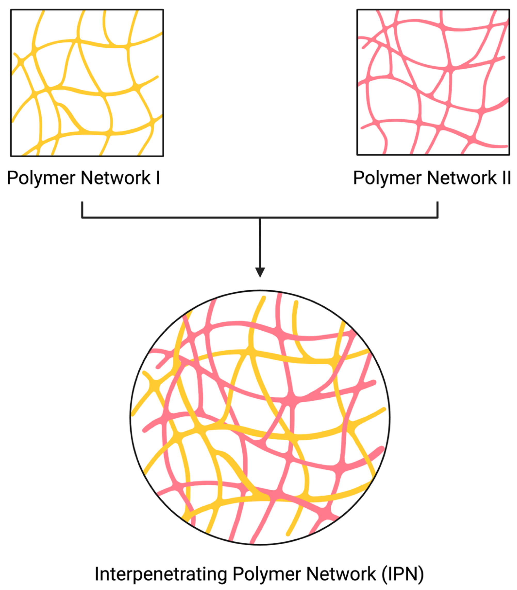
Figure 1. An illustration of IPN structure (made by BioRender.com).
Polymers 16 02050 g001
Figure 2. Physical forces that cause entanglements in IPNs (made by BioRender.com).
Polymers 16 02050 g002
Figure 3. Different hydrogel forms of IPNs (made by BioRender.com).
Polymers 16 02050 g003
Figure 4. Characterization methods for IPN structures (made by BioRender.com).
Polymers 16 02050 g004
Figure 5. A general view of a mid-IR spectrum (made by Biorender.com).
Polymers 16 02050 g005
Figure 6. A SEM image illustration of an IPN sample (made by Biorender.com).
Polymers 16 02050 g006
Figure 7. Crystalline and amorphous XRD pattern illustrations (made by Biorender.com).
Polymers 16 02050 g007
Figure 8. A generalized illustration of frequency sweep test graphs (made by Biorender.com).
Polymers 16 02050 g008
Figure 9. Illustration of different intensity bands at hydrogen bond region (made by Biorender.com).
Polymers 16 02050 g009
Table 1. Studies analyzed in this article.
Author/YearMaterialsMaterial FormIPN TypeApplicationPropertiesCharacterization Methods to Determine IPN Structure
Darge et al., 2022 [106]Sodium alginate and sulfobetaine methacrylate (SBMA)MicroneedlesSequential IPNDrug delivery
  • Desired immunochemotherapy agent release
  • Suitable mechanical properties for drug delivery
FTIR
Mechanical Testing
Agnihotri & Aminabhavi, 2005 [107]Gellan gum (GG) and poly(vinyl alcohol) (PVA)MicrospheresIPNControlled drug delivery
of a hypertension drug
  • Enhanced entrapment of drug into networks
  • Smooth microsphere surface
FTIR
DSC
Mechanical Testing
Mohamadnia et al., 2007 [108]Alginate and carrageenanBeadsIPNDrug delivery of a
corticosteroid drug
  • Controllable drug release with pH, temperature and ions
SEM
Bhattacharya et al., 2013 [109]Sodium carboxymethyl cellulose and
Sodium carboxymethyl xanthan gum
(SCMC-SCMXG)
BeadsIPNDrug delivery of an
anti-inflammatory drug
  • Successful drug release in alkaline medium
  • Reduced aftereffect for gastritis
XRD
Wahid et al., 2019 [34]Bacterial cellulose (BC), chitosan (CS)SlurrySemi IPNAntimicrobial applications
  • High mechanical and thermal stability
  • Antibacterial
FTIR
XRD
FESEM
Sampath et al., 2017 [112]Cellulose nanocrystal (CNC) and chitosan (CS)GelSemi IPNBiomedical applications
  • Enhanced mechanical properties
  • pH sensitivity
FTIR
Pan et al., 2018 [110]Sugar bagasse cellulose (SBC), carboxymethylcellulose (CMC) and
poly(N-isopropylacrylamide) (PNIPAm)
GelIPNDrug delivery
  • Sensitivity to pH and temperature
SEM
Tığlı & Gümüşderelioğlu, 2009 [113]Alginate (Alg) and chitosan (CS)-Semi IPNCartilage scaffold
  • Support proliferation
  • Maintain cell functionality
SEM
Fluorescence microscope
Wang et al., 2017 [100]p(N, N-dimethyl- acrylamide-co-4-acryloyldopamine),
p(N, N-dimethylacrylamide-co-3-acrylamidopheynlboronic acid) and
p(N, N-dimethylacrylamide-co-4-acryloyloxybenzaldehyde)
GelSemi IPN3D printing
  • Tunable mechanical properties by pH and solid content
  • Self-healing in both acidic and basic environment
  • Injectable
Rheology
Shariatzadeh et al., 2021 [101]Gelatin methacrylate (GelMA)
and hyaluronic acid (HA)
CryogelSequential IPNSoft tissue engineering
  • Highly porous
  • Hydrophile
  • Shape memory
  • Injectable
FTIR
Cui et al., 2023 [102]Oxidized alginate (OSA)/gelatin (Gel)/
cellulose nanofiber (CNF)
GelSemi IPNBone repair
  • Self-healing
  • Tunable water uptake capacity
  • Tunable degradation and swelling behavior
  • Injectable
FTIR
Mu et al., 2020 [103]Injectable platelet-rich fibrin (iPRF) and
gelatin nanoparticles (GNPs)
GelIPNBone healing
  • Mechanically tough
  • Bioactive
  • Self-healing
  • Degradable
  • Injectable
SEM
Kim et al., 2018 [111]Hyaluronic acid (HA)/poly (N-isopropylacrylamide) (PNIPAM)GelFull IPNTransdermal drug delivery
  • pH and temperature sensitive
Swelling ratio
Zhao et al., 2014 [105]Poly(ethylene glycol) methacrylate (PEGMA)/
N-isopropylacrylamide (NIPAm)/ methacrylated alginate (ALGMA)
GelIPNRelease studies
  • Injectable
  • Temperature and pH sensitive
Rheology
FTIR
Ngwabebhoh et al., 2016 [9]Chitosan and StarchGelSemi IPNWastewater treatment
  • Efficiently elimination of dye from aqueous solution
  • Non-toxic
  • Eco-friendly
FTIR
Disclaimer/Publisher’s Note: The statements, opinions and data contained in all publications are solely those of the individual author(s) and contributor(s) and not of MDPI and/or the editor(s). MDPI and/or the editor(s) disclaim responsibility for any injury to people or property resulting from any ideas, methods, instructions or products referred to in the content.