Article

Tailoring Crystallinity of Electrospun Plla Fibres by Control of Electrospinning Parameters

by
Olubayode Ero-Phillips
,
Mike Jenkins
and
Artemis Stamboulis
*
Biomaterials Group, School of Metallurgy & Materials, College of Engineering and Physical Sciences, University of Birmingham, Birmingham B15 2TT, UK
*
Author to whom correspondence should be addressed.
Polymers 2012, 4(3), 1331-1348; https://doi.org/10.3390/polym4031331
Submission received: 6 June 2012 / Accepted: 20 June 2012 / Published: 28 June 2012

Abstract

:
Poly(L-lactic acid) (PLLA) fibers were fabricated by electrospinning. The effects of various electrospinning process parameters on the thermal properties, especially the crystallinity of the electrospun fibers were investigated. Thermal analysis of the fibers revealed that they exhibited degree of crystallinity ranging from 23% to 46% while that for the as-received granules was approximately 37%, suggesting that the crystallinity of electrospun PLLA fibres can be controlled by optimizing the electrospinning process. This finding is very important because crystallinity affects polymer properties such as degradation, stiffness, yield stress, modulus and tensile strength, solubility, optical and electrical properties which will in turn affect the behavior of these materials when they are utilized in energy, environment, defense and security applications. The results presented in this paper show that the degree of crystallinity of the electrospun fibers decreased with increasing the polymer solution concentration. Furthermore, an optimum electrospinning voltage at which maximum degree of crystallinity can be obtained was observed. At voltages higher or lower than the optimum electrospinning voltage, the degree of crystallinity will decrease or increase, respectively. The effect of the needle tip to collector distance (NTCD) on the degree of crystallinity follows no predictable and consistent pattern.

Graphical Abstract

1. Introduction

Interest in electrospinning has grown exponentially over the last two decades due to the versatility of the technique. Electrospinning is capable of producing fibers with high strength [1] and large surface area-to-volume ratios [2]. An advantage of the process over conventional fiber forming methods is that only a small amount of polymer, as low as 50 mg, is required for electrospinning, in comparison to tens of kilogrammes of polymer required in conventional spinning methods [3]. However, in spite of the amount of research that has been carried out on electrospinning and electrospun fibers, studies on the effect of electrospinning on polymer properties have been limited. Most studies have focused on the effect of electrospinning parameters on electrospun fibre diameter [4,5,6,7] and the development of controlled morphology [8,9,10,11,12]. However, very few have focused on the effect of electrospinning on polymer properties, such as molecular orientation of polymer chains [2], polymer crystallization chain conformation [3] and polymer degradation [3,13].
Although electrospun fibers have potential applications in many different areas, their use as tissue engineering scaffold materials is probably the most popular application of electrospun fibers [14]. Furthermore, because most electrospun fibers are proposed for use in biomedical applications, it is imperative that an understanding of their properties, such as crystallinity and degradation kinetics, which will impact their functionality when in service, be properly developed.
Although non-biodegradable materials may also be used as tissue engineering scaffolds [15,16], biodegradable materials with controllable degradation rates are more commonly considered [17]. Specifically, poly(lactic acid), poly(glycolic acid) (PGA) and their copolymers are the most commonly researched polymers for tissue engineering because of their biocompatibility, bioresorbability, controllable degradation rates, and also because they have been approved by the Food and Drug Administration for biomedical applications [18]. Crystallinity has been reported to vary directly proportionally to the rate of degradation [19]. It is therefore crucial to have an understanding of the effect of electrospinning on polymer crystallinity.
Stephens et al. reported that the crystalline structure of Nylon-6 changed from α- to γ-crystalline structure due to the high stresses present during the electrospinning process, but no information was given about the effect of electrospinning on the degree of polymer crystallinity [3]. Regarding the effect of electrospinning on polymer crystallinity, it has been generally reported that electrospun fibers possess lower crystallinity than the degree of crystallinity of the as-received polymers and films cast from polymer solutions or melts. Bognitzki et al. reported that electrospun PLLA fibers had lower degree of crystallinity than fibers from melt or films from dilute solutions. This was attributed to the rapid deposition rate of the fibers during electrospinning, which allows very little time for crystallization to occur [20]. Zong et al. also reported lower degree of crystallinity in electrospun PLLA fibers as compared to as-received PLLA resin and attributed this also to the rapid solidification of the stretched polymer chains during the electrospinning process [21].
Other researchers have also compared the crystallinity of various electrospun fibers to the crystallinity of the pristine materials which they were electrospun from. Ramdhanie et al. electrospun blends of PLA and PGA to produce both aligned and random fibers [22]. They reported generally lower values of crystallinity [measured by differential scanning calorimetry (DSC)] for the PLA fibers than for the pristine PLA pellets, while some of the PGA electrospun fibers showed an increase in crystallinity. Deitzel et al. reported a wide angle X-ray diffraction (WAXD) study of electrospun polyethylene oxide (PEO) fibers and pristine PEO powder [23,24]. The (120) and (112) reflections of the electrospun fibres were weak and broad in comparison to the sharp peaks obtained for the powder, indicating that the crystalline microstructure of the electrospun fibres was not well developed. In their work on silk-like polymer with fibronectin functionality (SLPF), Buchko et al. also observed lower crystallinity for electrospun fibres than in the pristine material using wide-angle X-ray scattering (WAXS) and attributed this also to the rapid solidification of the electrospun fibres [25]. Ayutsede et al. obtained crystallinities of 48% and 39% for pristine Bombyx mori silk and electrospun fibres, respectively, using WAXD [26]. Similarly, Ribeiro et al. recently reported that electrospun PLLA fibres were nearly amorphous, and they attributed this to the rapid solidification of the electrospinning jet [27].
In the contrary, Zhao et al. suggested, that the degree of crystallinity of electrospun ethyl-cyanoethyl cellulose fibres was greatly influenced by the crystallization time as the jet travels from the capillary tip to the collector and the molecular orientation was brought about by the electrostatic field [28]. Specifically, the degree of crystallinity of the electrospun fibres increased with increasing voltage up to a maximum after which the degree of crystallinity began to decline.
More recently, Kongkhlang et al. reported the electrospinning of random fibres of polyoxymethylene (POM). The authors observed molecular orientation in the electrospun fibres, as well as a chain structure in which fully extended molecules aligned parallel to the needle axis. The presence of molecular orientation in the electrospun fibres was attributed to the substantial stretching forces imposed on the polymer solution by the electric field during the electrospinning process [2].
The high elongation strains and shear stresses that are exerted on polymer chains within the electric field during electrospinning are reported to cause the macromolecular chains to align along the fibre axis, leading to high degree of molecular orientation in the fibres [1,2,3,29,30]. Molecular orientation has been shown to be directly proportional to the degree of crystallinity [1,30]. It is therefore reasonable to suggest, that if electrospinning is able to cause molecular orientation of polymer chains, then it may be possible to influence the degree of crystallinity by carefully controlling the electrospinning parameters.
The crystalline properties of a material are important when considering the material for medical applications because the crystalline properties of a polymer have a direct bearing upon the degradation behavior in vivo [19,31,32,33]. A high or low degradation rate may be desirable, depending on the application of the material. It would therefore be beneficial if the degree of crystallinity of electrospun polymer fibres can be tailored by controlling the electrospinning process/parameters.
Although poly(lactic acid) is one of the most researched biodegradable polymers [34,35], there is no comprehensive systematic study in the literature about the effect of varying different electrospinning parameters on the degree of crystallinity of its electrospun fibres. The previous work on the study of crystallinity of electrospun PLLA in the literature [20,21,36] has all been carried out within a fixed set of parameters which limits their results to the parameters used. In a recent paper by Ribeiro et al. where a wider set of parameters (i.e., voltage and flow rate) was used, the degree of crystallinity of the electrospun fibres was controlled by an additional annealing process after electrospinning. In that case, a higher degree of crystallinity was achieved, but not directly during electrospinning [27]. This paper aims to investigate the possibility of controlling the degree of crystallinity of electrospun PLLA fibres in a single process by varying the electrospinning parameters.

2. Experimental Section

2.1. Materials

PLLA granules of molecular weight ≈220,000 g/mol were supplied by Goodfellow Chemicals, Cambridge, UK. Dichloromethane (DCM) (puriss. p.a., ACS reagent, ≥99.9%) was supplied by Sigma Aldrich, UK, while N,N-Dimethylformamide (DMF) (>99% assay) was supplied by Fisher Chemicals, UK. Four different solutions of 2, 5, 8 and 10 wt% concentration were made by dissolving PLLA granules in a 75:25 solution of Dichloromethane/N,N-Dimethylformamide.

2.2. Methods

2.2.1. Kinematic Viscosity

The kinematic viscosity of the different PLLA-DCM/DMF solutions described above was obtained using an Ostwald U-tube viscometer (Cannon Instrument Company, USA) enclosed in a water bath at 25 °C. The viscosities were calculated using the following formula supplied by the manufacturer
Polymers 04 01331 i001
Where v = kinematic viscosity (cSt), t = efflux time (s), and k = viscometer constant (0.01 cSt/s at 25 °C).

2.2.2. Electrospinning

The polymer solutions were electrospun using a Model ES1a electrospinning machine manufactured by Electrospinz Ltd., New Zealand. The electrospinning apparatus is shown in Figure 1. A static aluminium foil target was used to collect the non-woven electrospun fibres. A systematic variation of three electrospinning parameters, namely: polymer solution concentration, applied voltage and needle tip to collector distance (NTCD) was conducted. Briefly, during each electrospinning experiment, two of these parameters were held constant while the third one was varied. The electrospinning apparatus has been designed by the manufacturers to function with a gravity feed system which ensures the internal pressure of the droplet at the needle tip is constant [37]. Therefore, the system does not require a syringe pump to control the flow rate. The electrospinning was carried out at room temperature. The electrospun fibres were stored in a dessicator for one week to allow for complete evaporation of solvent. The 2 and 10 wt% solutions could not be electrospun under all varied electrospinning parameters due to their low and high viscosities, respectively (details discussed in Section 3.1). Additionally, a low yield of fibres from the 5 wt% and 8 wt% solution concentrations was also observed using 10 kV applied voltage, showing that 10 kV was too low for successful electrospinning of the polymer solution.
Figure 1. Schematic representation of ES1a electrospinning machine.
Polymers 04 01331 g001

2.2.3. Characterisation of Electrospun Polymer Fibres

The morphology of the electrospun fibres was obtained using a Philips XL-30 FEG SEM (SE mode) at an accelerating voltage of 20 kV. Four different specimens were cut from each sample and then mounted on an aluminium stub using a carbon adhesive film. The specimens were sputter coated with platinum using an Emscope SC500 (Emscope laboratories, Kent, UK) Sputter Coater at a current of 25 mA for 3 min.
A Perkin Elmer Differential Scanning Calorimeter (DSC 7) was used for the thermal analysis of the elctrospun polymer fibres. Approximately 6 mg of samples were crimped into standard DSC aluminium pans. The fibres were taken through a temperature scan of 25 °C to 200 °C at a heating rate of 10 °C/min, held at 200 °C for 2 min, followed by cooling to 25 °C at a cooling rate of 10 °C/min. With the aid of the Pyris™ software (Perkin Elmer) installed on the DSC equipment, the glass transition temperature (Tg) was calculated from the endothermic transition relating to the glass transition on DSC scans using the half-Cp (heat capacity) extrapolated method.
The values for the degree of crystallinity were calculated using the formula [38,39,40] below:
Polymers 04 01331 i002
where Xc (%) = Degree of crystallinity (%), Polymers 04 01331 i003 = Heat of fusion (J·g−1), Polymers 04 01331 i004 = Heat of cold crystallization (J·g−1) and, Polymers 04 01331 i005 = Heat of fusion for 100% crystalline material (93.6 J·g−1) [39].

3. Results and Discussion

3.1. Effect of Polymer Solution Parameters and Processing Conditions on the Electrospinning Process

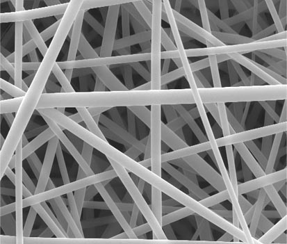
Several attempts were made to electrospin fibres from the 2 wt% PLLA solution (kinematic viscosity of 2.22 cSt), however a mixture of electrospraying and electrospinning was observed (Figure 2A). The lower polymer solution concentration resulted in a lower polymer chain entanglement density [41], which caused the chains to break up at the tip of the capillary due to the high surface tension [24]. Fibres, as well as some beads were obtained when the polymer solution concentration was increased to 5 wt% (Figure 2B), however, when the polymer solution concentration was increased to 8 wt%, bead-free fibres were obtained (Figure 2C). The 10 wt% polymer solution was too viscous with a kinematic viscosity of 142.57 cSt (Figure 3) and it was difficult to electrospin, even at very high electrospinning voltages.
Figure 2. SEM micrographs of electrospun polymer fibres obtained when electrospinning from (a) 2 wt%; (b) 5 wt%; and (c) 8 wt% polymer solutions at 20 kV and NCTD of 12.5 cm, respectively.
Polymers 04 01331 g002
The polymer solution concentration has a direct bearing upon the number of polymer chain entanglements within the solution and is one of the major factors which determine if fibres can be formed during electrospinning. As has been previously reported in the literature, there is an optimum range of polymer solution concentration within which uniform fibres can be electrospun when all other parameters are held constant [42], and in our study, this range of polymer solution concentration was found to between >2 wt% and <10 wt%.
When electrospinning was carried out at 10 kV, the amount of fibres produced was very small. The voltage applied during electrospinning induced charges into the polymer solution/melt and it was when the electrostatic forces created by these charges overcame the surface tension of the solution that electrospinning occurred. Hence, when the applied voltage was too low, the electrostatic forces were too low to initiate electrospinning. Similar to polymer solution concentration, the applied voltage has been shown to have a window within which electrospinning can be successful [4]. As mentioned above, electrospinning at 10 kV was not possible and therefore this is considered to be the minimum possible value.
Figure 3 shows the variation of viscosity with polymer solution concentration. The viscosity of the polymer solution increased with increasing polymer solution concentration as a result of increased number of polymer chain entanglements [41]. The increase in viscosity is dependent on polymer solution concentration [43].
Figure 3. Variation of kinematic viscosity with polymer solution concentration (where ν is kinematic viscosity and C is polymer solution concentration).
Polymers 04 01331 g003

3.2. Effect of Electrospinning on the Glass Transition Temperature (Tg)

The glass transition temperature, Tg, of the electrospun fibres under any of the experimental parameters and for all the polymer solutions was lower than that of the as-received granules (67 °C) and ranged from 52 °C to 63 °C (Table 1). Varying the voltage or the NTCD had no effect on the Tg of the electrospun fibres. The Tg generally increased when the polymer concentration was increased from 5 wt % to 8 wt%; this was due to increased polymer chain interaction, but the Tg remained lower compared to the as-received polymer. A decrease in Tg values has previously been also reported for electrospun fibres [21,44,45]. Zong et al. attributed the decrease in Tg in electrospun PLLA fibres to the plasticisation of the nanofibres by air and the large surface to volume ratio [21]. Similarly, Carrizales et al. reported a decrease of 20 °C for Tg of electrospun poly(methyl methacrylate) (PMMA) nanofibres during the 1st heating cycle of a DSC study compared to the Tg of the as-received PMMA powder. However, during the 2nd heating cycle of the electrospun nanofibres, it was reported that the Tg of the electrospun nanofibres was equal to that of the as-received PMMA powder. Carrizales et al. attributed this reduction in the Tg values of electrospun PMMA nanofibres to the plasticisation of the fibres by absorbed water, although their work was not clear about the source of the absorbed water [46]. We did not expect this to be the case in our studies because the electrospun fibres were properly dried prior to DSC characterisation. In addition, the glass transition temperatures measured during the 2nd DSC cycle (Table 1) were not much different to the 1st DSC cycle. Unfortunately, it is not clear from this work and the literature why there is a reduction in Tg after epectrospinning. Zong et al., Kim et al. and Cui et al. suggested that the reduction in Tg is possibly due to three factors: 1. High surface area [21], 2. Increase in segmental mobility as a result of decreased chain entanglements [13], and 3. Higher molecular orientation and stress within polymer chains [44]. It is possible that it is not only one reason that causes the reduction in Tg. From our results it is clear that the Tg increased with increasing the polymer concentration. This could possibly be related to segmental mobility however, this is still only a speculation.

3.3. Effect of Polymer Solution Concentration on Degree of Crystallinity

Figure 4 presents DSC scans of polymer fibres electrospun from 5 wt% and 8 wt% PLLA solutions at 20 kV and 10 cm NTCD. Just above the Tg, a hysteresis endothermic peak was observed (Figure 4) characteristic of volume relaxation due to the rapid structure formation of the fibres during electrospinning as has also been reported by Bognitzki et al. [20]. An exothermic peak observed around 75 °C was associated with “cold crystallization”. Cold crystallization has been reported the literature [21,36]. Cold crystallization is a transition that occurs at low temperature due to enhanced primary nucleation when heating the polymer above its glass transition temperature when polymer chains have enough energy to become mobile and achieve an ordered state via an exothermic reaction [22,40]. The occurrence and extent of this reaction is dependent on the processing history of the sample. Mezghani and Spruiell reported that generally, a crystalline or semi-crystalline polymer will have a lower tendency to undergo this transition than a less crystalline or amorphous polymer [38] and this will be reflected by a small ∆Hc for the crystalline/semi-crystalline polymer and a large ∆Hc for an amorphous polymer. In this study, the cold crystallization enthalpy (∆Hc), increased from 2.5 J·g−1 to 18.3 J·g−1 when the polymer solution concentration was increased from 5 wt% to 8 wt% and voltage and NTCD were held constant at 15 kV and 10 cm, respectively (Figure 4).
At low polymer solution concentrations, the polymer chains are more mobile due to relatively small number of chain entanglements resulting in higher molecular orientation during electrospinning compared to fibres resulting from higher solution concentrations. Also, the solidification process is expected to be slower for a low polymer solution concentration resulting in higher electric-field assisted molecular orientation during electrospinning [36]. Therefore, it can be said that low polymer concentration solutions can yield fibres with higher molecular orientation and most likely lower degree of cold crystallization. Using Equation 1 therefore the degree of crystallinity after electrospinning is expected to be higher. Conversely, higher polymer concentration solutions can yield fibres with reduced molecular orientation due to higher viscosity and most likely higher degree of cold crystallization and therefore generally lower degree of crystallinity. Based on the above explanation the reduction in the degree of crystallinity with increasing polymer solution concentration can be justified. However, in the exceptional case of fibers electrospun from the 8 wt% polymer solution concentration at a voltage of 25 kV, the degree of crystallinity increased compared to the fibers electrospun from the 5 wt% polymer concentration solution (Table 1). The reason for this will be discussed in Section 3.4.
Figure 4. (a) DSC scans of fibers electrospun from 5 wt% and 8 wt% solutions at 15 kV and 10 cm needle tip to collector distance (NTCD); (b) Cold crystallization region of scans shown in A.
Polymers 04 01331 g004
Table 1. Variation of Tg and Xc with electrospinning parameters. (n/a–not applicable; Xc was calculated during 1st heating cycle).
PLLA electrospinning solutionElectrospinning parametersTg (°C)Xc (%)PLLA electrospinning solutionElectrospinning parametersTg (°C)Xc (%)
NTCD (cm)Voltage (kV)1st heating cycle2nd heating cycleNTCD (cm)Voltage (kV)1st heating cycle2nd heating cycle
Granulen/an/a67n/a37.4n/an/an/a---
5%7.515525645.38%7.515616137.3
20545445.120646143.8
25525444.825636146.3
1015535443.91015606130.5
20525542.220606136.7
25535437.925626043.9
12.515555445.512.515606133.4
20535543.720586136.9
25545542.425646146.6
1515525546.61515606131.2
20555428.420606134.7
25565426.225636145.8

3.4. Effect of Voltage during Electrospinning on the Degree of Crystallinity

As shown in Figure 5, the degree of crystallinity of the fibers electrospun from the 5 wt% PLLA solution generally decreased with increasing voltage from 15 kV to 25 kV albeit in some cases, the reduction was only minimal. For instance, when the voltage was increased from 15 kV to 25 kV at a constant NTCD of 10 cm, the cold crystallization enthalpy, ∆Hc, increased from 2.5 J·g−1 to 6.7 J·g−1 (Figure 6). Consequently, the degree of crystallinity, Xc, calculated using Equation 1, decreased with increasing the voltage from 15 to 25 kV (Figure 5). On the contrary, the degree of crystallinity for the fibers electrospun from the 8 wt% solution concentration increased as the voltage was increased from 15 kV to 25 kV (Figure 7).
From Figure 5 and Figure 7, it is obvious that there was a voltage at which a maximum degree of crystallinity was obtained. We have called this voltage the optimum electrospinning voltage. For the fibers electrospun from the 5 wt% solution concentration, the maximum degree of crystallinity was obtained when electrospinning at 15 kV, while for the fibers electrospun from the 8 wt% solution concentration, the maximum degree of crystallinity was obtained when electrospinning at 25 kV.
Figure 5. Effect of voltage on the degree of crystallinity of fibers electrospun from 5 wt% solution concentration at different NTCDs.
Polymers 04 01331 g005
Figure 6. (a) DSC scans of fibers electrospun from 5 wt% solution at a constant NTCD of 10 cm and varying voltages; (b) Cold crystallization region of scans shown in A.
Polymers 04 01331 g006
Figure 7. Effect of voltage on the degree of crystallinity of fibers electrospun from 8 wt% solution concentration at different NTCDs.
Polymers 04 01331 g007
During the electrospinning process, as the jet travels from the needle tip to the collector, both molecular orientation induced by the electric field, and crystallization of the polymer occur [28]. Also, as the voltage is increased during electrospinning, the number of charges in the polymer solution increases and the effect of this is that there will be more stretching of the jet [4]. Furthermore, increasing the voltage will lead to an increase in the alignment of the polymer chains (i.e., molecular orientation) during electrospinning and consequently an increase in the degree of crystallinity of the electrospun fibers as reported by Zhao et al. [28]. However, this increase in the degree of crystallinity with an increase in electrospinning voltage is not expected to continue indefinitely, on the contrary, above a certain voltage (the optimum electrospinning voltage), the degree of crystallinity will reduce. This is because the degree of crystallinity is also influenced by the flight time of the jet, which at high voltages is short enough to leave insufficient time for the polymer to crystallize [47]. Increasing the voltage beyond the optimum electrospinning voltage will lead to a reduction in the flight time due to the higher acceleration of the jet, resulting in decreased crystallinity [28,47]. Generally, the degree of crystallinity will increase with increasing electrospinning voltage until the optimum electrospinning voltage is reached; above the optimum electrospinning voltage, the degree of crystallinity is reduced due to the effect of the acceleration imposed on the spun fibers by the electric field.
For the fibers electrospun from the 5 wt% solution concentration, as the maximum degree of crystallinity (Xc) was obtained at 15 kV, after which Xc began to decrease, it is likely that if a voltage lower than 15 kV was used to electrospin the polymer solution, the degree of crystallinity would increase with voltage up to a maximum at circa 15 kV. Above this voltage, reduction of the flight time of the jet occurs with a consequent decrease in the degree of crystallinity. It was attempted to electrospin the polymer solutions at lower voltages than 15 kV but this was not possible as has been explained in the Experimental Section. Consequently, it is believed that the range of voltages used in this study may have been above the optimum electrospinning voltage. Within this range, it is likely that increases in voltage will lead to increased acceleration of the jet to the extent that it reduces its flight time; the result of this is that there was insufficient time for crystallization and hence lower degree of crystallinity was observed beyond 15 kV.
For the fibers electrospun from the 8 wt% solution concentration, the degree of crystallinity reached a maximum at 25 kV when the voltage was increased from 15 kV to 25 kV (Figure 7). This suggests that the optimum voltage at which maximum degree of crystallinity can be obtained is at or close to 25 kV. It is therefore believed that the range of voltages used in this study fall below the optimum electrospinning voltage. It is very likely that further increases in voltage beyond 25 kV will lead to a reduction in the degree of crystallinity.
It was interesting to note, however, that the optimum voltage at which maximum degree of crystallinity can be obtained for fibers electrospun from the 5 wt% solution concentration was lower than that for fibers electrospun from 8 wt% solution concentration, i.e., 15 kV and 25 kV for the 5 wt% and 8 wt% solutions, respectively. As has been explained in the previous section, the polymer chains are more mobile in polymer solutions of low concentration; it is therefore believed, that this will make them more susceptible to influences of voltage at low voltage values. As the polymer solution concentration is increased however, there are more polymer chain entanglements, hence higher solution viscosity, resulting in the polymer chains exhibiting a higher resistance to stretching [48] during electrospinning. As a result, the probability of the electric field inducing molecular orientation in the electrospun fibers at low voltages will be small, except at higher voltages (in this study, this was observed to be approximately ≥ 25 kV).
A comparison of the degree of crystallinity of fibers electrospun from the 8 wt% solution at 25 kV with the degree of crystallinity of fibers electrospun from the 5 wt% solution at the same voltage shows that the degree of crystallinity was slightly higher for the first case. As explained above, at voltages above the optimum voltage at which maximum degree of crystallinity can be obtained the degree of crystallinity of the fibers will decrease. As the optimum voltage for fibers electrospun from the 5 wt% solution was at or close to 15 kV, any further increase in voltage will lead to a reduction in the flight time of the jet and consequently a decrease in the degree of crystallinity. However, for the fibers electrospun from the 8 wt% solution, because the optimum voltage was at circa 25 kV, the maximum degree of crystallinity was observed at this voltage.

3.5. Effect of NTCD on the Degree of Crystallinity

Ideally, increasing the NTCD should result in an increase in the flight time of the jet [4]. Since molecular orientation can be induced by the electric field as the jet travels from the needle tip to the collector, if the flight time of the jet is increased by increasing the NTCD [28], theoretically, it would be reasonable to suggest that the molecular orientation of the electrospun fibers could increase with a consequent increase in the degree of crystallinity. The recent work of Tong and Wong however, show that increasing the NTCD may not necessarily increase the flight time of the jet due to the complicated three-dimensional spiralling trajectory of the jet [4]. In this study, for fibers electrospun from the 5 wt% solution, no constant trend was observed in the variation of the degree of crystallinity (Xc) with changing NTCD, suggesting that the NTCD influences the degree of crystallinity of the electrospun fibers but not in a predictable and consistent way (Figure 5). Although the reason for this is not particularly clear, we suggest that it could be due to the inconsistency of trajectory of the polymer jet as Tong and Wang suggested [4]. However, it is interesting to note, that for the fibers electrospun from the 5 wt% solutions, at 15 kV which was the optimum electrospinning voltage for this solution, the degree of crystallinity remained fairly constant in the whole range of NTCD used in this study suggesting that the degree of crystallinity is not affected by variations in NTCD when electrospinning at or close to the optimum voltage. Similar observations as above were also made for fibers electrospun from the 8 wt% solution concentration (Figure 7). This has not been suggested before in the literature and therefore it is not possible to compare with any other similar example, however this is an indication that the acceleration voltage has a main role in the jet trajectory perhaps more important than the NTCD.

4. Conclusions

PLLA polymer fibers were electrospun from two different polymer solution concentrations, i.e., 5 wt% and 8 wt%, at varying voltages and NTCDs using an ES1a electrospinning machine manufactured by Electrospinz Ltd, New Zealand. The thermal characterisation as conducted by DSC revealed that the electrospun fibers exhibited crystallinities ranging from 23% to 46% while that for the as-received granules was approximately 37%. This suggests that by optimizing the solution and processing parameters during the electrospinning process, it may be possible to tailor the degree of crystallinity of PLLA. This can be a very important factor not only when considering an electrospun material for biomedical applications, but in other areas as well, because crystallinity affects polymer properties such as stiffness, yield stress, modulus and tensile strength, solubility, optical and electrical properties [49,50] which will in turn affect the behaviour of these materials when they are utilized in energy, environment, defence and security applications.
It was also observed that factors such as polymer solution concentration, electrospinning voltage and NTCD influenced the degree of crystallinity of the electrospun fibers. More specifically, the degree of crystallinity decreased with increasing the polymer solution concentration. In addition, an optimum electrospinning voltage at which maximum degree of crystallinity can be obtained was observed. At voltages higher or lower than the optimum electrospinning voltage, the degree of crystallinity will decrease or increase, respectively. It is believed that each polymer solution concentration would exhibit its individual optimum electrospinning voltage. The effect of the NTCD on the degree of crystallinity followed no predictable and consistent pattern, possibly due to inconsistency of trajectory of the polymer jet. However, it was interesting to observe that the degree of crystallinity was always maximum irrespective of the NTCD when electrospinning at the optimum electrospinning voltage.
In summary, in order to tailor the crystallinity of electrospun fibers, the polymer solution concentration and voltage should be carefully controlled.

References

  1. Baji, A.; Mai, Y.W.; Wong, S.C.; Abtahi, M.; Chen, P. Electrospinning of polymer nanofibers: Effects on oriented morphology, structures and tensile properties. Compos. Sci. Technol. 2010, 70, 703–718. [Google Scholar] [CrossRef]
  2. Kongkhlang, T.; Tashiro, K.; Kotaki, M.; Chirachanchai, S. Electrospinning as a new technique to control the crystal morphology and molecular orientation of polyoxymethylene nanofibers. J. Am. Chem. Soc. 2008, 130, 15460–15466. [Google Scholar] [CrossRef]
  3. Stephens, J.S.; Chase, D.B.; Rabolt, J.F. Effect of the electrospinning process on polymer crystallization chain conformation in nylon-6 and nylon-12. Macromolecules 2004, 37, 877–881. [Google Scholar] [CrossRef]
  4. Tong, H.W.; Wang, M. An investigation into the influence of electrospinning parameters on the diameter and alignment of poly(hydroxybutyrate-co-hydroxyvalerate) fibers. J. Appl. Polym. Sci. 2011, 120, 1694–1706. [Google Scholar] [CrossRef]
  5. Boland, E.D.; Wnek, G.E.; Simpson, D.G.; Pawlowski, K.J.; Bowlin, G.L. Tailoring tissue engineering scaffolds using electrostatic processing techniques: A study of poly(glycolic acid) electrospinning. J. Macromol. Sci. Pure Appl. Chem. 2001, 38, 1231–1243. [Google Scholar] [CrossRef]
  6. Sukigara, S.; Gandhi, M.; Ayutsede, J.; Micklus, M.; Ko, F. Regeneration of bombyx mori silk by electrospinning-part 1: Processing parameters and geometric properties. Polymer 2003, 44, 5721–5727. [Google Scholar] [CrossRef]
  7. Tan, S.H.; Inai, R.; Kotaki, M.; Ramakrishna, S. Systematic parameter study for ultra-fine fiber fabrication via electrospinning process. Polymer 2005, 46, 6128–6134. [Google Scholar] [CrossRef]
  8. He, W.; Ma, Z.W.; Teo, W.E.; Dong, Y.X.; Robless, P.A.; Lim, T.C.; Ramakrishna, S. Tubular nanofiber scaffolds for tissue engineered small-diameter vascular grafts. J. Biomed. Mater. Res. A 2009, 90A, 205–216. [Google Scholar] [CrossRef]
  9. Katta, P.; Alessandro, M.; Ramsier, R.D.; Chase, G.G. Continuous electrospinning of aligned polymer nanofibers onto a wire drum collector. Nano Lett. 2004, 4, 2215–2218. [Google Scholar] [CrossRef]
  10. Kidoaki, S.; Kwon, I.K.; Matsuda, T. Mesoscopic spatial designs of nano- and microfiber meshes for tissue-engineering matrix and scaffold based on newly devised multilayering and mixing electrospinning techniques. Biomaterials 2005, 26, 37–46. [Google Scholar] [CrossRef]
  11. Saraf, A.; Lozier, G.; Haesslein, A.; Kasper, F.K.; Raphael, R.M.; Baggett, L.S.; Mikos, A.G. Fabrication of nonwoven coaxial fiber meshes by electrospinning. Tissue Eng. C Methods 2009, 15, 333–344. [Google Scholar] [CrossRef]
  12. Sun, Z.C.; Zussman, E.; Yarin, A.L.; Wendorff, J.H.; Greiner, A. Compound core-shell polymer nanofibers by co-electrospinning. Adv. Mater. 2003, 15, 1929–1932. [Google Scholar] [CrossRef]
  13. Kim, J.S.; Lee, D.S. Thermal properties of electrospun polyesters. Polym. J. 2000, 32, 616–618. [Google Scholar] [CrossRef]
  14. Zhang, Y.Z.; Su, B.; Venugopal, J.; Ramakrishna, S.; Lim, C.T. Biomimetic and bioactive nanofibrous scaffolds from electrospun composite nanofibers. Int. J. Nanomed. 2007, 2, 623–638. [Google Scholar]
  15. Jagur-Grodzinski, J. Polymers for tissue engineering, medical devices, and regenerative medicine. Concise general review of recent studies. Polym. Adv. Technol. 2006, 17, 395–418. [Google Scholar] [CrossRef]
  16. Vacanti, J.; Vacanti, C.A. The History and Scope of Tissue Engineering. In Principles of Tissue Engineering, 3rd; Lanza, R., Langer, R., Vacanti, J., Eds.; Elsevier Academic Press: Burlington, VT, USA, 2007; pp. 1–6. [Google Scholar]
  17. Ma, P.X. Scaffolds for tissue fabrication. Mater. Today 2004, 7, 30–40. [Google Scholar]
  18. Jones, J.R. Scaffolds for Tissue Engineering. In Biomaterials, Artificial Organs and Tissue Engineering; Hench, L.L., Jones, J.R., Eds.; Woodhead Publishing Limited: Cambridge, UK, 2005; pp. 201–213. [Google Scholar]
  19. Middleton, J.C.; Tipton, A.J. Synthetic biodegradable polymers as orthopedic devices. Biomaterials 2000, 21, 2335–2346. [Google Scholar] [CrossRef]
  20. Bognitzki, M.; Czado, W.; Frese, T.; Schaper, A.; Hellwig, M.; Steinhart, M.; Greiner, A.; Wendorff, J.H. Nanostructured fibers via electrospinning. Adv. Mater. 2001, 13, 70–72. [Google Scholar] [CrossRef]
  21. Zong, X.H.; Kim, K.; Fang, D.F.; Ran, S.F.; Hsiao, B.S.; Chu, B. Structure and process relationship of electrospun bioabsorbable nanofiber membranes. Polymer 2002, 43, 4403–4412. [Google Scholar] [CrossRef]
  22. Ramdhanie, L.I.; Aubuchon, S.R.; Boland, E.D.; Knapp, D.C.; Barnes, C.P.; Simpson, D.G.; Wnek, G.E.; Bowlin, G.L. Thermal and mechanical characterization of electrospun blends of poly(lactic acid) and poly(glycolic acid). Polym. J. 2006, 38, 1137–1145. [Google Scholar] [CrossRef]
  23. Deitzel, J.M.; Kleinmeyer, J.D.; Hirvonen, J.K.; Beck Tan, N.C. Controlled deposition of electrospun poly(ethylene oxide) fibers. Polymer 2001, 42, 8163–8170. [Google Scholar] [CrossRef]
  24. Deitzel, J.M.; Kleinmeyer, J.; Harris, D.; Beck Tan, N.C. The effect of processing variables on the morphology of electrospun nanofibers and textiles. Polymer 2001, 42, 261–272. [Google Scholar]
  25. Buchko, C.J.; Chen, L.C.; Shen, Y.; Martin, D.C. Processing and microstructural characterization of porous biocompatible protein polymer thin films. Polymer 1999, 40, 7397–7407. [Google Scholar]
  26. Ayutsede, J.; Gandhi, M.; Sukigara, S.; Micklus, M.; Chen, H.-E.; Ko, F. Regeneration of bombyx mori silk by electrospinning. Part 3: Characterization of electrospun nonwoven mat. Polymer 2005, 46, 1625–1634. [Google Scholar]
  27. Ribeiro, C.; Sencadas, V.; Miguel Costa, C.; Gomez Ribelles, J.L.; Lanceros-Mendez, S. Tailoring the morphology and crystallinity of poly(L-lactide acid) electrospun membranes. Sci. Technol. Adv. Mater. 2011, 12, 015001:1–015001:9. [Google Scholar]
  28. Zhao, S.; Wu, X.; Wang, L.; Huang, Y. Electrospinning of ethyl-cyanoethyl cellulose/tetrahydrofuran solutions. J. Appl. Polym. Sci. 2004, 91, 242–246. [Google Scholar] [CrossRef]
  29. Arinstein, A.; Burman, M.; Gendelman, O.; Zussman, E. Effect of supramolecular structure on polymer nanofibre elasticity. Nat. Nanotechnol. 2007, 2, 59–62. [Google Scholar]
  30. Wong, S.C.; Baji, A.; Leng, S.W. Effect of fiber diameter on tensile properties of electrospun poly(epsilon-caprolactone). Polymer 2008, 49, 4713–4722. [Google Scholar] [CrossRef]
  31. Kister, G.; Cassanas, G.; Bergounhon, M.; Hoarau, D.; Vert, M. Structural characterization and hydrolytic degradation of solid copolymers of D,L-lactide-co-epsilon-caprolactone by raman spectroscopy. Polymer 2000, 41, 925–932. [Google Scholar] [CrossRef]
  32. Dong, Y.X.; Liao, S.; Ngiam, M.; Chan, C.K.; Ramakrishna, S. Degradation behaviors of electrospun resorbable polyester nanofibers. Tissue Eng. B Rev. 2009, 15, 333–351. [Google Scholar] [CrossRef]
  33. Hollinger, J.O.; Battistone, G.C. Biodegradable bone repair materials-synthetic-polymers and ceramics. Clin. Orthop. Relat. Res. 1986, 290–305. [Google Scholar]
  34. Gomes, M.E.; Reis, R.L. Biodegradable polymers and composites in biomedical applications: From catgut to tissue engineering—part 1—available systems and their properties. Int. Mater. Rev. 2004, 49, 261–273. [Google Scholar] [CrossRef]
  35. Rasal, R.M.; Janorkar, A.V.; Hirt, D.E. Poly(lactic acid) modifications. Prog. Polym. Sci. 2010, 35, 338–356. [Google Scholar] [CrossRef]
  36. Inai, R.; Kotaki, M.; Ramakrishna, S. Structure and properties of electrospun plla single nanofibres. Nanotechnology 2005, 16, 208–213. [Google Scholar] [CrossRef]
  37. Electrospinz. Frequently Asked Questions. 2008. Available online: http://www.electrospinz.co.nz/faq.php (accessed on 12 January 2012).
  38. Mezghani, K.; Spruiell, J.E. High speed melt spinning of poly(L-lactic acid) filaments. J. Polym. Sci. B Polym. Phys. 1998, 36, 1005–1012. [Google Scholar] [CrossRef]
  39. Fambri, L.; Pegoretti, A.; Fenner, R.; Incardona, S.D.; Migliaresi, C. Biodegradable fibres of poly(l-lactic acid) produced by melt spinning. Polymer 1997, 38, 79–85. [Google Scholar]
  40. Wunderlich, B. Thermal Analysis of Polymeric Materials; Springer: Berlin/Heidelberg, Germany, 2005. [Google Scholar]
  41. Shenoy, S.L.; Bates, W.D.; Frisch, H.L.; Wnek, G.E. Role of chain entanglements on fiber formation during electrospinning of polymer solutions: Good solvent, non-specific polymer-polymer interaction limit. Polymer 2005, 46, 3372–3384. [Google Scholar]
  42. Sill, T.J.; von Recum, H.A. Electro spinning: Applications in drug delivery and tissue engineering. Biomaterials 2008, 29, 1989–2006. [Google Scholar] [CrossRef]
  43. Young, R.J. Introduction to Polymers; Chapman and Hall: New York, NY, USA, 1981. [Google Scholar]
  44. Cui, W.G.; Li, X.H.; Zhu, X.L.; Yu, G.; Zhou, S.B.; Weng, J. Investigation of drug release and matrix degradation of electrospun poly(D,L-lactide) fibers with paracetanol inoculation. Biomacromolecules 2006, 7, 1623–1629. [Google Scholar] [CrossRef]
  45. Zong, X.H.; Ran, S.F.; Kim, K.S.; Fang, D.F.; Hsiao, B.S.; Chu, B. Structure and morphology changes during in vitro degradation of electrospun poly(glycolide-co-lactide) nanofiber membrane. Biomacromolecules 2003, 4, 416–423. [Google Scholar] [CrossRef]
  46. Carrizales, C.; Pelfrey, S.; Rincon, R.; Eubanks, T.M.; Kuang, A.X.; McClure, M.J.; Bowlin, G.L.; Macossay, J. Thermal and mechanical properties of electrospun PMMA, PVC, Nylon 6, and Nylon 6,6. Polym. Adv. Technol. 2008, 19, 124–130. [Google Scholar] [CrossRef]
  47. Ramakrishna, S.; Fujihara, K.; Teo, W.; Lim, T.; Ma, Z. An Introduction to Electrospinning and Nanofibres; World Scientific Publishing Co. Ltd.: Singapore, 2005. [Google Scholar]
  48. Jarusuwannapoom, T.; Hongrojjanawiwat, W.; Jitjaicham, S.; Wannatong, L.; Nithitanakul, M.; Pattamaprom, C.; Koombhongse, P.; Rangkupan, R.; Supaphol, P. Effect of solvents on electro-spinnability of polystyrene solutions and morphological appearance of resulting electrospun polystyrene fibers. Eur. Polym. J. 2005, 41, 409–421. [Google Scholar] [CrossRef]
  49. Billmeyer, F.W. Textbook of Polymer Science, 3rd ed; John Wiley & Sons, Inc.: Singapore, 1984. [Google Scholar]
  50. Hou, X.X.; Yang, X.P.; Zhang, L.Q.; Waclawik, E.; Wu, S.Z. Stretching-induced crystallinity and orientation to improve the mechanical properties of electrospun pan nanocomposites. Mater. Des. 2010, 31, 1726–1730. [Google Scholar] [CrossRef]