1. Introduction
Hypericin (Hyp) is one of the principal bioactive components of the St. John’s Wort plant (
Hypericum perforatum). Traditionally used as anti-bacterial, anti-inflammatory, anti-depressive, and anti-virus agents, hypericin has gained increasing interest recently due to its potential as a highly effective anti-tumor photosensitizer [
1]. Though hypericin can be obtained via chemical synthesis [
2], extraction from the plant itself is still an indispensable supply pathway. However, hypericin exists in a very low concentration with structural similar analogs (such as pseudohypericin) in the plant [
3]. This, together with its poor solubility and degradation upon exposure to heat and light, leaves its enrichment and purification from the plant a great challenge. This greatly inhibits the wide applications of hypericin in pharmaceutical fields. Establishing a facile method for producing efficient separation sorbents with high specificity and adsorption capacity towards hypericin is of significance and much desired [
4].
Molecular imprinting [
5,
6] is a well-known modern technology for the production of nanostructured materials capable of molecular recognition, with high selectivity and good chemical stability at a low cost. Surface molecularly imprinted polymers (SMIPs) [
7,
8] are synthesized by allowing polymerization to take place on substrate surface, in order to create recognition sites on/near the material surface. These recognition sites, alongside the MIPs advantages mentioned above, provide faster rebinding kinetics for the target molecules, and are effective for the separation and enrichment of natural bio-compounds, making this an attractive solution to the challenge [
9]. A variety of materials, including graphene [
10], carbon nanotubes [
11], silicon [
12], silica particles [
13], magnetic nanoparticles [
14,
15], inorganic material, and chips [
16], have been used as substrates for construction of SMIPs. Among these, magnetic nanospheres (MNSs) are one of the most popular substrates for the synthesis of core-shell structured SMIPs [
17]. Besides possessing advantages, such as high surface-to-volume ratio, low cost, regular shape with controllable size, and good mechanical stability, more significantly, their superparamagnetic property endows the final SMIPs facile magnetic response separation. This translates to potential applications in bio-imaging, drug delivery and therapeutics where the template is a drug [
18], such as hypericin. However, to the best of our knowledge, at present there are no reports of core-shell structured SMIP nanospheres based on MNSs for specific recognition of hypericin.
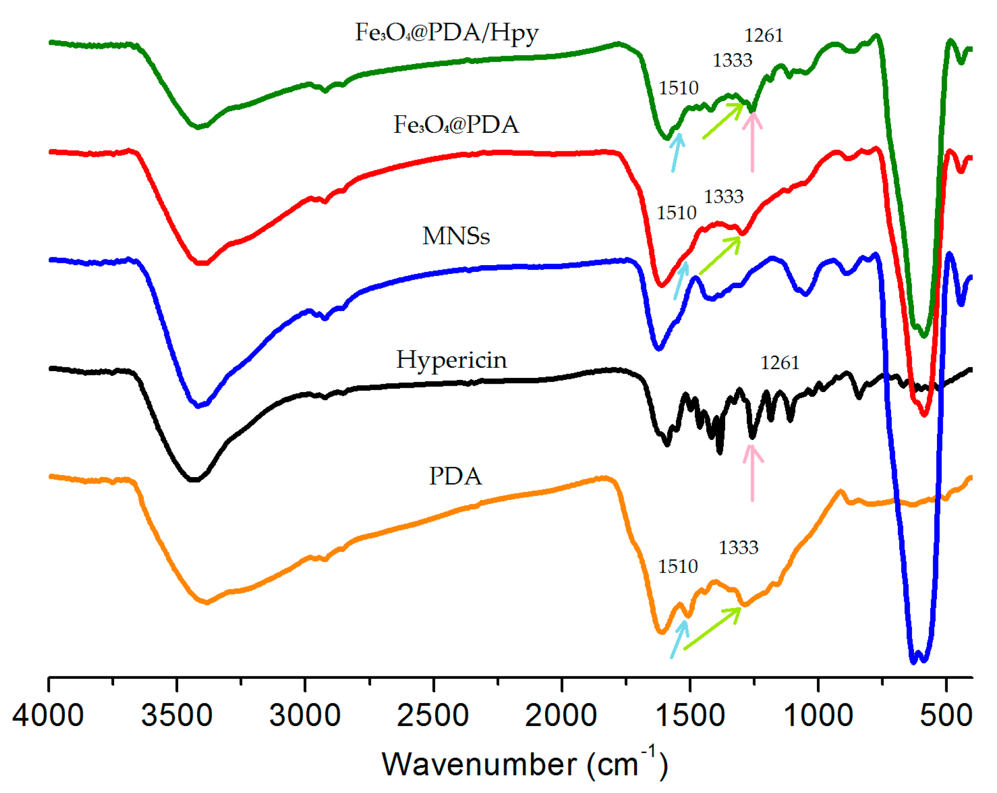
Traditionally, the MIP layer (shell) around MNS (core) is formed by self-assembly of functional monomers and templates, followed by copolymerization with cross-linkers. The copolymerization is normally initiated by heat or UV light, and performed in organic solvents, with the process extending over a prolonged period of time (overnight or longer) [
19]. Considering the increasing likelihood of hypericin to degrade during exposure to heat/light, it is obvious that the traditional procedure is not suitable for fabricating SMIPs for hypericin. Contrarily, dopamine can be oxidized and spontaneously self-polymerize, with oxygen as the oxidant under weak alkaline solution to yield polydopamine (PDA) [
20]. Though the molecular mechanism behind the formation of PDA is complicated and not well-understood, the self-polymerization of dopamine is very mild and the cross-linked PDA network can adhere to virtually any type of material surfaces. These unique features make dopamine a promising functional monomer for fabrication of core-shell structured SMIP nanoparticles without a cross-linker (also called bi- or multi-functional comonomer), which is generally required. In fact, a few SMIP nanoparticles that were successfully prepared using dopamine as a monomer have been reported [
14,
21], including Fe
3O
4@PDA NPs as a stationary phase for recognition of OT-CEC [
15] and SiO
2@PDA nanoparticles for protein recognition and separation [
13].
Inspired by the above-mentioned works, we envisioned that self-polymerization of dopamine on the surface of MNSs would provide a facile approach for fabrication of core-shell structured magnetic molecularly imprinted nanospheres for selective recognition of hypericin (Fe
3O
4@PDA/Hyp NSs). Herein, we investigated the feasibility for fabrication of Fe
3O
4@PDA/Hyp NSs by using dopamine as the only functional monomer. As depicted in
Scheme 1, MNSs suspended in weak alkaline Tris-HCl buffer solution was first mixed with a solution of hypericin in acetone, followed by the addition of dopamine. Dopamine self-polymerized on the surface of MNSs in the air to form a thin layer of PDA within which hypericin molecules were embedded via non-covalent hydrogen bonding and π–π interactions. Removal of hypericin from the PDA layer leaves the recognition sites behind, and Fe
3O
4@PDA/Hyp NSs were thus obtained. The recognition properties of so-prepared Fe
3O
4@PDA/Hyp NSs toward hypericin were then evaluated and screened by varying reaction conditions.
2. Experimental Section
2.1. Chemicals and Instrumentation
Dopamine hydrochloride (DA, 98%), polyethylene glycol 1000 (PEG 1000), and ethanolamine were purchased from Aladdin, Shanghai, China. FeCl
3·6H
2O, ammonium hydroxide (28%), and ethylene glycol were purchased from Xilong Chemical Industry, Sichuan, China. Emodin was purchased from Xi’an Tianfeng Biological Technology Co., Ltd. (Xi’an, China) Acetone was purchased from Rionlon Bohua Pharmaceutical & Chemical Co., Tianjin, China. Anhydrous sodium acetate was purchased from Tianjin Bodi Chemical Industry Co., Ltd., Tianjin, China. Protohypericin (Protohyp) and hypericin (Hyp) were synthesized according to the procedures developed in our lab [
22], and characterized by
1H-NMR (See
Figures S1–S3).
NMR spectra were recorded on a Bruker 500 MHz Spectrometer (Bruker, Fällanden, Switzerland) with working frequencies of 500 MHz for 1H in DMSO-d6 or MeOD-d4. The residual signals from DMSO-d6 (1H: δ 2.50 ppm), or MeOD-d4 (1H: δ 3.31 ppm) were used as internal standards. Transmission Electron Microscope (TEM) images were taken by an H-600 instrument (Hitachi Ltd., Tokyo, Japan, 80 kV). The samples were prepared by dropping a droplet of the sample solution onto a TEM grid (copper grid, 300 meshes, coated with carbon film). Dynamic light scattering (DLS) measurements were performed on a DelsaTM Nano system (Beckman Coulter, Brea, CA, USA). UV-Vis spectra were recorded with Shimadzu 1750 UV-Visible spectrophotometer (Shimadzu, Tokyo, Japan) at 298 K. The surface area and the porosity of the prepared NSs were measured by nitrogen physisorption (Autosorb-iQ, Quantachrome, Boynton Beach, FL, USA), based on the Brunauer–Emmet–Teller (BET) method (ASAP 2020, Micromeritics Inc., Norcross, GA, USA). Samples were vacuum-degassed at 50 °C for 9 h before the adsorption experiments.
2.2. Preparation of Fe3O4 Magnetic Nanospheres (MNSs)
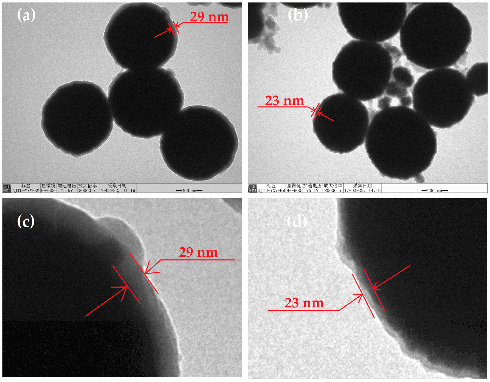
The MNSs were synthesized through the solvothermal approach according to the literature [
23]. Briefly, 1.68 g FeCl
3·6H
2O was dissolved in 50 mL ethylene glycol with vigorous stirring until the solid was dissolved, then 4.5 g anhydrous sodium acetate and 1.25 g PEG were added to the solution, with continuous stirring for one hour. The resultant mixture was transferred into a Teflon-lined stainless steel autoclave (with a volume of 100 mL) and placed in an oven at 200 °C for 10 h, then cooled to room temperature. The obtained precipitate was washed with ethanol and deionized water several times and collected by magnet. The final product was dispersed in ethanol for further use.
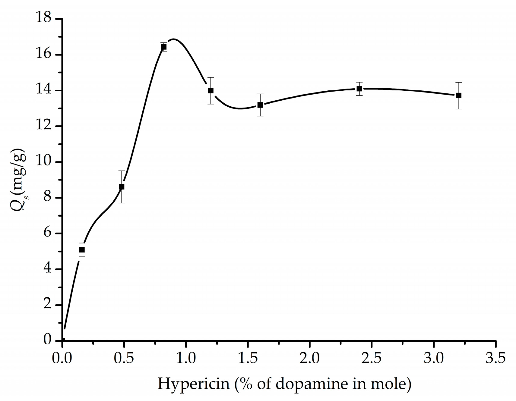
2.3. Preparation of Hypericin-Imprinted Nanospheres (Fe3O4@PDA/Hyp) and Non-Imprinted Nanospheres (Fe3O4@PDA)
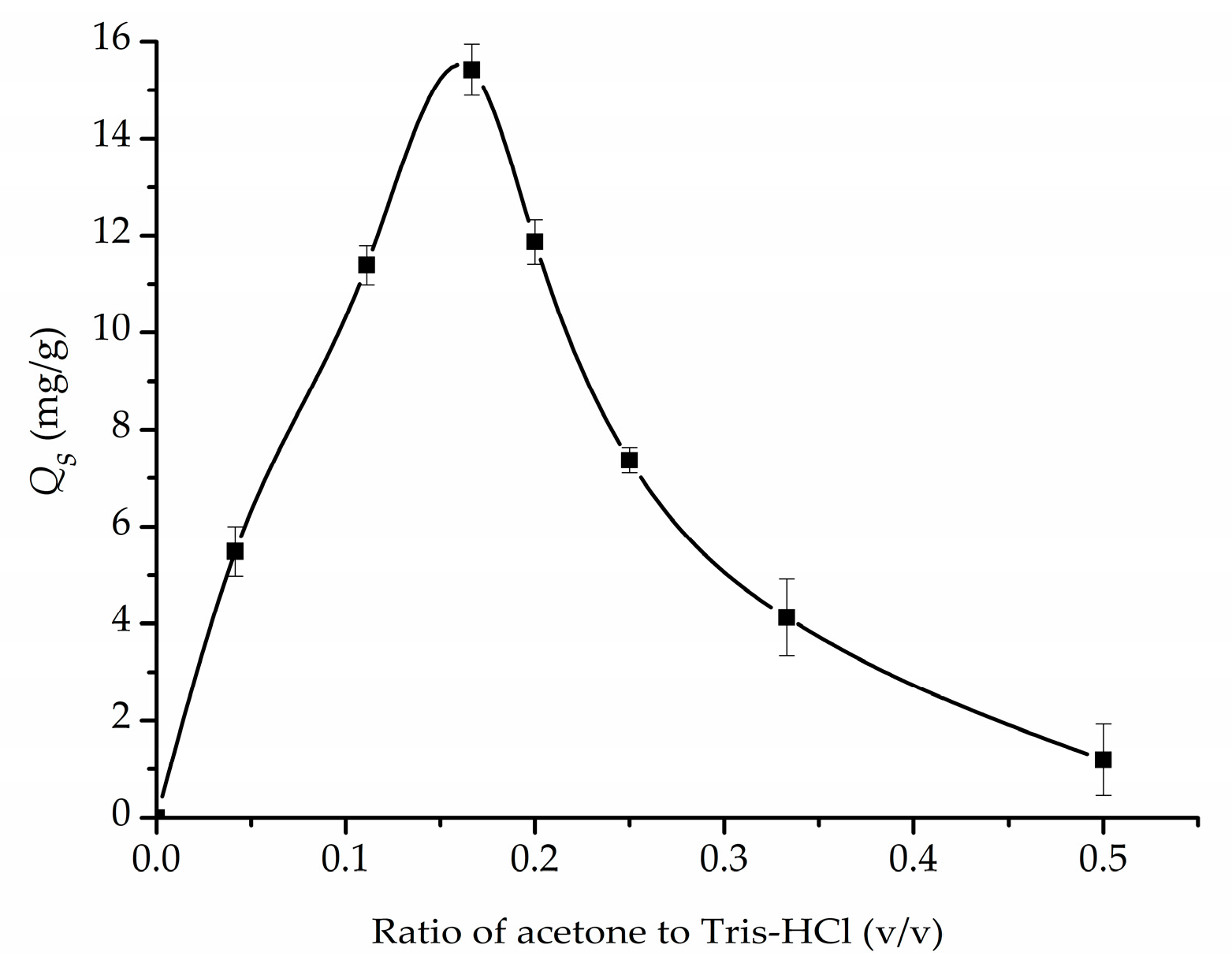
A typical procedure for preparation of Fe3O4@PDA/Hyp NSs was as following: 25 mg MNSs were dispersed in 10 mM Tris-HCl solution (pH = 8.0 unless specified) by ultrasonication for 10 min. Then hypericin dissolved in acetone was added to the suspension by mechanically stirring for 10 min, followed by the addition of dopamine. The mixture was stirred under air with a mechanical stirrer for 4 h. The solid was collected by magnetic separation and washed first with ultrapure water several times, then alternately with an acetone solution containing acetic acid (3% in volume), and ammonium hydroxide (3% in volume), to remove the embedded template, until no hypericin in the supernatant was detected using UV-vis spectrophotometer (Shimadzu, Tokyo, Japan) at 597 nm. Then the solid was treated with 2 μM ethanolamine to give Fe3O4@PDA/Hyp NSs. The final product was dispersed in ethanol for further use.
Fe3O4@PDA NSs were prepared and used as a control by following the same procedure as described for Fe3O4@PDA/Hyp NSs without the template.
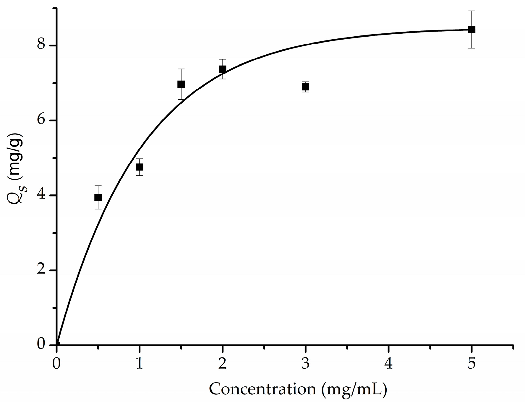
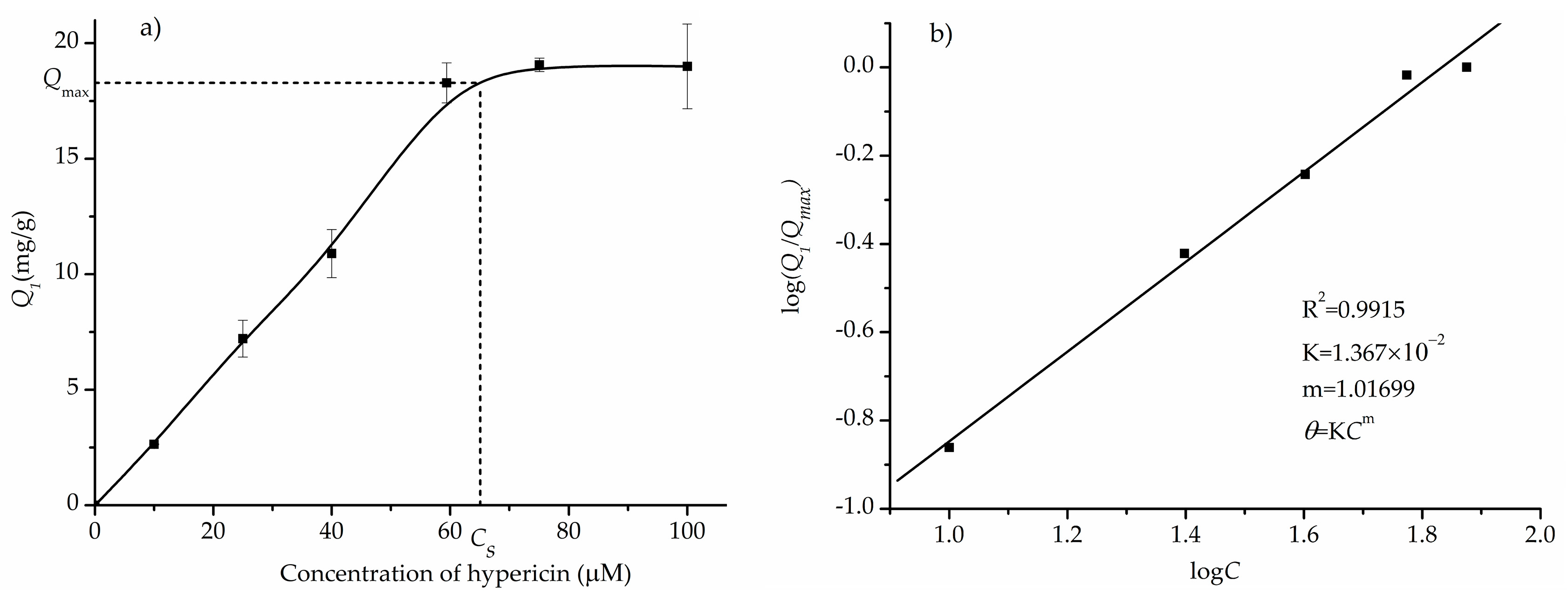
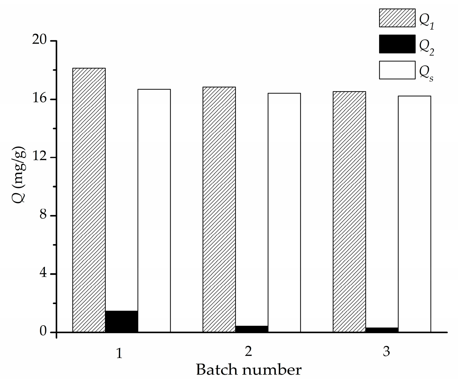
2.4. Determination of Static Adsorption Capacity of Fe3O4@PDA/Hyp for Hypericin (Q)
To a centrifuge tube of 10 mL, 4 mg of Fe
3O
4@PDA/Hyp or Fe
3O
4@PDA NSs were added into a 4 mL known concentration of hypericin (59.4 μM unless specified) acetone solution. The tube was shaken at room temperature for 24 h (unless specified) in the dark. Then a magnet was used for separation, and the concentration of hypericin in the supernatant was measured with a UV-Vis spectrophotometer at 597 nm (
Figures S6 and S7).
The adsorption capacity (
Q, μg/g) of the NSs (Fe
3O
4@PDA/Hyp or Fe
3O
4@PDA) towards the test molecule was calculated by the following equation:
where
C0 and
Ce represent the initial and equilibrium concentrations of the test molecule in acetone (μM), respectively;
M is the molecular weight of the test molecule;
V (L) is the volume of the solution, and
W is the dry weight of the NSs (g).
The specific adsorption capacity of Fe
3O
4@PDA/Hyp NSs (
Qs) towards hypericin is defined as the neat adsorption capacity of Fe
3O
4@PDA/Hyp over that of Fe
3O
4@PDA, and is calculated according to Equation (2):
where
Q1 and
Q2 are the static adsorption capacity of Fe
3O
4@PDA/Hyp and Fe
3O
4@PDA (μg/g) towards hypericin, respectively.
2.5. Dynamic Adsorption Test
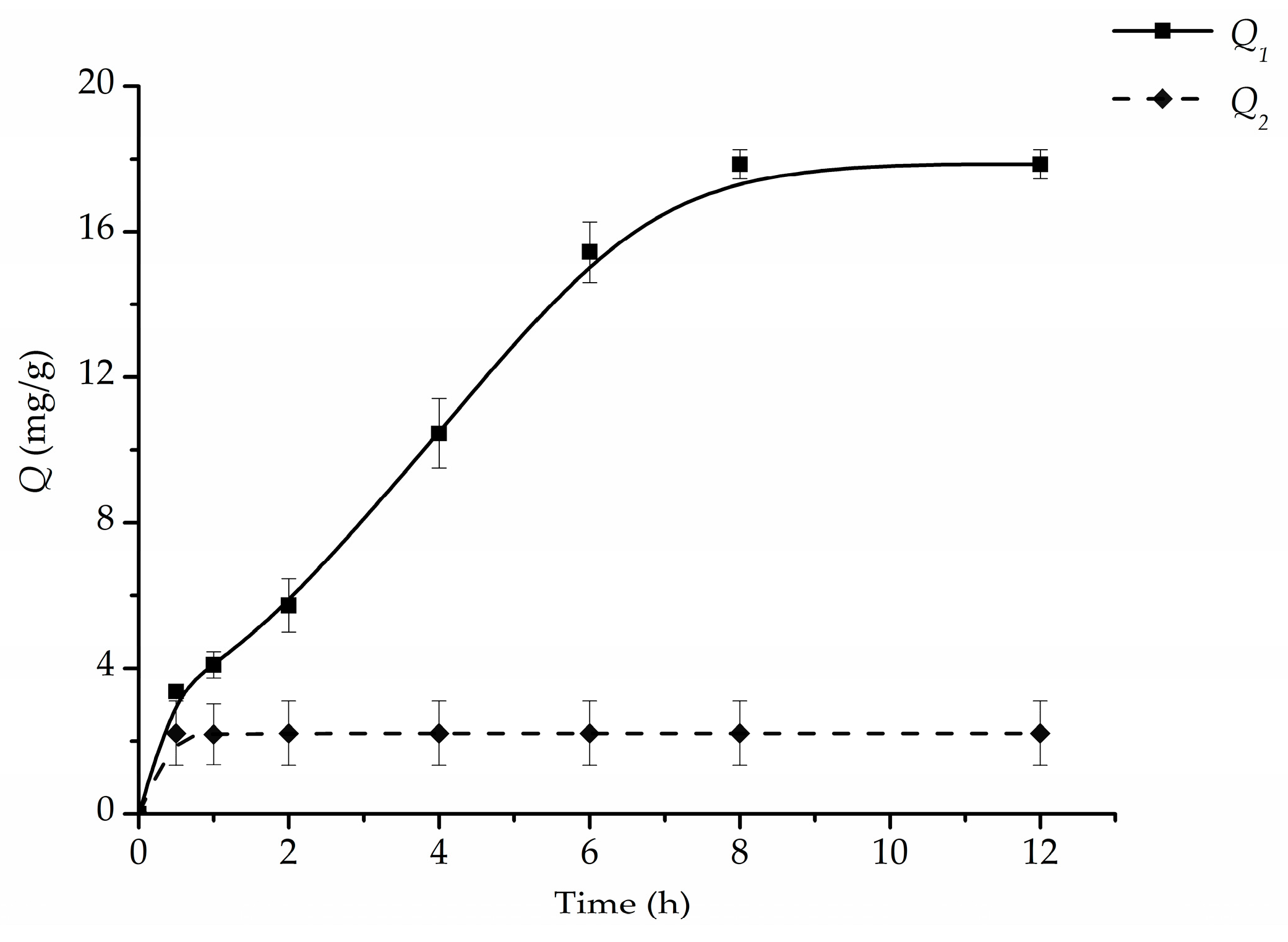
To investigate the adsorption kinetics of Fe3O4@PDA/Hyp (or Fe3O4@PDA) NSs, 4 mg of Fe3O4@PDA/Hyp (or Fe3O4@PDA) NSs were weighed into hypericin solution (59.4 μM, 4 mL) in a 10 mL centrifuge tube. The tubes were shaken at room temperature for the different time intervals (0.25, 1, 2, 4, 6, 8, and 12 h, respectively) in the dark. Then a magnet was used for the separation, and the concentration of hypericin in the supernatant was measured with a UV-Vis spectrophotometer at 597 nm.
2.6. Selectivity of Fe3O4@PDA/Hyp and Fe3O4@PDA for Hypericin
The binding selectivity of Fe
3O
4@PDA/Hyp and Fe
3O
4@PDA NSs was evaluated by measuring their binding capacities towards hypericin and two other molecules of protohypericin and emodin. 4 mg of the Fe
3O
4@PDA/Hyp or Fe
3O
4@PDA NSs were incubated respectively with 4 mL of hypericin, protohypericin, and emodin solution (59.4 μM in acetone) at 25 °C. After being incubated under continuously shaking for 24 h, the amounts of hypericin, protohypericin, and emodin bound to the Fe
3O
4@PDA/Hyp or Fe
3O
4@PDA NSs were measured, respectively. The binding selectivity of the NSs towards different molecules was compared using the “selectivity factor” (
SF) and “imprinting factor” (
IF) [
24] that can be defined by the following equations:
where
Q1’ is the adsorption capacity of the Fe
3O
4@PDA/Hyp NSs (μg/g) towards a non-template molecule.
where
QMIP and
QNIP is the adsorption capacity of the Fe
3O
4@PDA/Hyp and Fe
3O
4@PDA NSs (μg/g) towards a test molecule, respectively.
2.7. Adsorption–Extraction Cycles
One adsorption–extraction cycle consisted of loading the template, reaching equilibrium adsorption, followed by the extraction of the template. For the adsorption, 4 mg of Fe3O4@PDA/Hyp was added to 4 mL 59.4 μM template in acetone. The suspension was incubated with a shaker at 25 °C for 8 h. Then the NSs were collected with a magnet and washed following the extraction procedure.
2.8. Preparation of the Herb Extract Solution
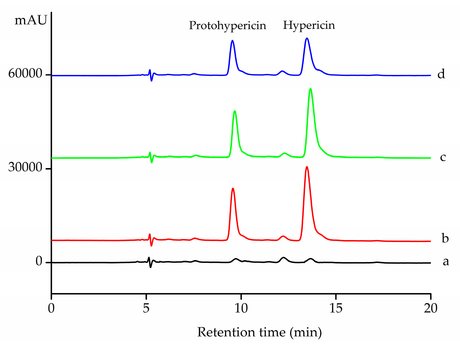
To prepare the herb extract, fresh flowers were picked up from Hypericum perforatum plant just before the extraction process. The extraction was performed in the dark. The procedure was as following: 10 g fresh flowers was charged to a 2-L beaker and immersed with distilled water at 50 °C for 2 h. Then the flowers were transferred to a 1000-mL flask and refluxed with 500 mL methanol-water mixture (80:20, v/v) for 6 h. The contents in the flask were cooled to room temperature and filtered. The filtrate was dried under reduced pressure. The residue was dissolved with acetone and filtered. The filtrate was combined and the volume was adjusted with a 25 mL volumetric flask.
2.9. HPLC Analysis
A total of 8 mL of the herb extract solution was mixed with 1 mL of hypericin and 1 mL of protohypericin solution (each with a concentration of 600 μM in acetone), to obtain an original solution for adsorption. To 4 mL of this final solution, 4 mg of the NSs (Fe3O4@PDA/Hyp or Fe3O4@PDA) was added. The mixture was shaken for 8 h. The supernatant was analyzed by HPLC (Shimadzu, Tokyo, Japan), with C18 reversed-phase column (5 μm, 4.6 mm × 150 mm) at 25 °C. The mobile phase consisted of 50% acetonitrile, 50% of the mixture of ammonium acetate-acetic acid buffer (0.3 M, pH = 6.96) and methanol (1:4, v/v); detection wavelength: 590 nm; flow rate: 0.4 mL/min; injection volume: 10 μL.


