Article

Host Specificity of Endophytic Fungi from Stem Tissue of Nature Farming Tomato (Solanum lycopersicum Mill.) in Japan

1
Graduate School of Agriculture, Tokyo University of Agriculture and Technology, Tokyo 183-8509, Japan
2
Department of Plant Pathology, Bangladesh Agricultural University, Mymensingh 2202, Bangladesh
3
Faculty of Agriculture, Tokyo University of Agriculture and Technology, Tokyo 183-8509, Japan
4
Graduate School of Bio-Applications and Systems Engineering, Tokyo University of Agriculture and Technology, Tokyo 183-8509, Japan
5
College of Agriculture, Ibaraki University, Ibaraki 310-8512, Japan
6
Department of Research, International Nature Farming Research Center, Nagano-ken 390-1401, Japan
*
Authors to whom correspondence should be addressed.
Agronomy 2020, 10(7), 1019; https://doi.org/10.3390/agronomy10071019
Submission received: 11 June 2020 / Revised: 7 July 2020 / Accepted: 12 July 2020 / Published: 15 July 2020
(This article belongs to the Special Issue Drivers of Microbial Diversity and Roles in Agroecosystems)

Abstract

:
To understand the distribution of the cultivable fungal community in plant tissues from nature farming tomato plants, we sampled plants of seven different tomato cultivars and recovered 1742 fungal isolates from 1895 stem tissues sampled from three sites in Japan. Overall, the isolation frequency was low (3–13%) and the isolation and colonization frequencies did not vary significantly as a function of the cultivar. The fungi were divided into 29 unique operational taxonomic units (OTUs) with 97% ITS gene sequence identity, the majority of which belong to Ascomycota (99.3%). The dominant genera of cultivable endophytic fungi were Fusarium (45.1%), Alternaria (12.8%), Gibberella (12.0%), and Dipodascus (6.8%). The alpha diversity of the fungal endophytes varied among tomato cultivars. Ordination analysis performed to investigate patterns of endophyte community assemblages on the various cultivars revealed that host cultivars had a significant impact on the endophyte community assemblages in all the study sites. Some of the taxa Fusarium, Alternaria, and Penicillium were found on all cultivars, while few were uniquely present in different cultivars. The dominant taxa may be adapted to the particular microecological and physiological conditions present in tomato stems.

Graphical Abstract

1. Introduction

It is now well known that endophytes, organisms that live asymptomatically within plant tissue, are very common and widely distributed in terrestrial ecosystems [1]. The plant and endophytes coevolved millions of years ago [1]. The composition of endophytic fungi is often diversified [2,3,4], and may vary as a function of host plant species [5,6], geographically distanced individuals of the same host species, and within the tissues or organs of a host plant [7]. In plant-endophyte mutualistic symbiosis, plants provide accommodation and diverse niches for endophytes [8]. In return, many endophytic fungi can improve the uptake and utilization of nutrients by the host to promote growth [9,10] and can enhance tolerance to biotic and abiotic stresses [11,12,13,14,15]. Hence, plant-endophytic symbiotic associations can play a significant role in the integrity and sustainability of agricultural ecosystems [16]. Therefore, an understanding of the nature, composition and distributions of endophytes is essential both for disclosing features of plant ecology and evolution in ecosystems and for developing further survey schemes for tapping into their functional potentials [17,18].
Conventional farming heavily relies on intensive applications of synthetic agrochemicals and chemical fertilizers, which have negative impacts on terrestrial ecosystems and human health [19]. The cultivation practice in nature farming systems influences plant-associated microbiome diversity and abundance and thus benefits plant growth, development, and health [20,21,22,23]. Therefore, to meet the rising global demands of increased eco-friendly farm production, there is a need to develop an agricultural practice that is sound and sustainable for the betterment of both the environment and humans living in it. The ‘Nature farming’, originated in Japan, is a concept that entails a farming practice that does not use synthetic chemical inputs in cultivating crops [24,25]. It is an ecological farming system based on sound agronomic principles and is the major certified organic farming method in Japan [26]. It differs from other organic farming systems primarily in its philosophical origin [27]. More or less similar or comparable farming approaches are in practice in different countries with different names such as ‘Permaculture’ in Australia [28,29], ‘Fertility Farming’ in USA [30], ‘Rishi Kheti’ and ‘Zero Budget Farming’ in India [31,32], Natural Farming in Japan [33], no-tillage farming, zero tillage farming or conservation agriculture in various countries [34]. Introducing nature farming to enhance the predominance of beneficial and effective microorganisms can help improve and maintain soil biological, chemical, and physical properties [35]. This farming concept encourages soil and water conservation, reduces chemical pollution, increases the abundance and diversity of plant and soil microbes, and improves the quality and safety of food supplies [24,26,35,36].
There are no systematic studies of the effects of nature farming practices on the culturable endophytic fungal communities in crop plants. Little is known about the endophyte communities associated with crop plants grown under nature farming, and no study has yet been done on the fungal endophytic communities of nature farming tomato plants. A previous study reported that endophytic microbial species abundance and diversity were significantly higher in organically farmed plants than in conventional plants [37]. While organic farming is not exactly the same as nature farming, but underlying principles overlap. In the current study, we aimed to identify culturable fungal isolates that are likely to be beneficial to plant growth, health, and yields and to reveal whether those endophytes vary in different tomato cultivars. This study represents basic information about the occurrence and species diversity of fungal endophytes in the stems of tomato plants growing in nature farming in Japan. The advantage of the culturable approach is that it allows isolation, identification, and consequently may contribute to further studies of function and application of endophytic fungi. [12,38,39]. As such, its findings might aid the development of novel, efficient, and sustainable strategies for improving crop quality and productivity while reducing the application of harmful chemicals.

2. Materials and Methods

2.1. Collection of Tomato Plant Samples

Nature farming tomato plants (Solanum lycopersicum L.) were collected from four different experimental farmlands (Site-C: the nature farming began in 1990, Site-D: the nature farming began in 1992 and Site-I: the nature farming began in 1999, distances between Site-C and Site-D: 1.8 km, between Site-D and Site-I: 0.4 km, between Site-I and Site-C: 1.6 km, respectively) in International Nature Farming Research Center (INFRC), Matsumoto, Nagano, Japan. The soil type in all experimental sites is andosol. In these experimental fields, instead of pesticides and chemical fertilizers, compost and green manure are applied to maintain the soil fertility and the ecological balance of farmlands. The tomato plants comprised several cultivars and were grown under different cultivation practices. Plant are of similar age (late fruiting stage) and three plants from each cultivar per cultivation practice per site were sampled in August 2019. After harvesting, the plants were packed in polythene, shipped in an ice box, stored in a refrigerator until use and processing was done within 72 h. Details of the cultivars are presented in Table 1.

2.2. Fungal Endophyte Isolation

We harvested whole plants and stems were cut from the lower part of the main stem from above 10 cm off the ground. The leaves, secondary branches and approximately 10 cm from the top were removed before endophyte isolation from the main stems to reduce heterogeneity among the samples. Asymptomatic tissues from the main stem of the tomato plants were used for endophyte isolation. Tissues were washed under running tap water to remove surface debris before surface sterilization and cut into ~5 cm long segments for ease in processing. Stem tissues were surface-sterilized by immersion in 0.5% sodium hypochlorite for 3 min before immersion in 70% ethanol for 3 min. After three washes in sterile distilled water and blotting with sterile filter paper, samples were cut into approximately 3 × 3 mm segments with a sterile scalpel. We placed around 50 segments (3 × 3 mm) from each plant and there were three plants for each cultivar per cultivation practice per site with a missing plant of the cultivar ‘Chako’ at site-D (~50 segments × 13 plants × 3 replications minus 1 missing plant), which made a total of 1895 segments (supposed to be 1900 segments but some plants had 1 or 2 missing segments due to mistake). To evaluate the efficacy of the surface sterilization procedure, the imprint method was used, i.e., pressing sterilized tissue segments gently onto potato dextrose agar (PDA) [40] to confirm that the fungal isolates only originated from the internal tissues of the tomato stem segments. Additionally, the water that was used to wash the surface sterilized tissues at the last step was incubated in 0.25 × PDA to observe any fungal growth. For fungal isolation, segments were plated in petri dishes containing media composed of 0.1 × PDA (PDB 2.4 g (Catalog # P6685, Difco) plus 17 g agar in 1 L water amended with 100 µg/mL streptomycin sulfate (Sigma, St Louis, MO, USA). Each plate contained five stem tissue fragments and was incubated at 25 °C in the dark and checked periodically for fungal growth. The growing margins (hyphal tips) of colonies from tissue segments were transferred to plates of 0.25 × PDA supplemented with streptomycin (100 µg/mL) [41]. The fungal isolates were counted and stored temporarily at 4 °C, and the mother culture was permanently stored at −80 °C in 80% sterile glycerol.

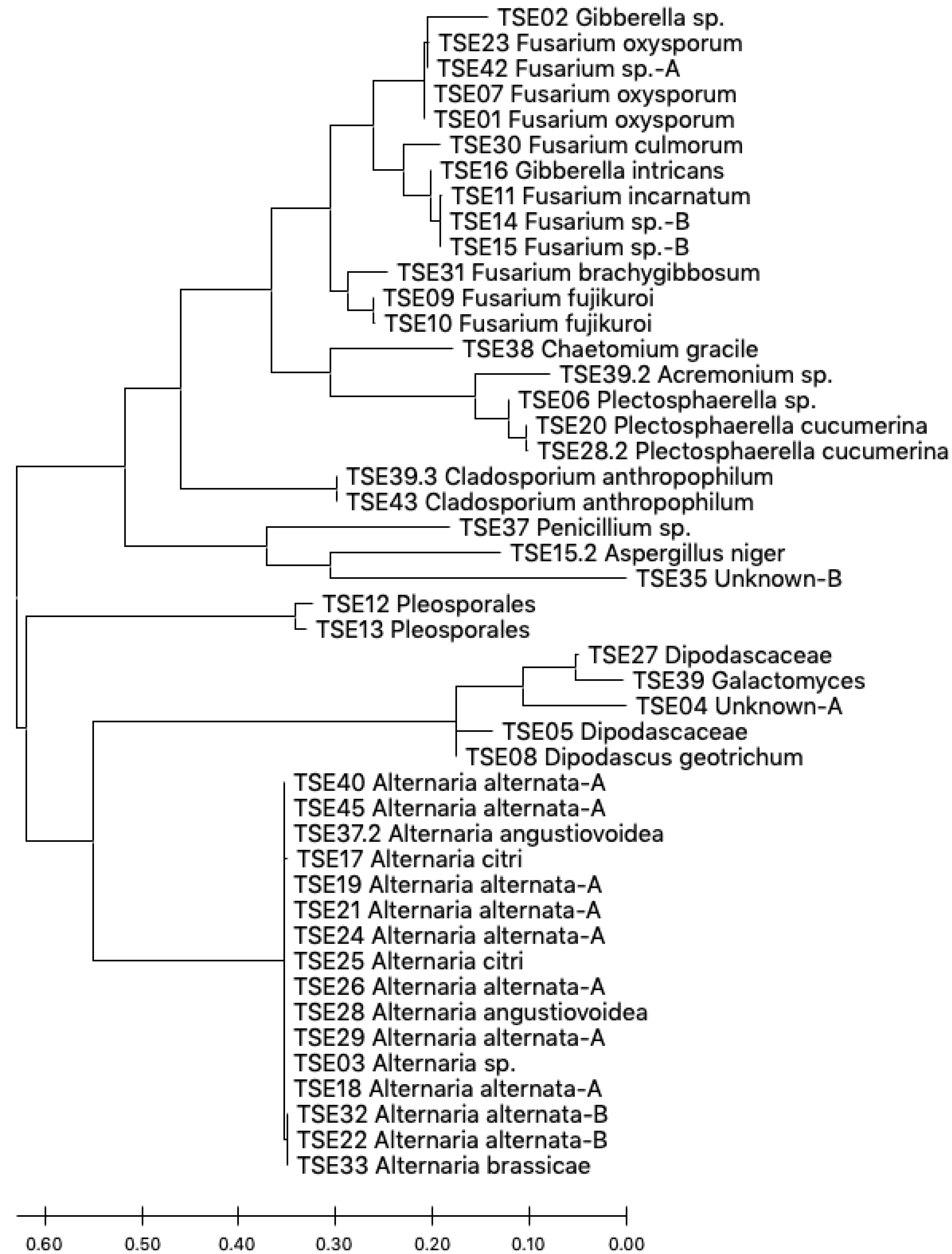
2.3. Endophyte Identification

We employed a combination of morphological and molecular techniques for the identification of fungal taxonomy. Fungi that showed similar morphological characteristics, such as colony characteristics and mycelial textures, in our culture plates were grouped, and one of them was identified as a representative strain. For molecular identification, fungal mycelium taken from an agar plate was transferred into a 50 mL falcon tube containing 20 mL PDB and incubated on a shaker for 7–21 days in the dark. At harvest, the fungal mycelium was trapped by a sterile mesh strainer and squeezed dry on a sterile paper towel and used for genomic DNA isolation by following a phenol:chloroform:isoamyl alcohol (PCI, 24:24:1) protocol [5]. Briefly, cryogenically ground fungal mycelia (approximately 100 mg) were lysed in 450 µL of extraction buffer (0.2 M Tris–HCl pH 7.5, 0.25 M EDTA, 0.2 M NaCl, 1% SDS) and mixed thoroughly with continuous shaking for 5 min. An equal volume of PCI solution was added to the lysate and incubated for 5 min after mixing and centrifuged at 20,000× g for 5 min. Then, 300 µL of the supernatant was transferred to a 1.5 mL tube. The genomic DNA was precipitated with an equal volume of iso-propanol at −20 °C for 15–30 min followed by centrifugation at 14,000× g for 20 min at 4 °C. The pellet was washed with 70% ethanol, air dried and dissolved in 50 μL of EB buffer (10 mM Tris-Cl, pH 8.5).
We amplified the ITS regions of the fungi using the universal primers ITS1 and ITS4 [42], using the PCR conditions as 95 °C for 3 min followed by 30 cycles of 95 °C for 30 s, 55 °C for 30 s, 72 °C for 30 s, and a final extension at 72 °C for 7 min. The 20 μL reaction mixture contained 10 µL of 2 × Promega Go Taq™ Master Mix, 10 µM of each primer, 1 µL of template DNA and 7 µL of water. Amplicons were purified by an AxyPrep Mag PCR Clean-up kit (Axygen Biosciences, Union City, CA, USA). Each sequencing reaction contained 1 µL of BigDye® Terminator sequence mix (Applied Biosystems, Foster City, CA, USA), 1.5 µL of 5 × Sequencing Buffer, 1 µL of the forward primer (3.2 µM), 4 µL cleaned PCR amplicons and 2.5 µL water. The cycling conditions were 35 cycles of 96 °C for 10 s, 50 °C for 5 s and 60 °C for 4 min.
The fungal ITS sequences were viewed for quality checking by using FinchTV 1.5 (Geospiza Inc., Seattle, WA; MacOS X), first ~20nt were trimmed, any ambiguous bases (N) were manually corrected by matching the chromatogram and average 500–800 bp high-quality sequences were aligned. The sequences were aligned as query sequences with the database UNITE using massBLASTer analysis options, which is specially compiled and used for fungal identification [43,44]. In all cases, the isolates were identified to the species level if their sequences were >98% similar to any identified accession from the database. When the similarity percentage was between 95–97%, only the genus name was accepted; for sequence identities <95%, the isolates were classified according to family or order or as “unknown fungus” [5]. The isolates that matched the same taxa were aligned using the MAFFT (multiple alignment using fast Fourier transform) algorithm [45], and percent similarity was obtained using the EMBL-EBI (http://www.ebi.ac.uk/Tools/msa/mafft/) platform. A ≥97% similarity threshold was used to distinguish taxa that yielded the same blast hit and noted them as “A” or “B” at the end of taxa name. The sequences were deposited in GenBank, and accession numbers were obtained (MT481755-MT481800).
Phylogenetic analysis of the isolated endophyte genera obtained from nature farming tomato plants. The evolutionary history was inferred by using the Maximum Likelihood method and the Tamura–Nei model [46]. The tree with the highest log likelihood (−8023.30) is shown. Initial tree(s) for the heuristic search were obtained automatically by applying Neighbor-Join and BioNJ algorithms to a matrix of pairwise distances estimated using the Maximum Composite Likelihood (MCL) approach, and then selecting the topology with a superior log likelihood value. This analysis involved 46 nucleotide sequences. The codon positions included were 1st+2nd+3rd+noncoding. There were a total of 1003 positions in the final dataset. Evolutionary analyses were conducted in MEGA X 10.1.5 [47].

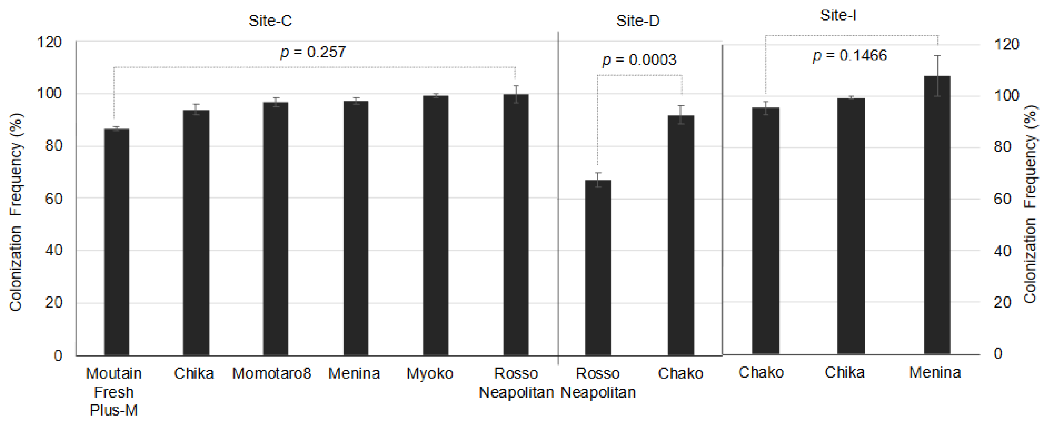
2.4. Colonization and Isolation Frequency

Colonization frequency (CF) was defined as the percentage of tissue fragments yielding an endophyte in culture; on the other hand, the isolation frequency (IF) was defined as the number of taxa isolated from a cultivar in a particular site and expressed as the percentage (%) of tissue planted. At first, we checked all data if these met the assumptions of normal distribution and homogeneity of variance using the Shapiro–Wilk normality test and Levene’s test, respectively. Colonization frequency (CF) data form Site-C and Site-I were found to be violating the assumption of normality and we used the Kruskal–Wallis (non-parametric) test for comparisons (Table S1). In all other cases, ANOVA tests were carried out to compare the CF and IF values between cultivation practices at site-D, which revealed no differences between them and their interaction with cultivars. Next, we conducted ANOVA tests among host genotypes at different sites and Tukey’s HSD test for multiple comparisons. We used the following formulae for calculating CF and IF.
CF (%) = (Number of segments shows endophyte growth/number of segments plated) × 100
IF (IF%) = (Number of unique taxa isolated/number of segments plated) × 100

2.5. Diversity and Community Composition

The diversity measures used were the Shannon–Wiener diversity index (H’), Fisher’s alpha diversity, evenness (H/S), Menhinick index and Simpson dominance. These were used to measure endophyte species diversity within samples (α-diversity). Since the results were consistent in all cases, only the Shannon–Wiener biodiversity, the most commonly used index, has been shown in the findings index (H’). An ANOVA test was performed to compare H’ values among host genotypes and cultivation practices.
We compared differences in community composition and structure among host cultivars (β-diversity) using analysis of similarity (ANOSIM) and PERMANOVA. The use of two different pairwise similarity measures was attempted because they take into account different types of information that can provide different insights for comparing communities [48]. We calculated (i) Jaccard’s index, which considers only the presence or absence of fungal taxa among samples, and (ii) the Bray–Curtis coefficient, which considers the abundance of taxa along with the presence or absence of particular fungal taxa [49].
Nonmetric multidimensional scale (NMDS) ordination was applied to visualize similarity measures of endophyte communities. Multidimensional scaling is used to graphically present interactions between objects in multidimensional space. NMDS is a popular and robust visual analysis tool applicable to various data types, and it is useful to a number of user-defined standardizations and transformations of the data, flexible with respect to which dissimilarity or similarity measure is used [48]. NMDS plots use rank-order information in a dissimilarity matrix [50]. The test statistic “R” and “pseudo-F” values were calculated as the difference of mean ranks between vs. within groups. Significance was calculated by 10,000 permutations of group membership. The diversity measures (α-diversity and β-diversity including NMDS plots) analyses were conducted using PAST ver. 3.26 [51].

3. Results

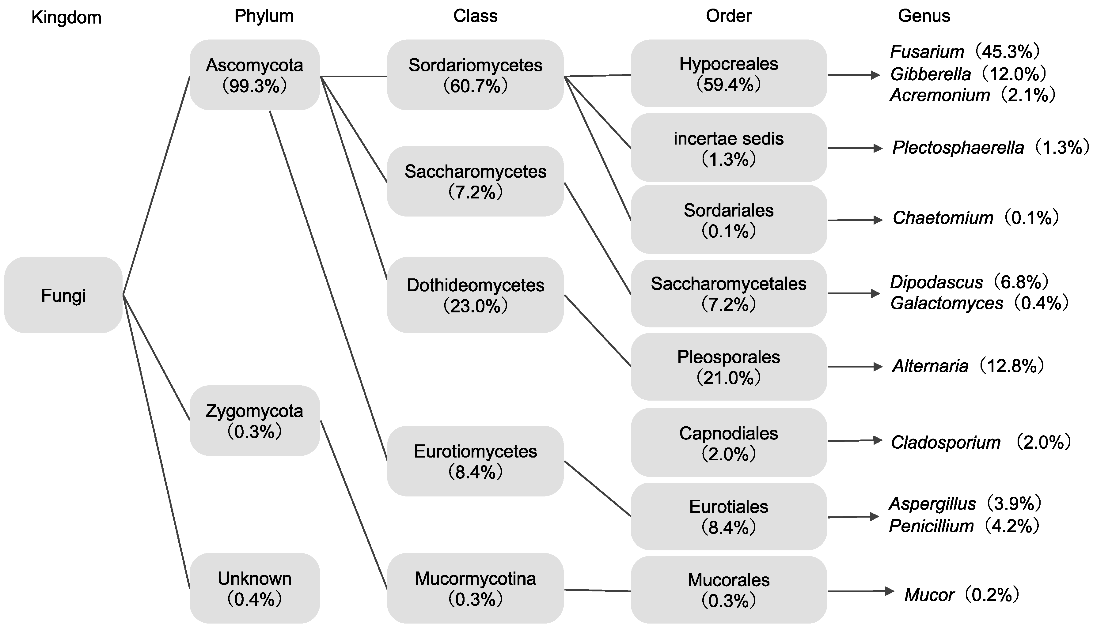
3.1. Isolation and Identification of Endophytes

The efficacy surface sterilization was confirmed by the absence of fungal or bacterial growth in tissue imprint, which indicated that the surface sterilization protocol was effective in eliminating epiphytic fungi. A total of 1742 isolates of fungal endophytes were recovered from 1895 stem segments of different cultivars of tomato plants from three sampling experimental sites (overall colonization frequency = 91.926%). Here, an isolate is defined as visible fungal growth from a (3 × 3 mm2) stem segment/plant/cultivar/location. The ANOVA test of colonization frequencies indicated that the number of fungal isolates recovered did not vary significantly as a function of cultivar except at site D, where ‘Chako’ yielded a significantly lower number of isolates than ‘Rosso Neapolitan’ (p = 0.003, Figure 1). Isolation frequency (IF), which we defined as the mean number of endophytic taxa per number of segments plated per cultivar in percentage, did not vary among cultivars at Site-D and Site-I (Figure 2). At Site-C, the Mountain Fresh Plus-M cultivar had the lowest IF, which was significantly different from that of Rosso Neapolitan. Overall, the isolation frequency was low (3–13%) in all cultivars regardless of the collection site (Figure 2). We did not observe any difference in endophyte CF or IF due to different cultivation practices at site D (Tables S2 and S3).
In aggregates across all sampling locations, 46 representative fungal isolates were selected for molecular identification based on morphological characteristics in cultures. Molecular identification using ITS rDNA sequences resulted in a total of 29 different endophytic fungal taxa that belonged to 12 different genera with few taxa not identified to the genus level. All endophytes recovered from tomato cultivars were members of Dikarya. Ninety-nine percent of endophyte isolates recovered here represented Ascomycota and were distributed in four classes of Pezizomycotina. The majority represented Hypocreales (59%), Pleosporales (21%), and Saccharomycetales (7%) (Figure 3). Overall, the most dominant fungal genera were Fusarium, Gibberella, Dipodacus, and Alternaria (Figure 3). Phylogenetically, the fungal endophytes recovered from our nature farming tomato plants did not seem highly diverse (Figure 4).

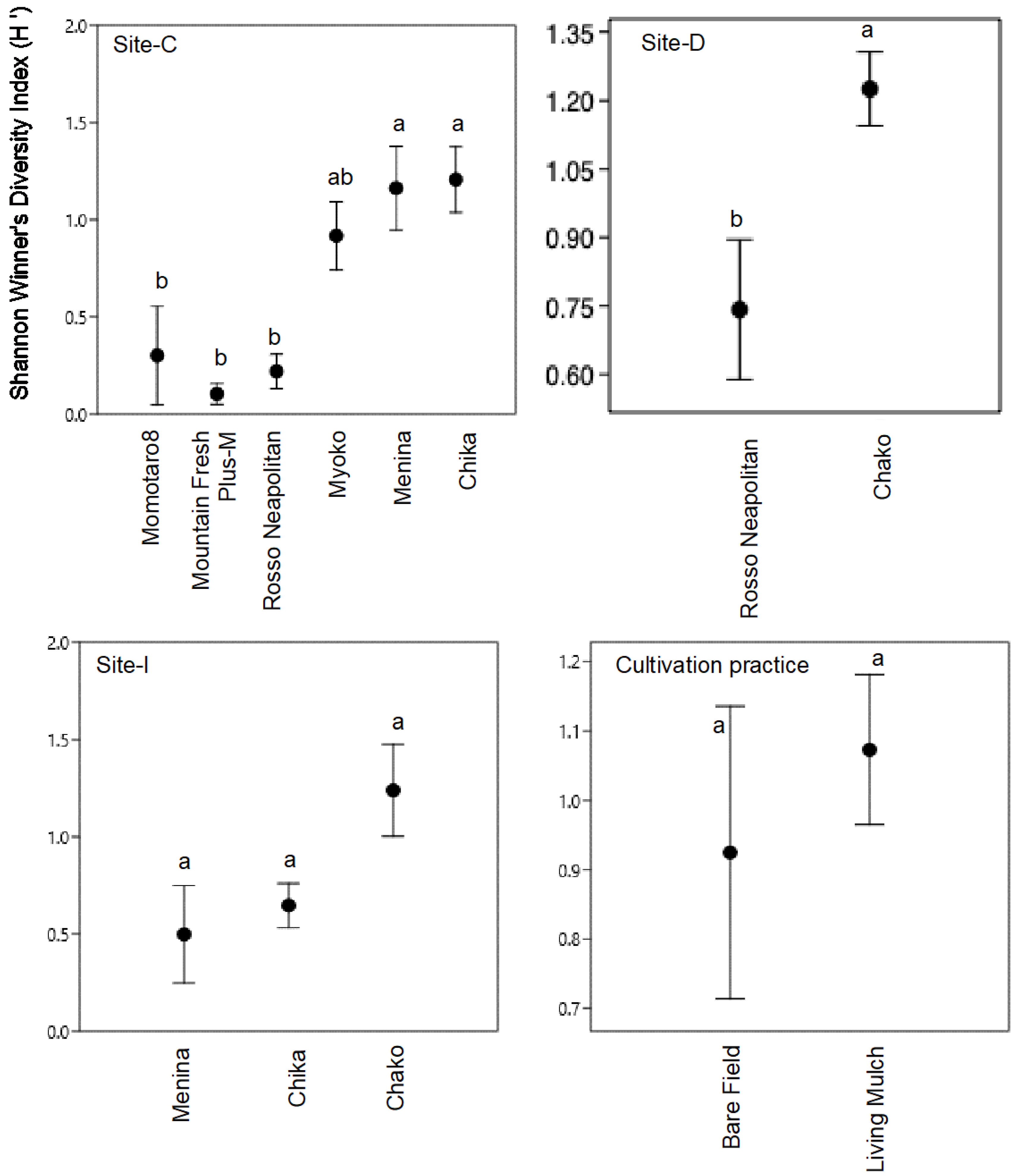
3.2. Diversity and Species Richness of Endophyte Communities (α-Diversity)

The diversity of each endophytic fungal community was assessed in relation to host cultivar and cultivation practice using the Shannon–Wiener diversity index (H’) value. We also did not detect any difference in alpha-diversity of fungal endophytes among the cultivars at Site-D and Site-I, but there was a variation at site-C. There was significantly higher fungal diversity recorded from the cultivars Menina and Chika (H’ ≥ 1.0) than from the cultivars Momotaro8, Mountain Fresh Plus-M, and Rosso Neapolitan (H’ ≤ 0.3) at site C (Figure 5). The diversity of the endophyte communities did not differ significantly between the plot with previously grass cultivated and bare plot without grass cultivation (Figure 5).

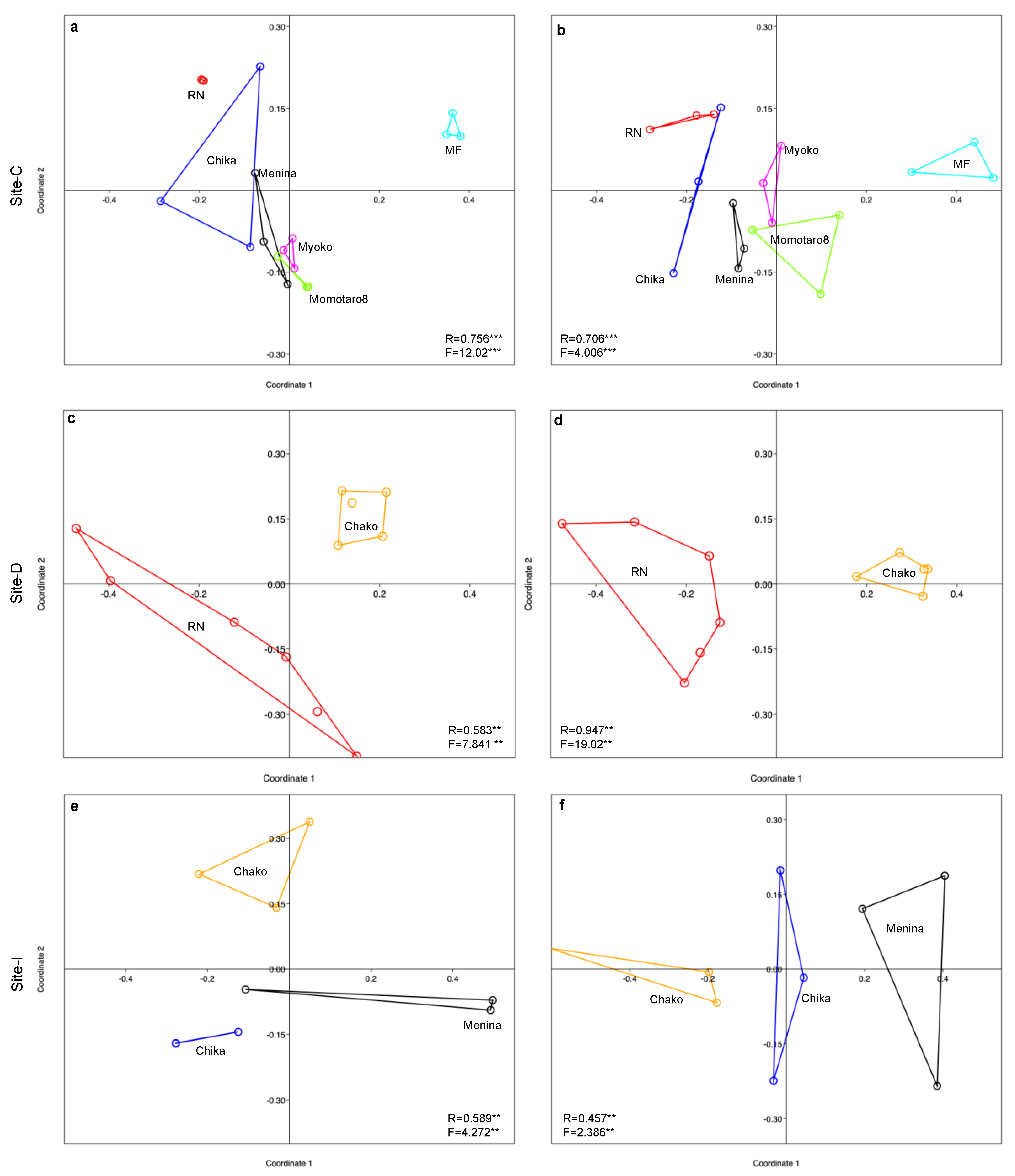
3.3. Variation in Endophyte Communities across Location, Host and Tissue (β-Diversity)

Ordination analysis was performed to investigate patterns of endophyte community assemblages on the various host cultivars. Two-dimensional nonmetric multidimensional scale (NMDS) as well as ANOSIM- and PERMANOVA-based clustering of fungal communities revealed that cultivation practice (that is, living mulch intercropping vs. bare field) did not influence the structures of endophyte communities at sampling site D (R = 0.069, p = 0.2603; Figure 6). However, host cultivars had a significant impact on the endophyte community assemblages in all study sites (Figure 7). The ANOSIM statistic R ranged from 0.45 to 0. 95, indicating a dissimilarity of the endophyte communities among the host cultivars and that the endophyte communities at particular locations appeared to be specific to particular host cultivars (Figure 7).

4. Discussion

Microbial symbionts of plants, including endophytes, have increasingly received attention in agriculture and biotechnology [4,53,54,55,56,57,58]. Fungal endophyte communities have been explored from a broad range of wild and agricultural plants. The object of our current study was to examine for the first time the composition, biodiversity and host affiliation of tomato plants grown under nature farming. Most published studies have focused on root or leaf endophytes and those from wild or conventionally cultivated crops, but limited numbers of studies have reported on crops grown with nature farming. The present study targeted stem fungal endophytes obtained from different tomato cultivar plants cultivated in nature farming. The resulting data set will contribute to the augmentation of the available knowledge on shoot-associated fungal endophytes of plants, especially those of nature farming.
The techniques used for endophyte isolation are important considerations [57]. The accuracy of identification and isolation is dependent on various factors that, in turn, may affect the usability and validity of datasets for comparison among groups. Too harsh surface sterilization procedures might kill all endophytes in plant tissues [58], whereas improper sterilization will allow epiphytes to proliferate and contaminate and thus reduce the ability to recover viable isolates grown on culture media. We used tissue imprinting to rule out the possibility of contaminants and optimize the surface sterilization procedure [59]. Again, we use only PDA media, which might also have an impact on the isolation of endophytes because some isolates might not grow on that media. However, since the same sterilization protocol, media and growth conditions were applied to all samples in this study, we expect that any bias related to the above-described was masked and gave us good confidence for comparable analyses. There are also problems in the identification of culturable endophytes using ITS amplification of DNA barcoding approaches [60,61]. Certain taxa can only be identified to the family or higher levels due to the absence of resembled sequences in GenBank [62]. In addition, the sequences deposited in GenBank may not have been precisely identified. For example, it has been reported that 96% of sequences listed as Colletotrichum gloeosporioides have been applied wrongly [62]. Given these limitations in the GenBank database, we used the UNITE database, which is specifically curated as described by other studies [44,63,64]. There are also concerns that ‘endophyte’ could be ‘latent pathogen’. Indeed, fungi isolated as endophytes have been shown to produce disease in the host upon inoculation [65,66,67,68]. In this paper, we did not evaluate the pathogenicity of any of the isolates obtained but we relied on the well-accepted definition of endophytes. To be specific, we used the term ‘endophyte’ to refer to the fungi that were present within the healthy plant tissue, rather than on the surface, without showing apparent disease symptoms or without necessarily implying any specific ecological functions [67,68,69].

4.1. Composition of Endophytes

On average, endophytes were isolated in culture from 92% of the tomato stem tissue segments plated. This seems to be very high colonization rather than those reported from other studies. This may be an implication of a possible higher colonization intensity of fungal endophytes in nature farming crops than conventional cropping or wild plants. Indeed, previous studies that compared fungal endophytes of tomato, soybean and grapevines grown organically reported higher overall colonization than those grown under conventional practice [12,70,71,72]. However, further studies to compare endophyte isolation from the same crop grown under different cultivation systems may reveal the potential differences and the cause of differences in colonization intensity.
DNA sequence data revealed 29 distinct fungal OTUs, of which the majority belonged to the phylum Ascomycota (92%). The predominance of Ascomycetes is likely to be characteristic of endophytic mycota identified from other plant species. Phylum Basidiomycetes have also been reported as endophytes in many plant species, although, as we found, in lower numbers and frequencies in this study [5,60]. Phylogenetically, the fungal endophytes recovered from our nature farming tomato plants seemed not highly diverse (Figure 7). Fusarium was the most dominant genus in all tomato cultivars, and most of them were very closely related and are grouped into three common evolutionary groups. Another dominant genus was Alternaria, and several species were very closely related and had a common origin. The dominant endophytic fungi identified in this study contained several isolates of the genera Fusarium, Alternaria, Aspergillus, and Penicillium; most of them represent common saprophytes or plant pathogens. However, they were also reported to be common plant symbionts playing significant roles in plant growth promotion, such as nutrient acquisition, tolerance to harsh environments, and defense against pathogens and herbivores [14,73,74,75,76,77].
Our results are in accordance with previous studies [21,75], which reported the frequent occurrence of species of Alternaria sp., Aspergillus, Chaetomium, Fusarium, and Penicillium sp., isolated from tissues of tomato plants. Xia et al. [21] also identified certain other endophytic fungi, such as Nigrospora, Periconia, and Hypoxylon, from both organic and conventionally grown tomato in the USA that were not detected in our study. This indicates that internationally, Nicotiana species may be colonized by a wider range of endophytic fungal taxa.

4.2. Diversity and Structuring of the Endophyte Community

Endophytic composition and diversity can be shaped by several factors, such as host genotypes, tissue origin, local environmental conditions, nutrient availability and interactions with soil fungi and bacteria [5,76,77,78,79,80,81]. Therefore, we predicted that endophytic composition and diversity would exhibit substantial variation among different tomato cultivars. The Shannon–Wiener diversity index (H’) values, which consider both richness (the number of species) and evenness (relative abundance) of the individuals present in a sample [82], were not significantly different among cultivars in two locations, but we did observe a difference at one location (Site-C), implying that host genotype may or may not impact the diversity of endophytes relying on geography, farming practice or other factors.
The isolation frequency as well as the diversity of the endophytes was relatively low. Previous studies also indicated that culturable, horizontally transmitted endophytes in aboveground plant parts are often negatively correlated with factors such as UV radiation and aridity [59,83] and that endophytes colonizing stem and leaf tissues of these plants are not as diverse as those inhibiting belowground tissue [5,61,84,85,86]. We do not extend our hypothesis that root tissues might have higher endophytic diversity since we did not study endophyte colonization in root tissues, but further study may reveal this fact.
Within a particular sampling location, we attempted to examine the effect of host cultivars on endophyte community structures by comparing three different indices of community similarity (β-diversity), each of which conveys different information and explanations. The Bray–Curtis coefficient uses quantitative species abundance data, while Jaccard’s similarity measure takes into account only binary presence–absence data [49,87]. Irrespective of the community similarity index employed, we observed a clear clustering of endophyte communities associated with different tomato genotypes at any of the sample locations (Figure 7). This implied that the endophyte community structure can be highly influenced by host genotypes. Regarding cultivation practices, we did not observe any clustering due to different cultivation practices, indicating neutral effects of cultivation practices on endophyte community structures at any particular location.
This is the first report of endophytic fungi from nature farming tomato from multiple sites. The results obtained in this work are in agreement with reports on fungal endophytes in which higher colonization is described with few species observed frequently (dominant). Some of the taxa Fusarium, Alternaria, and Penicillium were found on all cultivars, while few were uniquely present in different cultivars. The dominant taxa may be adapted to the particular microecological and physiological conditions present in tomato stems. However, it should be noted that our inferences have limitations because we focused only on cultivable fungi, and specifically, on those that could be cultured on PDA, we are likely to have underestimated the numbers of fungi present in our samples. Previous research indicates that plants harbor many species of endophytes, including other microbiomes that remain undetected in culture-based approaches but can be detected in culture-independent approaches [88,89,90]. Such tools may be particularly important for crops growing in natural or organic systems since less human intervention in cropping is expected to increase the diversity and community composition of plant-associated microbes. Therefore, culture-independent identification approaches should ideally be combined with culture-dependent methods. Future work could also include a study of the association (if any) of these fungi, particularly the dominant fungal genera such as Fusarium, Alternaria, Penicillium and Aspergillus, on the growth and development of their host. Nevertheless, information on endophytic fungal assemblages in plants could serve as a database reference for assessing fungal diversity from other geographical locations. Various diversity indices exhibited the diversity of the fungal endophytic population in relation to the abovementioned factors. Our study will help in understanding the ecology and community structure of endophytic fungal associations in nature farming crops. We did not investigate the sexual reproduction of any of the isolates obtained in the current study but future study might consider examining the production of sexual spore along with other mycological investigations for better understanding the endophyte, their characteristics and roles in a host.
The roles of fungal endophytes are diverse and vary widely. They have been described as assisting plants in the uptake of nutrition and protecting against herbivory, plant pathogens and environmental stress [54,55]. Furthermore, nature is a great source of diverse chemical compounds with huge bioactive prospects that cannot be recreated in laboratories. Endophytic fungi are relatively less explored natural resources that can be a promising source of secondary metabolites. Future experiments might undertake works focusing on identifying the potential roles of endophyte candidates and detecting the metabolites, as a first step towards evaluating their eco-physiological importance in sustainable agriculture.

Supplementary Materials

The following are available online at https://www.mdpi.com/2073-4395/10/7/1019/s1, Table S1. Results of tests for normality of distribution and homogeneity statistics, Table S2: Two-way ANOVA test showing the effect of cultivation practice and cultivar on fungal endophyte colonization frequency from the stem of nature farming tomato (Solanum lycopersicum) at Site-D in Japan, Table S3: Two-way ANOVA test showing the effect of cultivation practice and cultivar on fungal endophyte isolation frequency from the stem of nature farming tomato (Solanum lycopersicum) at Site-D in Japan.

Author Contributions

Conceptualization: Y.O., M.Y. and S.O.; methodology, K.M.G.D. and S.O.; formal analysis: K.M.G.D. and Y.O.; investigation: Y.O. and K.M.G.D.; resources: E.M., Q.X.; writing—original draft preparation: K.M.G.D., Y.O. and M.K.; writing—review and editing: M.K., M.Y. and S.O.; supervision: M.Y. and S.O.; project administration: Q.X., E.M. and S.O.; funding acquisition, S.O. All authors have read and agreed to the published version of the manuscript.

Funding

This work was partially supported by the Yanmar Environmental Sustainability Support Association.

Acknowledgments

We would like to thank K. Suzuki (Tokyo University of Agriculture and Technology (TUAT), Tokyo, Japan) for her technical support. We also thank A. Harada and T. Miki (International Nature Farming Research Center) for providing tomato samples.

Conflicts of Interest

The authors declare no conflicts of interest.

References

  1. Hardoim, P.; Van Overbeek, L.S.; Berg, G.; Pirttilä, A.M.; Compant, S.; Campisano, A.; Döring, M.; Sessitsch, A. The Hidden World within Plants: Ecological and Evolutionary Considerations for Defining Functioning of Microbial Endophytes. Microbiol. Mol. Biol. Rev. 2015, 79, 293–320. [Google Scholar] [CrossRef] [PubMed] [Green Version]
  2. Gazis, R.; Chaverri, P. Diversity of fungal endophytes in leaves and stems of wild rubber trees (Hevea brasiliensis) in Peru. Fungal Ecol. 2010, 3, 240–254. [Google Scholar] [CrossRef]
  3. U’Ren, J.M.; Dalling, J.W.; Gallery, R.E.; Maddison, D.R.; Davis, E.C.; Gibson, C.M.; Arnold, A.E. Diversity and evolutionary origins of fungi associated with seeds of a neotropical pioneer tree: A case study for analysing fungal environmental samples. Mycol. Res. 2009, 113, 432–449. [Google Scholar] [CrossRef] [PubMed]
  4. Arnold, A.E. Understanding the diversity of foliar endophytic fungi: Progress, challenges, and frontiers. Fungal Biol. Rev. 2007, 21, 51–66. [Google Scholar] [CrossRef]
  5. Dastogeer, K.M.G.; Li, H.; Sivasithamparam, K.; Jones, M.; Wylie, S.J. Host Specificity of Endophytic Mycobiota of Wild Nicotiana Plants from Arid Regions of Northern Australia. Microb. Ecol. 2017, 75, 74–87. [Google Scholar] [CrossRef]
  6. Liu, J.; Zhao, J.; Wang, G.; Chen, J. Host identity and phylogeny shape the foliar endophytic fungal assemblages of Ficus. Ecol. Evol. 2019, 9, 10472–10482. [Google Scholar] [CrossRef] [Green Version]
  7. Fang, K.; Miao, Y.-F.; Chen, L.; Zhou, J.; Yang, Z.-P.; Dong, X.-F.; Zhang, H.-B. Tissue-Specific and Geographical Variation in Endophytic Fungi of Ageratina adenophora and Fungal Associations with the Environment. Front. Microbiol. 2019, 10, 2919. [Google Scholar] [CrossRef] [Green Version]
  8. Petrini, O. Fungal Endophytes of Tree Leaves. In Microbial Ecology of Leaves; Andrews, J.H., Hirano, S.S., Eds.; Springer: New York, NY, USA, 1991; pp. 179–197. [Google Scholar]
  9. Martínez-Medina, A.; Roldan, A.; Albacete, A.; Pascual, J. The interaction with arbuscular mycorrhizal fungi or Trichoderma harzianum alters the shoot hormonal profile in melon plants. Phytochemestry 2011, 72, 223–229. [Google Scholar] [CrossRef]
  10. Yadav, A.N.; Verma, P.; Kour, D.; Rana, K.L.; Kumar, V.; Singh, B.; Chauahan, V.S.; Sugitha, T.; Saxena, A.K.; Dhaliwal, H.S. Plant microbiomes and its beneficial multifunctional plant growth promoting attributes. Int. J. Environ. Sci. Nat. Resour. 2017, 3, 1–8. [Google Scholar] [CrossRef]
  11. Rodriguez, R.J.; Redman, R.S.; Henson, J.M. The Role of Fungal Symbioses in the Adaptation of Plants to High Stress Environments. Mitig. Adapt. Strat. Glob. Chang. 2004, 9, 261–272. [Google Scholar] [CrossRef]
  12. Azad, K.; Kaminskyj, S. A fungal endophyte strategy for mitigating the effect of salt and drought stress on plant growth. Symbiosis 2016, 68, 73–78. [Google Scholar] [CrossRef]
  13. Cui, J.-L.; Wang, Y.-N.; Jiao, J.; Gong, Y.; Wang, J.-H.; Wang, M. Fungal endophyte-induced salidroside and tyrosol biosynthesis combined with signal cross-talk and the mechanism of enzyme gene expression in Rhodiola crenulata. Sci. Rep. 2017, 7, 12540. [Google Scholar] [CrossRef] [PubMed] [Green Version]
  14. Dastogeer, K.M.G.; Li, H.; Sivasithamparam, K.; Jones, M.; Du, X.; Ren, Y.; Wylie, S.J. Metabolic responses of endophytic Nicotiana benthamiana plants experiencing water stress. Environ. Exp. Bot. 2017, 143, 59–71. [Google Scholar] [CrossRef]
  15. Dastogeer, K.M.G.; Li, H.; Sivasithamparam, K.; Jones, M.; Wylie, S.J. Fungal endophytes and a virus confer drought tolerance to Nicotiana benthamiana plants through modulating osmolytes, antioxidant enzymes and expression of host drought responsive genes. Environ. Exp. Bot. 2018, 149, 95–108. [Google Scholar] [CrossRef] [Green Version]
  16. Nair, D.N.; Padmavathy, S. Impact of endophytic microorganisms on plants, environment and humans. Sci. World J. 2014, 2014, 250693. [Google Scholar] [CrossRef] [Green Version]
  17. Higginbotham, S.J.; Arnold, A.E.; Ibáñez, A.; Spadafora, C.; Coley, P.D.; Kursar, T.A. Bioactivity of Fungal Endophytes as a Function of Endophyte Taxonomy and the Taxonomy and Distribution of Their Host Plants. PLoS ONE 2013, 8, e73192. [Google Scholar] [CrossRef] [PubMed]
  18. Sandberg, D.C.; Battista, L.J.; Arnold, A.E. Fungal endophytes of aquatic macrophytes: Diverse host-generalists characterized by tissue preferences and geographic structure. Microb. Ecol. 2014, 67, 735–747. [Google Scholar] [CrossRef]
  19. Kremen, C.; Miles, A. Ecosystem Services in Biologically Diversified versus Conventional Farming Systems: Benefits, Externalities, and Trade-Offs. Ecol. Soc. 2012, 17, 40. [Google Scholar] [CrossRef]
  20. Shaikh, N.F.; Gachande, B.D. Effect of organic bio-booster and inorganic inputs on rhizosphere mycoflora population and species diversity of wheat. Int. J. Sci. Res. 2015, 4, 295–302. [Google Scholar]
  21. Xia, Y.; Sahib, M.R.; Amna, A.; Opiyo, S.O.; Zhao, Z.; Gao, Y.G. Culturable endophytic fungal communities associated with plants in organic and conventional farming systems and their effects on plant growth. Sci. Rep. 2019, 9, 1669. [Google Scholar] [CrossRef]
  22. Hartman, K.; Van Der Heijden, M.G.; Wittwer, R.A.; Banerjee, S.; Walser, J.-C.; Schlaeppi, K. Cropping practices manipulate abundance patterns of root and soil microbiome members paving the way to smart farming. Microbiome 2018, 6, 14. [Google Scholar] [CrossRef] [PubMed]
  23. Lupatini, M.; Korthals, G.W.; De Hollander, M.; Janssens, T.K.S.; Kuramae, E.E. Soil Microbiome Is More Heterogeneous in Organic Than in Conventional Farming System. Front. Microbiol. 2017, 7, 2064. [Google Scholar] [CrossRef] [PubMed] [Green Version]
  24. Okada, M. The Basis of Paradise–Kyusei Nature Farming. Atami (Japan); Sekai Kyusei Kyo Press: Atami, Japan, 1993; pp. 331–393. [Google Scholar]
  25. Okada, M. Health and the New Civilization; Church of World Messianity: Los Angeles, CA, USA, 1991; p. 84. [Google Scholar]
  26. Xu, H.-L. Nature Farming. Res. Signpost 2006, 37, 661. [Google Scholar] [CrossRef]
  27. Hsieh, S.C. Concept and Practice of Natural Farming in the Subtropics. In Proceedings of the Sustainable Farming: New Technology for Survival, Dusit Resort, Pattaya, Bangkok, Thailand, 11–14 May 1993. [Google Scholar]
  28. Hemenway, T. Gaia’s Garden: A Guide to Home-Scale Permaculture; Chelsea Green Publishing: White River Junction, VT, USA, 2009. [Google Scholar]
  29. Mollison, B. Permaculture: A Designer’s Manual; Tagari Publications: Sisters Creek, Australia, 1988. [Google Scholar]
  30. Turner, N.; Rateaver, B. Fertility Farming; Faber and Faber Ltd.: London, UK, 1951. [Google Scholar]
  31. Khadse, A.; Rosset, P. Zero Budget Natural Farming in India—From inception to institutionalization. Agroecol. Sustain. Food Syst. 2019, 43, 848–871. [Google Scholar] [CrossRef]
  32. Naachimuthu, K.P. Sustainable Agriculture—The Indian Way. J. Rural Ind. Dev. 2015, 3, 25–32. [Google Scholar]
  33. Fukuoka, M. The Natural Way of Farming: The Theory and Practice of Green Philosophy; Bookventure: Ishpeming, MI, USA, 1985. [Google Scholar]
  34. Derpsch, R.; Friedrich, T.; Kassam, A.; Li, H. Current status of adoption of no-till farming in the world and some of its main benefits. Int. J. Agric. Biol. Eng. 2010, 3, 1–25. [Google Scholar]
  35. Xu, H.-L. Nature Farming: History, principles and perspectives. J. Crop Prod. 2001, 3, 1–10. [Google Scholar] [CrossRef]
  36. Van Quyen, N.; Sharma, S. Relative effect of organic and conventional farming on growth, yield and grain quality of scented rice and soil fertility 1: Relative wirkung von organischem und konventionellem ackerbau auf wachstum, ertrag und kornqualität von reis und auf die bodenqualität. Arch. Agron. Soil Sci. 2003, 49, 623–629. [Google Scholar]
  37. Xia, Y.; DeBolt, S.; Dreyer, J.; Scott, D.; Williams, M.A. Characterization of culturable bacterial endophytes and their capacity to promote plant growth from plants grown using organic or conventional practices. Front. Plant Sci. 2015, 6, 490. [Google Scholar] [CrossRef] [Green Version]
  38. Abdel-Monaim, M.F.; Abdel-Gaid, M.A.; Zayan, S.A.; Nassef, D.M. Enhancement of Growth Parameters and Yield Components in Eggplant using Antagonism of Trichoderma spp. Against Fusarium Wilt Disease. Int. J. Phytopathol. 2014, 3, 33–40. [Google Scholar] [CrossRef] [Green Version]
  39. Berg, G.; Grube, M.; Schloter, M.; Smalla, K. The plant microbiome and its importance for plant and human health. Front. Microbiol. 2014, 5, 491. [Google Scholar] [CrossRef] [PubMed] [Green Version]
  40. Schulz, B.; Wanke, U.; Draeger, S.; Aust, H.-J. Endophytes from herbaceous plants and shrubs: Effectiveness of surface sterilization methods. Mycol. Res. 1993, 97, 1447–1450. [Google Scholar] [CrossRef]
  41. Strobel, G.A.; Hess, W.M.; Ford, E.; Sidhu, R.S.; Yang, X. Taxol from fungal endophytes and the issue of biodiversity. J. Ind. Microbiol. Biotechnol. 1996, 17, 417–423. [Google Scholar] [CrossRef]
  42. White, T.J.; Bruns, T.D.; Lee, S.B.; Taylor, J.W. Amplification and Direct Sequencing of Fungal Ribosomal RNA Genes for Phylogenetics. In PCR Protocols: A Guide to Methods and Applications; Innis, M.A., Gelfand, D.H., Sninsky, J.J., White, T.J., Eds.; Academic Press: Cambridge, MA, USA, 1990; pp. 315–322. [Google Scholar]
  43. Abarenkov, K.; Nilsson, R.H.; Larsson, K.-H.; Alexander, I.J.; Eberhardt, U.; Erland, S.; Høiland, K.; Kjøller, R.; Larsson, E.; Pennanen, T.; et al. The UNITE Database for Molecular Identification of Fungi—Recent Updates and Future Perspectives. New Phytol. 2010, 186, 281–285. [Google Scholar] [CrossRef]
  44. Nilsson, R.H.; Larsson, K.H.; Taylor, A.F.S.; Bengtsson-Palme, J.; Jeppesen, T.S.; Schigel, D.; Kennedy, P.; Picard, K.; Glöckner, F.O.; Tedersoo, L.; et al. The UNITE database for molecular identification of fungi: Handling dark taxa and parallel taxonomic classifications. Nucleic Acids Res. 2019, 47, D259–D264. [Google Scholar] [CrossRef]
  45. Katoh, K.; Misawa, K.; Kuma, K.I.; Miyata, T. MAFFT: A novel method for rapid multiple sequence alignment based on fast Fourier transform. Nucleic Acids Res. 2002, 30, 3059–3066. [Google Scholar] [CrossRef] [PubMed] [Green Version]
  46. Tamura, K.; Nei, M. Estimation of the number of nucleotide substitutions in the control region of mitochondrial DNA in humans and chimpanzees. Mol. Biol. Evol. 1993, 10, 512–526. [Google Scholar] [CrossRef]
  47. Kumar, S.; Stecher, G.; Li, M.; Knyaz, C.; Tamura, K. MEGA X: Molecular Evolutionary Genetics Analysis across Computing Platforms. Mol. Biol. Evol. 2018, 35, 1547–1549. [Google Scholar] [CrossRef]
  48. Quinn, G.P.; Keough, M.J. Multidimensional Scaling and Cluster Analysis. In Experimental Design and Data Analysis for Biologists; Quinn, G.P., Keough, M.J., Eds.; Cambridge University Press: Cambridge, UK, 2002; pp. 473–493. [Google Scholar]
  49. Wolda, H. Similarity indices, sample size and diversity. Oecologia 1981, 50, 296–302. [Google Scholar] [CrossRef]
  50. Gauch, H.G. Multivariate Analysis in Community Ecology; Cambridge University Press: Cambridge, UK, 1982; p. 312. [Google Scholar]
  51. Hammer, Ø.; Harper, D.A.T.; Ryan, P.D. Past: Paleontological statistics software package for education and data analysis. Palaeontol. Electron. 2001, 4, 9. [Google Scholar]
  52. Hibbett, D.S.; Binder, M.; Bischoff, J.F.; Blackwell, M.; Cannon, P.F.; Eriksson, O.E.; Huhndorf, S.; James, T.; Kirk, P.M.; Lücking, R.; et al. A higher-level phylogenetic classification of the Fungi. Mycol. Res. 2007, 111, 509–547. [Google Scholar] [CrossRef]
  53. Aamir, M.; Rai, K.K.; Zehra, A.; Kumar, S.; Yadav, M.; Shukla, V.; Upadhyay, R.S. Fungal endophytes: Classification, diversity, ecological role, and their relevance in sustainable agriculture. In Microbial Endophytes; Kumar, A., Singh, V.K., Eds.; Woodhead Publishing: Cambridge, MA, USA, 2020; pp. 291–323. [Google Scholar]
  54. Dastogeer, K.M.G.; Wylie, S.J. Plant–Fungi Association: Role of Fungal Endophytes in Improving Plant Tolerance to Water Stress. In Plant-Microbe Interactions in Agro-Ecological Perspectives; Singh, D.P., Singh, H.B., Prabha, R., Eds.; Springer Nature Singapore Pte Ltd.: Singapore, 2017; pp. 143–159. [Google Scholar]
  55. Rodriguez, R.J.; White, J.F., Jr.; Arnald, A.E.; Redman, R.S. Fungal endophytes: Diversity and functional roles. New Phytol. 2009, 182, 314–330. [Google Scholar] [CrossRef] [PubMed]
  56. Sharma, S.; Rani, V.; Saini, R.; Verma, M.L. Bioprospecting and Biotechnological Applications of Microbial Endophytes. In Microbial Technology for Health and Environment; Arora, P.K., Ed.; Springer: Singapore, 2020; pp. 191–228. [Google Scholar]
  57. Hyde, K.D.; Soytong, K. The Fungal Endophyte Dilemma. Fungal Divers. 2008, 33, 163–173. [Google Scholar]
  58. Ownley, B.H.; Griffin, M.R.; Klingeman, W.E.; Gwinn, K.D.; Moulton, J.K.; Pereira, R.M. Beauveria bassiana: Endophytic colonization and plant disease control. J. Invertebr. Pathol. 2008, 98, 267–270. [Google Scholar] [CrossRef] [PubMed]
  59. Arnold, A.E.; Lutzoni, F. Diversity and host range of foliar fungal endophytes: Are tropical leaves biodiversity hotspots? Ecology 2007, 88, 541–549. [Google Scholar] [CrossRef] [PubMed]
  60. Pinruan, U.; Rungjindamai, N.; Choeyklin, R.; Lumyong, S.; Hyde, K.D.; Jones, E.B.G. Occurrence and diversity of basidiomycetous endophytes from the oil palm, Elaeis guineensis in Thailand. Fungal Divers. 2010, 41, 71–88. [Google Scholar] [CrossRef]
  61. Márquez, S.S.; Bills, G.F.; Acuña, L.D.; Zabalgogeazcoa, I. Endophytic mycobiota of leaves and roots of the grass Holcus lanatus. Fungal Divers. 2010, 41, 115–123. [Google Scholar] [CrossRef]
  62. Cai, L.; Hyde, K.D.; Taylor, P.W.J.; Weir, B.S.; Waller, J.; Abang, M.M.; Zhang, J.Z.; Yang, Y.L.; Phoulivong, S.; Liu, Z.Y.; et al. A polyphasic approach for studying Colletotrichum. Fungal Divers. 2009, 39, 183–204. [Google Scholar]
  63. Blaalid, R.; Kumar, S.; Nilsson, R.H.; Abarenkov, K.; Kirk, P.M.; Kauserud, H.; Nilsson, R.H. ITS1 versus ITS2 as DNA metabarcodes for fungi. Mol. Ecol. Resour. 2013, 13, 218–224. [Google Scholar] [CrossRef] [Green Version]
  64. De Beeck, M.O.; Lievens, B.; Busschaert, P.; Declerck, S.; Vangronsveld, J.; Colpaert, J.V. Comparison and Validation of Some ITS Primer Pairs Useful for Fungal Metabarcoding Studies. PLoS ONE 2014, 9, e97629. [Google Scholar] [CrossRef] [Green Version]
  65. Dastogeer, K.M.G.; Li, H.; Sivasithamparam, K.; Jones, M.; Wylie, S.J. A simple and rapid in vitro test for large-scale screening of fungal endophytes from drought-adapted Australian wild plants for conferring water deprivation tolerance and growth promotion in Nicotiana benthamiana seedlings. Arch. Microbiol. 2017, 199, 1357–1370. [Google Scholar] [CrossRef]
  66. Slippers, B.; Wingfield, M. Botryosphaeriaceae as endophytes and latent pathogens of woody plants: Diversity, ecology and impact. Fungal Biol. Rev. 2007, 21, 90–106. [Google Scholar] [CrossRef]
  67. Photita, W.; Lumyong, S.; Lumyong, P.; McKenzie, E.H.C.; Hyde, K.D. Are some endophytes of Musa acuminata latent pathogens? Fungal Divers. 2004, 16, 131–140. [Google Scholar]
  68. Saikkonen, K.; Faeth, S.H.; Helander, M.; Sullivan, T.J. Fungal endophytes: A Continuum of Interactions with Host Plants. Annu. Rev. Ecol. Syst. 1998, 29, 319–343. [Google Scholar] [CrossRef]
  69. Stone, J.; Petrini, O. Endophytes of forest trees: A model for fungus-plant interactions. In Plant Relationships Part B; Springer: Berlin/Heidelberg, Germany, 1997; pp. 129–140. [Google Scholar]
  70. Murphy, B.R.; Nieto, L.M.; Doohan, F.M.; Hodkinson, T.R. Profundae diversitas: The uncharted genetic diversity in a newly studied group of fungal root endophytes. Mycology 2015, 6, 139–150. [Google Scholar] [CrossRef] [PubMed] [Green Version]
  71. Da Costa Stuart, A.K.; Stuart, R.M.; Pimentel, I.C. Effect of agrochemicals on endophytic fungi community associated with crops of organic and conventional soybean (Glycine max L. Merril). Agric. Nat. Resour. 2018, 52, 388–392. [Google Scholar] [CrossRef]
  72. Radic, T.; Likar, M.; Hančević, K.; Bogdanović, I.; Pasković, I. Occurrence of root endophytic fungi in organic versus conventional vineyards on the Croatian coast. Agric. Ecosyst. Environ. 2014, 192, 115–121. [Google Scholar] [CrossRef]
  73. Gond, S.K.; Verma, V.C.; Kumar, A.; Kumar, V.; Kharwar, R.N. Study of endophytic fungal community from different parts of Aegle marmelos Correae (Rutaceae) from Varanasi (India). World J. Microbiol. Biotechnol. 2007, 23, 1371–1375. [Google Scholar] [CrossRef]
  74. Bae, H.; Roberts, D.P.; Lim, H.-S.; Strem, M.D.; Park, S.-C.; Ryu, C.-M.; Melnick, R.; Bailey, B.A. EndophyticTrichodermaIsolates from Tropical Environments Delay Disease Onset and Induce Resistance AgainstPhytophthora capsiciin Hot Pepper Using Multiple Mechanisms. Mol. Plant-Microbe Interact. 2011, 24, 336–351. [Google Scholar] [CrossRef] [Green Version]
  75. Cannon, P.F.; Simmons, C.M. Diversity and host preference of leaf endophytic fungi in the Iwokrama Forest Reserve, Guyana. Mycology 2002, 94, 210–220. [Google Scholar] [CrossRef]
  76. Harman, G. Multifunctional fungal plant symbionts: New tools to enhance plant growth and productivity. New Phytol. 2011, 189, 647–649. [Google Scholar] [CrossRef]
  77. Paparu, P.; Dubois, T.; Coyne, D.; Viljoen, A. Dual inoculation of Fusarium oxysporumendophytes in banana: Effect on plant colonization, growth and control of the root burrowing nematode and the banana weevil. Biocontrol Sci. Technol. 2009, 19, 639–655. [Google Scholar] [CrossRef]
  78. Larran, S.; Monaco, C.; Alippi, H. Endophytic fungi in leaves of Lycopersicon esculentum Mill. World J. Microbiol. Biotechnol. 2001, 17, 181–184. [Google Scholar] [CrossRef]
  79. Küngas, K.; Bahram, M.; Põldmaa, K. Host tree organ is the primary driver of endophytic fungal community structure in a hemiboreal forest. FEMS Microbiol. Ecol. 2019, 96. [Google Scholar] [CrossRef] [PubMed]
  80. Christian, N.; Sedio, B.E.; Florez-Buitrago, X.; Ramírez-Camejo, L.A.; Rojas, E.I.; Mejía, L.C.; Palmedo, S.; Rose, A.; Schroeder, J.W.; Herre, E.A. Host affinity of endophytic fungi and the potential for reciprocal interactions involving host secondary chemistry. Am. J. Bot. 2020, 107, 219–228. [Google Scholar] [CrossRef] [Green Version]
  81. Murphy, B.R.; Hodkinson, T.R. Endophyte Ecology, Diversity and Utilisation; Taylor & Francis: Abingdon, UK, 2018. [Google Scholar]
  82. Colwell, R.K.; Chao, A.; Gotelli, N.J.; Lin, S.-Y.; Mao, C.X.; Chazdon, R.; Longino, J.T. Models and estimators linking individual-based and sample-based rarefaction, extrapolation and comparison of assemblages. J. Plant Ecol. 2012, 5, 3–21. [Google Scholar] [CrossRef] [Green Version]
  83. U’Ren, J.M.; Lutzoni, F.; Miadlikowska, J.; Laetsch, A.D.; Arnold, A.E. Host and geographic structure of endophytic and endolichenic fungi at a continental scale. Am. J. Bot. 2012, 99, 898–914. [Google Scholar] [CrossRef] [PubMed] [Green Version]
  84. Lau, M.K.; Arnold, A.E.; Johnson, N.C. Factors influencing communities of foliar fungal endophytes in riparian woody plants. Fungal Ecol. 2013, 6, 365–378. [Google Scholar] [CrossRef]
  85. Sun, Y.; Wang, Q.; Lu, X.; Okane, I.; Kakishima, M. Endophytic fungal community in stems and leaves of plants from desert areas in China. Mycol. Prog. 2012, 11, 781–790. [Google Scholar] [CrossRef]
  86. Kumar, D.S.S.; Hyde, K.D. Biodiversity and tissue-recurrence of endophytic fungi in Tripterygium wilfordii. Fungal Divers. 2004, 17, 69–90. [Google Scholar]
  87. Magurran, A.E. Measuring Biological Diversity; John Wiley & Sons: Hoboken, NJ, USA, 2013. [Google Scholar]
  88. Higgins, K.L.; Coley, P.D.; Kursar, T.A.; Arnold, A.E. Culturing and direct PCR suggest prevalent host generalism among diverse fungal endophytes of tropical forest grasses. Mycology 2011, 103, 247–260. [Google Scholar] [CrossRef] [Green Version]
  89. Zimmerman, N.B.; Vitousek, P.M. Fungal endophyte communities reflect environmental structuring across a Hawaiian landscape. Proc. Natl. Acad. Sci. USA 2012, 109, 13022–13027. [Google Scholar] [CrossRef] [PubMed] [Green Version]
  90. Ben Chobba, I.; Elleuch, A.; Ayadi, I.; Khannous, L.; Namsi, A.; Cerqueira, F.; Drira, N.; Gharsallah, N.; Vallaeys, T. Fungal diversity in adult date palm (Phoenix dactylifera L.) revealed by culture-dependent and culture-independent approaches. J. Zhejiang Univ. Sci. B 2013, 14, 1084–1099. [Google Scholar] [CrossRef] [PubMed] [Green Version]
Figure 1. Colonization frequency (CF) of fungal endophyte of the stem of nature farming tomato (Solanum lycopersicum) at three experimental sites in the INFRC. The CF refers to the mean number of tissues colonized in a cultivar and expressed as percentage (%) of tissue planted in PDA plate. The error bar indicates the standard error (SE) of the mean. The p-values were obtained from Kruskal–Wallis (Site-C and I) or ANOVA (Site-D) to indicate significant differences.
Agronomy 10 01019 g001
Figure 2. Isolation frequency (IF) of fungal endophytes from the stem of nature farming tomato (Solanum lycopersicum) at three different locations in Japan. The IF refers to the mean number of taxa isolated from a cultivar and expressed as percentage (%) of tissue plated in PDA plates. The error bar indicates the standard error (SE) of the mean. The p-values were obtained from ANOVA (Site-D and Site-I) and by post hoc test using Tukey’s HSD test (Site-C) to indicate significant differences (different letters above the bars).
Agronomy 10 01019 g002
Figure 3. Schematic representation of phylogenetic placement of fungal taxa identified from ITS sequences of endophytes isolated from nature farming tomato plants in three experimental sites in INFRC. The classification follows Hibbett et al. [52].
Agronomy 10 01019 g003
Figure 4. Phylogenetic analysis of the isolated endophyte genera obtained from nature farming tomato plants. The evolutionary history was inferred using the Maximum Likelihood method and Tamura–Nei model [46]. The tree with the highest log likelihood (−8023.30) is shown. Initial tree(s) for the heuristic search were obtained automatically by applying Neighbor-Join and BioNJ algorithms to a matrix of pairwise distances estimated using the Maximum Composite Likelihood (MCL) approach, and then selecting the topology with superior log likelihood value. This analysis involved 46 nucleotide sequences. The codon positions included were 1st+2nd+3rd+noncoding. There were a total of 1003 positions in the final dataset. Evolutionary analyses were conducted in MEGA X [47]. The “A” or “B” at the end of the taxa name are given to distinguish taxa of the same name with an ITS sequence dissimilarity of >3%.
Agronomy 10 01019 g004
Figure 5. Fungal endophyte biodiversity analysis. The effects of host cultivar and cultivation practice on fungal endophyte biodiversity as measured by the Shannon–Wiener index (H’). Endophytes were isolated from the stems of different tomato cultivars grown under nature farming conditions and sampled from three experimental sites in INFRC. The Within each site, different letters (a,b) above the bars indicate significant differences (p < 0.05, ANOVA and Tukey’s HSD tests).
Agronomy 10 01019 g005
Figure 6. Fungal endophyte communities as influenced by cultivation practice. Nonmetric multidimensional scaling (NMDS) and ANOSIM analysis of endophyte communities recovered from site-D from two cultivation practices as indicated by different colors as well as labels. For clustering analysis, two different community similarity indices were computed: (a) Bray–Curtis coefficient, which compares fungal taxa presence or absence along with the abundance among groups, and (b) Jaccard’s index uses only presence and absence data for comparing fungal community similarity among groups. The ANOSIM statistic R and The PERMANOVA statistic F values and the corresponding p-values indicating the significance of dissimilarity were obtained by permutation of group membership, with 9999 replicates.
Agronomy 10 01019 g006
Figure 7. Fungal endophyte communities as influenced by host cultivars. Nonmetric multidimensional scaling (NMDS), one-way ANOSIM and one-way PERMANOVA analyses of endophyte communities recovered from several tomato cultivars as indicated by colors and labels at different locations in Japan. For clustering analysis, two different community similarity indices were computed: (a,c,e) Bray–Curtis coefficient, which compares fungal taxa presence or absence along with the abundance among groups, and (b,d,f) Jaccard’s index uses only presence and absence data for comparing fungal community similarity among groups. The ANOSIM statistic R values; large positive R (up to 1) signifies dissimilarity between groups. F indicates the pseudo-F values. *** and ** represent the significance of dissimilarity (R) at p < 0.001 and p < 0.01, respectively, obtained by permutation of group membership, with 9999 replicates. RN = Rosso Neapolitan, MF = Mountain Fresh Plus-M.
Agronomy 10 01019 g007
Table 1. Natural farming tomato (Solanum lycopersicum) cultivars used for the isolation of stem endophytes. These sites belong to International Nature Farming Center (INFC); 5632-1 Hata, Matsumoto, Nagano, Japan.
SiteCultivation PracticeCultivar
Site-CLiving Mulch (Grass Cultivation)Chika, Menina, Momotaro8, Mountain Fresh Plus-M, Myoko, Rosso Neapolitan
Site-DLiving Mulch (Grass Cultivation)Chako, Rosso Neapolitan
Site-DBare FieldChako, Rosso Neapolitan
Site-IGreen ManuringChako, Chika, Menina

