Abstract
Pseudostellaria heterophylla, in the family Caryophyllaceae, is an important Chinese medicinal plant commonly used to treat various diseases in children and valued for its ornamental properties. In this study, nodal segments were obtained from wild plants and used as explants to develop an efficient micropropagation protocol for this species. Murashige and Skoog (MS) medium supplemented with 1.5 mg·L−1 6-benzyladenine (6-BA) was the most suitable medium for inducing axillary buds and enhancing their growth, and MS medium containing 0.1 mg·L−1 indole-3-butyric acid (IBA) was the most effective for inducing in vitro rooting. To reduce labor, time, and cost, microshoots were rooted under ex vitro conditions. Pretreatments of the shoots with 100 mg·L−1 naphthaleneacetic acid (NAA) for 1 min ensured successful rooting in 86.7% of shoots. Comparison of the leaf microstructure between in vitro- and ex vitro-rooted plantlets revealed abnormal stomatal apparatus in the former. The stomatal apparatus of ex vitro plantlets were normal, although the stomatal density was reduced, which indicated that these plantlets were more likely to be able to adapt to environmental conditions in the field. We identified the optimal medium for P. heterophylla multiplication with respect to increased rooting efficiency of micropropagated shoots under ex vitro conditions. This results presented here will be helpful for agricultural cultivation of P. heterophylla.
1. Introduction
Pseudostellaria heterophylla (Miq.) Pax, commonly known as Tai Zi Shen, is a perennial herb with a spindle-shaped root in the Caryophyllaceae. It is a traditional Chinese medicinal plant used to treat disorders such as night sweats and loss of appetite in children [1,2,3]. It can replace Panax ginseng as a medicine, and has the effects of tonifying Qi and spleen, replenishing body fluids, moistening lungs, and enriching essence [4,5,6]. It is also highly valued as a garden ornamental plant [3]. However, the over-exploitation of wild P. heterophylla populations and destruction of the environment have led to a gradual decrease in the natural occurring populations of this plant [7]. Because of the thick seed coat, the seeds of P. heterophylla are difficult to germinate, and the plants are mainly propagated using roots [8]. However, long-term asexual reproduction results in high disease incidence rate and degradation of mother plants, which seriously affects their quality and yield [8,9,10]. Moreover, asexual reproduction has a low reproduction rate and requires a large number of mother plants, which makes it difficult to meet the market demand [11]. In contrast, plant tissue culture is not limited by seasons and environment, and can overcome the shortcomings of traditional breeding techniques. It has thus become a reliable method for plant propagation, especially the propagation of rare and endangered species [12,13,14,15,16]. Plant tissue culture has been widely used to detoxify and rejuvenated a variety of plants, including species in the Caryophyllaceae. For example, micropropagated seedlings were successfully cultured in vitro from apical buds in Dianthus caryophyllus and Silene cretacea [17,18]. Therefore, implementing plant tissue culture technology to propagate seedlings of P. heterophylla could provide an effective method for large-scale multiplication.
In vitro rooting technology is more complex, time-consuming, and costly, while ex vitro rooting can help microshoots acclimate to the external environment in advance and thereby reduce the time, labor, and cost of tissue culture [19,20,21,22]. Comparative analysis of leaf micromorphology of plantlets obtained by in vitro and ex vitro rooting could elucidate the growth characteristics of plants under different environmental conditions, which could help improve the survival rate of in vitro-regenerated plants under ex vitro conditions [23]. To the best of our knowledge, there is no research on ex vitro rooting in micropropagated shoots of P. heterophylla.
The main objective of this study was to develop a protocol that combines in vitro propagation and ex vitro-rooting in P. heterophylla. To reduce cost, labor, and time, an ex vitro rooting technique was developed using microshoots obtained by in vitro propagation. Then, leaf stoma micromorphology was compared between the plantlets produced by in vitro and ex vitro rooting. The results of this study provide a theoretical basis for understanding the adaptability of micropropagated P. heterophylla seedlings to environmental conditions.
2. Materials and Methods
2.1. Explant Preparation
P. heterophylla material was collected from Nanshan, Rizhaozhuang Village, Liuge Town, Haiyang City, in May 2018, identified by Flora of Shandong Province [24] and planted in the greenhouse of the Life Science College of Qufu Normal University. Nodal segments of healthy P. heterophylla were selected, the leaves were removed, and the segments were washed with detergent for 10 min and rinsed with running water. The segments were soaked with 75% alcohol for 30 s, rinsed with sterile water four times, sterilized with 0.1% mercury (HgCl2) for 3 min, and rinsed 3‒5 times with sterilized water.
2.2. Culture Establishment and Conditions
The sterilized explants were inoculated into Murashige and Skoog (MS) basic medium. The medium was prepared by adding sucrose 3% and agar 0.6% to the MS basic medium, adjusting the pH to 5.8‒6.0, and sterilizing at 121 °C for 15 min. Sterilized explants were inoculated into the prepared medium and cultured at 25 ± 1 °C with illumination of 40‒50 μmol·m–2·s–1 photosynthetic photon flux density (PPFD), produced by cool-white fluorescent lamps, for 12 h/d.
2.3. Shoot Multiplication
The sterile seedlings, cultured as described in Section 2.2, with optimal growth were selected, cut into 0.5 to 1.0 cm single-stem segments on filter paper, and inoculated onto the MS medium with different concentrations of 6-benzyladenine (6-BA), kinetin (KT) and thidiazuron (TDZ) (0.5, 1.0, 1.5, 2.0, 2.5 mg·L−1: Table 1). Culture conditions were the same as in Section 2.2, and the induction rate of axillary buds, the number of axillary buds, the number of primary branches, and plant height were recorded after 4 weeks of growth.

Table 1.
Effects of plant growth regulators (PGRs) on axillary bud induction of Pseudostellaria heterophylla.
2.4. In Vitro and Ex Vitro Rooting
The micropropagated shoots obtained from MS basic medium were inoculated on MS basic medium supplemented with naphthaleneacetic acid (NAA) and indole-3-butyric acid (IBA) (0.1, 0.2, 0.3 mg·L−1: Table 2) for inducing roots, with MS basic medium as a control. To study the effect of auxin on ex vitro rooting, micropropagated shoots obtained from MS basic medium were cut into single stem sections and pretreated with 100 mg·L−1 NAA, IBA, or water (control) for 1 min. The treated explants were inserted into the mixed soil (nutrient soil and perlite mixed in a ratio of 1:2) and cultured under the same conditions as described in Section 2.2. The whole tray was covered with a perforated transparent plastic cap to keep it moist, and water was sprayed once a day. The caps were gradually opened for a specific time every day after 2 weeks, and finally removed after 3 weeks. After 4 weeks, rooting rate, root length, rooting number, plant height, callus diameter, and transplant survival rate were recorded.
Table 2.
Effects of different concentrations of auxin on rooting in vitro of P. heterophylla.
2.5. Foliar Micromorphology
The leaves of 4-week-old healthy plantlets cultured under in vitro and ex vitro conditions were selected. The second, third, and fourth leaf from the shoot base were removed, and the leaf epidermis was peeled following Johansen’s protocol (1940) [25]. The peeled epidermis was stained with 10% safranin under dark conditions for two days, and temporary pieces were made to study the growth and development of the stomata under 10× and 40× microscope (OLYMPUS BX51, Japan). Three leaves were randomly selected to calculate the number of stomata in the same field of view, and each treatment was repeated three times.
2.6. Statistical Analysis
There were 15 explants per treatment, and each experiment was repeated three times. The results are presented as means ± standard errors. SPSS ver.19.0 (IBM, Armonk, NY, USA) was used for analysis of data, and significant of differences among the mean values were calculated using Duncan’s multiple range tests.
3. Results
3.1. Effect of Plant Growth Regulators (PGRs) on Axillary Bud Induction
To explore the effects of different cytokinins on the induction of axillary buds in P. heterophylla, different concentrations of 6-BA, KT and TDZ were added to the MS basic medium. The axillary bud induction rate of P. heterophylla was 100% in all treatments, but the axillary bud number in the control was significantly lower than that in other treatments (Table 1). The axillary buds were induced sooner by treatment with 6-BA than by other treatments and appeared in the first week of culture (Figure 1A). Application of 1.5 mg·L−1 6-BA exhibited the best induction effect (Figure 1B), with an average of 11.6 buds per explant. The number of axillary buds was also high in treatments with 2.0 mg·L−1 6-BA, but the number of primary branches was significantly lower than that in treatments with 1.5 mg·L−1 6-BA. The microshoots with a higher reproduction coefficient were shorter and had a relatively vigorous growth, whereas those with a lower reproduction coefficient were taller and the shoots were thinner. The addition of different concentrations of NAA (0.1, 0.2, 0.3 mg·L−1) to the medium supplemented with 1.5 mg·L−1 6-BA had no significant effect on the proliferation of axillary buds (results not shown). Therefore, MS medium containing 1.5 mg·L−1 6-BA was the optimal medium for induction and growth of axillary buds in P. heterophylla.
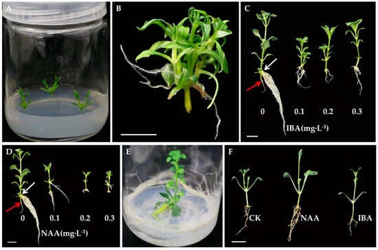
Figure 1.
Tissue culture process of Pseudostellaria heterophylla (A) Axillary buds induced after one week; (B) Four-week-old micro-shoots developed in medium supplemented with 1.5 mg·L−1 of 6-benzyladenin (BA); (C,D) In vitro rooting on Murashige and Skoog (MS) medium with different concentrations of indole-3-butyric acid (IBA) and naphthaleneacetic acid (NAA) (white arrows indicated the site of adventitious roots growth, and red arrows indicated the roots formed at the base of the stem); (E) Spindle-shaped root appeared after three months of culturing; (F) Ex vitro rooting in treatments with 100 mg·L−1 of NAA and IBA (scale = 1 cm).
3.2. Effects of Different Concentrations of Auxin on Rooting In Vitro
To identify conditions for optimal root growth of tissue culture seedlings, the well-developed microshoots of P. heterophylla were inoculated on rooting media with different concentrations of NAA and IBA (Table 2). Adventitious roots mainly appeared above the nodes (white arrow showed), fewer and shorter roots developed at the shoot base of microshoots cultured on the MS basic medium (Figure 1C,D, red arrow showed). Additionally, the rooting rate was significantly lower than that of microshoots cultured in auxin-added media. Both NAA and IBA significantly promoted in vitro rooting of P. heterophylla. The highest rooting rate (100%), root number (3.7), and root length (17.3 mm) were obtained in treatments with MS medium supplemented with 0.1 mg·L−1 IBA (Figure 1C). In the MS medium with 0.1 mg·L−1 NAA, the number of induced roots was 3.4, and the fibrous roots were longer and more abundant compared with the main roots. The treatments with 0.2 mg·L−1 NAA and 0.3 mg·L−1 NAA induced fewer and shorter roots than the treatment with 0.1 mg·L−1 NAA, and caused callus formation (Figure 1D). Spindle-shaped roots were visible after three months of culturing in IBA-added media (Figure 1E). In conclusion, MS medium with 0.1 mg·L−1 IBA was the most efficient for the induction of in vitro rooting in P. heterophylla.
3.3. Effects of Different Concentrations of Auxin on Ex Vitro Rooting
To reduce the time, cost, and labor of the micropropagation process, stem segments of the microshoots were inserted into the nutrient soil mixed with perlite at a ratio of 1:2. The effects of NAA and IBA on ex vitro rooting in P. heterophylla were observed after four weeks (Figure 1F). The best results were observed in treatment with 100 mg·L−1 NAA; the rooting rate was 86.7%, six roots developed per explant, the average root length was 18.8 mm, and plant height was taller than those in other treatments (Table 3). Thus, NAA was the most suitable auxin for ex vitro rooting of P. heterophylla.
Table 3.
Effects of different types of auxin on ex vitro rooting of P. heterophylla.
3.4. Comparison of In Vitro and Ex Vitro Rooting
The effects of in vitro (Figure 1C) and ex vitro (Figure 1F) rooting of P. heterophylla on growth were significantly different. The number of roots obtained by ex vitro culturing was significantly greater than that developed by in vitro (Table 4). Although the rooting rate of the in vitro treatment was higher than that of the ex vitro treatment (Table 4), the root systems of in vitro cultured seedlings were poorly developed, and the survival rate of transplants was low (data not shown). Most importantly, the plants cultivated by ex vitro rooting, which was similar to that occurring naturally, did not form any callus at the base of the cuttings, whereas the plants cultured in vitro developed a callus (Figure 1D, Table 4). These results indicated that ex vitro rooting of P. heterophylla was more suitable for root development than in vitro rooting.
Table 4.
Comparison of in vitro and ex vitro rooting.
3.5. Comparative Analysis of Leaf Micromorphology
The comparison of the leaf stoma micromorphology under in vitro and ex vitro rooting conditions revealed that the leaf development of plantlets in both treatments have undergone significant changes. Morphologically, the leaves were pale green in in vitro-rooted plantlets and bright green in ex-vitro rooted plantlets. The number of contiguous stomata was greater in the leaves of in vitro-rooted plantlets, but they were rarely observed in ex vitro-rooted plantlets (Figure 2C). In the in vitro-rooted plantlets, the stomata were open (Figure 2A,C), while those of the ex vitro-rooted plantlets were closed and uniform in size and distribution (Figure 2B,D). Stomatal density was significantly higher in the leaves of in vitro plantlets (58.4 ± 9.63) than in the leaves of ex vitro plantlets (26.9 ± 2.23).
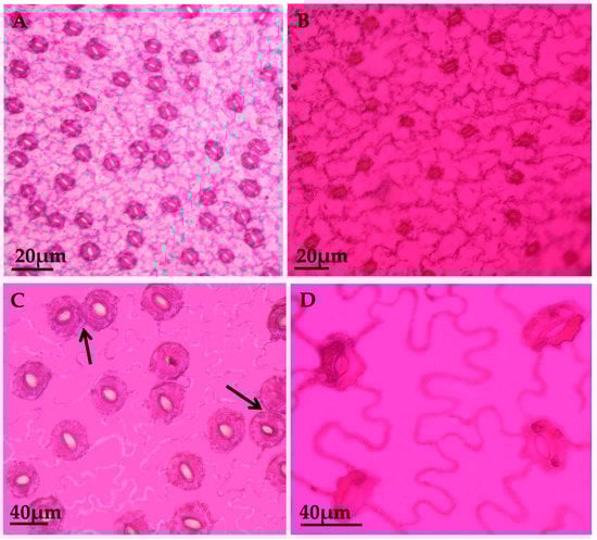
Figure 2.
Foliar epidermal peels of abaxial surfaces of P. heterophylla (A) Leaf stomata of In vitro-raised plants; (B) Leaf stomata of ex vitro-raised plants after four weeks of culture; (C,D) Magnified view of stomata from in vitro and ex vitro plantlets, respectively (arrows indicate contiguous stomata).
4. Discussion
Plant growth regulators (PGRs) are important for tissue culture and plant growth [26,27,28]. Different PGRs exhibit different effects on the induction and proliferation of axillary buds in P. heterophylla. Compared with KT and TDZ, 6-BA was more effective in stimulating proliferation of axillary buds in stem nodes, and 1.5 mg·L−1 6-BA provided the optimal concentration for axillary induction. Similar results have been reported for the induction of adventitious buds using Crocus vernus bulbs as explants [29], and for the induction of axillary buds using Caralluma edulis segments [30].
To develop a protocol for the fast propagation of P. heterophylla plants with a well-developed root system, we performed both in vitro and ex vitro rooting. In the in vitro-rooted plantlets, IBA was more effective than other auxins in root induction. Similarly, IBA was more efficient than other auxins in the in vitro rooting of Randia dumetorum [31], Anthocephalus cadamba [32], and Morinda officinalis [33]. Although the NAA-induced rooting ratio was higher than that of IBA, the number of roots induced by NAA was less than that of IBA (Table 2), and the roots induced by NAA formed a callus. This was in line with a report by Zhang et al. [34], who found that NAA can induce callus formation in Beta vulgaris. Therefore, IBA was more conducive to root induction than NAA in P. heterophylla.
The highest rooting rate of ex vitro-rooted plantlets was obtained from the treatment with 100 mg·L−1 NAA for 1 min. Similar results were reported by Yan et al. [35] for Siratia grosvenorii nodes that had been dipped in 100 mg·L−1 NAA for 1 min. The rooting rate of P. heterophylla dipped in NAA was 86.7%. Drisya Ravi et al. [36] obtained a similar rooting rate of 83.3% in Moringa oleifera ex vitro microshoots after they were treated for 30 s with 50 μM NAA. The low rooting rate of ex vitro plantlets treated with NAA was attributed to drastic changes in the environment as the in vitro plantlets were transferred from a sterile environment with high temperature, high humidity, and weak light to a natural environment inhabited by bacteria. Weak or young seedlings were unable to adjust to the unsuitable environmental conditions and eventually expired [30,37,38,39,40].
The root morphologies of in vitro and ex vitro P. heterophylla were significantly different. In vitro grown roots were thick and fragile, lacking root hairs. Kataoka [41] studied the root morphology of Carica papaya and found that roots growing in agar medium showed structural abnormalities and often lacked root hairs, which had adverse effects on their growth in field soil. The roots of Lonicera caerulea microcuttings developed by Karhu [42] through an in vitro method exhibited the same abnormalities. In contrast, the ex vitro roots assumed full growth after transplantation, the plantlets had a fully developed rooting system, and no callus was formed at the root base (Figure 1F). Similarly, ex vitro-rooted plantlets of Siraitia grosvenorii developed lateral roots and lacked callus at the base of the microshoots [35]. During the rooting process, ex vitro-rooted plantlets gradually adapted to the natural environment [19]. Therefore, ex vitro rooting is more suitable than in vitro for root development in P. heterophylla.
In production at commercial-scale, the formation of callus adversely affects the growth of plants. Perlite is a fibrous material that can enhance root growth and development and has been found to reduce callus formation successfully in S. grosvenorii [35]. Li [43] suggested that perlite, due to the presence of large spaces, light weight, good air permeability, and water retention capacity, facilitated the survival of ex vitro-rooted Euphorbia pulcherrima microshoots, and reported that the survival rate reached 83.8%. The ex vitro rooting medium used in the present study was perlite, and the resulting plantlets of P. heterophylla developed almost no callus at their base (Figure 1F), indicating that perlite is suitable for large-scale propagation of P. heterophylla in field conditions.
The micromorphological studies on the foliar apparatus of in vitro- and ex vitro-rooted plantlets revealed developmental changes in stomatal structure and stomatal density that took place as the plantlets were transferred from in vitro to ex vitro conditions. These changes in stomatal structure were responsible for establishing proper biochemical and physiological metabolisms during microshoot development [44,45]. The contrast in leaf color between in vitro and ex vitro P. heterophylla plantlets expressed differences in the state of development of photosynthetic tissue. According to Kozai et al. [46] and Gaspar et al. [47], underdeveloped photosynthetic organs occurred in in vitro microshoots due to an insufficient carbon source in the culture medium. On the other hand, plantlets in the field environment had a completely developed photosynthetic tissue.
The stomata on leaves of in vitro-developed P. heterophylla plants were contiguous and open, the stomatal density was high (58.4 ± 9.63), and the stomatal function was abnormal. Meanwhile, the ex vitro plants exhibited closed stomata, and the stomatal density was low (26.9 ± 2.23) compared with that of the in vitro plants. Previous studies on Passiflora edulis [48], Morinda coreia [23], and Oldenlandia corymbosa [45] reported stomatal differences between in vitro and ex vitro plantlets that were consistent with our results on in vitro and ex vitro plantlets of P. heterophylla. Plants developed in vitro had weaker water control mechanisms, leading to inefficient physiological and metabolic processes, which hampered plants’ survival during field transplanting [48]. The impaired stomatal development was explained by high hydration of in vitro-grown plants [49]. Saez et al. [50] believed that the low light conditions of the in vitro environment were the cause of stomatal damage. Therefore, ex vitro rooting favored the growth of plantlets, improved their quality, and saved the labor, energy, and cost associated with tissue culture.
5. Conclusions
This experiment described a simple and efficient protocol for P. heterophylla micropropagation. A large number of micropropagated plantlets could be obtained from MS medium containing 1.5 mg·L−1 6-BA by the plant tissue culture technique within a month, plantlets treated with 100 mg·L−1 NAA for 1 min under the ex vitro-rooting conditions have the highest quality and are suitable for large-scale commercial production. The study on leaf morphology revealed the adaptability of micropropagated plants to the outside environment. In addition, the ex vitro rooting does not require sterilization and combines rooting and transplanting processes, which ultimately reduces the cost of cultivation, saves time, and conserves labor invested for plant acclimatization. Therefore, the present method is suitable for the large-scale agricultural production of P. heterophylla plants.
Author Contributions
Conceptualization, B.L. and F.W.; methodology, B.L. and F.W.; software, F.W., X.X. and H.W.; validation, B.L.; formal analysis, B.L., F.W. and X.Q.; investigation, F.W., X.X. and. X.Q.; resources, B.L.; data curation, F.W., H.W. and X.Q.; writing—original draft preparation, F.W. and B.L.; writing—review and editing, B.L. and F.W.; visualization, F.W. and H.W.; supervision, B.L.; project administration, B.L.; funding acquisition, B.L. All authors have read and agreed to the published version of the manuscript.
Funding
This work was funded by the National Natural Science Foundation of China (Grant 31700624).
Conflicts of Interest
The authors declare no conflict of interest.
References
- Yin, S.S.; Gao, W.Y.; Liang, Y.Y.; Wang, J.; Liu, H.; Wei, C.L.; Zuo, B.M. Influence of sucrose concentration and phosphate source on biomass and metabolite accumulation in adventitious roots of Pseudostellaria heterophylla. Acta Physiol. Plant. 2013, 35, 1579–1585. [Google Scholar] [CrossRef]
- Xu, W.Y.; Zhu, H.T.; Tan, N.H.; Tang, J.; Zhang, Y.J.; Cerny Ron, L.; Du, L.C. An in vitro system to study cyclopeptide heterophyllin B biosynthesis in the medicinal plant Pseudostellaria heterophylla. Plant Cell Tissue Organ Cult. 2012, 108, 137–145. [Google Scholar] [CrossRef]
- Yan, P.L.; Wang, Z.H.; Jin, Z.F.; Gao, Y.; Qiu, F.T. Study on tissue culture and rapid propagation of Pseudostellaria heterophylla. Anhui Agri. Sci. Bull. 2016, 22, 50–52. (In Chinese) [Google Scholar]
- Deng, Y.; Han, B.X.; Hu, D.J.; Zhao, J.; Li, S.P. Qualitation and quantification of water soluble non-starch polysaccharides from Pseudostellaria heterophylla in China using saccharide mapping and multiple chromatographic methods. Carbohydr. Polym. 2018, 199, 619–627. [Google Scholar] [CrossRef] [PubMed]
- Liang, Y.Y.; Liao, L.; Zuo, B.M.; Gao, W.Y. Study on adventitious roots induction and proliferation culture of Pseudostellaria heterophylla. North. Hortic. 2013, 2, 147–149. (In Chinese) [Google Scholar]
- Peng, Y.S.; Chen, R.; Yang, R.D. Analysis of heavy metals in Pseudostellaria heterophylla in Baiyi Country of Wudang District. J. Geochem. Explor. 2016, 176, 57–63. [Google Scholar] [CrossRef]
- Yin, S.S.; Liang, Y.Y.; Gao, W.Y.; Wang, J.; Jing, S.S.; Zhang, Y.; Liu, H. Influence of medium salt strength and nitrogen source on biomass and metabolite accumulation in adventitious root cultures of Pseudostellaria heterophylla. Acta Physiol. Plant. 2013, 35, 2623–2628. [Google Scholar] [CrossRef]
- Ye, Z.Y.; Wang, Y.Y.; Tian, H.Q. Regeneration of plantlets and tetraploidy induction in Pseudostellaria heterophylla. Acta Biol. Cracov. Ser. Bot. 2009, 51, 13–18. [Google Scholar]
- Ma, X.M.; Wu, C.F.; Wang, G.R. Application of artificial seeds in rapid multiplication of Pseudostellaria heterophylla. Afr. J. Biotechnol. 2011, 10, 15744–15748. [Google Scholar] [CrossRef]
- Gao, W.; Zhang, J.S.; Zhang, J.H. Detection and control of Taizishen mosaic virus. Virol. Sin. 1993, 8, 390–393. (In Chinese) [Google Scholar]
- Yu, C.X.; Zheng, X.F.; Deng, Y.H.; Huang, G. Tissue culture for Pseudostellaria heterophylla. Guizhou Agric. Sci. 2004, 32, 16–17. (In Chinese) [Google Scholar]
- Francis, S.V.; Senapati, S.K.; Rout, G.R. Rapid clonal propagation of Curculigo orchioides Gaertn., an endangered medicinal plant. In Vitro Cell. Dev. Biol. Plant 2007, 43, 140–143. [Google Scholar] [CrossRef]
- Joshi, P.; Dhawan, V. Axillary multiplication of Swertia chirayita (Roxb. Ex Fleming) H. Karst., a critically endangered medicinal herb of temperate Himalayas. In Vitro Cell. Dev. Biol. Plant 2007, 43, 631–638. [Google Scholar] [CrossRef]
- Sivanesan, I.; Jeong, B.R. Direct shoot regeneration from nodal explants of Sida cordifolia Linn. In Vitro Cell. Dev. Biol. Plant 2007, 43, 436–441. [Google Scholar] [CrossRef]
- Offord Catherine, A.; Tyler Joanne, L. In vitro propagation of Pimelea spicata R.Br (Thymelaeaceae), an endangered species of the Sydney region, Australia. Plant Cell Tissue Organ Cult. 2009, 98, 19–23. [Google Scholar] [CrossRef]
- Goncalves, S.; Fernandes, L.; Romano, A. High-frequency in vitro propagation of the endangered species Tuberaria major. Plant Cell Tissue Organ Cult. 2010, 101, 359–363. [Google Scholar] [CrossRef]
- Ali, A.; Afrasiab, H.; Naz, S.; Rauf, M.; Iqbal, J. An efficient protocol for in vitro propagation of carnation (Dianthus caryophyllus). Pak. J. Bot. 2008, 40, 111–121. [Google Scholar]
- Kritskaya, T.A.; Kashin, A.S.; Spivak, V.A.; Firstov, V.E. Features of clonal micropropagation of Silene cretacea (caryophyllaceae) in in vitro culture. Russ. J. Dev. Biol. 2016, 47, 359–366. [Google Scholar] [CrossRef]
- Benmahioul, B.; Dorion, N.; Kaid-Harche, M.; Daguin, F. Micropropagation and ex vitro rooting of pistachio (Pistacia vera L.). Plant Cell Tissue Organ Cult. 2012, 108, 353–358. [Google Scholar] [CrossRef]
- Baskaran, P.; Van Staden, J. Rapid in vitro micropropagation of Agapanthus praecox. S. Afr. J. Bot. 2013, 86, 46–50. [Google Scholar] [CrossRef]
- Pate, l.A.K.; Agarwal, T.; Phulwaria, M.; Kataria, V.; Shekhawat, N.S. An efcient in vitro plant regeneration system from leaf of mature plant of Leptadenia reticulata (Jeewanti): A life giving endangered woody climber. Ind. Crop. Prod. 2014, 52, 499–505. [Google Scholar] [CrossRef]
- Liu, B.L.; Fang, H.Z.; Meng, C.R.; Chen, M.; Chai, Q.D.; Zhang, K.; Liu, S.J. Establishment of a rapid and efficient micropropagation system for succulent plant Haworthia turgida haw. HortScience 2017, 52, 1278–1282. [Google Scholar] [CrossRef]
- Shekhawat Mahipal, S.; Manokari, M.; Kannan, N. Micromorphological response towards altered environmental conditions in subsequent stages of in vitro propagation of Morinda coreia. Environ. Exp. Biol. 2017, 15, 37–46. [Google Scholar]
- Chen, H.B.; Zheng, Y.J.; Li, F.Z. Flora of Shandong Province; Qingdao Publishing House: Qingdao, China, 1990; Volume 1, pp. 1133–1137. [Google Scholar]
- Johansen, D.A. Plant Microtech.; McGraw Hill Book Co.: New York, NY, USA, 1940; Volume 1, pp. 182–197. [Google Scholar]
- Frello, S.; Venerus, E.; Serek, M. Regeneration of various species of Crassulaceae, with special reference to Kalanchoë. J. Hortic. Sci. Biotechnol. 2002, 77, 204–208. [Google Scholar] [CrossRef]
- Kordi, M.; Kaviani, B.; Hashemabadi, D. In vitro propagation of Kalanchoe blossfeldiana using BA and NAA. Eur. J. Exp. Biol. 2013, 3, 285–288. [Google Scholar]
- Peeters Anton, J.M.; Gerards, W.; Barendse Gerard, W.M.; Wullems George, J. In vitro flower bud formation in tobacco: Interaction of hormones. Plant Physiol. 1991, 97, 402–408. [Google Scholar] [CrossRef]
- Sivanesan, I.; Jana, S.; Jeong, B.R. In vitro shoot regeneration and microcorm development in Crocus vernus (L.) Hill. Pak. J. Bot. 2014, 46, 693–697. [Google Scholar]
- Patel Ashok, K.; Phulwaria, M.; Rai Manoj, K.; Gupta Amit, K.; Shekhawat, S.; Shekhawat, N.S. In vitro propagation and ex vitro rooting of Caralluma edulis (Edgew.) Benth. & Hook. f.: An endemic and endangered edible plant species of the Thar Desert. Sci. Hortic. 2014, 165, 175–180. [Google Scholar]
- Begum, F.; Islam Kazi, M.D.; Paul, R.N.; Mehedi, M.; Rani, S. In vitro propagation of emetic nut Randia dumetorum (Lamb.). Indian J. Exp. Biol. 2003, 41, 1479–1481. [Google Scholar]
- Joshi, A.; Mathur, N. In vitro propagation and conservation of Anthocephalus cadamba through apical bud and nodal explants—A valuable medicinal plant. CIBTech J. Biotechnol. 2015, 4, 8–18. [Google Scholar]
- Deng, Z.C.; Jin, H.; He, H. An efficient micropropagation system for Morinda officinalis How. (Rubiaceae), an Endangered Medicinal Plant. J. Agric. Sci. Technol. 2015, 17, 1609–1618. [Google Scholar]
- Zhang, C.L.; Chen, D.F.; Elliott Malcolm, C.; Slater, A. Efficient procedures for callus induction and adventitious shoot organogenesis in sugar beet (Beta vulgaris L.) breeding lines. In Vitro Cell. Dev. Biol. Plant 2004, 40, 475–481. [Google Scholar] [CrossRef]
- Yan, H.B.; Liang, C.X.; Yang, L.T.; Li, Y.R. In vitro and ex vitro rooting of Siratia grosvenorii, a traditional medicinal plant. Acta Physiol. Plant. 2010, 32, 115–120. [Google Scholar] [CrossRef]
- Drisya Ravi, R.S.; Siril, E.A.; Nair, B.R. The effect of silver nitrate on micropropagation of Moringa oleifera Lam. An important vegetable crop of tropics with substantial nutritional value. Physiol. Mol. Biol. Plants 2019, 25, 1311–1322. [Google Scholar] [CrossRef]
- Xu, Z.H.; Bai, Z.Y.; Lin, Y.; Wang, X.Y.; Zhao, S.P. Anatomical observation on root of test-tube seedlings in Rosa. Acta Hortic. Sin. 1998, 25, 405–407. (In Chinese) [Google Scholar]
- Chen, Z.L.; Xue, H.; Feng, Y.; Yan, Z.L.; Qi, L. First probe into of a certain factors affecting the tissue culture seedlings of the Jujuba under outer test-tube root formation. J. Yanan Univ. 1996, 15, 46–49. (In Chinese) [Google Scholar]
- Phulwaria, M.; Shekhawat, N.S.; Rathore, J.S.; Singh, R.P. An efficient in vitro regeneration and ex vitro rooting of Ceropegia bulbosa Roxb.—A threatened and pharmaceutical important plant of Indian Thar Desert. Ind. Crops Prod. 2013, 42, 25–29. [Google Scholar] [CrossRef]
- Rathore, J.S.; Rai Manoj, K.; Phulwaria, M.; Shekhawat, N.S. A liquid culture system for improved micropropagation of mature Acacia nilotica (L.) Del. ssp. indica and ex vitro rooting. Proc. Natl. Acad. Sci. India Sect. B Biol. Sci. 2014, 84, 193–200. [Google Scholar] [CrossRef]
- Kataoka, I. Influence of rooting substrates on the morphology of papaya root formed in vitro. Jpn. J. Trop. Agric. 1994, 38, 251–257. [Google Scholar]
- Karhu, S.T. Rooting of blue honeysuckle microshoots. Plant Cell Tissue Organ Cult. 1997, 48, 153–159. [Google Scholar] [CrossRef]
- Li, H.X. Study on rooting technique of Euphorbia pulcherrima in vitro. Gansu Agric. Sci. Technol. 2000, 11, 46–47. (In Chinese) [Google Scholar]
- Carvalho Luisa, C.; Leonor Osório, M.; Manuela Chaves, M.; Amâncio, S. Chlorophyll fluorescence as an indicator of photosynthetic functioning of in vitro grapevine and chestnut plantlets under ex vitro acclimatization. Plant Cell Tissue Organ Cult. 2001, 67, 271–280. [Google Scholar] [CrossRef]
- Revathi, J.; Manokari, M.; Shekhawat, M.S. Optimization of factors affecting in vitro regeneration, flowering, ex vitro rooting and foliar micromorphological studies of Oldenlandia corymbosa L.: A multipotent herb. Plant Cell Tissue Organ Cult. 2018, 134, 1–13. [Google Scholar] [CrossRef]
- Kozai, T.; Iwabuchi, K.; Watanabe, K.; Watanabe, I. Photoautotrophic and photomixotrophic growth of strawberry plantlets in vitro and changes in nutrient composition of the medium. Plant Cell Tissue Organ Cult. 1991, 25, 107–115. [Google Scholar]
- Gaspar, T.; Franck, T.; Bisbis, B.; Kevers, C.; Jouve, L.; Hausman, J.F.; Dommes, J. Concepts in plant stress physiology. Application to plant tissue cultures. Plant Growth Regul. 2002, 37, 263–285. [Google Scholar] [CrossRef]
- Manokari, M.; Shekhawat, M.S. Comprehensive analysis of in vitro to feld transition of micromorphology and leaf architecture in Passiflora edulis Sims. f. flavicarpa Deg. Ind. J. Plant Physiol. 2017, 22, 240–246. [Google Scholar] [CrossRef]
- Machado, M.P.; Silva, A.L.L.; Biasi, L.A.; Deschamps, C.; Filho, J.C.B.; Zanette, F. Influence of calcium content of tissue on hyperhydricity and shoot tip necrosis of in vitro regenerated shoots of Lavandula angustifolia Mill. Braz. Arch. Biol. Technol. 2014, 57, 636–643. [Google Scholar] [CrossRef]
- Saez, P.L.; Bravo, L.A.; Latsagne, M.I.; Rios, D.G. Increased light intensity during in vitro culture improves water loss control and photosynthetic performance of Castanea sativa grown in ventilated vessels. Sci. Hortic. 2012, 138, 7–16. [Google Scholar] [CrossRef]


© 2020 by the authors. Licensee MDPI, Basel, Switzerland. This article is an open access article distributed under the terms and conditions of the Creative Commons Attribution (CC BY) license (http://creativecommons.org/licenses/by/4.0/).