Article

Growth Promotion of Rapeseed (Brassica napus L.) and Blackleg Disease (Leptosphaeria maculans) Suppression Mediated by Endophytic Bacteria

1
The AgroBioTech Research Centre (ABT RC), Slovak University of Agriculture in Nitra, 949 01 Nitra, Slovakia
2
Faculty of Biotechnology and Food Sciences, Slovak University of Agriculture in Nitra, Trieda Andreja Hlinku 2, 949 01 Nitra, Slovakia
3
Institute of Forest Ecology, Slovak Academy of Sciences, Detached Branch Mlynany Arboretum, Vieska nad Zitavou 178, 949 01 Nitra, Slovakia
*
Author to whom correspondence should be addressed.
Agronomy 2021, 11(10), 1966; https://doi.org/10.3390/agronomy11101966
Submission received: 30 August 2021 / Revised: 17 September 2021 / Accepted: 21 September 2021 / Published: 29 September 2021

Abstract

:
Rapeseed is an important oil crop strongly dependent on high agrochemical inputs. Some pathogens, including Leptosphaeria maculans, cause blackleg disease and can drastically decrease yields. Microbial inoculants seem to be a promising solution to these problems. However, a selection of potent bacterial strains able to improve growth and/or suppress disease is needed. Endophytic bacteria (n = 38) isolated from rapeseed plants with exceptionally good growth were screened for plant growth promoting (PGP) traits and L. maculans antifungal activity. A majority of isolates (35) showed the ability to produce siderophores, 17 isolates solubilized phosphate, and 28 isolates inhibited the growth of L. maculans. The six most promising isolates belonging to Bacillus genera were characterized in detail and compared to two previously published PGP strains. Plant growth measured as total weight and root length of rapeseed seedlings was stimulated by all isolates in comparison to control. The best isolate, 1L6, preliminary identified as Bacillus pumilus showed the highest phosphate solubilization, IAA and HCN production, and growth promotion of plants. Isolates with high antifungal activity in screening showed good potential to suppress disease on plants, with 87% reduction of lesions caused by L. maculans. These strains are good candidates to be explored under field use either solely or in combination.

1. Introduction

Rapeseed is widely cultivated in temperate climate regions mainly for the production of oily seeds used in human and animal nutrition [1]. Methylated rapeseed oil is a biofuel which is mixed into diesel in an effort to decrease fossil fuel usage and thus suppress climatic change [2]. However, rapeseed cultivation is heavily dependent on high inputs of agrochemicals, mainly fertilizers and pesticides that contradict sustainable agriculture intentions [3]. Microbial inoculants are intensively studied due to their promising properties to improve plant nutrition and health, which allows a reduction of agrochemical use [4,5].
However, with increased production of oilseed rape, blackleg disease, also called phoma stem canker, caused by Leptosphaeria maculans has become a disease of major economic importance in rapeseed production. It is considered to be a serious global plant disease. It occurs in epidemic proportions in most of the rapeseed producing regions in Europe [6,7]. Climate change may also exacerbate the spread of the disease, as under experimental conditions, higher temperatures (15 °C vs. 10 °C or 5 °C) have been shown to reduce spore germination time and increase mycelium penetration efficiency [8,9]. Several strategies such as crop rotation, chemical control, sanitation, and resistant cultivars have been advised for blackleg control [10]. The perceived health and environmental risks of fungicide use resulted in increased interest in alternative disease management strategies. Biocontrol seems to be a viable alternative which involves harnessing disease-suppressive microorganisms to improve plant health [11].
Microorganisms play a key role in the health and development of crops [12] by different direct or indirect mechanisms. Beneficial functions attributed to endophytic bacteria include plant growth promotion (PGP) by supplying nutrients (e.g., nitrogen fixation or phosphate solubilization), increased tolerance to biotic and abiotic stressors, detoxification of harmful compounds, and the production of bioactive compounds [13,14]. Direct biocontrol activity attributed to endophytic bacteria include the production of extracellular antibiotics, siderophores, and cell wall-degrading enzymes (chitinase, cellulose, β-1,3-glucanase) (Labuschagne et al., 2016). Various endophytic microorganisms have been categorized as plant growth promoting bacteria (PGPB) and/or biocontrol agents (BCAs). They are currently used in the formulation of diverse bioproducts (e.g., biofertilizers and biofungicides) in order to modify and/or introduce beneficial bacteria into the plant microbiome for agricultural purposes [15]. In the screening for potential inoculants of plants, auxin production, phosphate solubilization, siderophores release, and activity against pathogens belong to the most frequently reported traits of endophytic bacteria [16,17,18].
Indole-3-acetic acid (IAA), which is known for its key function in plant-bacterial interactions, is generally considered to be the most important phytohormone from the auxin group [19]. It affects the division and differentiation of plant cells, increases the rate of root growth and lateral root growth, affects photosynthesis and the biosynthesis of metabolites, antioxidant enzyme activity, and resistance to stress conditions [20].
Phosphorus (P) is one of the basic elements necessary for plant growth as it directly or indirectly affects all biological processes [21]. The availability of P to the root system is often minimal, despite the fact that its concentration may be high in some soils, because only 0.1% of P is in a form available for use in plants [22]. Due to the increasing problems with the content of available P in the soil, the application of P fertilizers is required in agriculture. This can lead to environmental problems such as eutrophication of water or contamination by heavy metals co-founded in P fertilizer [23,24].
Iron (Fe) is an essential micronutrient for plants and microorganisms. Under aerobic conditions (especially in calcareous soils), the solubility of iron is low, which limits its supply to various life forms [25]. Bacteria have developed active Fe absorption strategies by which they are able to overcome nutritional limitations by using chelating agents called siderophores, which bind Fe3+ and transport it to the root surface where it is reduced to Fe2+ and absorbed by the plant [26]. The ability to produce siderophores provides competitive advantages for endophytic bacteria during the colonization of plant tissues and elimination of other microorganisms [27]. Microbial siderophores are important to plant pathology as determinants of biocontrol activity and/or ecological factors influencing the iron nutrition of plants. They are also important in induced systemic resistance [28].
Hydrogen cyanide (HCN) production is another important attribute of endophytic bacteria. It can inhibit the function of many enzymes or protein carriers and, in specific cases, is likely to inhibit the growth of certain organisms. Despite the fact that, according to Ramette et al. [29], HCN plays a role in suppressing diseases, it is not a universal biological control tool because a there is no direct evidence about the relationship between HCN production and biocontrol of phytopathogens [30]. Previous studies of PGPB confirmed the presence of HCN-producing strains in the rhizosphere and in the plant [31]. Rijavec and Lapanje [32] suggested the role of HCN in the regulation of the availability of phosphates for rhizobacteria and microbial plant hosts. Production of hydrogen cyanide increases with increasing concentrations of iron and phosphates, suggesting their specific relation [33].
Many studies described a successful isolation of PGPB that greatly improved plant growth. However, there is constant effort to isolate new, more effective, better adapted strains of microorganisms for agricultural use [34]. Internal plant tissue is commonly targeted for this as it is predicted that endophytes are well adapted to plants and they will survive in this specific environment [35]. We hypothesize that endophytes isolated from rapeseed should have a better ability to colonize their hosts than endophytes from non-related plant species or bacteria from other environments.
The objective of this study was to evaluate the plant growth properties of endophytic bacteria isolated from rapeseed by analysis of IAA, HCN and siderophore production as well as phosphate solubilization, and also by analysis of growth and pathogen development in inoculated rapeseed plants in comparison with previously described PGPB.

2. Materials and Methods

2.1. Plant Samples

Plants of winter rapeseed (Brassica napus L., variety Sidney) used for the isolation of endophytic bacteria were collected from several plots in the experimental farm of the Slovak University of Agriculture in Nitra (SUA) located in Kolíňany, Slovakia (40°26′46″ N, 79°58′56″ W). Conventional farming practices, including mineral fertilization and pesticide use, were used on the rapeseed canopy. Plants were collected in various stages, BBCH 15 (growth of 2–4 true leaves), BBCH 55 (flowering), and BBCH 85 (maturity) during the 2017/2018 season. Plants of rapeseed without any disease symptoms and with exceptionally good growth according to visual evaluation of plant height and biomass were selected for endophyte evaluation. Plants were carefully taken with a block of soil and transferred to the laboratory of Department of Microbiology, SUA in Nitra.

2.2. Isolation of Endophytic Bacteria

Roots, stems, and leaves were repeatedly washed under tap water to remove adhering soil particles, debris and epiphytic microorganisms. The plants were cut, and individual parts were subjected to three-step surface sterilization using 99% ethanol for 1 min, then 3.125% sodium hypochlorite solution for 6 min, and 99% ethanol for 30 s followed by a 3-times repeated final wash in sterile distilled water [36]. Ten grams of sterilized plant material was homogenized with 90 mL of sterile physiological solution by blender, inoculated onto tryptone soya agar plates, and incubated at 30 °C for 24 h. After incubation, colonies of each morphological type were re-inoculated and primarily screened for PGP traits.

2.3. Primary Screening of Endophytic Bacteria

Primary screening comprised qualitative methods for assessing production of siderophores (CAS medium) [37] and phosphate solubilization (Pikovskaya medium) [38].
To test the biocontrol activity of isolates, an antifungal bioassay against L. maculans (isolate Kmi16JM002 obtained from Brassica napus lesion in 2016) was done on potato dextrose agar. Hundred microliters of bacterial culture were dispensed carefully on the plates and incubated at 30 °C for 24 h. The control plates were not inoculated by any bacteria. Mycelial plugs of L. maculans (5 mm diameter) were cut out from the edges of an actively growing colony and placed mycelial side down on the agar, at the center of the assay plates. The plates were incubated under light at 25 °C. The percentage of inhibition (%) was calculated using the equation published by Fernando and Pierson [39]: R1 − R2 = R1/100, where R1 is radius of mycelium growth on the control plate and R2 is radius of mycelium growth on the experimental plate.
Bacterial isolates tested in primary screening were identified using MALDI TOF mass spectrometry. One colony from each bacterial strain was harvested and deposited on a MALDI target plate by sterile toothpick and left to dry. After drying, two microliters of matrix solution (saturated α-cyano-4-hydroxycinnamic acid, 50% acetonitrile, 2.5% trifluoroacetic acid) were added and allowed to co-crystallize with the sample. Samples were analyzed with a MALDI-TOF MS Microflex LT mass spectrometer (Bruker Daltonics, Bremen, Germany). Acquired spectra were compared to MALDI biotyper database.
Six isolates with the highest combined scores from primary screening were subjected to molecular identification and detailed enzymatic and physiological characterization using Biolog GEN III plates, quantitative measurement of PGP traits and in planta assay. Two previously described plant growth promoting bacterial strains, namely Bacillus velezensis QST713, an active ingredient of Serenade ASO (Bayer CropScience GmbH), and Pseudomonas simiae WCS417 were used for comparison of the assessed parameters of endophytic isolates.

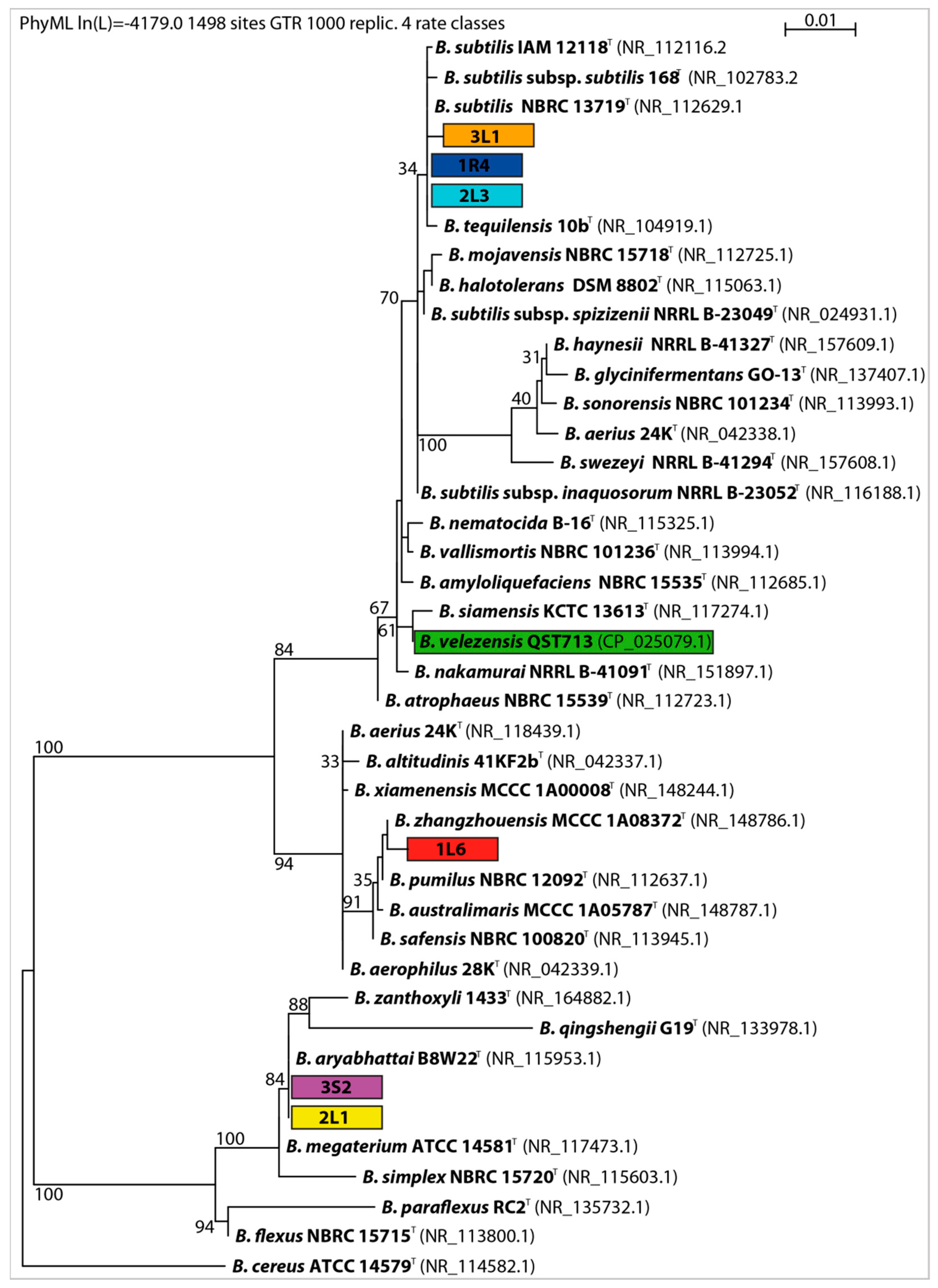
2.4. Molecular Characterization

Approximately 50 mg of bacterial culture was placed in 150 μL of PrepMan solution (Life Technologies, Carlsbad, CA, USA). The 16S rRNA gene of the DNA was amplified using universal primers 27 F (5′-AGAGTTTGATCCTGGCTCAG-3′) and 1492R (5′-TACGGTTACCTTGTTACGACTT-3′). PCR products were sequenced by the MacroGen Company, Seoul, Korea. The homology of partial 16S rRNA sequences was BLAST-checked against the GenBank (www.genbank.com, accessed on 13 August 2021). 16S rRNA gene database, limited to type strains. Sequences of type strains with similarity higher than 96% were selected for phylogenetic analysis. The DNA sequences were aligned by MUSCLE [40], and the maximum likelihood phylogenetic tree was constructed using phyML [41] in the Seaview4 environment [42].

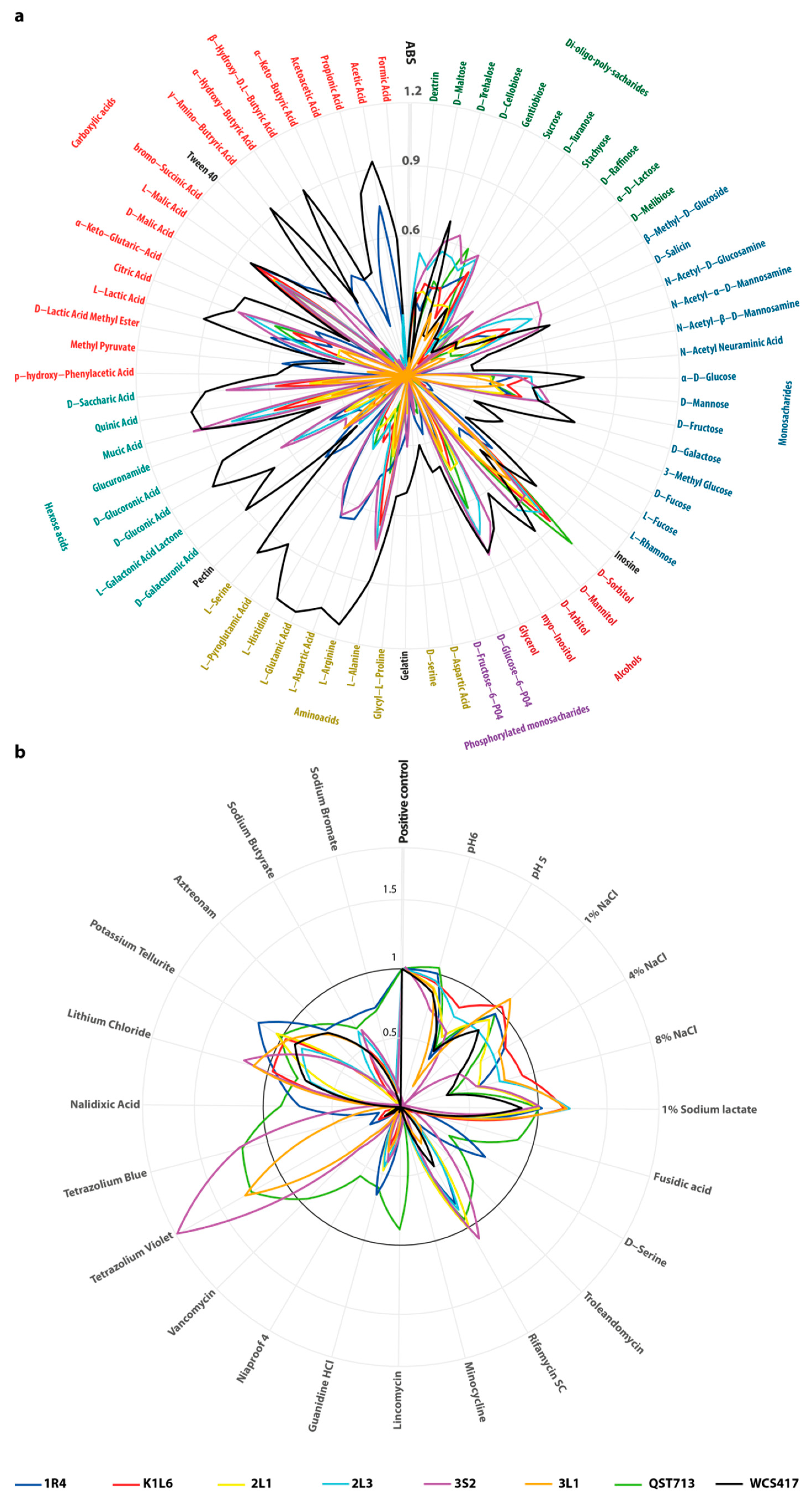
2.5. Enzymatic and Physiological Characterization

Catalase, urease, and cytochrome c oxidase of the bacterial isolates were assayed according to Collins et al. [43].
Physiological evaluation was done using the Biolog Gen III system. Microplates were incubated at 33 °C and optical density at 570 nm was measured after 24 h on the GloMax Multi Detection System (Promega, USA). Optical density values of all wells were corrected using the negative control well. Sensitivity wells were compared to the positive control well.

2.6. Quantitative Analysis of PGP Traits

IAA production was determined according to the method of Kumar [44]. Endophytic bacterial isolates (100 µL of bacterial culture with density 0.5 McFarland) were inoculated into NB (Nutrient Broth) media containing 0.1% (v/v) L-tryptophan and incubated at 30 °C for 7 days with vigorous shaking. Each day, 1 mL of sample was centrifuged at 10,000 rpm for 10 min and mixed with 400 µL of Salkowski reagent (35% HClO4, FeCl3). Negative control, represented by non-inoculated media, was spiked by an exact amount of IAA (0–200 mg·L−1) to obtain calibration curve points. For IAA concentration, optical density at 535 nm was measured after 30 min incubation of samples at room temperature, and compared to the calibration curve.
Phosphate solubilization was quantified by method of Nautial [45]. Bacteria were cultivated in NBRIP growth medium (National Botanical Research Institute’s phophate). After centrifugation, supernatant was acidified to pH below 2 by 4.5 M H2SO4, and 100 µL of the mixture was pipetted into a microplate well. Acidified supernatant was mixed with 40 µL of reagent A containing ammonium molybdate and potassium antimonyl-tartrate, 80 µL of reagent B containing ascorbic acid, and 280 µL of water. Optical density was measured at 882 nm and phosphorus concentration was calculated using the calibration curve.
Quantification of HCN production was performed according to Rijavec and Lapanje [32]. Bacteria were cultivated in LB broth with glycine for seven days. Each day, one milliliter of culture was centrifuged, 190 µL of culture supernatant was mixed with 10 µL of methemoglobine reagent in a microplate. The mixture was incubated for 30 min and the optical density at 422 nm was measured. Hydrogen cyanide production was calculated using the calibration curve.
Siderophore production was quantified using the protocol of Arora and Verma [46]. One-half milliliter of LB broth supernatant was mixed with 0.5 mL of CAS reagent, incubated for 20 min, and measured at 630 nm. Siderophore production was calculated as (Ar-As) ×100/Ar; where Ar is the absorbance of non-inoculated media and As is the absorbance of the sample.
For all quantitative analyses, bacterial isolates were cultivated in triplicate. All optical density measurements were performed using the LB 943 Multimode Reader Mithras (Berthold Technologies GmbH, Bad Wildbad, Germany).

2.7. In Planta Assay

Six endophytic isolates together with two reference plant growth-promoting strains were used for inoculation of rapeseed seedlings. Seeds of rapeseed cultivar ES Astrid were used for in planta assay.
Bacteria were cultivated on a Petri dish with TSA medium and incubated at 30 °C, 24 h. The bacterial inoculum was prepared by suspension of the bacterial colony in sterile water and the suspension was diluted to approximately 1.5 × 10⁸ CFU·mL−1 using a calibrated turbidimeter (DensiLa meter, Lachema, Brno, Czech Republic). In all in planta experiments, sterile water instead of the bacterial suspension treatment was used as negative control.

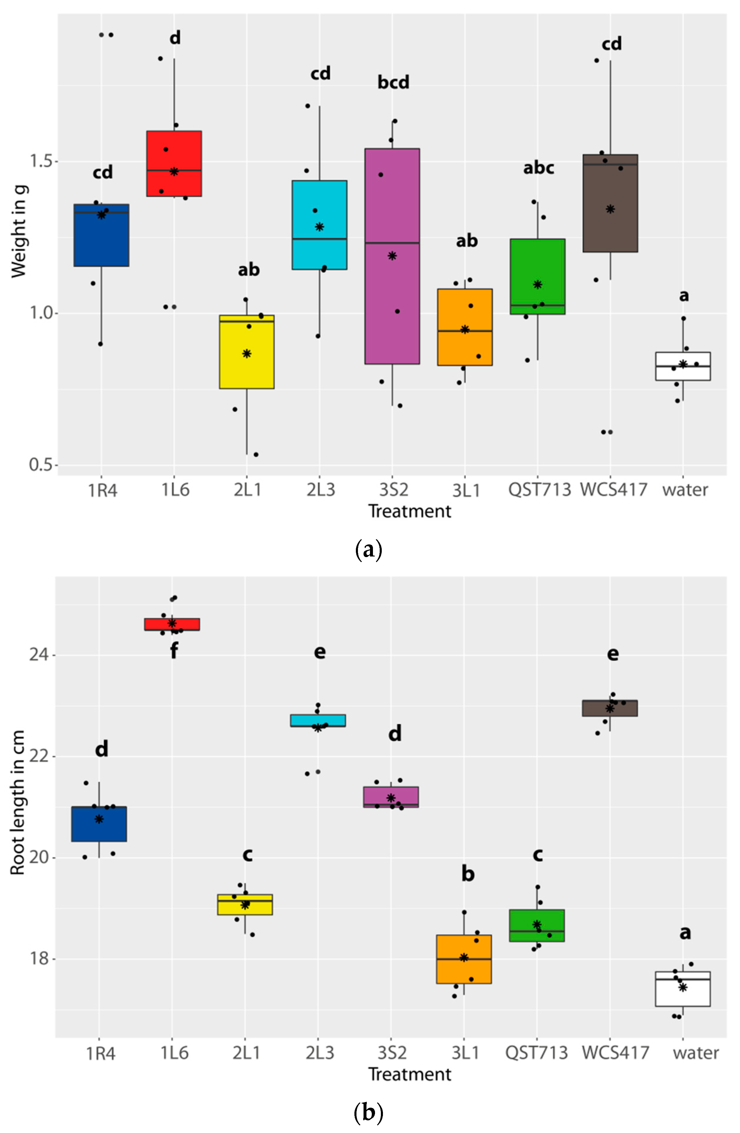
2.8. Growth Promotion Experiment

Six pots filled with autoclave sterilized soil were prepared for each treatment. Two rapeseed seeds were sown in a single pot and plants were thinned to one plant per pot after germination (7 d). Plants were grown in an environmental chamber under controlled environmental conditions (14 h day/10 h night cycle, 23 °C day/18 °C night, and 50% relative humidity). Pots were randomly distributed in the chamber and their positions were changed every 3 days to ensure the same conditions for all plants. Eleven days after sowing, plants were inoculated with 0.5 mL of bacterial suspension per plant using a hand sprayer. Plants were evaluated when they achieved an age of 25 days (i.e., 14 days after inoculation). Plants were removed from the pots and rinsed with water. The fresh root length and total biomass weight were measured and statistically evaluated.

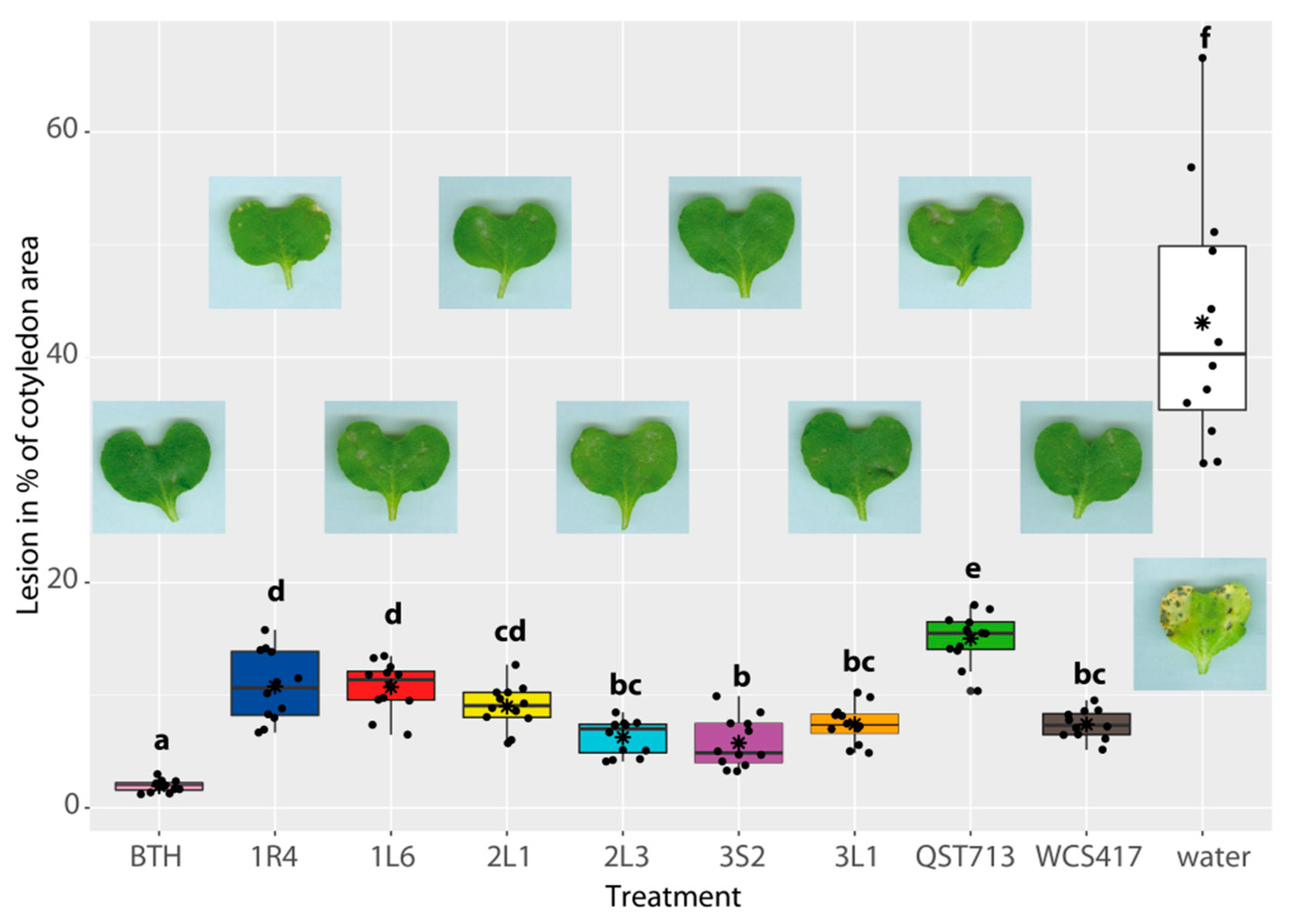
2.9. Leptosphaeria Suppression Experiment

Leptosphaeria maculans was cultivated on V8 agar (200 mL of vegetable juice V8; 3 g CaCO3; 20 g agar in 1 L). The petri dish with the culture was flooded with deionized sterile water to release the spores. The suspension was filtered and diluted to 1 × 10⁵ spores per milliliter.
Pots with plants were prepared and inoculated in the same manner as in the growth promotion experiment. In addition, benzothiadiazole (BTH), a plant systemic resistance inducer, was used as the positive control treatment in the 60 mM concentration. Due to the previously examined high variability of the lesion area, the number of replicates was increased to twelve. The true leaves were removed regularly to retain cotyledon growth. On the third day after inoculation by bacterial suspension (plant age 14 days), cotyledon leaves were infiltrated by 0.1 mL of L. maculans spore suspension using a syringe without needle [47]. Eleven days after infiltration (age 25 days), the plants were picked up and the cotyledons were scanned. Areas of L. maculans lesions were scored using ImageJ software [48].

2.10. Statistical Analysis

The differences among strains were evaluated using analysis of variance (ANOVA) followed by LSD test. Analysis of residues using the Shapiro–Wilk test confirmed the normal distribution required for ANOVA. Relations between all pairs of parameters (PGP traits, in planta assay results, Biolog) measured on selected isolates and reference strains were analyzed using linear regression. All statistical tests and graphs were performed using the R statistical environment [49] and the ggplot2 library [50].

3. Results

3.1. Primary Screening

A total of 38 isolates of bacteria were isolated from surface sterilized roots, stems, and leaves of Brassica napus (Table 1). According to MALDI-TOF identification, the isolates belonged to 15 genera. The most common genera were Bacillus (n = 8), followed by Pantoea (n = 4), Pseudomonas (n = 4), Microbacterium (n = 3), Acinetobacter (n = 3), and Serratia (n = 3). Low MALDI biotyper scores together with high species complexity for the most of isolates did not allow precise identification to the species level. Only 17 isolates showed the ability to dissolve phosphates on Pikovskaya agar, while 35 out of 38 were positively screened for siderophore production. Only isolates 2S3, 3S3, and 3S4 were negative. Antifungal activity against L. maculans was detected in 28 isolates, and reached more than 60% reduction of colony growth in 8 isolates (1R7, 1L5, 2S1, 2L3, 3S2, 3S4, 3L1, and 3L2). There was not any apparent relation between origin of isolates or phenological stage and the ability to produce siderophores, phosphate solubilization, or antifungal activity. However, a high portion of well-scored isolates belonged to Bacillus genera. Thus, six isolates of Bacillus spp. with promising combinations of all screened properties were selected for quantitative analysis and in planta experiments.

3.2. PGP Traits and Physiological Characterization of Selected Isolates

All isolates were previously identified as Bacillus species, however 16S rRNA sequence analysis was used for more precise phylogenetic placement (Figure 1). Isolates shared high 16S rRNA sequence homology with Bacillus subtilis, B. pumilus, and B. aryabhattai, according to BLAST. Isolates 1R4 and 2L3 shared the same 16S rRNA sequence as isolates 2L1 and 3S2. All isolates were catalase-positive, while only 1R4, 2L3, and WCS413 were urease positive, and 1L6, 2L1 and 3S2 were oxidase-negative (Table 2). Physiological analysis on Biolog Gen III plates showed a very different pattern of substrate utilization in the case of strain WCS413. Its growth was superior to other isolates in several wells with amino acids, carboxylic acids, and hexose acids (Figure 2). As other bacteria were phylogenetically more closely related, the differences were smaller. Isolates 1R4 and 3S2 showed higher utilization of some sources like L-glutamic, L-aspartic acid, L-histidine, and some hexose acids, and isolate 1R4 was also able to utilize acetic acid. On the other hand, isolate 1R4 was much more susceptible to the stressor wells of the Biolog plate than all other isolates belonging to Bacillus genera. Only strain QST713 showed resistance to lincomycin.
After 7 days of incubation in Pikovskaya broth, the amount of solubilized phosphates reached 284–456 mg·L−1 with significant differences between the isolates. The value reached by isolate 1L6 was significantly higher than the values of other isolates, including the reference PGPB strains. This isolate also showed significantly higher IAA and HCN production. The production of IAA by endophytic isolates was in the range of 32–58 mg·kg−1, while the reference PGPB produced between 37 and 50 mg·kg−1. Maximal increase of IAA production was measured after three days of incubation for all isolates except WCS417, which achieved maximum production on the fifth day. The reference strains scored 2nd (QST7) and 6th (WCS413) in HCN production (Table 2). Pseudomonas simiae WCS417 produced significantly less siderophores than other isolates. Isolates 1R4, 3S2, and 3L1, together with QST713, produced significantly higher amounts than other isolates (above 80%). When related to biology results, phosphate solubilization correlated with use of pectin (R = 0.78, P = 0.023), D-lactic acid methyl ester (R = 0.81, P = 0.014), or inhibition by D-serine (R = 0.85, P = 0.007). There was significant correlation of IAA production with use of inosine (R = 0.76, P = 0.028), D-sorbitol (R = −0.91, P = 0.002), methyl pyruvate (R = 0.93, P = 0.001), hydroxy-butyric acid (R = 0.88, P = 0.003), and others in the Biolog assay. Siderophores correlated negatively with use of inosine (R = −0.91, P = 0.002), serine (R = −0.83, P = 0.01), acetoacetic acid (R = −0.86, P = 0.006), and many others. Production of HCN significantly correlated only with resistance to sodium butyrate (Supplementary Materials, Table S1).

3.3. Plant Growth Promoting Properties of Isolates

The average total biomass weight of control plants was 0.8 g (Figure 3a). Despite the higher biomass weight of the plants treated with 2L1, 3L1, and QST7, these treatments did not promote plant growth significantly. All other isolates improved seedling growth significantly compared to the control, however only isolate 1L6 (1.5 g) promoted biomass yield significantly more than QST7 (1.1 g). Plant weight significantly correlated with the phosphate solubilization ability of used isolate (R = 0.85, P = 0.008).
Length of roots was in the range 17–25 cm, and it was more variable than total biomass weight. All inoculated plants scored better than the control (Figure 3b). Four endophytic isolates achieved better root growth than QST7 (18 cm). Only plants treated with isolate 1L6 had significantly longer roots (25 cm) than those treated with WCS413 (23 cm). We observed more root hairs on the roots of all inoculated plants as compared to the control (unmeasured observation). The root lengths of treated plants correlated with phosphate solubilization (R = 0.85, P = 0.004) and IAA production (R = 0.69, P = 0.036). Correlations of both plant growth parameters with Biolog substrate utilization were weak, with maximal value for D serine in relation to root length (R = 0.77, P = 0.024)

3.4. Suppression of L. maculans

Infiltration of non-inoculated plants by spore suspension of L. maculans resulted in phoma leaf spot lesions covering 43% of the cotyledon area. All isolates suppressed development of L. maculans better than QST713, which lowered the damaged area to 15% (Figure 4). However, only 3S2 (5.8%), 2L3 (6.3%), and 3L1 (7.4%) achieved comparable results to WCS417 (7.4%). Plants treated with BTH had only occasional lesions covering less than 2% of area, representing by a significant margin the lowest damage among all treatments. Correlation between PGP traits and plant damage by L. maculans was insignificant. A strong negative correlation (i.e., smaller area of lesions was related to higher utilization) was found for certain carbohydrates, such as dextrin (R = −0.91, P = 0.002), D-trehalose (R = −0.77, P = 0.024), D-melibiose (R = −0.90, P = 0.003), D-fructose (R = −0.79, P = 0.019), and D-turanose (R = −0.79, P = 0.020) in the Biolog assay.

4. Discussion

The scientific community is continuously searching for new PGP and biocontrol bacteria in effort to increase the plant yield and to lower the necessity of agrochemicals. Endophytes interact more closely with their host plants compared to microbes from soil or other plants, and they can be isolated from their host plant and cultured in vitro [51]. Thus, they are very commonly targeted in an effort to isolate new strains of PGP and/or bacteria. Some studies rely on common plant sources of endophytes [52,53] while others use less common plants or plants from specific environments [54,55,56]. Endophytic bacteria of various Brassica species were isolated and there are also studies about the microbial community of Brassica plants [5]. Kloepper et al. [57] described the selection of PGP bacteria suitable for inoculation, growth promotion, and yield increase of rapeseed as early as 1988. De Freitas et al. [58] found that inoculation of plants with Bacillus sp. and Xanthomonas maltophilia isolates originated from the rapeseed rhizosphere had positive effects on plant growth, but not on plant P content. Bertrand et al. [59] isolated several strains of Gram negative bacteria associated with rapeseed that showed plant growth promotion. Recently Shmidt et al. [60] reported isolation and PGP testing of bacterial and fungal endophytes from healthy and phoma-diseased plants of rapeseed. None of their bacterial endophytes (Achromobacter, Pseudomonas, Serratia, Enterobacter, or Stenotrophomonas) promoted growth of rapeseed in controlled P-limiting conditions; however, they significantly reduced the incidence of Sclerotinia disease. In case of L. maculans, their isolates were not able to lower the damaged areas of cotyledons. In contrast, our assay brings promising results, as our isolates either promoted growth or suppressed the pathogen. Isolate 3S2 suppressed L. maculans damage by 87%. We suppose that better results are based on source plant selection, as we isolated endophytes only from plants showing superior growth in exceptionally good conditions, possibly naturally colonized by beneficial bacteria.
The selection of PGP isolates based on PGP traits is becoming a common practice. Although not all PGP strains need to show their ability in in-vitro tests, such an attitude can eliminate most non-perspective isolates from further testing.
The ability to produce IAA is considered to be an important plant growth promotion property of bacteria, and it is widely tested in new strain prospecting. High variability in IAA production among bacterial species or strains has been reported [61]. Our isolates produced IAA in the range of 26–58 mg·L−1 which seems to be higher than the range reported by Khan et al. [62] or Kumar et al. [63], who used the same method of evaluation. As the IAA pathway depends on L-tryptophan [64], its concentration in the medium had a heavy impact on IAA production. Al Kahtani et al. [54] used several concentrations of L-tryptophan in their study and reported IAA production in range of approximately 12–33 or 24–57 mg·L−1, corresponding to concentrations of 1 and 5 g·L−1 of tryptophan, respectively. Isolate 1L6, with the highest IAA production, phosphate solubilization, and siderophore production, also greatly improved plant growth and the length of roots, which pointed to the relationship between these parameters and plant growth. However, some bacteria were successfully tested as PGPB even without a high ability to produce IAA in laboratory conditions, indicating that some other mechanisms can be employed in plant growth promotion. Moreover, in the case of IAA, high levels may have negative effects on plant growth, and the production of high levels is often a key characteristic of plant pathogens [65].
In preliminary screening, the majority of isolates showed signs of siderophore production which is in concordance with many studies [17,66,67,68], as this ability is widespread within various bacteria. Bacterial genera such as Klebsiella, Stenotrophomonas, Rhizobium, Herbaspirillum, and Citrobacter showed low production of siderophores, while others like Burkholderia, Enterobacter, or Streptomyces were strong producers [26,27,69]. Variability in siderophore production between genera may explain the significantly lower production by Pseudomonas simiae WCS413 in comparison to other isolates which belonged to Bacillus genera. Differences were also found in siderophore production dynamics when WCS 413 produced its maximum amount after five of cultivation, while Bacillus spp. isolates achieved the maximum after three days. However, Pseudomonas isolates were strong siderophore producers in some studies [70,71], as were Bacillus isolates [72,73,74]. All our selected isolates can be ranked as good producers in comparison to the results of Arora and Verma [46] who found siderophore production in the range of 8.33% to 69.81% in their isolates. There is a predicted relation between siderophore production and the ability of bacteria to suppress the growth of pathogens, including microscopic fungi [75,76]. According to our primary results, siderophore production was not connected to the suppression of L. maculans, as there were siderophore production positive isolates without any antifungal activity, and vice versa. Regression analysis of quantitative data also did not confirm the relation between biocontrol and siderophore producing ability of isolates.
Screening for phosphate solubilizing bacteria is commonly based on the formation of a depletion halo on Pikovskaya agar. However, we used an additional liquid culture test for the precise quantification of P solubilization, as recommended by Liu et al. [77]. In primary screening, we found solubilization ability in a similar amplitude as reported in scientific studies dealing with PGP bacteria [78,79]. After 7 days of cultivation in liquid medium, our endophytic isolates were able to solubilize P in the range of 70–400 mg·L−1. Other studies reported amounts in the range of 10–800 mg·L−1, where solubilization ability greater than 200 mg·L−1 can be considered as high [80].
Catalase is involved in oxidation stress suppression, thus catalase positive bacteria survive better in the ecosystem, and within the plant they can even protect against oxidative stress [81]. All of our isolates were catalase positive, but only three were urease positive. Urease is an important enzyme that catalyzes urea to ammonium, and this way bacteria applied into soil can indirectly improve plant growth [82]. Physiological profiling using the Biolog system is not widely adopted in microbial inoculant research. This technique was used in analysis of various PGP bacteria by Wozniak et al. [83], who stated that the metabolic and phenotypic properties of plant growth-promoting endophytes are correlated. We also found interesting relations between the utilization of certain carbohydrates and the ability to suppress L. maculans. Analysis of possible metabolic pathways under this relationship is beyond the scope of this article, but leaf surface sugars play an important role in plant colonization by bacteria.
There is an enormous number of studies describing the isolation of PGP bacteria, but their authors usually avoid direct comparison of their isolates with established ones, although isolates can be provided for scientific use for free. We used two of the widely studied strains that have well described PGP ability.
Bacillus velezensis QST713 (formerly known as B. subtilis or B. amyloliquefaciens QST713) was proven both to suppress various fungal pathogens and to support the growth of many plant species including Brassica spp. [84,85,86]. This bacterium is sold in the Serenade ASO formulation, which also contains the secondary metabolites of this bacterium, and these two components showed significantly better results than either component alone [86]. In our study, all other isolates were more efficient against L. maculans. Moreover, plants treated by this strain did not grow exceptionally well in comparison to other treatments despite its previously published PGP ability. As we used autoclaved soil, the applied isolates did not need to be competent among other microbiota, which may be an important limitation in field conditions [87]. Also, the application technique of bioagens can be critical for their effectiveness [88].
Pseudomonas simiae WCS417 is one of the best studied plant growth promoters and it has been under scientific examination for more than 30 years [89]. It was used on several Brassica species for growth promotion and systemic resistance induction [90,91] and it also showed plant growth promotion in our assay. Despite the fact that our strain 1L6 showed slightly longer roots, the root hairs were not as developed as in the WCS417 treatment. Some endophytic isolates seem to be at least comparable or better than these reference PGP bacteria. They can be used either for growth promotion or blackleg disease suppression. Due to the possibility of different modes of action of various endophytic isolates, their combination within a single formulation could provide a synergistic effect and/or could broaden the spectrum of environmental conditions under which the bioagents provide the desirable effect.

5. Conclusions

Thirty-eight isolates of endophytic bacteria obtained from rapeseed plants were screened for PGP traits, and six of them were further evaluated for PGP properties and their biocontrol potential in an in planta assay. Isolate Bacillus sp. 1L6 demonstrated plant growth properties comparable to well established PGP bacterial strains. Isolate Bacillus sp. 3S2 showed strong antifungal activity and significantly decreased the leaf area damaged by L. maculans. Further testing is necessary to confirm their PGP and blackleg disease-suppression properties in the field conditions. In the case of satisfactory results, they can be used alone or in combination in rapeseed cultivation systems.

Supplementary Materials

The following are available online at https://www.mdpi.com/article/10.3390/agronomy11101966/s1, Table S1: Correlation analysis of PGP traits, in planta growth promotion of rapeseed, Leptosphaeria maculans lesions, and Biolog substrates use.

Author Contributions

Conceptualization, N.L., J.M. (Juraj Medo), J.M. (Jana Maková), J.M. (Jaroslav Michalko) and S.J.; methodology, N.L., R.A., J.M. (Juraj Medo), J.M. (Jana Maková), S.J. and J.P.; software, J.M. (Juraj Medo) and N.L.; validation, N.L., J.M. (Juraj Medo) and J.M. (Jana Maková); formal analysis, N.L. and J.M. (Juraj Medo); investigation, N.L., J.M. (Juraj Medo), J.M. (Jana Maková), R.A. and J.P.; resources, J.M. (Juraj Medo), J.M. (Jana Maková), S.J., R.A., N.L. and J.M. (Jaroslav Michalko); data curation, N.L. and J.M. (Juraj Medo); writing—original draft preparation, N.L. and J.M. (Juraj Medo); writing—review and editing, all authors; visualization, J.M. (Juraj Medo) and N.L.; supervision, S.J., J.M. (Juraj Medo) and J.M. (Jana Maková); project administration, J.M. (Juraj Medo), J.M. (Jana Maková) and S.J.; funding acquisition, J.M. (Jaroslav Michalko) All authors have read and agreed to the published version of the manuscript.

Funding

This research was financially supported by a project of the Ministry of Education, Science, Research and Sport of the Slovak Republic, grant no. VEGA2/0100/18, and the Operational Program Integrated Infrastructure within the Demand-Driven Research for the Sustainable and Innovative Food project, Drive4SIFood 313011V336, co-financed by the European Regional Development Fund. All experiments were carried out in an AgroBioTech Research Centre built in accordance with the project Building “AgroBioTech” Research Centre ITMS 26220220180.

Institutional Review Board Statement

Not applicable.

Informed Consent Statement

Not applicable.

Data Availability Statement

All data are available upon to request correspondence author.

Acknowledgments

Authors would like to thank to Henrieta Blaškovičová and Daniela Košťálová for their help in laboratory.

Conflicts of Interest

The authors declare no conflict of interest.

References

  1. Carré, P.; Pouzet, A. Rapeseed market, worldwide and in Europe. OCL 2014, 21, D102. [Google Scholar] [CrossRef]
  2. Konur, O. Rapeseed oil-based biodiesel fuels: A review of the research. In Biodiesel Fuels Based Edible Nonedible Feedstocks Wastes Algae; Konur, O., Ed.; CRC Press: Boca Raton, FL, USA, 2021; pp. 497–514. [Google Scholar]
  3. Bunzel, K.; Kattwinkel, M.; Schauf, M.; Thrän, D. Energy crops and pesticide contamination: Lessons learnt from the development of energy crop cultivation in Germany. Biomass Bioenergy 2014, 70, 416–428. [Google Scholar] [CrossRef]
  4. Souza, R.d.; Ambrosini, A.; Passaglia, L.M. Plant growth-promoting bacteria as inoculants in agricultural soils. Genet. Mol. Biol. 2015, 38, 401–419. [Google Scholar] [CrossRef] [PubMed]
  5. Card, S.D.; Hume, D.E.; Roodi, D.; McGill, C.R.; Millner, J.P.; Johnson, R.D. Beneficial endophytic microorganisms of Brassica–A review. Biol. Control 2015, 90, 102–112. [Google Scholar] [CrossRef]
  6. West, J.S.; Kharbanda, P.; Barbetti, M.; Fitt, B.D. Epidemiology and management of Leptosphaeria maculans (phoma stem canker) on oilseed rape in Australia, Canada and Europe. Plant Pathol. 2001, 50, 10–27. [Google Scholar] [CrossRef] [Green Version]
  7. Huang, Y.-J.; Qi, A.; King, G.J.; Fitt, B.D. Assessing quantitative resistance against Leptosphaeria maculans (phoma stem canker) in Brassica napus (oilseed rape) in young plants. PLoS ONE 2014, 9, e84924. [Google Scholar] [CrossRef] [Green Version]
  8. Toscano-Underwood, C.; West, J.S.; Fitt, B.D.; Todd, A.; Jedryczka, M. Development of phoma lesions on oilseed rape leaves inoculated with ascospores of A-group or B-group Leptosphaeria maculans (stem canker) at different temperatures and wetness durations. Plant Pathol. 2001, 50, 28–41. [Google Scholar] [CrossRef] [Green Version]
  9. Huang, Y.; Fitt, B.D.; Hall, A. Survival of A-group and B-group Leptosphaeria maculans (phoma stem canker) ascospores in air and mycelium on oilseed rape stem debris. Ann. Appl. Biol. 2003, 143, 359–369. [Google Scholar] [CrossRef]
  10. Deb, D.; Khan, A.; Dey, N. Phoma diseases: Epidemiology and control. Plant Pathol. 2020, 69, 1203–1217. [Google Scholar] [CrossRef]
  11. Syed Ab Rahman, S.F.; Singh, E.; Pieterse, C.M.J.; Schenk, P.M. Emerging microbial biocontrol strategies for plant pathogens. Plant Sci. 2018, 267, 102–111. [Google Scholar] [CrossRef] [Green Version]
  12. Berg, G. Plant–microbe interactions promoting plant growth and health: Perspectives for controlled use of microorganisms in agriculture. Appl. Microbiol. Biotechnol. 2009, 84, 11–18. [Google Scholar] [CrossRef] [PubMed]
  13. Hardoim, P.R.; van Overbeek, L.S.; van Elsas, J.D. Properties of bacterial endophytes and their proposed role in plant growth. Trends Microbiol. 2008, 16, 463–471. [Google Scholar] [CrossRef] [PubMed]
  14. Compant, S.; Clément, C.; Sessitsch, A. Plant growth-promoting bacteria in the rhizo-and endosphere of plants: Their role, colonization, mechanisms involved and prospects for utilization. Soil Biol. Biochem. 2010, 42, 669–678. [Google Scholar] [CrossRef] [Green Version]
  15. Riaz, U.; Murtaza, G.; Anum, W.; Samreen, T.; Sarfraz, M.; Nazir, M.Z. Plant Growth-Promoting Rhizobacteria (PGPR) as biofertilizers and biopesticides. In Microbiota and Biofertilizers; Springer: Berlin/Heidelberg, Germany, 2021; pp. 181–196. [Google Scholar]
  16. Ahmad, F.; Ahmad, I.; Khan, M. Screening of free-living rhizospheric bacteria for their multiple plant growth promoting activities. Microbiol. Res. 2008, 163, 173–181. [Google Scholar] [CrossRef] [PubMed]
  17. Boubekri, K.; Soumare, A.; Mardad, I.; Lyamlouli, K.; Hafidi, M.; Ouhdouch, Y.; Kouisni, L. The Screening of potassium-and phosphate-solubilizing actinobacteria and the assessment of their ability to promote wheat growth parameters. Microorganisms 2021, 9, 470. [Google Scholar] [CrossRef] [PubMed]
  18. Raymaekers, K.; Ponet, L.; Holtappels, D.; Berckmans, B.; Cammue, B.P. Screening for novel biocontrol agents applicable in plant disease management—A review. Biol. Control 2020, 144, 104240. [Google Scholar] [CrossRef]
  19. Duca, D.; Lorv, J.; Patten, C.L.; Rose, D.; Glick, B.R. Indole-3-acetic acid in plant–microbe interactions. Antonie Van Leeuwenhoek 2014, 106, 85–125. [Google Scholar] [CrossRef]
  20. Etesami, H.; Alikhani, H.A.; Hosseini, H.M. Indole-3-acetic acid (IAA) production trait, a useful screening to select endophytic and rhizosphere competent bacteria for rice growth promoting agents. MethodsX 2015, 2, 72–78. [Google Scholar] [CrossRef]
  21. Raghothama, K.G. Phosphorus and plant nutrition: An overview. Phosphorus Agric. Environ. 2005, 46, 353–378. [Google Scholar]
  22. Frossard, E.; Condron, L.M.; Oberson, A.; Sinaj, S.; Fardeau, J. Processes governing phosphorus availability in temperate soils. J. Environ. Qual. 2000, 29, 15–23. [Google Scholar] [CrossRef] [Green Version]
  23. Pizzol, M.; Smart, J.C.; Thomsen, M. External costs of cadmium emissions to soil: A drawback of phosphorus fertilizers. J. Clean. Prod. 2014, 84, 475–483. [Google Scholar] [CrossRef]
  24. Huang, J.; Xu, C.-C.; Ridoutt, B.G.; Wang, X.-C.; Ren, P.-A. Nitrogen and phosphorus losses and eutrophication potential associated with fertilizer application to cropland in China. J. Clean. Prod. 2017, 159, 171–179. [Google Scholar] [CrossRef]
  25. Colombo, C.; Palumbo, G.; He, J.-Z.; Pinton, R.; Cesco, S. Review on iron availability in soil: Interaction of Fe minerals, plants, and microbes. J. Soils Sedim. 2014, 14, 538–548. [Google Scholar] [CrossRef]
  26. Lemanceau, P.; Bauer, P.; Kraemer, S.; Briat, J.-F. Iron dynamics in the rhizosphere as a case study for analyzing interactions between soils, plants and microbes. Plant Soil 2009, 321, 513–535. [Google Scholar] [CrossRef]
  27. Loaces, I.; Ferrando, L.; Scavino, A.F. Dynamics, diversity and function of endophytic siderophore-producing bacteria in rice. Microb. Ecol. 2011, 61, 606–618. [Google Scholar] [CrossRef] [PubMed]
  28. Höfte, M.; Bakker, P.A. Competition for iron and induced systemic resistance by siderophores of plant growth promoting rhizobacteria. In Microbial Siderophores; Springer: Berlin/Heidelberg, Germany, 2007; pp. 121–133. [Google Scholar]
  29. Ramette, A.; Frapolli, M.; Défago, G.; Moënne-Loccoz, Y. Phylogeny of HCN synthase-encoding hcnBC genes in biocontrol fluorescent pseudomonads and its relationship with host plant species and HCN synthesis ability. Mol. Plant Microbe Interact. 2003, 16, 525–535. [Google Scholar] [CrossRef] [Green Version]
  30. Blom, D.; Fabbri, C.; Connor, E.; Schiestl, F.; Klauser, D.; Boller, T.; Eberl, L.; Weisskopf, L. Production of plant growth modulating volatiles is widespread among rhizosphere bacteria and strongly depends on culture conditions. Environ. Microbiol. 2011, 13, 3047–3058. [Google Scholar] [CrossRef]
  31. Bakker, A.W.; Schippers, B. Microbial cyanide production in the rhizosphere in relation to potato yield reduction and Pseudomonas spp-mediated plant growth-stimulation. Soil Biol. Biochem. 1987, 19, 451–457. [Google Scholar] [CrossRef]
  32. Rijavec, T.; Lapanje, A. Hydrogen cyanide in the rhizosphere: Not suppressing plant pathogens, but rather regulating availability of phosphate. Front. Microbiol. 2016, 7, 1785. [Google Scholar] [CrossRef] [PubMed] [Green Version]
  33. von Rohr, M.R.; Furrer, G.; Brandl, H. Effect of iron and phosphate on bacterial cyanide formation determined by methemoglobin in two-dimensional gradient microcultivations. J. Microbiol. Methods 2009, 79, 71–75. [Google Scholar] [CrossRef]
  34. Santos, M.S.; Nogueira, M.A.; Hungria, M. Microbial inoculants: Reviewing the past, discussing the present and previewing an outstanding future for the use of beneficial bacteria in agriculture. AMB Express 2019, 9, 1–22. [Google Scholar] [CrossRef]
  35. Afzal, I.; Shinwari, Z.K.; Sikandar, S.; Shahzad, S. Plant beneficial endophytic bacteria: Mechanisms, diversity, host range and genetic determinants. Microbiol. Res. 2019, 221, 36–49. [Google Scholar] [CrossRef]
  36. Sun, L.; Qiu, F.; Zhang, X.; Dai, X.; Dong, X.; Song, W. Endophytic bacterial diversity in rice (Oryza sativa L.) roots estimated by 16S rDNA sequence analysis. Microb. Ecol. 2008, 55, 415–424. [Google Scholar] [CrossRef] [PubMed]
  37. Schwyn, B.; Neilands, J. Universal chemical assay for the detection and determination of siderophores. Anal. Biochem. 1987, 160, 47–56. [Google Scholar] [CrossRef]
  38. Gupta, R.; Singal, R.; Shankar, A.; Kuhad, R.C.; Saxena, R.K. A modified plate assay for screening phosphate solubilizing microorganisms. J. Gen. Appl. Microbiol. 1994, 40, 255–260. [Google Scholar] [CrossRef]
  39. Fernando, W.; Pierson III, L. The effect of increased phenazine antibiotic production on the inhibition of economically important soil-borne plant pathogens by pseudomonas a ureofaciens 30–84. Arch. Phytopathol. Plant Prot. 1999, 32, 491–502. [Google Scholar] [CrossRef]
  40. Edgar, R.C. MUSCLE: Multiple sequence alignment with high accuracy and high throughput. Nucleic Acids Res. 2004, 32, 1792–1797. [Google Scholar] [CrossRef] [PubMed] [Green Version]
  41. Guindon, S.; Dufayard, J.-F.; Lefort, V.; Anisimova, M.; Hordijk, W.; Gascuel, O. New algorithms and methods to estimate maximum-likelihood phylogenies: Assessing the performance of PhyML 3.0. Syst. Biol. 2010, 59, 307–321. [Google Scholar] [CrossRef] [Green Version]
  42. Gouy, M.; Guindon, S.; Gascuel, O. SeaView version 4: A multiplatform graphical user interface for sequence alignment and phylogenetic tree building. Mol. Biol. Evol. 2010, 27, 221–224. [Google Scholar] [CrossRef] [Green Version]
  43. Collins, C.H.; Grange, J.; Falkinham, J. Collins and Lyne’s Microbiological Methods, 8th ed.; Hodder Arnold: London, UK, 2003; p. 480. [Google Scholar]
  44. Kumar, V.; Kumar, V.; Umrao, V.; Singh, M. Effect of GA3 and IAA on growth and flowering of carnation. Hortflora Res. Spectr. 2012, 89, 69. [Google Scholar]
  45. Nautiyal, C.S. An efficient microbiological growth medium for screening phosphate solubilizing microorganisms. FEMS Microbiol. Lett. 1999, 170, 265–270. [Google Scholar] [CrossRef] [PubMed]
  46. Arora, N.K.; Verma, M. Modified microplate method for rapid and efficient estimation of siderophore produced by bacteria. 3 Biotech 2017, 7, 1–9. [Google Scholar] [CrossRef] [PubMed] [Green Version]
  47. Šašek, V.; Nováková, M.; Dobrev, P.I.; Valentová, O.; Burketová, L. β-aminobutyric acid protects Brassica napus plants from infection by Leptosphaeria maculans. Resistance induction or a direct antifungal effect? Eur. J. Plant Pathol. 2012, 133, 279–289. [Google Scholar] [CrossRef]
  48. Schneider, C.A.; Rasband, W.S.; Eliceiri, K.W. NIH Image to ImageJ: 25 years of image analysis. Nat. Methods 2012, 9, 671–675. [Google Scholar] [CrossRef]
  49. R Core Team. R: A Language and Environment for Statistical Computing; R Foundation for Statistical Computing: Vienna, Austria, 2020. [Google Scholar]
  50. Wickham, H. ggplot2: Elegant Graphics for Data Analysis; Springer: Berlin/Heidelberg, Germany, 2016. [Google Scholar]
  51. Maggini, V.; Mengoni, A.; Gallo, E.R.; Biffi, S.; Fani, R.; Firenzuoli, F.; Bogani, P. Tissue specificity and differential effects on in vitro plant growth of single bacterial endophytes isolated from the roots, leaves and rhizospheric soil of Echinacea purpurea. BMC Plant Biol. 2019, 19, 284. [Google Scholar] [CrossRef] [Green Version]
  52. Costa, L.E.D.O.; Queiroz, M.V.D.; Borges, A.C.; Moraes, C.A.D.; Araújo, E.F.D. Isolation and characterization of endophytic bacteria isolated from the leaves of the common bean (Phaseolus vulgaris). Braz. J. Microbiol. 2012, 43, 1562–1575. [Google Scholar] [CrossRef]
  53. Forchetti, G.; Masciarelli, O.; Alemano, S.; Alvarez, D.; Abdala, G. Endophytic bacteria in sunflower (Helianthus annuus L.): Isolation, characterization, and production of jasmonates and abscisic acid in culture medium. Appl. Microbiol. Biotechnol. 2007, 76, 1145–1152. [Google Scholar] [CrossRef] [PubMed]
  54. ALKahtani, M.D.F.; Fouda, A.; Attia, K.A.; Al-Otaibi, F.; Eid, A.M.; Ewais, E.E.-D.; Hijri, M.; St-Arnaud, M.; Hassan, S.E.-D.; Khan, N.; et al. Isolation and Characterization of Plant Growth Promoting Endophytic Bacteria from Desert Plants and Their Application as Bioinoculants for Sustainable Agriculture. Agronomy 2020, 10, 1325. [Google Scholar] [CrossRef]
  55. Fouda, A.; Eid, A.M.; Elsaied, A.; El-Belely, E.F.; Barghoth, M.G.; Azab, E.; Gobouri, A.A.; Hassan, S.E.-D. Plant Growth-Promoting Endophytic Bacterial Community Inhabiting the Leaves of Pulicaria incisa (Lam.) DC Inherent to Arid Regions. Plants 2021, 10, 76. [Google Scholar] [CrossRef] [PubMed]
  56. Tarroum, M.; Ben Romdhane, W.; Ali, A.A.M.; Al-Qurainy, F.; Al-Doss, A.; Fki, L.; Hassairi, A. Harnessing the Rhizosphere of the Halophyte Grass Aeluropus littoralis for Halophilic Plant-Growth-Promoting Fungi and Evaluation of Their Biostimulant Activities. Plants 2021, 10, 784. [Google Scholar] [CrossRef] [PubMed]
  57. Kloepper, J.; Hume, D.; Scher, F.; Singleton, C.; Tipping, B.; Laliberte, M.; Frauley, K.; Kutchaw, T.; Simonson, C.; Lifshitz, R. Plant growth-promoting rhizobacteria on canola (rapeseed). Plant Dis. 1988, 72, 42–46. [Google Scholar] [CrossRef]
  58. De Freitas, J.; Banerjee, M.; Germida, J. Phosphate-solubilizing rhizobacteria enhance the growth and yield but not phosphorus uptake of canola (Brassica napus L.). Biol. Fertil. Soils 1997, 24, 358–364. [Google Scholar] [CrossRef]
  59. Bertrand, H.; Nalin, R.; Bally, R.; Cleyet-Marel, J.-C. Isolation and identification of the most efficient plant growth-promoting bacteria associated with canola (Brassica napus). Biol. Fertil. Soils 2001, 33, 152–156. [Google Scholar] [CrossRef]
  60. Schmidt, C.; Mrnka, L.; Lovecká, P.; Frantík, T.; Fenclová, M.; Demnerová, K.; Vosátka, M. Bacterial and fungal endophyte communities in healthy and diseased oilseed rape and their potential for biocontrol of Sclerotinia and Phoma disease. Sci. Rep. 2021, 11, 3810. [Google Scholar] [CrossRef]
  61. Sridevi, M.; Mallaiah, K.V. Bioproduction of indole acetic acid by Rhizobium strains isolated from root nodules of green manure crop, Sesbania sesban (L.) Merr. Brief Rep. 2007, 5, 178–182. [Google Scholar]
  62. Khan, A.L.; Halo, B.A.; Elyassi, A.; Ali, S.; Al-Hosni, K.; Hussain, J.; Al-Harrasi, A.; Lee, I.-J. Indole acetic acid and ACC deaminase from endophytic bacteria improves the growth of Solanum lycopersicum. Electron. J. Biotechnol. 2016, 21, 58–64. [Google Scholar] [CrossRef] [Green Version]
  63. Kumar, V.; Singh, P.; Jorquera, M.A.; Sangwan, P.; Kumar, P.; Verma, A.; Agrawal, S. Isolation of phytase-producing bacteria from Himalayan soils and their effect on growth and phosphorus uptake of Indian mustard (Brassica juncea). World J. Microbiol. Biotechnol. 2013, 29, 1361–1369. [Google Scholar] [CrossRef]
  64. Spaepen, S.; Vanderleyden, J.; Remans, R. Indole-3-acetic acid in microbial and microorganism-plant signaling. FEMS Microbiol. Rev. 2007, 31, 425–448. [Google Scholar] [CrossRef] [Green Version]
  65. Kunkel, B.N.; Chen, Z. Virulence strategies of plant pathogenic bacteria. Prokaryotes 2006, 2, 421–440. [Google Scholar]
  66. Kumar, P.; Thakur, S.; Dhingra, G.; Singh, A.; Pal, M.K.; Harshvardhan, K.; Dubey, R.; Maheshwari, D. Inoculation of siderophore producing rhizobacteria and their consortium for growth enhancement of wheat plant. Biocatal. Agric. Biotechnol. 2018, 15, 264–269. [Google Scholar] [CrossRef]
  67. Husen, E. Screening of soil bacteria for plant growth promotion activities in vitro. Indones. J. Agric. Sci. 2003, 4, 27–31. [Google Scholar] [CrossRef]
  68. Cinkocki, R.; Lipková, N.; Javoreková, S.; Petrová, J.; Maková, J.; Medo, J.; Ducsay, L. The Impact of Growth-Promoting Streptomycetes Isolated from Rhizosphere and Bulk Soil on Oilseed Rape (Brassica napus L.) Growth Parameters. Sustainability 2021, 13, 5704. [Google Scholar] [CrossRef]
  69. Krewulak, K.D.; Vogel, H.J. Structural biology of bacterial iron uptake. Biochim. Biophys. Acta BBA Biomembr. 2008, 1778, 1781–1804. [Google Scholar] [CrossRef] [Green Version]
  70. Subramanian, J.; Satyan, K. Isolation and selection of fluorescent pseudomonads based on multiple plant growth promotion traits and siderotyping. Chil. J. Agric. Res. 2014, 74, 319–325. [Google Scholar] [CrossRef] [Green Version]
  71. Kotasthane, A.S.; Agrawal, T.; Zaidi, N.W.; Singh, U. Identification of siderophore producing and cynogenic fluorescent Pseudomonas and a simple confrontation assay to identify potential bio-control agent for collar rot of chickpea. 3 Biotech 2017, 7, 1–8. [Google Scholar] [CrossRef] [Green Version]
  72. Zhao, L.; Xu, Y.; Lai, X.-H.; Shan, C.; Deng, Z.; Ji, Y. Screening and characterization of endophytic Bacillus and Paenibacillus strains from medicinal plant Lonicera japonica for use as potential plant growth promoters. Braz. J. Microbiol. 2015, 46, 977–989. [Google Scholar] [CrossRef]
  73. Santoyo, G.; Moreno-Hagelsieb, G.; del Carmen Orozco-Mosqueda, M.; Glick, B.R. Plant growth-promoting bacterial endophytes. Microbiol. Res. 2016, 183, 92–99. [Google Scholar] [CrossRef] [PubMed]
  74. Perez-Rosales, E.; Alcaraz-Meléndez, L.; Puente, M.E.; Vázquez-Juárez, R.; Quiroz-Guzmán, E.; Zenteno-Savín, T.; Morales-Bojórquez, E. Isolation and characterization of endophytic bacteria associated with roots of jojoba (Simmondsia chinensis (Link) Schneid). Curr. Sci. 2017, 396–401. [Google Scholar]
  75. Wahyudi, A.T.; Astuti, R.P.; Widyawati, A.; Mery, A.; Nawangsih, A.A. Characterization of Bacillus sp. strains isolated from rhizosphere of soybean plants for their use as potential plant growth for promoting rhizobacteria. J. Microbiol. Antimicrob. 2011, 3, 34–40. [Google Scholar]
  76. Arya, N.; Rana, A.; Rajwar, A.; Sahgal, M.; Sharma, A. Biocontrol efficacy of siderophore producing indigenous Pseudomonas strains against Fusarium Wilt in Tomato. Natl. Acad. Sci. Lett. 2018, 41, 133–136. [Google Scholar] [CrossRef]
  77. Liu, Z.; Li, Y.C.; Zhang, S.; Fu, Y.; Fan, X.; Patel, J.S.; Zhang, M. Characterization of phosphate-solubilizing bacteria isolated from calcareous soils. Appl. Soil Ecol. 2015, 96, 217–224. [Google Scholar] [CrossRef]
  78. Stephen, J.; Jisha, M. Gluconic acid production as the principal mechanism of mineral phosphate solubilization by Burkholderia sp. (MTCC 8369). J. Trop. Agric. 2011, 49, 99–103. [Google Scholar]
  79. Surapat, W.; Pukahuta, C.; Rattanachaikunsopon, P.; Aimi, T.; Boonlue, S. Characteristics of phosphate solubilization by phosphate-solubilizing bacteria isolated from agricultural chili soil and their efficiency on the growth of chili (Capsicum frutescens L. cv. Hua Rua). Chiang Mai J. Sci. 2013, 40, 11–25. [Google Scholar]
  80. Chaiharn, M.; Lumyong, S. Screening and optimization of indole-3-acetic acid production and phosphate solubilization from rhizobacteria aimed at improving plant growth. Curr. Microbiol. 2011, 62, 173–181. [Google Scholar] [CrossRef] [PubMed]
  81. Santos, A.d.A.; Silveira, J.A.G.d.; Bonifacio, A.; Rodrigues, A.C.; Figueiredo, M.d.V.B. Antioxidant response of cowpea co-inoculated with plant growth-promoting bacteria under salt stress. Braz. J. Microbiol. 2018, 49, 513–521. [Google Scholar] [CrossRef] [PubMed]
  82. Nosheen, A.; Bano, A. Potential of plant growth promoting rhizobacteria and chemical fertilizers on soil enzymes and plant growth. Pak. J. Bot. 2014, 46, 1521–1530. [Google Scholar]
  83. Woźniak, M.; Gałązka, A.; Tyśkiewicz, R.; Jaroszuk-Ściseł, J. Endophytic Bacteria Potentially Promote Plant Growth by Synthesizing Different Metabolites and their Phenotypic/Physiological Profiles in the Biolog GEN III MicroPlateTM Test. Int. J. Mol. Sci. 2019, 20, 5283. [Google Scholar] [CrossRef] [Green Version]
  84. Altuntaş, Ö. A comparative study on the effects of different conventional, organic and bio-fertilizers on broccoli yield and quality. Appl. Ecol. Environ. Res. 2018, 16, 1595–1608. [Google Scholar] [CrossRef]
  85. McGehee, C.S.; Raudales, R.E.; Elmer, W.H.; McAvoy, R.J. Efficacy of biofungicides against root rot and damping-off of microgreens caused by Pythium spp. Crop Prot. 2019, 121, 96–102. [Google Scholar] [CrossRef]
  86. Lahlali, R.; Peng, G.; McGregor, L.; Gossen, B.D.; Hwang, S.F.; McDonald, M. Mechanisms of the biofungicide Serenade (Bacillus subtilis QST713) in suppressing clubroot. Biocontrol Sci. Technol. 2011, 21, 1351–1362. [Google Scholar] [CrossRef]
  87. Bach, E.; dos Santos Seger, G.D.; de Carvalho Fernandes, G.; Lisboa, B.B.; Passaglia, L.M.P. Evaluation of biological control and rhizosphere competence of plant growth promoting bacteria. Appl. Soil Ecol. 2016, 99, 141–149. [Google Scholar] [CrossRef]
  88. Seema, K.; Mehta, K.; Singh, N. Studies on the effect of plant growth promoting rhizobacteria (PGPR) on growth, physiological parameters, yield and fruit quality of strawberry cv. chandler. Chandler. J. Pharmacogn. Phytochem. 2018, 7, 383–387. [Google Scholar]
  89. Pieterse, C.M.J.; Berendsen, R.L.; de Jonge, R.; Stringlis, I.A.; Van Dijken, A.J.H.; Van Pelt, J.A.; Van Wees, S.C.M.; Yu, K.; Zamioudis, C.; Bakker, P.A.H.M. Pseudomonas simiae WCS417: Star track of a model beneficial rhizobacterium. Plant Soil 2021, 461, 245–263. [Google Scholar] [CrossRef]
  90. van der Wolf, J.M.; Michta, A.; van der Zouwen, P.S.; de Boer, W.J.; Davelaar, E.; Stevens, L.H. Seed and leaf treatments with natural compounds to induce resistance against Peronospora parasitica in Brassica oleracea. Crop Prot. 2012, 35, 78–84. [Google Scholar] [CrossRef]
  91. Haney, C.H.; Wiesmann, C.L.; Shapiro, L.R.; Melnyk, R.A.; O’Sullivan, L.R.; Khorasani, S.; Xiao, L.; Han, J.; Bush, J.; Carrillo, J.; et al. Rhizosphere-associated Pseudomonas induce systemic resistance to herbivores at the cost of susceptibility to bacterial pathogens. Mol. Ecol. 2018, 27, 1833–1847. [Google Scholar] [CrossRef]
Figure 1. Maximum likelihood phylogenetic tree of Bacillus spp. isolates used in PGP trait analysis. Tree was generated using the GTR model, BioNJ starting tree, optimized nucleotide frequencies, and best of NNI and SPR tree searching, followed by 1000 bootstraps. Numbers above branches are bootstrap values.
Agronomy 11 01966 g001
Figure 2. Biolog GEN III physiological analysis of selected endophytic bacteria from rapeseed and reference PGP strains. (a) Substrate utilization in absorbance unit, (b) relative growth (positive control is 1) of isolates in inhibition testing wells of a Biolog plate.
Agronomy 11 01966 g002
Figure 3. Boxplot of plant growth promotion ability of selected endophytic isolates from rapeseed and reference strains of PGP bacteria: (a) weight of seedlings 14 days after spray inoculation; (b) length of roots 14 days after spray inoculation. Boxes indicate 1st and 3rd quartiles, horizontal line is median, asterisk is average, whiskers indicate 1.5 times theinterquartile range, and dots are data points. Boxes accompanied with the same letter are not significantly different at α = 0.05 according to Fisher LSD test.
Agronomy 11 01966 g003
Figure 4. Boxplot of damaged area of rapeseed cotyledons after application of selected endophytic isolates from rapeseed and reference strains of PGP bacteria followed by infiltration by Leptosphaeria maculans. Boxes indicate 1st and 3rd quartiles, horizontal line is median, asterisk is average, whiskers indicate 1.5 times theinterquartile range, and dots are data points. Boxes accompanied with the same letter are not significantly different at α = 0.05 according to Fisher LSD test.
Agronomy 11 01966 g004
Table 1. Bacterial isolates obtained from surface sterilized plants of rapeseed (Brassica napus). Origin, identification according to MALDI TOF, siderophore production (SP), phosphate solubilization (PS), and antifungal activity (AFA) against Leptosphaeria maculans.
StrainOriginBBCH StageMALDI Best MatchMALDI Score *SPPSAFA
1R1root15Pseudomonas mendocina DSM 50017T1.997+09.26
1R2root15Erwinia amylovora CFBP 1232T1.912+019.63
1R3root15Acinetobacter baumannii B3891.632+00
1R4root15Bacillus pumilus DSM 17941.998+1.5660
1R5root15Microbacterium liquefaciens HKI 113741.808+00
1R6root15Chryseobacterium joostei LMG 18212T1.769+1.9760
1R7root15Staphylococcus xylosus DSM 202671.984+068.89
1S1stem15Xanthomonas codiaei DSM 18812TB1.444+00
1S2stem15Citrobacter freundii 22054_11.945+1.2814.81
1S3stem15Flavobacterium hibernum DSM 12611T2.033+051.85
1S4stem15Enterobacter ludwigii DSM 16688T2.085+1.3918.52
1S5stem15Pseudomonas graminis DSM 11363T1.398+00
1L1leaf15Pantoea agglomerans DSM 85701.795+09.26
1L2leaf15Microbacterium liquefaciens DSM 20638T1.686+019.63
1L3leaf15Bacillus licheniformis DSM 13T1.718+05.56
1L4leaf15Pseudomonas aeruginosa 8147_22.162+00
1L5leaf15Acinetobacter junii DSM 149682.023+062.22
1L6leaf15Bacillus pumilus DSM 3541.896+1.0938.89
1L7leaf15Serratia marcescens DSM 124851.926+00
1L8leaf15Xanthomonas codiaei DSM 18812TB1.995+010.37
2R1root55Microbacterium liquefaciens DSM 20638T1.828+1.4857.04
2R2root55Staphylococcus lutrae DSM 102461.437+1.3746.3
2R3root55Serratia marcescens DSM 301222.192+00
2S1stem55Pantoea eucrina DSM 24231T1.814+060.74
2S2stem55Serratia fonticola CCUG 385702.114040.74
2L1leaf55Bacillus subtilis DSM 56111.998+1.1844.81
2L2leaf55Chryseobacterium joostei LMG 18212T1.818+1.310
2L3leaf55Bacillus subtilis 107_W_7_QSA1.962+1.5962.22
3R1root85Pantoea agglomerans CCM 24061.697+1.180
3R2root85Pseudomonas oleovorans DSM 501881.598+1.510.37
3S1stem85Acinetobacter calcoaceticus DSM 30006T1.653+00
3S2stem85Bacillus megaterium DSM 32T DSM2.118+1.0981.48
3S3stem85Klebsiella oxytoca ATCC 700324 THL1.4151.113.33
3S4stem85Pantoea agglomerans DSM 8570 DSM2.0881.2864.07
3S5stem85Bacillus megaterium DSM 32T DSM1.477+1.727.04
3S6stem85Enterobacter kobei S58 ADRIA1.773+00
3L1leaf85Bacillus subtilis DSM 5552 DSM1.982+1.1361.85
3L2leaf85Sphingobium chlorophenolicum DSM 7098T HAM1.407+064.81
* MALDI scores of 2.000–2.299 indicate secure genus identification and probable species identification; 1.700–1.999 indicate probable genus identification; <1.7 indicate no reliable MALDI identification; +/− sign indicates strain’s positivity/negativity in siderophore production.
Table 2. 16S rRNA homology of selected endophytic isolates of rapeseed, their enzymatic characteristics, and PGP trait comparison.
Isolate1R41L62L12L33S23L1QST713WCS417
Identification/Best BLAST hitB. subtilis NR_112116.2
100%
B. pumilus NR_148786.1
99.73%
B. arybhattai
NR_115953.1
100%
B. subtilis
NR_112116.2
100%
B. arybhattai
NR_115953.1
100%
B. subtilis NR_112116.2
99.88%
Bacillus velezensisPseudomonas simiae
16S rRNA AccessionMZ947229MZ947231MZ947227MZ947228MZ947230MZ947232CP025079.1CP007637.1
Catalase++++++++
Urease+++
Oxidase+-++++
HCN (mg·L−1)124.5 ± 0.21 a 1221.8 ± 1.55 g158.2 ± 0.77 c170.1 ± 0.57 d170.6 ± 0.98 d173.6 ± 0.74 e198.0 ± 0.57 f145.3 ± 0.74 b
IAA (mg·L−1)31.8 ± 0.83 b58.0 ± 0.77 g26.6 ± 0.10 a40.4 ± 0.63 e31.6 ± 0.36 b38.7 ± 0.66 d36.5 ± 0.46 c49.9 ± 0.63 f
Solubilized P (mg·L−1)350.2 ± 2.34 e457.5 ± 6.37 g305.4 ± 2.66 b337.7 ± 0.58 d363.9 ± 2.52 f284.8 ± 0.98 a322.6 ± 0.58 c366.9 ± 0.98 f
Siderophores (%)87.5 ± 0.82 f67.1 ± 1.46 b70.2 ± 1.48 c78.4 ± 0.68 d87.1 ± 0.43 f84.5 ± 0.33 ef83.0 ± 1.57 e59.8 ± 1.22 a
1 Values are averages followed by standard deviation. Values accompanied with the same letter (within the row) are not significantly different at α = 0.05 according to Fisher LSD test; +/− sign indicates strain’s positive/negative reaction in the respective enzyme assay.
Publisher’s Note: MDPI stays neutral with regard to jurisdictional claims in published maps and institutional affiliations.

