Article

Chemical Composition, Antibacterial, Enzyme-Inhibitory, and Anti-Inflammatory Activities of Essential Oil from Hedychium puerense Rhizome

1
Key Laboratory of Plant Resource Conservation and Germplasm Innovation in Mountainous Region (Ministry of Education), Collaborative Innovation Center for Mountain Ecology & Agro-Bioengineering (CICMEAB), Institute of Agro-Bioengineering, College of Life Sciences, Guizhou University, Guiyang 550025, China
2
National & Local Joint Engineering Research Center for the Exploition of Homology Resources of Southwest Medicine and Food, Guizhou University, Guiyang 550025, China
*
Author to whom correspondence should be addressed.
Agronomy 2021, 11(12), 2506; https://doi.org/10.3390/agronomy11122506
Submission received: 16 November 2021 / Revised: 8 December 2021 / Accepted: 8 December 2021 / Published: 10 December 2021

Abstract

:
Hedychium puerense, a perennial rhizomatous herb, is used as an ornamental, medicinal, and edible plant in Yunnan Province, China. Essential oils from Hedychium plants are widely used in perfumes and traditional medicine, but there are no studies on the constituents and bioactivities of H. puerense essential oil (EO). Therefore, this study was designed to explore the chemical composition, antibacterial, enzyme-inhibitory, and anti-inflammatory activities of H. puerense rhizome EO. The gas chromatography with flame ionization or mass selective detection (GC-FID/MS) results indicated that H. puerense EO was mainly composed of linalool (26.5%), β-pinene (18.6%), γ-terpinene (12.1%), terpinen-4-ol (7.7%), α-pinene (5.8%), sabinene (4.9%), E-nerolidol (4.1%), and p-cymene (3.6%). For biological activities, H. puerense EO displayed broad-spectrum antibacterial properties against Enterococcus faecalis, Bacillus subtilis, Staphylococcus aureus, Proteus vulgaris, Pseudomonas aeruginosa, and Escherichia coli with diameter of inhibition zone (DIZ) values ranging from 7.44 to 10.30 mm, a minimal inhibitory concentration (MIC) of 3.13–6.25 mg/m), and a minimal bactericidal concentration (MBC) of 3.13–12.50 mg/mL. Moreover, the EO significantly inhibited acetylcholinesterase (AChE) (IC50 = 0.94 ± 0.02 mg/mL) and butyrylcholinesterase (BChE) (IC50 = 1.32 ± 0.06 mg/mL) activities, and exhibited a moderate inhibitory effect on α-glucosidase (IC50 = 5.42 ± 0.32 mg/mL) and tyrosinase (IC50 = 3.23 ± 0.21 mg/mL). Furthermore, the EO significantly suppressed the secretion of the pro-inflammatory mediator, nitric oxide (NO) (99.23 ± 0.26%), cytokines tumor necrosis factor-α (TNF-α) (97.14 ± 0.11%), and interleukin-6 (IL-6) (82.42 ± 0.16%) in lipopolysaccharide (LPS)-stimulated RAW264.7 cells at 250 μg/mL without cytotoxicity. Hence, H. puerense EO can be considered a bioactive, natural product that has great potential for utilization in the fields of food, cosmetics, and pharmaceutics.

1. Introduction

Essential oils are a mixture of secondary metabolites with a volatile nature and strong odor obtained from aromatic plants [1,2]. An estimated 3000 essential oils were documented, approximately 300 of which have important commercial value and are mainly used in agricultural, cosmetic, perfume, sanitary, food, and pharmaceutical fields for their wide range of biological and pharmacological properties, including antioxidant, anti-inflammatory, antifungal, antibacterial, antiviral, anticarcinogenic, antimutagenic, antidiabetic, antiprotozoal, and analgesic activities [3,4,5,6,7]. Since the negative impact of synthetic products on health and the environment has become a serious global problem, there is an urgent need for healthier and more natural alternatives, such as essential oils [4,5,6].
The genus Hedychium of the Zingiberaceae family consists of about 93 species and is mainly distributed throughout tropical and warm regions of India, China, and Southeast Asia [8,9]. The species of Hedychium are commonly referred to as “ginger lily,” “garland lily,” or “butterfly ginger,” and are well known for their various uses in the fragrance, ornamental, paper, cosmetics, and food industries [10]. Additionally, Hedychium species are used in ethnomedicine to treat nausea, leishmaniasis, asthma, bronchitis, diarrhea, gastric diseases, flu, and snake bites [10,11,12]. In particular, the essential oils from the rhizomes of Hedychium plants possess great potential for wide applications and were utilized in perfumes and traditional medicine [9,10]. The most ubiquitous constituents in Hedychium rhizomes essential oils are monoterpenes and sesquiterpenes, including 1,8-cineole, linalool α-pinene, and β-pinene [10]. According to previous reports, Hedychium rhizomes essential oils possess various biological activities, including antifungal, antioxidant, anthelmintic, and insecticidal properties; especially, their antibacterial activity is well confirmed [11,13,14,15,16].
Hedychium puerense Y. Y. Qian is a perennial rhizomatous herb that belongs to the Hedychium genus, and its native range is Yunnan Province, China [17,18,19]. H. puerense is widely cultivated as an ornamental plant because of its beautiful and aromatic flowers. Its rhizome is used as a flavoring ingredient and in traditional Chinese medicine for the treatment of stomach pain, indigestion, and arthritis [20]. Essential oils from Hedychium plants were proven to possess a great potential for applications in the food, cosmetics, and pharmaceutical fields. However, to our knowledge, there are no studies on the phytochemical and biological activities of H. puerense, which may hinder its exploitation in the industry. Therefore, this study was designed to explore the chemical composition, antibacterial, enzyme inhibitory, and anti-inflammatory activities of essential oils from H. puerense rhizome.

2. Materials and Methods

2.1. Plant Material

The rhizome of H. puerense was collected in August 2019 from Yun County, Lincang City, Yunnan Province, China. The species of this plant was identified by Prof. Guoxiong Hu from the College of Life Sciences, Guizhou University. A voucher specimen (Voucher No: HP20190811) was deposited in National and Local Joint Engineering Research Center for the Exploitation of Homology Resources of Southwest Medicine and Food, Guizhou University.

2.2. Extraction of Essential Oil

To obtain EO, the fresh, finely chopped rhizome of H. puerense (2.0 kg) was subjected to hydrodistillation in an all-glass Clevenger-type apparatus for 4 h. The EO was dehydrated over anhydrous Na2SO4 and filtered. After filtration, the extracted EO (4.23 g, 0.21% w/w) was stored in a sealed glass vial at 4 °C.

2.3. Analysis of Essential Oil

The chemical composition of the EO was analyzed using an Agilent 6890 gas chromatograph (GC) (Agilent Technologies, Santa Clara, CA, USA) fitted with a flame ionization detector (FID) and a fused silica capillary HP-5MS column (60 m × 0.25 mm, 0.25 μm film thickness). EO (1 μL) was injected in split mode (split ratio: 1:20) with helium as a carrier gas at a flow rate of 1 mL/min. The GC operating parameters were as follows: maintained at 70 °C (2 min), increased to 180 °C at 2 °C/min (55 min), then raised to 310 °C at 10 °C/min (13 min), and maintained at 310 °C (14 min). Gas chromatography–mass spectrometry (GC–MS) analysis was carried out by using an Agilent 6890 gas chromatograph coupled with an Agilent 5975C mass spectrometric detector (Agilent Technologies, Santa Clara, CA, USA). GC column and parameters were the same as in GC-FID analysis. The MS settings were as follows: ionization voltage of 70 eV, scan range of m/z 29 to 500 amu, ion source temperature at 230 °C, and interface temperature at 280 °C. The relative abundance (%) of each composition was determined by the peak area. The retention time of n-alkanes (C8–C22) was used to calculate the retention index (RI). Identification of the EO components was confirmed by comparison of the mass spectrum and RI in NIST 2017 (National Institute of Standards and Technology, Gaithersburg, MD, USA) and Wiley 275 (Wiley, New York, NY, USA) databases.

2.4. Antibacterial Activity

The antibacterial activity of EO was tested against three Gram-positive bacterial strains (Staphylococcus aureus ATCC 6538P, Enterococcus faecalis ATCC 19433, and Bacillus subtilis ATCC 6633) and three Gram-negative bacterial strains (Proteus vulgaris ACCC 11002, Pseudomonas aeruginosa ATCC 9027, and Escherichia coli CICC 10389).
The diameter of the inhibition zone (DIZ) was determined with the disc diffusion method described by Chen et al. [21]. Briefly, 100 μL of bacterial suspension (1 × 10⁶ CFU/mL) was evenly spread on Mueller-Hinton agar medium. Subsequently, the filter paper discs (6 mm in diameter) containing 20 μL of EO (100 mg/mL diluted in ethyl acetate) or streptomycin (100 μg/mL diluted in distilled water) were placed on the surface of the agar medium. Streptomycin was used as a positive control. Ethyl acetate and distilled water were used as negative controls for EO and streptomycin, respectively. After incubation at 37 °C for 24 h, the diameter of the inhibition zone (DIZ), including the 6 mm disk, was measured and recorded.
The minimal inhibitory concentration (MIC) and minimal bactericidal concentration (MBC) values were assayed by the double broth dilution method [22]. Briefly, 100 μL of bacterial suspensions (2 × 10⁵ CFU/mL) and two-fold serially diluted sample solutions (100 μL) were distributed into each well of the 96-well plates. After incubation for 24 h at 37 °C, resazurin solutions (20 μL, 0.1 mg/mL) were added to each well and incubated at 37°C for 2 h in the dark. The MIC values were defined as the minimum concentration of EO without color change. To determine MBC values, the culture (10 μL) from the wells without color change was evenly spread on the Mueller-Hinton agar medium and cultured for an additional 24 h at 37 °C. The MBC values were determined as the lowest concentration of EO without bacterial growth.

2.5. Enzyme Inhibitory Activities

The α-glucosidase inhibitory effect of EO was determined according to the method described by Silva et al. with slight modification and used acarbose as a positive reference [23]. Sample solution (30 μL), α-glucosidase solution (10 μL, 0.8 U/mL), and phosphate buffer (60 μL, pH 6.8) were added into each well of 96-well plates. After 15 min incubation at 37 °C, the reaction was initiated by addition of the p-Nitrophenyl-α-D-glucopyranoside (p-NPG) substrate (10 μL, 1 mM) and incubated for 15 min at 37 °C. Subsequently, Na2CO3 solution (80 μL, 0.2 M) was added to stop the reaction and, finally, absorbance values were recorded at 405 nm using a Varioskan Lux Multimode microplate reader (Thermo Fisher Scientific, Waltham, MA, USA). The inhibitory properties of α-glucosidase were expressed by IC50 values.
The tyrosinase inhibitory activity was performed according to the protocol described by Zardi-Bergaoui et al. with marginal modifications [24]. An amount of 70 μL EO solution or arbutin solution (positive control) was mixed with 100 μL of tyrosinase solution (100 U/mL) and added to each well of the 96-well plates. After incubation at 37 °C for 5 min, the reaction was started by adding the L-tyrosine substrate (80 μL, 5.5 mM) and incubating for 30 min at 37 °C. Finally, the absorbance was read at 492 nm and the inhibition of tyrosinase was expressed by IC50 values.
The inhibitory activities on acetylcholinesterase (AChE) and butyrylcholinesterase (BChE) were determined using Ellman’s method with some modifications [25]. An amount of 10 μL AChE or BuChE solution (pH 8.0, 0.5 U/mL) was mixed with sample solution (50 μL) and added to each well of the 96-well plates. After 15 min incubation at 4 °C, the reaction was initiated by adding 20 μL of 5,5′-dithiobis-(2-nitrobenzoic acid) (DTNB) solution (pH 8.0, 2 mM) and 20 μL of acetylthiocholine (ATCI) or butyrylthiocholine (BTCI) solution (2 mM), and then incubated for 30 min at 37 °C. Finally, the absorbance was measured at 405 nm and galanthamine was used as a positive control. The inhibitory activity of cholinesterase was expressed as IC50 values.

2.6. Anti-Inflammatory Activity

The cytotoxic effect was estimated against murine macrophages (RAW264.7) and murine fibroblast cells (L929) using the MTT method with some modifications [26]. The RAW264.7 and L929 cell lines were cultured in DMEM medium and RPMI 1640 medium, respectively, and supplemented with 10% fetal bovine serum, 100 μg/mL streptomycin, 100 U/mL penicillin, and 2 mM glutamine. The EO was dissolved in DMSO, and then was two-fold serially diluted with medium, and the final DMSO concentration was no higher than 0.5%. The cell suspensions (100 μL) were distributed into 96-well plates (2 × 10 4 cells per well) and cultured for 24 h in an incubator with 5% CO2 at 37 °C. Subsequently, the diluted EO solutions (100 μL) were added to each well and cultured for 24 h. Then, 10 μL MTT solutions (5 mg/mL in PBS) were added and incubated for an additional 4 h. After dissolving the formazan crystal with DMSO (150 μL), the absorbance was measured at 490 nm to evaluate the cell viability.
The RAW264.7 cell suspensions (100 μL) were distributed into 96-well plates (2 × 10⁴ cells per well) and incubated for 24 h. Subsequently, the cells were exposed to 100 μL of fresh medium containing series concentrations of EO. Following incubation for 2 h, 100 μL of lipopolysaccharide (LPS) solutions was added into each well at the final concentration of 1 μg/mL and cultured for an additional 24 h. The changes in the cellular morphology of RAW264.7 cells were viewed by a Leica DMi8 microscope (Leica Microsystems, Wetzlar, Germany). After collecting the supernatant by centrifugation, NO detection kit was used to detect the accumulation amount of NO in the supernatant according to the manufacturer’s instructions (Beyotime, Shanghai, China). The production of pro-inflammatory cytokines (IL-6 and TNF-α) of RAW264.7 cells was determined using respective ELISA determination kits according to the manufacturer’s protocol (Multi Sciences Biotech Co., Ltd., Hangzhou, China). Dexamethasone (DXM, 20 μg/mL) was used as a positive control.

2.7. Statistical Analysis

All experimental results were expressed as mean ± standard deviation (SD) from at least three parallel experiments. Statistical analysis was conducted using SPSS 25.0 software (IBM Corp., Armonk, NY, USA). The significant difference between the two groups was compared via one-way analysis of variance (ANOVA) and Fischer’s LSD post hoc test at p < 0.05.

3. Results and Discussion

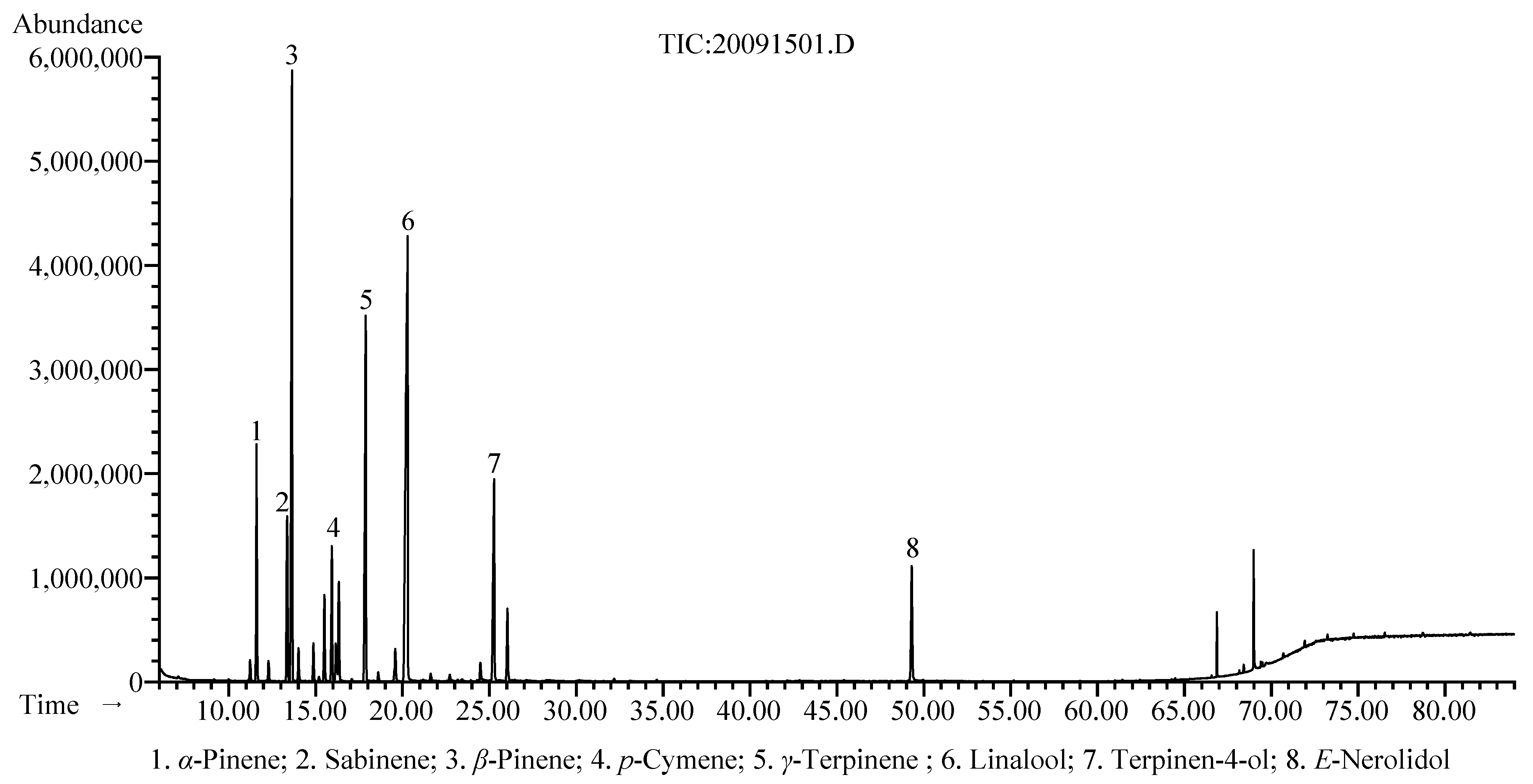
3.1. Chemical Composition

The extraction yield of the hydrodistilled EO was 0.21% (w/w) based on the fresh weight of H. puerense fresh rhizome. As shown in Table 1, based on GC-MS/FID analysis, thirty-four chemical components were identified, and accounted for 98.2% of the total EO. The main compounds of H. puerense EO were linalool (26.5%), β-pinene (18.6%), γ-terpinene (12.1%), terpinen-4-ol (7.7%), α-pinene (5.8%), sabinene (4.9%), E-nerolidol (4.1%), and p-cymene (3.6%), which are presented in Figure 1. As the most predominant component of H. puerense EO, linalool was proven to be the main marker compound present in many other Hedychium species, such as H. flavesens, H. coronarium, H. stenopetalum, H. flavum, H. aurantiacum, H. matthewiii, H. spicatum, H. forrestii, H. larsenii, etc. [9,10,14] Linalool is widely used as a fragrance ingredient in perfumes, cosmetics, shampoos, soaps, and household cleaners; it was also employed in food and beverages for aroma and flavoring [9,27,28]. Additionally, linalool has several well-documented bioactivities, including antimicrobial, anti-inflammatory, anti-hyperlipidemic, analgesic, anticancer, anxiolytic, and neuroprotective effects [28,29]. Hence, the H. puerense rhizome EO can be used as a new source of linalool and has a high potential for applications in the fields of food, cosmetics, and pharmaceuticals.

3.2. Antibacterial Activity

As shown in Table 2, the EO at the concentration of 100 mg/mL displayed a broad-spectrum antibacterial property against both Gram-negative and Gram-positive bacterial strains with the diameter of inhibition zones (DIZ) values ranging from 7.44 to 10.30 mm. According to previous studies, MIC values below 5 mg/mL are considered to possess strong antibacterial effects [30]. Therefore, the EO showed a strong antibacterial property against Enterococcus faecalis (MIC = 3.13 mg/mL, MBC = 3.13 mg/mL), Bacillus subtilis (MIC = 3.13 mg/mL, MBC = 3.13 mg/mL), and Proteus vulgaris (MIC = 3.13 mg/mL, MBC = 6.25 mg/mL), as well as exhibited moderate antibacterial activity against Staphylococcus aureus (MIC = 6.25 mg/mL, MBC = 12.50 mg/mL), Pseudomonas aeruginosa (MIC = 6.25 mg/mL, MBC = 12.50 mg/mL), and Escherichia coli (MIC = 6.25 mg/mL, MBC = 12.50 mg/mL). When EO had a larger DIZ value, its MIC and MBC values became smaller, and thus the DIZ results of EO showed a correlation with the results of MIC and MBC. Linalool, as the most predominant component of the H. puerense rhizome EO, is well known for its remarkable antibacterial property and is documented as a broad-spectrum antibacterial agent against various bacterial strains such as Salmonella sp., Staphylococcus aureus, Listeria monocytogenes, Pseudomonas aeruginosa, Shewanella putrefaciens, and Escherichia coli [31,32,33,34]. In addition, linalool showed an outstanding growth inhibition effect against S. putrefaciens through interfering with the function and integrity of the cell membrane and wall, inducing the leakage of cellular materials, and causing energy and metabolic dysfunction [31]. According to the previous studies, other main compounds in H. puerense EO, including β-pinene, γ-terpinene, terpinen-4-ol, α-pinene, sabinene, E-nerolidol, and p-cymene, were proven to have a significant antibacterial activity [35,36,37,38,39,40,41]. Hence, the presence of these major compounds could explain the significant antibacterial activity of the EO. These results indicated that H. puerense EO could be used as a new source of natural antibacterial agents in the food, cosmetics, and pharmaceutical industries.

3.3. Enzyme Inhibitory Activity

The inhibitory activities of H. puerense EO against α-glucosidase, tyrosinase, acetylcholinesterase (AChE), and butyrylcholinesterase (BChE) were investigated and the results were presented in Table 3.
H. puerense EO showed a moderate α-glucosidase inhibitory activity (IC50 = 5.42 ± 0.32 mg/mL). Certain α-glucosidase inhibitors, including acarbose, can reduce blood glucose and insulin levels after meals by delaying the intake of carbohydrates; therefore, they are widely used to treat type 2 diabetes [42]. Pre-diabetes is the preceding condition of diabetes; most of those affected by this condition will eventually develop diabetes, but this can be prevented through the effective treatment of pre-diabetes [43]. Acarbose at lower doses is reported to possess a beneficial effect on the management of pre-diabetes, but its use is limited by its side effects, which include diarrhea, flatulence, and abdominal distension [44,45]. Therefore, it is very promising to screen natural products that inhibit the activity of α-glucosidase as a complementary/alternative therapy for pre-diabetes. According to a previous study, linalool, as the most predominant constituent of the EO, showed a significant antidiabetic effect and enhanced insulin resistance [46]. Additionally, the α-glucosidase inhibitory properties of α-pinene were confirmed in the previous report [47]. Hence, H. puerense EO can be used as a new source of natural products for the treatment of diabetes.
As depicted in Table 3, H. puerense EO exhibited a moderate tyrosinase inhibitory activity (IC50 = 3.23 ± 0.21 mg/mL). Tyrosinase is a copper-containing oxidase involved in the biosynthesis of mammalian melanogenesis and the enzymatic browning in fruits [48,49]. Due to concerns regarding the toxicity and side effects of synthetic inhibitors, researchers are encouraged to look for safe and effective natural tyrosinase inhibitors. According to the research of Chao et al., the α-pinene and 1,8-cineole, which are the major and minor components of H. puerense EO, significantly inhibited tyrosinase activity and melanin content in the α-melanocyte-stimulating hormone-induced B16 melanoma cell line [50]. Furthermore, sabinene was reported to have a tyrosinase inhibitory effect [51]. Thus, the moderate tyrosinase inhibitory activity of H. puerense EO could be attributed to the presence of these compounds.
H. puerense EO showed a significant AChE and BChE inhibitory activity with the lower IC50 values of 0.94 ± 0.02 mg/mL and 1.32 ± 0.06 mg/mL, respectively. AChE and BChE are involved in the hydrolysis of acetylcholine, thus cholinesterase inhibitors were used to prevent and treat Alzheimer’s disease by inhibiting the decomposition of acetylcholine and enhancing cholinergic neurotransmission [52,53]. The anti-cholinesterase activity of the main components of H. puerense EO, including linalool, β-pinene, γ-terpinene, terpinen-4-ol, α-pinene, and p-cymene were reported in the previous studies [54,55,56,57,58]. Therefore, the significant anti-cholinesterase effect of H. puerense EO could be attributed to these major compounds, and used as a new source of natural cholinesterase inhibitors in the pharmaceutical industry.

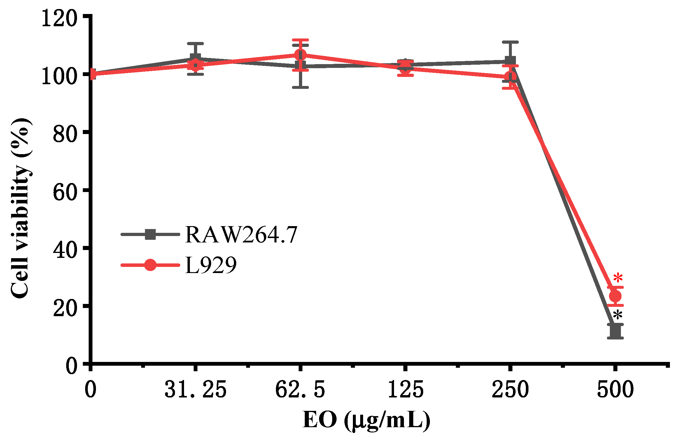
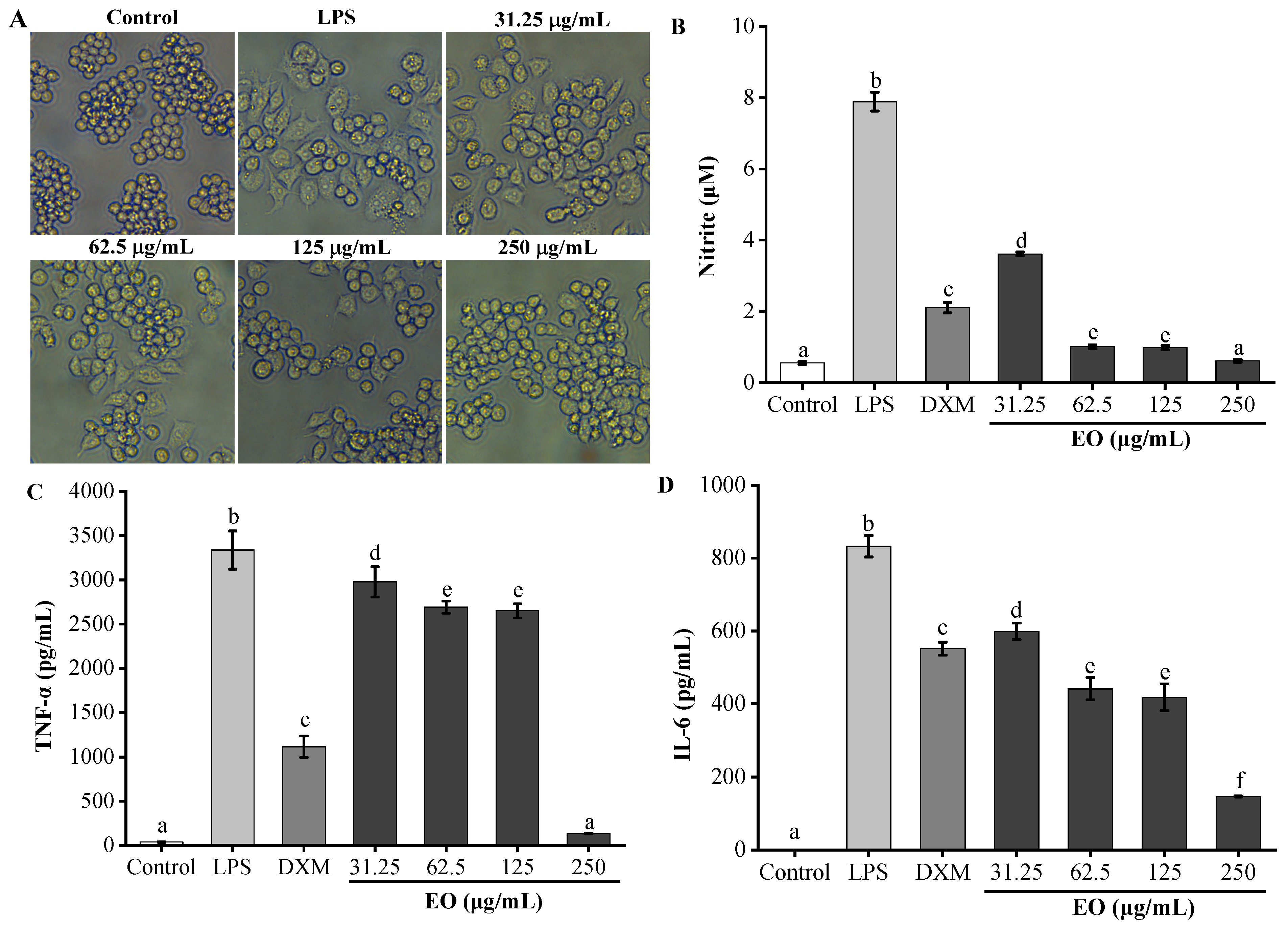
3.4. Anti-Inflammatory Activity

The cytotoxicity of H. puerense EO on RAW264.7 and L929 cell lines was measured by the MTT assay after incubation with different doses of EO (0, 31.25, 62.5, 125, 250, 500 μg/mL) for 24 h. As shown in Figure 2, H. puerense EO, in concentrations from 31.25−250 μg/mL, did not cause any significant cytotoxic effect on RAW264.7 and L929 cell lines in comparison with untreated cells. Hence, the no cytotoxic concentration range of EO (31.25−250 μg/mL) was used in subsequent experiments.
The anti-inflammatory effect of H. puerense EO was detected in LPS-stimulated RAW264.7 cells. The morphological changes of RAW264.7 macrophages treated with LPS and EO were viewed under phase-contrast microscopy (Figure 3A). The normal cells in the control group were circular with a smooth surface. RAW264.7 macrophages treated with LPS became larger in size and irregular in shape, while there were fewer changes in cell morphology in the EO-treated group. In addition, H. puerense EO significantly inhibited the production of pro-inflammatory mediator (NO) and cytokines (TNF-α and IL-6) in LPS-stimulated RAW264.7 macrophages (Figure 3B–D). Dexamethasone (DXM, 20 μg/mL) was used as a positive reference. As presented in Figure 3B, compared with the LPS group (7.88 ± 0.27 μM), EO significantly reduced the production of NO. Particularly, the NO production in RAW264.7 cells pretreated with EO at doses of 62.5, 125, and 250 μg/mL decreased to 1.01 ± 0.05 μM (93.84 ± 0.93%), 0.98 ± 0.06 μM (94.16 ± 1.07%), and 0.61 ± 0.03 μM (99.23 ± 0.26%), respectively, which were lower values than those of DXM (2.11 ± 0.15 μM, 78.76 ± 2.31%). Moreover, when compared to the LPS group (3335.91 ± 216.38 pg/mL), EO significantly reduced the production of TNF-α in LPS-induced RAW264.7 cells at concentrations of 31.25 μg/mL (2910.24 ± 62.13 pg/mL), 62.5 μg/mL (2689.24 ± 68.85 pg/mL), 125 μg/mL (2649.58 ± 80.41 pg/mL), and 250 μg/mL (131.58 ± 3.51 pg/mL) (Figure 3C). Especially, the inhibition rate of EO at 250 μg/mL (97.14 ± 0.11%) exceeded that of DXM (67.36 ± 3.67% at 20 μg/mL). As depicted in Figure 3D, the accumulation of IL-6 in the culture supernatants of the EO pretreatment group was clearly reduced in comparison with the LPS group (832.51 ± 29.29 pg/mL). In particular, when the concentrations of EO were 62.5 μg/mL (441.36 ± 30.58 pg/mL, 46.98 ± 3.67%), 125 μg/mL (417.90 ± 36.80 pg/mL, 49.80 ± 4.42%), and 250 μg/mL (146.36 ± 1.35 pg/mL, 82.42 ± 0.16%), the inhibitory effect of EO on LPS-induced IL-6 secretion exceeded that of DXM (551.63 ± 17.62 pg/mL, 33.74 ± 2.12%). The pro-inflammatory mediator (NO) and cytokines (TNF-α and IL-6) play an essential role in inflammation disorders; therefore, inhibiting their production has become a therapeutic strategy for treating inflammatory diseases [59]. In the previous study, linalool, as the most predominant component of H. puerense EO, suppressed the secretion of LPS-induced IL-6 and TNF-α both in vitro and in vivo and could be a potential candidate to treat inflammation-related diseases [60]. Furthermore, other main components of EO, including β-pinene, γ-terpinene, terpinen-4-ol, α-pinene, sabinene, E-nerolidol, and p-cymene possess anti-inflammatory properties, as shown in previous studies [36,61,62,63,64,65]. Therefore, the anti-inflammatory effect of the EO may be attributed to these main ingredients. The above results indicated that H. puerense EO could provide natural anti-inflammatory agents for the cosmetics and pharmaceutical fields.

4. Conclusions

To the best of our knowledge, this is the first study on the chemical composition and bioactivities of H. puerense EO. Based on GC-MS/FID analysis, thirty-four chemical components were identified in H. puerense EO. The EO displayed broad-spectrum antibacterial properties against Enterococcus faecalis, Bacillus subtilis, Staphylococcus aureus, Proteus vulgaris, Pseudomonas aeruginosa, and Escherichia coli. Moreover, the EO exhibited a significant inhibitory capability in AChE and BChE, and moderate inhibitory effect on α-glucosidase and tyrosinase. Meanwhile, it significantly suppressed the secretion of pro-inflammatory mediator (NO) and cytokines (TNF-α and IL-6) in LPS-stimulated RAW264.7 macrophages without any cytotoxicity. Hence, H. puerense EO has antibacterial, enzyme-inhibitory, and anti-inflammatory activities in vitro, and this finding encourages further studies on the antibacterial and anti-inflammatory molecular mechanisms and in vivo activities of EO. It could be considered a bioactive natural product and has a high potential for utilization in the food, cosmetics, and pharmaceutical industries.

Author Contributions

Conceptualization, M.T. and X.L.; methodology, Y.H.; software, M.T.; validation, H.W. and M.Z.; formal analysis, M.T.; investigation, Y.H.; resources, X.L.; data curation, Y.H.; writing—original draft preparation, M.T.; writing—review and editing, M.T.; visualization, M.T.; supervision, H.W. and M.Z.; project administration, M.T.; funding acquisition, M.T. All authors have read and agreed to the published version of the manuscript.

Funding

This research was funded by the Guizhou Science and Technology Program (Qian Ke He Zhi Cheng (2020) 1Y133, Qian Ke He Ji Chu-ZK (2021) Yiban 150 and (2021) Yiban 520); Guizhou University Introduced Talent Research Project (Gui Da Ren Ji He Zi (2019) 29).

Institutional Review Board Statement

Not applicable.

Informed Consent Statement

Not applicable.

Data Availability Statement

The data presented in this study are available on request from the corresponding author.

Conflicts of Interest

The authors declare no conflict of interest.

References

  1. Bakkali, F.; Averbeck, S.; Averbeck, D.; Idaomar, M. Biological effects of essential oils–A review. Food Chem. Toxicol. 2008, 46, 446–475. [Google Scholar] [CrossRef] [PubMed]
  2. Blowman, K.; Magalhães, M.; Lemos, M.F.L.; Cabral, C.; Pires, I.M. Anticancer properties of essential oils and other natural products. Evid. Based Complement. Alternat. Med. 2018, 2018, 3149362. [Google Scholar] [CrossRef]
  3. Burt, S. Essential oils: Their antibacterial properties and potential applications in foods—A review. Int. J. Food Microbiol. 2004, 94, 223–253. [Google Scholar] [CrossRef]
  4. Raut, J.S.; Karuppayil, S.M. A status review on the medicinal properties of essential oils. Ind. Crop. Prod. 2014, 62, 250–264. [Google Scholar] [CrossRef]
  5. Buchbauer, G.; Bohusch, R. Biological activities of essential oils: An update. In Handbook of Essential Oils: Science, Technology, and Applications; Baser, K.H.C., Buchbauer, G., Eds.; CRC Press/Taylor & Francis Group: Boca Raton, FL, USA, 2010; pp. 281–321. [Google Scholar]
  6. Harris, R. Phytotherapeutic uses of essential oils. In Handbook of Essential Oils: Science, Technology, and Applications; Baser, K.H.C., Buchbauer, G., Eds.; CRC Press/Taylor & Francis Group: Boca Raton, FL, USA, 2010; pp. 381–431. [Google Scholar]
  7. Bayala, B.; Bassole, I.H.N.; Scifo, R.; Gnoula, C.; Morel, L.; Lobaccaro, J.M.; Simpore, J. Anticancer activity of essential oils and their chemical components—A review. Am. J. Cancer Res. 2014, 4, 591–607. [Google Scholar]
  8. The Plant List. Version 1.1. Available online: http://www.theplantlist.org/1.1/browse/A/Zingiberaceae/Hedychium/ (accessed on 3 September 2021).
  9. Prakash, O.; Chandra, M.; Punetha, H.; Pant, A.K.; Rawat, D.S. Chapter 84-Spiked Ginger Lily (Hedychium spp.) Oils. In Essential Oils in Food Preservation, Flavor and Safety; Preedy, V., Ed.; Academic Press: London, UK, 2016; pp. 737–750. [Google Scholar]
  10. Sakhanokho, H.F.; Rajasekaran, K. Hedychium Essential Oils: Composition and Uses. In Essential Oil Research; Malik, S., Ed.; Springer: Cham, Switzerland, 2019; pp. 49–60. [Google Scholar]
  11. Hartati, R.; Suganda, A.G.; Fidrianny, I. Botanical, phytochemical and pharmacological properties of Hedychium (Zingiberaceae)—A review. Procedia Chem. 2014, 13, 150–163. [Google Scholar] [CrossRef] [Green Version]
  12. Tushar; Basak, S.; Sarma, G.C.; Rangan, L. Ethnomedical uses of Zingiberaceous plants of Northeast India. J. Ethnopharmacol. 2010, 132, 286–296. [Google Scholar] [CrossRef]
  13. Ray, A.; Jena, S.; Kar, B.; Sahoo, A.; Panda, P.C.; Nayak, S.; Mahapatra, N. Volatile metabolite profiling of ten Hedychium species by gas chromatography mass spectrometry coupled to chemometrics. Ind. Crop. Prod. 2018, 126, 135–142. [Google Scholar] [CrossRef]
  14. Ray, A.; Jena, S.; Dash, B.; Kar, B.; Halder, T.; Chatterjee, T.; Ghoshb, B.; Pandac, P.C.; Nayak, S.; Mahapatra, N. Chemical diversity, antioxidant and antimicrobial activities of the essential oils from Indian populations of Hedychium coronarium Koen. Ind. Crop. Prod. 2018, 112, 353–362. [Google Scholar] [CrossRef]
  15. Lima, A.S.; Costa Junior, H.N.P.; Costa-Junior, L.M.; Monteiro, O.S.; Maia, J.G.S.; da Rocha, C.Q. Anthelmintic effect of essential rhizome oil from Hedychium coronarium Koenig (Zingiberaceae) introduced in Northeastern Brazil. Acta Trop. 2021, 218, 105912. [Google Scholar] [CrossRef]
  16. Tavares, W.R.; Barreto, M.C.; Seca, A.M.L. Uncharted source of medicinal products: The case of the Hedychium genus. Medicines 2020, 7, 23. [Google Scholar] [CrossRef]
  17. Qian, Y.Y. Two New Species of Hedychium from Yunnan, China. Acta Phytotaxon. Sin. 1996, 34, 443–446. [Google Scholar]
  18. Wu, D.L.; Larsen, K. Zingiberaceae. In Flora of China; Wu, Z.Y., Raven, P.H., Eds.; Science Press: Beijing, China; Missouri Botanical Garden Press: St. Louis, MO, USA, 2000; Volume 24, pp. 322–377. [Google Scholar]
  19. Plants of the World Online. Available online: http://plantsoftheworldonline.org/taxon/urn:lsid:ipni.org:names:991085-1 (accessed on 4 September 2021).
  20. Ye, Y.S.; Zhou, X.; Huang, J.P. Ex Situ Flora of China (Zingiberaceae); China Forestry Publishing House: Beijing, China, 2020; pp. 437–439. [Google Scholar]
  21. Chen, Q.; Zhao, X.G.; Lu, T.Y.; Yang, Y.; Hong, Y.; Tian, M.Y.; Zhou, Y. Chemical Composition, Antibacterial, and Anti-Inflammatory Activities of Essential Oils from Flower, Leaf, and Stem of Rhynchanthus beesianus. BioMed. Res. Int. 2021, 2021, 5562461. [Google Scholar] [CrossRef]
  22. Tian, M.Y.; Wu, X.H.; Lu, T.Y.; Zhao, X.G.; Wei, F.; Deng, G.D.; Zhou, Y. Phytochemical analysis, antioxidant, antibacterial, cytotoxic, and enzyme inhibitory activities of Hedychium flavum rhizome. Front. Pharmacol. 2020, 11, 572659. [Google Scholar] [CrossRef] [PubMed]
  23. Silva, T.C.; Justino, A.B.; Prado, D.G.; Koch, G.A.; Martins, M.M.; de Souza Santos, P.; de Morais, S.A.L.; Goulart, L.R.; Cunha, L.C.S.; de Sousa, R.M.F.; et al. Chemical composition, antioxidant activity and inhibitory capacity of α-amylase, α-glucosidase, lipase and non-enzymatic glycation, in vitro, of the leaves of Cassia bakeriana Craib. Ind. Crop. Prod. 2019, 140, 111641. [Google Scholar] [CrossRef]
  24. Zardi-Bergaoui, A.; Jelizi, S.; Flamini, G.; Ascrizzi, R.; Jannet, B.H. Comparative study of the chemical composition and bioactivities of essential oils of fresh and dry seeds from Myoporum insulare R. Br. Ind. Crop. Prod. 2018, 111, 232–237. [Google Scholar] [CrossRef]
  25. Ellman, G.L.; Courtney, K.D.; Andres, V.; Featherston, R.M. A new and rapid colorimetric determination of acetylcholinesterase activity. Biochem. Pharmacol. 1961, 7, 88–95. [Google Scholar] [CrossRef]
  26. Mosmann, T. Rapid colorimetric assay for cellular growth and survival: Application to proliferation and cytotoxicity assays. J. Immunol. Methods 1983, 65, 55–63. [Google Scholar] [CrossRef]
  27. Letizia, C.S.; Cocchiara, J.; Lalko, J.; Api, A.M. Fragrance material review on linalool. Food Chem. Toxicol. 2003, 41, 943–964. [Google Scholar] [CrossRef]
  28. Kamatou, G.P.P.; Viljoen, A.M. Linalool—A Review of a Biologically Active Compound of Commercial Importance. Nat. Prod. Commun. 2008, 3, 1183–1192. [Google Scholar] [CrossRef] [Green Version]
  29. Pereira, I.; Severino, P.; Santos, A.C.; Silva, A.M.; Souto, E.B. Linalool bioactive properties and potential applicability in drug delivery systems. Colloids Surf. B Biointerfaces 2018, 171, 566–578. [Google Scholar] [CrossRef]
  30. Monteiro, P.C.; Majolo, C.; Chaves, F.C.M.; Bizzo, H.R.; Almeida O’Sullivan, F.L.; Chagas, E.C. Antimicrobial activity of essential oils from Lippia sidoides, Ocimum gratissimum and Zingiber officinale against Aeromonas spp. J. Essent. Oil Res. 2021, 33, 152–161. [Google Scholar] [CrossRef]
  31. Guo, F.; Liang, Q.; Zhang, M.; Chen, W.; Chen, H.; Yun, Y.; Zhong, Q.; Chen, W. Antibacterial Activity and Mechanism of Linalool against Shewanella putrefaciens. Molecules 2021, 26, 245. [Google Scholar] [CrossRef]
  32. Prakash, A.; Vadivel, V.; Rubini, D.; Nithyanand, P. Antibacterial and antibiofilm activities of linalool nanoemulsions against Salmonella Typhimurium. Food Biosci. 2019, 28, 57–65. [Google Scholar] [CrossRef]
  33. Silva, V.A.; Sousa, J.P.; Guerra, F.Q.S.; Pessôa, H.L.F.; Freitas, A.F.R.; Alves, L.B.N.; Lima, E.O. Antibacterial activity of Ocimum basilicum essential oil and linalool on bacterial isolates of clinical importance. IJPP 2015, 7, 1066–1071. [Google Scholar]
  34. Liu, X.; Cai, J.; Chen, H.; Zhong, Q.; Hou, Y.; Chen, W.; Chen, W. Antibacterial activity and mechanism of linalool against Pseudomonas aeruginosa. Microb. Pathog. 2020, 141, 103980. [Google Scholar] [CrossRef]
  35. Rivas da Silva, A.C.; Lopes, P.M.; Barros de Azevedo, M.M.; Costa, D.C.M.; Alviano, C.S.; Alviano, D.S. Biological activities of α-pinene and β-pinene enantiomers. Molecules 2012, 17, 6305–6316. [Google Scholar] [CrossRef] [Green Version]
  36. Salehi, B.; Upadhyay, S.; Erdogan Orhan, I.; Kumar Jugran, A.; Jayaweera, L.D.S.; Dias, D.A.; Sharopov, F.; Taheri, Y.; Martins, N.; Baghalpour, N.; et al. Therapeutic Potential of α- and β-Pinene: A Miracle Gift of Nature. Biomolecules 2019, 9, 738. [Google Scholar] [CrossRef] [Green Version]
  37. Yoshitomi, K.; Taniguchi, S.; Tanaka, K.; Uji, Y.; Akimitsu, K.; Gomi, K. Rice terpene synthase 24 (OsTPS24) encodes a jasmonate-responsive monoterpene synthase that produces an antibacterial γ-terpinene against rice pathogen. J. Plant Physiol. 2016, 191, 120–126. [Google Scholar] [CrossRef]
  38. Miladi, H.; Zmantar, T.; Kouidhi, B.; Al Qurashi, Y.M.A.; Bakhrouf, A.; Chaabouni, Y.; Mahdouani, K.; Chaieb, K. Synergistic effect of eugenol, carvacrol, thymol, p-cymene and γ-terpinene on inhibition of drug resistance and biofilm formation of oral bacteria. Microb. Pathog. 2017, 112, 156–163. [Google Scholar] [CrossRef]
  39. Zhang, Y.; Feng, R.; Li, L.; Zhou, X.; Li, Z.; Jia, R.; Song, X.; Zou, Y.; Yin, L.; He, C.; et al. The Antibacterial Mechanism of Terpinen-4-ol Against Streptococcus agalactiae. Curr. Microbiol. 2018, 75, 1214–1220. [Google Scholar] [CrossRef]
  40. Arunkumar, R.; Nair, S.A.; Rameshkumar, K.B.; Subramoniam, A. The essential oil constituents of Zornia diphylla (L.) Pers, and anti-inflammatory and antimicrobial activities of the oil. Rec. Nat. Prod. 2014, 8, 385–393. [Google Scholar]
  41. Fernandes, F.H.; Guterres, Z.D.; Violante, I.M.P.; Lopes, T.F.S.; Garcez, W.S.; Garcez, F.R. Evaluation of mutagenic and antimicrobial properties of brown propolis essential oil from the Brazilian Cerrado biome. Toxicol. Rep. 2015, 2, 1482–1488. [Google Scholar] [CrossRef] [Green Version]
  42. van de Laar, F.A.; Lucassen, P.L.; Akkermans, R.P.; van de Lisdonk, E.H.; Rutten, G.E.; van Weel, C. α-Glucosidase inhibitors for patients with type 2 diabetes: Results from a Cochrane systematic review and meta-analysis. Diabetes Care 2005, 28, 154–163. [Google Scholar] [CrossRef] [Green Version]
  43. Khan, R.M.M.; Chua, Z.J.Y.; Tan, J.C.; Yang, Y.; Liao, Z.; Zhao, Y. From Pre-Diabetes to Diabetes: Diagnosis, Treatments and Translational Research. Medicina 2019, 55, 546. [Google Scholar] [CrossRef] [Green Version]
  44. Kim, J.G.; Jo, S.H.; Ha, K.S.; Kim, S.C.; Kim, Y.C.; Apostolidis, E.; Kwon, Y.I. Effect of long-term supplementation of low molecular weight chitosan oligosaccharide (GO2KA1) on fasting blood glucose and HbA1c in db/db mice model and elucidation of mechanism of action. BMC Complement. Altern. Med. 2014, 14, 272. [Google Scholar] [CrossRef] [PubMed] [Green Version]
  45. Zhao, X.G.; Chen, Q.; Lu, T.Y.; Wei, F.; Yang, Y.; Xie, D.; Wang, H.J.; Tian, M.Y. Chemical Composition, Antibacterial, Anti-Inflammatory, and Enzyme Inhibitory Activities of Essential Oil from Rhynchanthus beesianus Rhizome. Molecules 2020, 26, 167. [Google Scholar] [CrossRef] [PubMed]
  46. More, T.A.; Kulkarni, B.R.; Nalawade, M.L.; Arvindekar, A.U. Antidiabetic activity of linalool and limonene in streptozotocin-induced diabetic rat: A combinatorial therapy approach. Int. J. Pharm. Pharm. Sci. 2014, 6, 59–163. [Google Scholar]
  47. Sahin Basak, S.; Candan, F. Effect of Laurus nobilis L. Essential Oil and its Main Components on α-glucosidase and Reactive Oxygen Species Scavenging Activity. Iran. J. Pharm. Res. 2013, 12, 367–379. [Google Scholar]
  48. Chang, T.S. An updated review of tyrosinase inhibitors. Int. J. Mol. Sci. 2009, 10, 2440–2475. [Google Scholar] [CrossRef] [Green Version]
  49. Masum, M.N.; Yamauchi, K.; Mitsunaga, T. Tyrosinase inhibitors from natural and synthetic sources as skin-lightening agents. Rev. Agric. Sci. 2019, 7, 41–58. [Google Scholar] [CrossRef] [Green Version]
  50. Chao, W.W.; Su, C.C.; Peng, H.Y.; Chou, S.T. Melaleuca quinquenervia essential oil inhibits α-melanocyte-stimulating hormone-induced melanin production and oxidative stress in B16 melanoma cells. Phytomedicine 2017, 34, 191–201. [Google Scholar] [CrossRef]
  51. Matsuura, R.; Ukeda, H.; Sawamura, M. Tyrosinase inhibitory activity of citrus essential oils. J. Agric. Food Chem. 2006, 54, 2309–2313. [Google Scholar] [CrossRef]
  52. Pavlić, B.; Teslić, N.; Zengin, G.; Đurović, S.; Rakić, D.; Cvetanović, A.; Gunes, A.K.; Zeković, Z. Antioxidant and enzyme-inhibitory activity of peppermint extracts and essential oils obtained by conventional and emerging extraction techniques. Food Chem. 2021, 338, 127724. [Google Scholar] [CrossRef] [PubMed]
  53. Grutzendler, J.; Morris, J.C. Cholinesterase inhibitors for Alzheimer’s disease. Drugs 2001, 61, 41–52. [Google Scholar] [CrossRef]
  54. Howes, M.J.; Perry, N.S.L.; Houghton, P.J. Plants with traditional uses and activities, relevant to the management of Alzheimer’s disease and other cognitive disorders. Phytother. Res. 2003, 17, 1–18. [Google Scholar] [CrossRef]
  55. Perry, N.S.L.; Houghton, P.J.; Theobald, A.; Jenner, P.; Perry, E.K. In-vitro inhibition of human erythrocyte acetylcholinesterase by Salvia lavandulaefolia essential oil and constituent terpenes. J. Pharm. Pharmacol. 2000, 52, 895–902. [Google Scholar] [CrossRef]
  56. Miyazawa, M.; Watanabe, H.; Kameoka, H. Inhibition of acetylcholinesterase activity by monoterpenoids with a p-menthane skeleton. J. Agric. Food Chem. 1997, 45, 677–679. [Google Scholar] [CrossRef]
  57. Kim, S.W.; Kang, J.; Park, I.K. Fumigant toxicity of Apiaceae essential oils and their constituents against Sitophilus oryzae and their acetylcholinesterase inhibitory activity. J. Asia Pac. Entomol. 2013, 16, 443–448. [Google Scholar] [CrossRef]
  58. Seo, S.M.; Kim, J.; Kang, J.; Koh, S.H.; Ahn, Y.J.; Kang, K.S.; Park, I.K. Fumigant toxicity and acetylcholinesterase inhibitory activity of 4 Asteraceae plant essential oils and their constituents against Japanese termite (Reticulitermes speratus Kolbe). Pestic. Biochem. Physiol. 2014, 113, 55–61. [Google Scholar] [CrossRef]
  59. Owona, B.A.; Njayou, N.F.; Laufer, S.; Moundipa, P.F.; Schluesener, H.J. A fraction of stem bark extract of Entada africana suppresses lipopolysaccharide-induced inflammation in RAW 264.7 cells. J. Ethnopharmacol. 2013, 149, 162–168. [Google Scholar] [CrossRef] [PubMed]
  60. Huo, M.; Cui, X.; Xue, J.; Chi, G.; Gao, R.; Deng, X.; Guan, S.; Wei, J.; Soromou, L.W.; Feng, H.; et al. Anti-inflammatory effects of linalool in RAW 264.7 macrophages and lipopolysaccharide-induced lung injury model. J. Surg. Res. 2013, 180, E47–E54. [Google Scholar] [CrossRef]
  61. Ramalho, T.R.; Filgueiras, L.R.; Pacheco de Oliveira, M.T.; Oliveira, M.T.; Lima, A.L.; Bezerra-Santos, C.R.; Jancar, S.; Piuvezam, M.R. Gamma-Terpinene modulation of LPS-Stimulated macrophages is dependent on the PGE2/IL-10 axis. Planta Med. 2016, 82, 1341–1345. [Google Scholar] [CrossRef] [PubMed]
  62. Hart, P.H.; Brand, C.; Carson, C.F.; Riley, T.V.; Prager, R.H.; Finlay-Jones, J.J. Terpinen-4-ol, the main component of the essential oil of Melaleuca alternifolia (tea tree oil), suppresses inflammatory mediator production by activated human monocytes. Inflamm. Res. 2000, 49, 619–626. [Google Scholar] [CrossRef]
  63. Valente, J.; Zuzarte, M.; Liberal, J.; Gonçalves, M.J.; Lopes, M.C.; Cavaleiro, C.; Cruz, M.T.; Salgueiro, L. Margotia gummifera essential oil as a source of anti-inflammatory drugs. Ind. Crop. Prod. 2013, 47, 86–91. [Google Scholar] [CrossRef]
  64. Tung, Y.T.; Chua, M.T.; Wang, S.Y.; Chang, S.T. Anti-inflammation activities of essential oil and its constituents from indigenous cinnamon (Cinnamomum osmophloeum) twigs. Bioresour. Technol. 2008, 99, 3908–3913. [Google Scholar] [CrossRef]
  65. Bonjardim, L.R.; Cunha, E.S.; Guimarães, A.G.; Santana, M.F.; Oliveira, M.G.B.; Serafini, M.R.; Araújo, A.A.S.; Antoniolli, A.R.; Cavalcanti, S.C.H.; Santos, M.R.V.; et al. Evaluation of the anti-inflammatory and antinociceptive properties of p-cymene in mice. Z. Naturforsch. C J. Biosci. 2012, 67, 15–21. [Google Scholar] [CrossRef] [PubMed]
Figure 1. GC-MS chromatogram of H. puerense essential oil.
Agronomy 11 02506 g001
Figure 2. Cytotoxicity of H. puerense EO on RAW264.7 and L929 cells. Cell viability was measured using the MTT assay after incubation with different concentrations of EO (0, 31.25, 62.5, 125, 250, 500 μg/mL) for 24 h. The results were presented as the mean ± standard deviation (SD). * p < 0.05, cell viability of EO treatment group compared with untreated control group.
Agronomy 11 02506 g002
Figure 3. Effects of H. puerense EO on cells morphology and NO, IL-6, and TNF-α production in LPS-stimulated RAW264.7 cell line. Cells were pretreated with indicated concentrations of EO and exposed to LPS (1 μg/mL) for 24 h. (A) Changes in cellular morphology were viewed under phase-contrast microscopy. (B) The levels of NO in the supernatant were detected using a NO detection kit. (C,D) The levels of IL-6 and TNF-α in the culture medium were measured using respective ELISA determination kits. Data are represented as the mean ± SD from three independent experiments. a–f The statistical difference between the different samples is shown in letters above bars; the same letters above bars showed no statistical difference and different letters in the same above bars indicate a significant difference (p < 0.05).
Agronomy 11 02506 g003
Table 1. Chemical components of the essential oil from H. puerense rhizome.
Compounds aRI bRI cRT (min)% AreaIdentification d
Octane8008007.074tr eMS, RI
Tricyclene92692511.132tr eMS, RI
α-Thujene92892911.2240.5MS, RI
α-Pinene93793711.5995.8MS, RI
Camphene95395212.290.5MS, RI
Sabinene97797413.3574.9MS, RI
β-Pinene98397913.64118.6MS, RI
β-Myrcene99299114.0130.8MS, RI
α-Phellandrene1009100514.871.0MS, RI
3-Carene1014101115.1860.1MS, RI
α-Terpinene1020101715.5042.2MS, RI
p-Cymene 1028102515.9313.6MS, RI
Limonene1032103016.1631.3MS, RI
1,8-Cineole1035103216.3352.6MS, RI
α-Ocimene1048104717.0760.1MS, RI
γ-Terpinene 1062106017.88112.1MS, RI
4-Thujanol1070107018.3260.0MS, RI
Linolool oxide1075107418.5960.2MS, RI
α-Terpinolene1092108819.5851.1MS, RI
Linalool1104109920.29426.5MS, RI
1,3,8-p-Menthatriene1116111921.047tr eMS, RI
Fenchol1118112021.19tr eMS, RI
p-Menth-2-en-1-ol1125112621.6190.2MS, RI
trans-p-2-Menthen-1-ol1143114022.7130.2MS, RI
Camphor1150114523.173tr eMS, RI
Camphene hydrate1154114823.430.1MS, RI
Pinocarvone1168116424.2990.0MS, RI
endo-Borneol1171116724.4850.5MS, RI
Terpinen-4-ol1183117725.2727.7MS, RI
α-Terpineol1195119026.0412.2MS, RI
Bornyl acetate1290128532.1810.1MS, RI
cis-β-Farnesene1459144442.841tr eMS, RI
E-Nerolidol1568156449.3134.1MS, RI
Coronarin E2161213666.8740.8MS, RI
Monoterpene hydrocarbons 52.7
Oxygenated monoterpenes 40.5
Sesquiterpene hydrocarbons tr e
Oxygenated sesquiterpenes 4.1
Diterpenes 0.8
Total identified 98.2
a Compounds listed in order of elution from the HP-5MS column. b Retention indices (RI) calculated based on the n-alkanes (C8–C22) on the HP-5MS column. c Retention indices (RI) from databases (NIST 2017 and Wiley 275). d Identification: MS, matching mass spectral similarity with databases (NIST 2017 and Wiley 275); RI, comparison of calculated retention indices with those in databases (Wiley 275 and NIST 2017). e tr: trace (trace < 0.1%).
Table 2. Antibacterial activity of the essential oil from H. puerense rhizome.
Bacterial Strains aEOStreptomycin
DIZ b (mm)MIC c (mg/mL)MBC c (mg/mL)DIZ b (mm)MIC c (μg/mL)MBC c (μg/mL)
Gram positive
E. faecalis10.30 ± 0.323.133.137.01 ± 0.2912.5025.00
B. subtilis9.80 ± 0.883.133.1318.39 ± 0.711.563.13
S. aureus7.44 ± 0.346.2512.5022.03 ± 0.610.390.78
Gram negative
P. vulgaris10.16 ± 0.433.136.2514.83 ± 0.510.781.56
P. aeruginosa7.76 ± 0.396.2512.5010.17 ± 0.433.1312.50
E. coli7.73 ± 0.146.2512.5016.25 ± 0.890.783.13
a Bacterial strains: Enterococcus faecalis (ATCC 19433), Bacillus subtilis (ATCC 6633), Staphylococcus aureus (ATCC 6538P), Proteus vulgaris (ACCC 11002), Pseudomonas aeruginosa (ATCC 9027), and Escherichia coli (CICC 10389). b DIZ: The diameter of the inhibition zones (mm) included the disk diameter (6 mm). Essential oil solution diluted with ethyl acetate (tested volume: 20 μL, 100 mg/mL); Streptomycin solution diluted with distilled water (tested volume: 20 μL, 100 μg/mL) as positive reference. c MIC: Minimal inhibitory concentration; MBC: Minimal bactericidal concentration.
Table 3. The enzyme inhibitory activity of the essential oil from H. puerense rhizome.
SamplesEnzyme Inhibitory Activity (IC50, mg/mL) 1
α-GlucosidaseTyrosinaseAcetylcholinesteraseButyrylcholinesterase
EO5.42 ± 0.32 a3.23 ± 0.21 a0.94 ± 0.02 a1.32 ± 0.06 a
Acarbose0.23 ± 0.03 b
Arbutin0.26 ± 0.01 b
Galanthamine *0.45 ± 0.03 b6.94 ± 0.29 b
1 IC50: Concentration reducing enzyme activities by 50%. a,b Different letters in the same column represent significant differences (p < 0.05). * Galanthamine: IC50 (μg/mL).
Publisher’s Note: MDPI stays neutral with regard to jurisdictional claims in published maps and institutional affiliations.

