Article

A Tool Derived from the Vicia faba Micronucleus Assay, to Assess Genotoxicity, Cytotoxicity or Biostimulation of Novel Compounds Used in Agriculture

1
SAS Bois Valor, 13 Rue Jean Mermoz, 81160 Saint-Juery, France
2
Laboratoire Écologie Fonctionnelle et Environnement, Université de Toulouse, CNRS, 31062 Toulouse, France
*
Author to whom correspondence should be addressed.
Agronomy 2021, 11(2), 321; https://doi.org/10.3390/agronomy11020321
Submission received: 21 December 2020 / Revised: 1 February 2021 / Accepted: 8 February 2021 / Published: 11 February 2021

Abstract

:
The increased use of biostimulants in conventional agriculture and organic farming requires the implementation of rapid tests to determine their effectiveness in enhancing plant growth and protection against abiotic stresses. However, their innocuity to plant health has rarely been demonstrated. We used the Vicia faba Micronucleus Assay, as described by the standard AFNOR EN ISO 29200(2020-05) to reveal biostimulant, genotoxic and cytotoxic effects of four commercialized wood-based products by comparing mitotic indices and micronucleus frequencies with respect to the controls. Neither genotoxicity, as measured by micronucleus frequency (MN), nor cytotoxicity, assessed by Mitotic index counts, was observed. Additionally, one of these stimulants (BHS®) conferred protective effects against contaminants (maleic hydrazide or lead nitrate). We describe that plotting micronuclei frequency against mitotic indices allows discrimination between cytotoxic/genotoxic effects from growth levels. Vicia faba experiments were successfully transposed to other agronomical important crops such as corn and sunflower. This technique can be valuable to industrials, to assess growth, potential cytoxicity and genotoxicity effects of any new biostimulant or organic.

1. Introduction

Novel types of substances, referred to as “biostimulants”, have characteristics of plant growth promoters, and have been shown to enhance nutrient assimilation [1], yield [2,3], nutrient use and availability in soils, to stimulate soil microbial activity [4], as well as improving tolerance to abiotic stress conditions [5]. Biostimulants can be applied to high value-added crops used for organic market gardening, orchards, horticulture, to increase quality and yield, while reducing fertilizer use and environmental contamination [6,7,8]. They contribute to seed priming by reducing abiotic stress conditions during early stages of germination and seedling growth [9] and enhance cadmium tolerance in wheat plants [10]. Considering the increased development of these commercial products, the European Commission adopted Regulation 2019/1009 on 5 June 2019, that went into force in July 2019 [11]. The European Commission adopted Regulation 2019/1009 on 5 June 2019, which went into force in July 2019 [11]. This regulation considers the need to make use of recycled or organic materials for fertilizing purposes. The scope of the harmonization has thus been extended in order to include recycled and organic materials; furthermore, fertilizer of mineral or organic origin could contain contaminants such as cadmium, which could pose a risk to human, animal or plant health. Du Jardin identified 7 major classes of plant biostimulants [12], categorized by Pylack et al. [7] as Biopreparations, Biostimulants and Microbial inoculants respectively. They are usually classified on the basis of the source of the initial raw material as reviewed by Bulgari et al. [5] Marine resources, namely algae and seaweeds, have attracted major attention considering their high content in phytohormones, hormone-like substances, vitamins, amino acids as well as micro and macroelements that are likely to stimulate plant growth [3]. Seaweed-based extracts have been used alone in pear field trials, and resulted in reducing the fertilization pressure whilst increasing fruit yield and quality [8]. Arbuscular mycorrhizal fungi, Trichoderma spp has been shown to enhance communication with the plant root system, regulating root branching and nutrient uptake capacity [13]. The combined use of seaweeds and Arbuscular fungi extracts [14], resulted in additive and synergistic effects on biomass, flower development, protein and carbohydrate accumulation. Humic substances, naturally formed by chemical and biological transformations of plant or animal materials and microbial metabolism have long been used in agriculture as plant growth promoters [15,16]. They are known to cause morphological, physiological and biochemical modifications in higher plants [17,18,19,20]. The development of metabolomics has prompted scientists to study alterations of the plant metabolite fingerprints after exposure to humic substances to unravel metabolisms and identify genes involved in the regulation process [21,22]. This could pave the way to devise strategies to deviate metabolisms and to enhance growth and/or abiotic stresses.
Alternatives to conventional biostimulants have made use of wastes from agriculture [23] and from cities [24], nanoparticles and nanomaterials [25], composts [26], sewage sludge [27] and waste-water from processing industries [4]. These novel alternatives may present potential risks to living organisms. Sewage sludge has indeed been proven to contain heavy metals such as hexavalent chromium (CrVI) [28]. Co-composting of the sewage sludge with palm wastes was demonstrated to remove the toxicity induced by Cr(VI) [29]. Similarly, olive mill waste waters contain significant levels of phenolic compounds and undecomposed organic matter that exerts negative effects on soil biology. Biooxydation of these waste waters with organic matter derived from municipal solid wastes decreased the phenolic content of the olive mill wastes and promoted corn growth and nutrient accumulation [30]. There is growing concern for the safety of these biostimulants, and data are now available on the impacts of these products on living organisms. The cytotoxicity, genotoxicity and toxicity of biostimulants to non-target species and to organisms in water bodies was reported [31]. Furthermore, different types of nanoparticles, that are released into the environment, have been shown to be genotoxic in both terrestrial and aquatic organisms [32,33,34].
Biostimulants may also be derived from wood wastes [35]. However, recovery of organic substances from these raw materials is a lengthy process and produces heterogeneous results on plant development. An alternative consists in producing artificial humic substances recovered from wood waste during crate or paper manufacturing. These humic substances have been termed biomimetic humic substances (BHS® Bois Valor Company), and are prepared by degrading poplar lignocellulosic materials through mechanical and chemical manufacturing processing [36].
The standardized genotoxicity test developed on Vicia faba [37] is widely used to assess cytotoxicity [38], genotoxicity of soils [39], composts [40], sludge [41], wastes [42], fertilizers and chemical substances in waters and effluents [43,44]. Trace metals or organic molecules cause irreversible damage in the cell. Micronucleus appear during cell division by breakage of a part of the chromosome (clastogenic effect) leading to a small chromosome fragment or by failure of the whole chromosome migration (aneugenic effect) during anaphase [45]. A mild cytotoxic effect reduces the number of dividing cells and consequently decreases the number of micronuclei. In the case of severe cytotoxicity, cell division can be totally blocked, and will result in a concomitant reduction of the mitotic index. The endorsement of the Micronucleus Assay, as stipulated in the AFNOR EN ISO 29200(2020-05) norm, implies that cells in the root apical zone should be in division and that the mitotic index (number of dividing cells/1000 cells) should be superior, or equal to 20‰.
In this report, we have used both the mitotic index and micronuclei counts as indicators of cell division, cytotoxicity and genotoxicity. We used the Vicia faba micronucleus assay to assess the effects of four biosourced biostimulant commercial products. The mitotic index was therefore used to evaluate the potential biostimulant effect or cytotoxic effect of the tested product. Putative genotoxic effects were evaluated by measuring the extent of micronucleus frequency. We have transposed this technique to field crops such as maize and sunflower. The novelty of this study relies on plotting micronuclei results against mitotic indices that can distinguish between genotoxicity, cytotoxicity and biostimulation effects.

2. Material and Methods

2.1. Plant Culture

Broad bean (Vicia faba, L., 1753; Aguadulce, Caillard, France), sunflower (Helianthus annuus) and maize (Zea mays) seeds, stored at 4 °C under dry conditions, were used in this study. Before experimentations, seeds were soaked for six hours in tap water to remove the integument and were allowed to germinate in a tray, lined with wet filter paper, in a germination chamber at 24 °C for four days. All tests were performed with plantlets grown hydroponically in an aerated non-circulating solution, in a phytotron under controlled conditions: Day/Night temperature 24 ± 1 °C/with relative humidity 70% and 16 h/8 h light/dark photoperiod under daylight fluorescent lamps providing 350 µmol m−2s−1 (Philips 600 W, Eindhoven, Netherlands).

2.2. Hydroponic Conditions

Nutrient solutions were prepared in deionized water at the following final concentrations: KNO3 5 mM; Ca (NO3)2 4H2O 5 mM; KH2PO4 2 mM; MgSO4 1.5 mM; Fe-EDTA 268.8 µM; MnSO4, H2O 8.9 µM; ZnSO4, 7H2O 1.7 µM; CuSO4, 5H2O 1.0 µM; H3BO4 24.3 µM; Na2MoO4, 2H2O 0.1 µM [46]. The pH of the nutrient solution was adjusted to 5.0.
The primary root of germinated seedlings was cut 0.5 cm from the apex to stimulate secondary root growth during hydroponic culture. Seedlings were transferred to an aerated Hoagland solution and left for 4 days without renewal of the solution. The volume was adjusted daily and brought to the initial volume with distilled water. When secondary roots appeared, five young plantlets were floated over 580 mL nutrient solution, containing the required test molecule.
Four compounds were purchased from professional agri-supplier distributors that were tested for their potential effects on Vicia faba plantlets, according to the commercial recommendations of industrial suppliers for field applications. These products are: Biomimetic Humic Substances (BHS®), Fulvic acid (FA) used at 0.25 and 0.5%, Product “A” (Leonardite extract) and Product B (Osiryl). The final carbon concentrations for the 4 products were thus different (Table 1).
The total incubation period runs over 56 h for all of them, except for the Maleic Hydrazide (MH positive control) where the experimental design is composed of a first 24 h phase of adaptation, followed by an 8 h exposure step and a last 24 h recovery phase.

2.3. Humic Substances and Fulvic Acids

Biomimetic Humic Substances (BHS®) certified for organic farming (ECOCERT, Isle Jourdain, France) and Fulvic acids (FA) in process of certification have been used in this study. They were produced by biosourcing poplar wood wastes and are commercialized by the Bois Valor Company in Albi, France. Biomimetic Humic Substances (BHS®) were extracted by physico-chemical extraction of poplar wood waste, and are made up of 45% humic acids (HA) and 55% fulvic acid (FA). Fulvic acids were extracted from BHS®. The initial BHS® carbon concentration was 1.5% and was diluted to a final concentration of 0.5% fulvic acids (FA), after lowering the pH to 2 and 15 min centrifugation at 3000 G. After extraction, the FA carbon concentration was 0.825% and was diluted to reach 0.5% and 0.25% with respect to the carbon concentration in the final nutrient solution. For experimentations, BHS® and FA were diluted so as to achieve the same equivalent percentage of carbon of 0.0075% in the final nutrient solution.

2.4. Other Commercial Products

The two other commercial products tested and marketed by competing companies are termed “A” and “B”. The commercial product “A” (Humifirst produced by Trade Corps—Belgium) is extracted with KOH from American Leonardite. It is obtained from coal mines and consists of 30 to 80% of oxidized lignite [47]. The carbon concentration of product “A” was 7.28% (Humic acid 12% w/w; fulvic acid 3% w/w) and was diluted to a final concentration of 0.12% in the nutrient solution. The last product “B” (Osiryl produced by Frayssinet, France), sold as a root growth stimulator, certified for organic farming (ECOCERT, Isle Jourdain, France), consists of sulfonated lignin obtained by depolymerization of lignocellulosic compounds. Sulfonated lignins are water-soluble anionic polyelectrolyte polymers and are by-products of the production of wood pulp using sulphite pulping. The initial carbon concentration of B was 23% and the final concentration was brought to 0.033% in the nutrient solution. This product is highly viscous (density = 1.32) and the dilution was done by weighing.

2.5. Effects of BHS® on Vicia faba Roots Exposed to Contaminants

Two known genotoxic products were used to assess effects of BHS®: an organic molecule, namely maleic hydrazide (MH) used at a final concentration of 1.12 mg. L−1 (10−5 M) and lead (II) nitrate as 1 mg of Pb per L−1 (4.82 10−6 M) of nutrient solution. These two molecules are used in the Micronucleus Assay and are recognized by the norm as positive controls [37]. Maleic hydrazide or 1,2-dihydro-3,6-pyridazinedione enters into the composition of formulated products used as herbicides and as a plant growth inhibitor that interferes on mitosis in plants. It is also used as anti-germinative agent on potatoes, carrots and onions during storage, by controlling the cellular multiplication of meristems. Lead (Pb) is known to induce oxidative stress and to increase micronuclei frequency. One negative control without MH and without Pb, and two positive controls with MH (MH control) or with Pb (Pb control), were necessary to confirm the test. The different conditions that have been used to assess protective effects of BHS® on Vicia faba roots exposed to MH or Pb are defined in Table 2.

2.6. Growth Measurement

At the end of the experiments, all root systems were scanned in a water layer on a transparent tray (30 cm × 20 cm) with an Epson Expression 836 × L scanning system. Root pictures were analyzed with the image processing software, WinRhizo® Pro 2017a (Regent Instruments, Quebec, QC, Canada). WinRHIZO is an image analysis software specifically designed for length, area and volume measurement of the root systems. The dry weight of aerial parts and roots were then determined after 24 h drying at 105 °C.

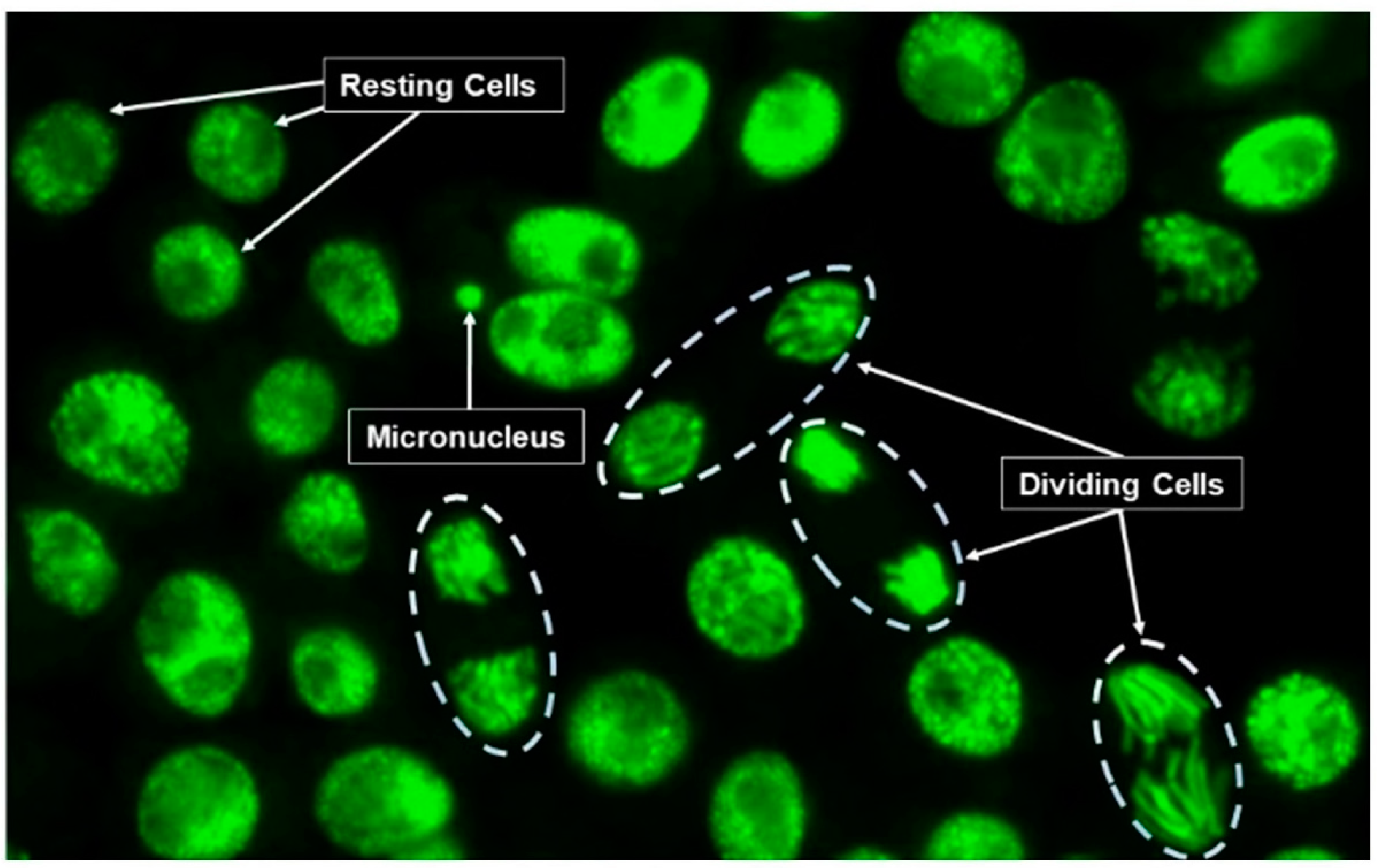
2.7. Micronucleus Assay

All experiments were performed as described by [48], modified by [39], and established by the standard AFNOR EN ISO 29200(2020-05) [37]. Ten apices of secondary roots (about 5 mm from the end of the root) were retained for the Micronucleus Assay. All root cell samples were fixed in Carnoy solution (25% acetic acid and 75% ethanol, v: v) during one night at 4 °C, after which they were washed three times with distilled water. At this stage, roots can be preserved in 70% ethanol at 4 °C for future observations but they should be rehydrated with demineralized water before proceeding. Secondary root apices were hydrolyzed with 1N hydrochloric acid at 60 °C for 7 min. It is important to remove the first mm corresponding to the meristematic region of the root cap and to retain only the following second mm, which is made up of daughter cells [48,49]. Staining of DNA was carried out by placing one drop of acridine orange (CAS 65-61-2) at a final concentration of 0.0375 g. L−1 on each apex of secondary roots. Samples were then crushed between the slide and the coverslip before observations under a fluorescence microscope with a 400× magnification. Cell counts were performed on digitized photos using the free software ImageJ (Rasband, W.S., ImageJ, U. S. National Institutes of Health, Bethesda, ML, USA) and micronuclei (MN), dividing and resting cells were counted on a total of 1000 cells per root, as illustrated in Figure 1.
One slide with three root apices was prepared for each of five replicates. A total of 3000 cells per replicate were counted for analysis, accounting for a total of 15,000 cells scored for each condition. The percentage of mitotic cells, called mitotic index (MI), was calculated. Only roots with a MI greater than 20‰ were considered when scoring micronuclei in cells (AFNOR EN ISO 29200 (2020-05)) [37]. MN and MI were calculated as shown below:
MN   =   M i c r o n u c l e i   R e s t i n g   c e l l s   ×   1000           MI   =   D i v i d i n g   c e l l s T o t a l   c e l l s   ×   1000

2.8. Statistical Analysis

As the data presented a normal distribution, one-way ANOVA and average comparison tests (Tukey) with a 95% confidence interval were performed using Sigma Plot 12.0.

3. Results and Discussion

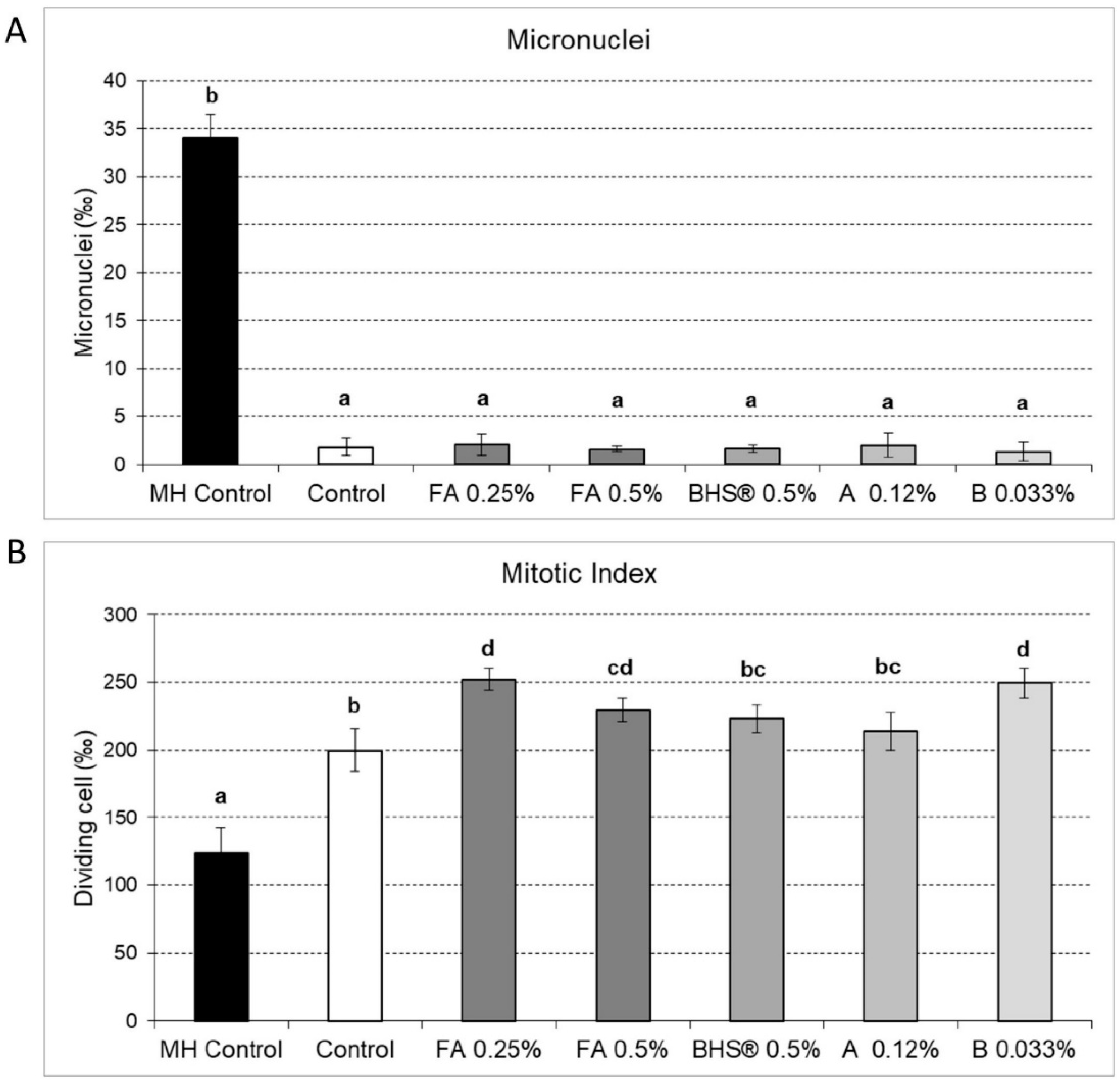
3.1. Impacts of Biostimulants on Micronuclei Formation and Cell Division Frequency in Vicia Faba Roots Cells

Fifty-six hours later, either exposure to the 4 commercial products, or to maleic hydrazide as positive control (MH Control) or without treatment as the negative control (Control), micronuclei (MN) and diving cells (mitotic index MI) were scored in the root apices of Vicia faba, respectively (Figure 2A,B), and expressed as a ‰ of the total number of cells.
The micronuclei values for all commercial products and for the control are close (2–3‰) and display no significant differences. This demonstrates the absence of genotoxicity of commercial biostimulant products (Figure 2A). In contrast, plants exposed to the known genotoxic product (MH) as a herbicide, and used as a positive control, displayed a 17-fold increase in the number of micronuclei compared to the control, indicating significant genotoxicity and damage to cell DNA.
The corresponding mitotic index (MI) of all biostimulants tested (Figure 2B), as well as those of the MH-treated plantlets (Figure 2B: MH control), were higher than 20‰ [37]. Fulvic acids provided at 0.25% and 0.5%, as well as product “B” at 0.033%, showed significant enhanced cell division frequencies with respect to the control plants with no biostimulants, indicating their role in cell division. However, the mitotic index of root cells exposed to MH, showed a significant reduction in the number of dividing cells with respect to the controls, indicating a significant cytotoxicity effect.
Micronuclei are formed if cells are actively dividing after exposure to genotoxicants and observed in non-dividing cells. A decrease in the mitotic index (MI) as compared to the control indicates a toxicity effect and automatically reduces the number of micronuclei observed. However, the absence or the decrease of the MN frequencies could mislead interpretations or conclusions relating to the safety of the tested molecule if cells are not actively divided. Indeed, a toxic molecule could have shorter time impacts than a genotoxic one on the development of MN. The comparison of the MI for all treatments indicated the absence of toxic effects, since all values were higher than those of the unexposed control (Figure 2B). Furthermore, the MI in root tips of plants treated with FA 0.25%, and B product provided at 0.033% were significantly higher by 25% than the control. The comparison of mitotic indices can demonstrate the potential of a molecule or a mixture of compounds present in a commercial product towards “biostimulant” activity on root apex cell division.
In summary, the AFNOR EN ISO 29200 (2020-05) Micronucleus assay [37] provides three types of data on molecule effects towards dividing cells of root apices:
  • Genotoxic effects, if the micronuclei number is greater than the control (MN > MN Control);
  • Cytotoxic effects, if the mitotic index is less than the control (MI < MI control);
  • Biostimulant effects, if the mitotic index is greater than control (MI > MI Control).

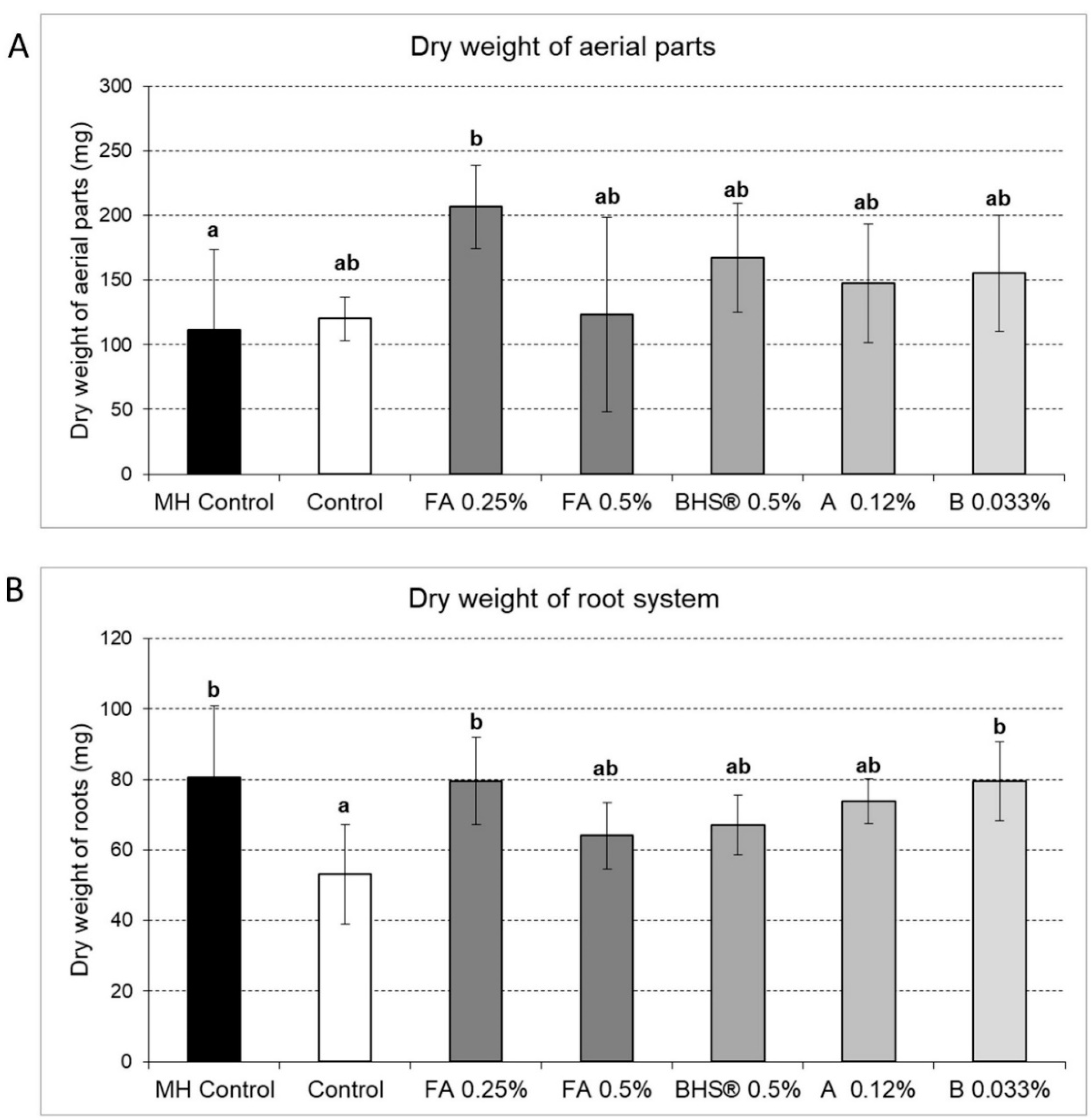
3.2. Impacts of Biostimulants on Root and Plant Aerial Biomass

Using the same previous experiments as described above, plants were harvested and dry matter for aerial parts and roots were weighed (Figure 3).
The dry weights of the aerial part are distributed into three groups (Figure 3A): the first group with the positive MH control is lower than the negative control but not statistically different. The second group is composed of FA 0.5%, BHS® 0.5%, products A and B, showing no significant differences. FA 0.25% stands alone and reaches the highest value (200 mg) with a statistically significant difference from the others reaching 1.7 times the biomass of the control. This is in agreement with [50], where it is described that a product with «biostimulant» activity generally promotes an increase in biomass and growth relative to a control plant.
For root biomass, we observed three groups in increasing order (Figure 3B). A first group consisted only of the control (53 mg), followed by the second group comprised of FA 0.5% (64 mg), BHS® 0.5% (67 mg) and product A (73.7 mg). Finally, the last group, consisting of HM Control (80 mg), and both FA 0.25% and product B (79.5 mg), reached the highest root weight, i.e., 1.5 times the control. Root exposure to MH resulted in a 17-fold increase of the MN frequencies (Figure 2A), in spite of its antimitotic effect, as seen by only a 1.6 fold reduction in the MI (Figure 2B). Indeed, MH is known to inhibit mitosis and is applied to vegetables at 10−2 M. A dose test showed that the antimitotic effect on root growth was observed at 10−3 M, well above the 10−5 M dose used in this study (data not shown). The effect of the HM antimitotic effect at 10−5 M is less than what it is at 10−2 M and could explain the 1.45-fold increase of the root system of MH controls with respect to the negative control. Mitotic index results are in agreement with only two out of four treatments for root and aerial part dry matter (FA 0.25% and B at 0.033%). In both cases, higher percentages of dividing cells are accompanied by an increase in biomass. Biostimulant effects are marked by root meristematic cell division rate and biomass increases, contrarily to the MH control.
Mitotix index analysis has several advantages, and in addition to being a useful marker to predict plant dysfunction, it can be used to:
-
Test growth effects within 10 days contrarily to those implying longer growth experimentation durations needed for biomass production;
-
Compare agronomical efficiencies of different commercial products;
-
Define the optimal dose required for the same product within short spans of time.
Some authors have shown that exposure of Vicia faba to 510 micromoles of Cd per kg of soil induces micronuclei after only 5 days of cultivation without affecting plant growth [51]. Plant growth was affected by cadmium only after longer exposure periods. Indeed, after 155 days, biomass increases were more substantial, but with a significant decrease in the number of flowers and inhibition of fruit setting. Other authors have related an induction of micronuclei on Vicia faba roots exposed to 20, 200 and 2000 µM Cd, but with a visible effect on dry matter only for the highest Cd concentrations [52]. These results clearly demonstrate that the consequences of Cd on micronuclei occurrence is faster, earlier and more sensitive than the reduction of biomass production.

3.3. Impacts of Biostimulants on Length and Surface Area of the Root System

The total length (cm) and area (cm2) of the root system was measured at the end of experiments and are represented in Figure 4A,B.
The length and surface of roots are distributed into three groups. The first group, consisting of the MH control and control without any treatment, is characterized by the lowest values with no significant differences (Figure 4A,B). A second group, displaying slightly higher values, was observed with FA at 0.5%, BHS® at 0.5%, product A and product B. The most significant length and surface area of the root system was observed for the lower concentration of FA at 0.25% (Figure 4). The other treatments showed no biostimulant effects at these doses and under these experimental conditions, with results nearing those of the control. However, it needs to be confirmed whether a longer exposure time would enhance the measured differences between these treatments.
Dry weight increase could be the consequences of other interactive physiological processes following the application of biostimulants. These could be due, for instance, to increased lignification, photosynthesis, secondary metabolites synthesis, that are later events and that contribute to plant build up.
The comparison of different “biostimulant” products, obtained by measuring the mitotic index, is similar to that observed for biomass accumulation or root system growth. In view of these different results, in the short term, the incubation with FA 0.25% appeared to be the most efficient biostimulant concentration. This method seems to be entirely appropriate for rapid evaluation and anticipation of biostimulant effects of different products as well as optimizing the best concentration to be applied.

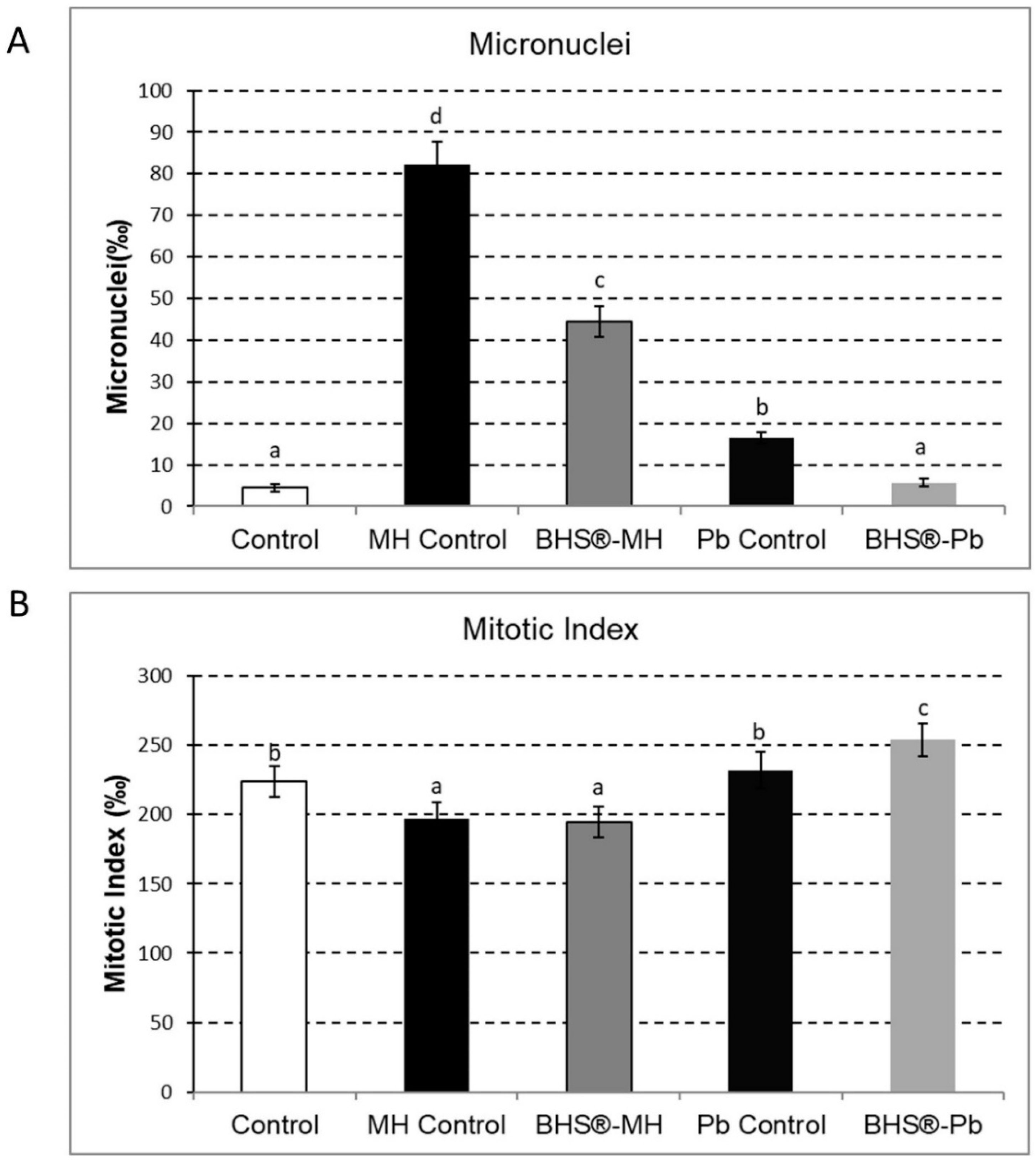
3.4. Protective Effects of Biostimulants towards Exposure to Contaminants

Some companies have suggested in their advertising material that biostimulants could have protective effects against organic and inorganic pollutants, but to date, there is no scientific evidence to support this. In this study, we assessed whether BHS® had protective effects against organic (MH) and metallic (Pb) contaminants using the micronucleus test. This experimentation followed the scheme described in Table 2. The scoring of the mitotic index and the number of micronuclei are presented in Figure 5A,B respectively. The micronuclei count shows, that after 56 h of experimentation, the plantlets exposed 8 h to MH only (MH Control) or to Pb only (Pb Control) displayed significant higher numbers of micronuclei, respectively 18.3 and 3.7 times more than that of the control. In the presence of BHS®, the number of micronuclei for plants exposed to MH was reduced by 46% as compared to the MH control and by 35% for Pb exposure compared to the Pb control. The presence of BHS® strongly reduced the genotoxic effect of Pb and MH, as witnessed by the MN frequencies, with a greater protective effect for Pb.
The values of MI were all well above 20‰ for all conditions tested. The MI of control plantlets relative to those treated with MH (MH Control) is higher. On the contrary, there was no significant difference between the MI of control plantlets and lead control (Pb Control). The pre-treatment with BHS® in the case of MH and Pb displayed different responses. The MI of MH control and BHS®-MH were both around 190‰ with no significant statistical differences, but were lower than that of control plantlets. This can be explained by a slight decrease in cell division, implying a cytotoxic effect of MH.
As far as MI is concerned, we notice that there were no significant differences in the MI of the unexposed controls (224‰) and Pb-control (232‰) as shown in Figure 5B. BHS® pre-treatment plantlets followed by exposure to Pb resulted in an increase of mitotic index from 232‰ to 254‰. The increase of cell division as seen by the MI results show that pre-treating root cells before lead exposure offers a protective effect, enabling recovery of cell divisions in root meristematic cells. In spite of the slight enhancement of cell division rates, there is a significant reduction in the MN frequencies when plantlets are pre-treated with BHS®. This protective effect on micronucleus formation is accompanied by an insignificant effect of the MI for MH showing that cell divisions were not affected by MH in the presence of BHS®.
In the presence of BHS® and for lead, the number of micronuclei was not only reduced by 35%, but there was a 9.5% increase in the MI. In the case of lead (Pb), it has been demonstrated that Pb promotes micronuclei formation through the induction of a strong oxidative stress associated with DNA breakdown. It does not act directly on chromosomal DNA but indirectly by triggering the production of Reactive Oxygen Species (ROS) and/or lipid-soluble by-products of oxidative stress. Indeed, the use of an antioxidant molecule, like Vitamin E, inhibits the effects of oxidative stress due to Reactive Oxygen Species in the presence of Pb as well as reducing the percentage of micronucleus frequency [53]. The micronuclei reduction indicates a protective effect of BHS® against oxidative stress. This protective effect could be at the origin of the significant mitotic index increase in the presence of Pb. Indeed, the increase of the MI with Pb should have automatically increased the number of micronuclei, which would have shown the protective effects of BSH. Portuondo-Farías et al. have shown the protective effect of humic substances from vermicompost against Pb in beans growing on 100 mg of Pb per Kg of soil [54].
We made use of the two components of the micronucleus assay to discriminate between genotoxic effects (based on the MN score) from the biostimulant and cytotoxic effects (measured by the MI) by plotting micronuclei frequency as a function of the mitotic index on Vicia faba root tips (Figure 6A,B).
Results are presented with respect to the position of the control (circle points) using the coordinate pair on the abscissa and ordinate axes (MN; IM) as 4.5‰/224‰. Compared to control plants (circle points MN 4.5‰), exposure of plantlets to lead nitrate (1 mg/L) increased the number of micronuclei by a factor of 3.7 without significantly affecting the mitotic index (Figure 6A—triangle point 16.5‰/232‰). The effect of lead nitrate (1 mg/L) therefore had a significant genotoxic effect with an increase in the MN frequency, but no toxic effect at this concentration with no significant difference in MI, with respect to controls. To test the potential protective effect of BHS®, plantlets were exposed during 24 h to BHS®, then 8 h to Pb and again 24 h to BHS® to allow cell division and thus the development of micronuclei (Figure 6A—square point 5.75‰/254‰). Treatment with BHS® greatly reduced the number of micronuclei that nearly reached the control level while significantly increasing the mitotic index by 10% (254‰). However, the increase in the mitotic index should normally have favored the formation of micronuclei if there had been genotoxic effects. Under our conditions, the increase in the MI with a reduction of the MN clearly demonstrated the protective properties of BHS® against lead. These protective properties of humic or fulvic substances have already been demonstrated by several authors on lettuce against Cd [55] and on radish against Cd, Cu and Zn [56], but not directly on the rate of division in the root apex. The salicylic acid post-treatment of Nigella sativa, exposed to another toxic heavy-metal (cadmium), increases the mitotic index while reducing chromosomal abnormalities [57]. Salicylic acid has a protective action against the genotoxic and toxic effect of heavy metals, as does BHS®.
Another experiment was carried out with MH under the same conditions with the same control (4.5‰/224‰). The exposure of plantlets to MH strongly increased the number of micronuclei by a factor of 18 (82.25‰), showing genotoxic effects but also significantly reduced the mitotic index by a factor of 1.14, indicating toxic effects (Figure 6B—triangle point 82.25‰/197‰). The treatment of plantlets with BSH® halved the number of micronuclei but without any modification of the mitotic index level with respect to controls (Figure 6B—square point 44.5‰/195‰). MH is known to be used as an herbicide that acts on plants in two ways: MH inhibits DNA replication (phase S of the cell division cycle), hence the antimitotic property and its anti-germinative action, which reduces or inhibits the mitotic index depending on the concentration used. MH has plant-specific mutagenic/clastogenic properties, that induces the appearance of micronuclei after cell division, thus revealing the damage caused to DNA [58]. However, no DNA-strand break was observed by comet assay [59]. BHS® reduced the mutagenic/clastogenic effects but had no protective action against the inhibition of DNA replication and therefore against MI reduction.

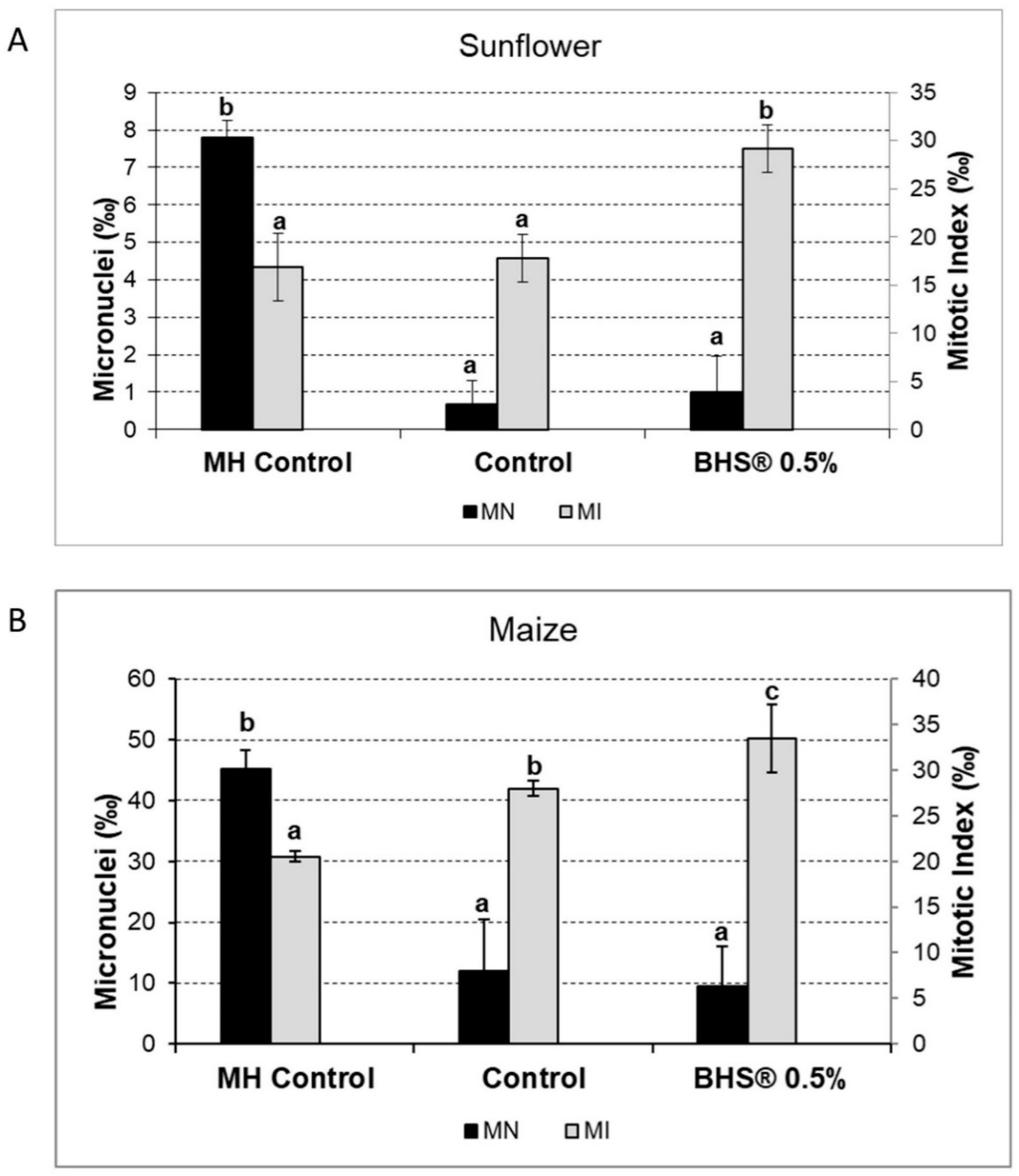
3.5. Extension to Other Agronomic Crops

The micronuclei test was initially developed on broad beans, because the large cells facilitate micronucleus observation by microscopy. However, this technique has been applied to other plants, such as onions [60,61]. We extrapolated the methodology to agronomically important crops, such as sunflower (Helianthus annuus) and maize (Zea mays), to test the effect of BHS® 0.5% (Figure 7). For sunflower (Figure 7A), the higher values of MN obtained with MH Control (7.8‰) compared to those of the Control and BHS® (0.5%) was in conformity with the expected results, i.e., less than 1‰ as for results obtained in Vicia faba (Figure 2A). The MI of the Control (16.8‰) and MH Control (17.8‰) were similar with a mean value around 17‰, while the MI with BHS® were 1.64-fold higher (28‰). This shows that BHS® stimulated cell division in sunflower rootlets.
For maize, the MN value was higher for MH Control (45.13‰) than for the control and for BHS® (both close to 10‰), and corresponding to 10 times those of Vicia faba (Figure 2A). The MI fell into three statistically different categories. The control had an MI of 28‰. Addition of MH reduced the MI to 20.5‰, indicating a cytotoxic effect. On the contrary, BHS® increased the MI 1.2 fold (33.5‰) with a statistically significant biostimulation effect on the plantlets.
It should be noted that mitotic index values obtained for plants exposed to MH were equivalent to the control for sunflower and was lower for maize with a reduction of 28% of the control value. These results indicate that MH (10−5 M) induced slight toxicity and we concluded that corn seems to be more sensitive than sunflower or broad bean. This slight toxicity could be due to the difference in uronic content of root tissues and to the cationic exchange capacity between monocotyledons and dicotyledons [62]. Plant exposure to BHS® (0.5%) increased significantly the mitotic index by 1.56 times for sunflower, 1.26 times for maize and 1.125 times for the Vicia faba control value. The biostimulant effect on cell multiplication of root apices was statistically significant for all plants tested.
This micronulei test can be adapted to different plants being easy to carry out over spans of 10 days. The method can be used to evaluate effects of different commercially formulated products or new molecules by measuring both MI and MN frequencies. The test provides quick information on the cytotoxicity of products (MI < Control), its genotoxicity (MN > control) and its biostimulant effect (MI > control). It is also possible to verify the protective effect of biostimulants against organic or metallic entities by pre-incubating the roots with the product to be tested.

4. Conclusions

Biostimulants used in this study did not reveal any genotoxic effects on broad beans. Fulvic acid used at 0.25% was found to be more efficient than at 0.5% in matter of root and aerial biomass of plants.
This work shows that the Micronucleus Assay, as described by the AFNOR EN ISO 29200 (2020-05) norm, can be used to assess cytotoxic or genotoxic or growth effects. We report here, for the first time, that lengthy cultures to assess plant growth can be replaced by the use of mitotic index counts, to measure, within 10 days, the effects of bio-sourced materials on growth. To our knowledge, this is the first report whereby micronucleus frequency measurements are linked to mitotic indices using them as reliable markers to measure and to differentiate genotoxic or toxic effects of biostimulants and to evaluate growth impacts on plant cells. This could be useful to determine rapidly whether any newly developed product could have marketing opportunities, with proven arguments to convince their potential clients. This study with Vicia faba also demonstrates that humic biostimulants, like BHS®, confer protective effects against an organic molecule like maleic hydrazide, used as an anti-germinative product or trace metal element like lead that can be found in soil or in fertilizers. This method performed on Vicia faba, maize and sunflower plantlets could be transposed to other agronomical crops or market gardening.

Author Contributions

For research articles with several authors, a short paragraph specifying their individual contri-butions must be provided. The following statements should be used “Conceptualization, J.S., J.K. and E.P.; methodology, L.C. and P.K.; software, L.C. and J.S.; validation, J.S., L.C., and P.K.; formal analysis, J.S. and P.K.; investigation, J.S. and J.K.; resources, M.M; data curation, J.S.; writing—original draft preparation, J.S., J.K., E.P., P.K. and L.C.; writing—review and editing, J.K. and J.S.; visualization, L.C. and P.K. supervision, J.S., and J.K. project administration, J.K. and J.S.; funding acquisition, M.M. This experimentation was financed by the private company Bois Valor. All authors have read and agreed to the published version of the manuscript.

Funding

This research received no external funding.

Data Availability Statement

The data presented in this study are available on request from the corresponding author.

Acknowledgments

All authors are indebted to the deeply regretted late, Philippe MORARD at the Engineering Faculty of Life Sciences of Toulouse (Toulouse INP-ENSAT) for his vision on the value enhancement of agro resources and bio-sourcing of poplar wood wastes. He was the first to design industrial processes towards extraction of Biomimetic Humic Substances (BHS®), their characterization and their effect on different plant species. We would like to thank the Bois Valor company for seconding staff to help us carry out the experimentations.

Conflicts of Interest

The funders had no role in the design of the study; in the collection, analyses, or interpretation of data; in the writing of the manuscript, or in the decision to publish the results.

References

  1. De Pascale, S.; Rouphael, Y.; Colla, G. Plant biostimulants: Innovative tool for enhancing plant nutrition in organic farming. Eur. J. Hortic. Sci. 2018, 82, 277–285. [Google Scholar] [CrossRef]
  2. Laurent, E.-A.; Ahmed, N.; Durieu, C.; Grieu, P.; Lamaze, T. Marine and fungal biostimulants improve grain yield, nitrogen absorption and allocation in durum wheat plants. J. Agric. Sci. 2020, 158, 279–287. [Google Scholar] [CrossRef]
  3. Kocira, S.; Szparaga, A.; Findura, P.; Treder, K. Modification of Yield and Fiber Fractions Biosynthesis in Phaseolus vulgaris L. by Treatment with Biostimulants Containing Amino Acids and Seaweed Extract. Agronomy 2020, 10, 1338. [Google Scholar] [CrossRef]
  4. Ioppolo, A.; Laudicina, V.A.; Badalucco, L.; Saiano, F.; Palazzolo, E. Wastewaters from citrus processing industry as natural biostimulants for soil microbial community. J. Environ. Manag. 2020, 273, 111137. [Google Scholar] [CrossRef]
  5. Bulgari, R.; Franzoni, G.; Ferrante, A. Biostimulants Application in Horticultural Crops under Abiotic Stress Conditions. Agronomy 2019, 9, 306. [Google Scholar] [CrossRef] [Green Version]
  6. Bulgari, R.; Cocetta, G.; Trivellini, A.; Vernieri, P.; Ferrante, A. Biostimulants and crop responses: A review. Biol. Agric. Hortic. 2014, 31, 1–17. [Google Scholar] [CrossRef]
  7. Pylak, M.; Oszust, K.; Frąc, M. Review report on the role of bioproducts, biopreparations, biostimulants and microbial inocu-lants in organic production of fruit. Rev. Environ. Sci. Biotechnol. 2019, 18, 597–616. [Google Scholar] [CrossRef] [Green Version]
  8. Daniel, E.C.; Fabio, G. An assessment of seaweed extracts: Innovation for sustainable agriculture. Agronomy 2020, 10, 1433. [Google Scholar] [CrossRef]
  9. Masondo, N.A.; Kulkarni, M.G.; Finnie, J.F.; Van Staden, J. Influence of biostimulants-seed-priming on Ceratotheca triloba germination and seedling growth under low temperatures, low osmotic potential and salinity stress. Ecotoxicol. Environ. Saf. 2018, 147, 43–48. [Google Scholar] [CrossRef]
  10. Alzahrani, Y.; Rady, M.M. Compared to antioxidants and polyamines, the role of maize grain-derived organic biostimulants in improving cadmium tolerance in wheat plants. Ecotoxicol. Environ. Saf. 2019, 182, 109378. [Google Scholar] [CrossRef] [PubMed]
  11. European Union. Regulation 2019/1009 of the European Parliament. Available online: https://eur-lex.europa.eu/eli/reg/2019/1009/oj (accessed on 4 November 2020).
  12. Du Jardin, P. Plant Biostimulants: Definition, concept, main categories and regulation. Sci. Hortic. 2015, 196, 3–14. [Google Scholar] [CrossRef] [Green Version]
  13. Szczałba, M.; Kopta, T.; Gąstoł, M.; Sekara, A. Comprehensive insight into arbuscular mycorrhizal fungi, Trichoderma spp. and plant multilevel interactions with emphasis on biostimulation of horticultural crops. J. Appl. Microbiol. 2019, 127, 630–647. [Google Scholar] [CrossRef] [PubMed] [Green Version]
  14. González-González, M.F.; Ocampo-Alvarez, H.; Santacruz-Ruvalcaba, F.; Sánchez-Hernández, C.V.; Casarrubias-Castillo, K.; Becerril-Espinosa, A.; Castañeda-Nava, J.J.; Hernández-Herrera, R.M. Physiological, Ecological, and Biochemical Implications in Tomato Plants of Two Plant Biostimulants: Arbuscular Mycorrhizal Fungi and Seaweed Extract. Front. Plant Sci. 2020, 11, 999. [Google Scholar] [CrossRef]
  15. Canellas, L.P.; Olivares, F.L.; Aguiar, N.O.; Jones, D.L.; Nebbioso, A.; Mazzei, P.; Piccolo, A. Humic and fulvic acids as biostimulants in horticulture. Sci. Hortic. 2015, 196, 15–27. [Google Scholar] [CrossRef]
  16. Rodrigues, M.; Baptistella, J.L.C.; Hörz, D.C.; Bortolato, L.M.; Mazzafera, P. Organic Plant Biostimulants and Fruit Quality—A Review. Agronomy 2020, 10, 988. [Google Scholar] [CrossRef]
  17. Chen, Y.; Aviad, T. Humic Substances in Soil and Crop Sciences: Selected Readings. Humic Subst. Soil Crop Sci. Sel. Read. 1990, 161–186. [Google Scholar] [CrossRef]
  18. Vaughan, D.; Macdonald, I. Some effects of humic acid on cation uptake by parenchyma tissue. Soil Biol. Biochem. 1976, 8, 415–421. [Google Scholar] [CrossRef]
  19. Eyheraguibel, B. Caractérisation des Substances Humiques Biomimétiques: Effets sur les Végétaux; INPT: Toulouse, France, 2004; Volume 3. [Google Scholar]
  20. Baldotto, M.A.; Muniz, R.C.; Baldotto, L.E.B.; Dobbss, L.B. Root growth of Arabidopsis thaliana (L.) Heynh. treated with humic acids isolated from typical soils of Rio de Janeiro state, Brazil. Rev. Ceres 2011, 58, 504–511. [Google Scholar] [CrossRef] [Green Version]
  21. Canellas, N.O.A.; Olivares, F.L.; Canellas, L.P. Metabolite fingerprints of maize and sugarcane seedlings: Searching for markers after inoculation with plant growth-promoting bacteria in humic acids. Chem. Biol. Technol. Agric. 2019, 6, 1–10. [Google Scholar] [CrossRef]
  22. Ertani, A.; Nardi, S.; Francioso, O.; Pizzeghello, D.; Tinti, A.; Schiavon, M. Metabolite-Targeted Analysis and Physiological Traits of Zea Mays L. in Response to Application of a Leonardite-Humate and Lignosulfonate-Based Products for Their Evaluation as Potential Biostimulants. Agronomy 2019, 9, 445. [Google Scholar] [CrossRef] [Green Version]
  23. Donno, D.; Beccaro, G.L.; Mellano, M.G.; Canterino, S.; Cerutti, A.K.; Bounous, G. Improving the nutritional value of kiwifruit with the application of agroindustry waste extracts. J. Appl. Bot. Food Qual. 2013, 86, 11–15. [Google Scholar] [CrossRef]
  24. Antón-Herrero, R.; García-Delgado, C.; Alonso-Izquierdo, M.; Cuevas, J.; Carreras, N.; Rivilla, B.; Camacho-Arévalo, R.; Eymar, E. New Uses of Treated Urban Waste Digestates on Stimulation of Hydroponically Grown Tomato (Solanum lycopersicon L.). Waste Biomass Valorization 2020, 1–13. [Google Scholar] [CrossRef]
  25. Juárez-Maldonado, A.; Ortega-Ortíz, H.; Morales-Díaz, A.B.; González-Morales, S.; Morelos-Moreno, Á.; La Fuente, M.C.-D.; Sandoval-Rangel, A.; Cadenas-Pliego, G.; Benavides-Mendoza, A. Nanoparticles and Nanomaterials as Plant Biostimulants. Int. J. Mol. Sci. 2019, 20, 162. [Google Scholar] [CrossRef] [Green Version]
  26. Spaccini, R.; Cozzolino, V.; Di Meo, V.; Savy, D.; Drosos, M.; Piccolo, A. Bioactivity of humic substances and water extracts from compost made by ligno-cellulose wastes from biorefinery. Sci. Total. Environ. 2019, 646, 792–800. [Google Scholar] [CrossRef] [PubMed]
  27. De Moraes, E.R.; De Camargo, R.; Lana, R.M.Q.; Madeiros, M.H.; Menezes, F.G.; Giorgenon, E.P. Yield and biometry of fertilized sugar cane with organomineral fertilizer of sewage sludge and biostimulant. Biosci. J. 2020, 36, 1564–1576. [Google Scholar] [CrossRef]
  28. El Fels, L.; Zamama, M.; El Asli, A.; Hafidi, M. Assessment of biotransformation of organic matter during co-composting of sewage sludge-lignocelullosic waste by chemical, FTIR analyses, and phytotoxicity tests. Int. Biodeterior. Biodegrad. 2014, 87, 128–137. [Google Scholar] [CrossRef]
  29. El Fels, L.; Hafidi, M.; Silvestre, J.; Kallerhoff, J.; Merlina, G.; Pinelli, E. Efficiency of co-composting process to remove genotoxicity from sewage sludge contaminated with hexavalent chromium. Ecol. Eng. 2015, 82, 355–360. [Google Scholar] [CrossRef]
  30. Palumbo, G.; Schiavon, M.; Nardi, S.; Ertani, A.; Celano, G.; Colombo, C.M. Biostimulant Potential of Humic Acids Extracted From an Amendment Obtained via Combination of Olive Mill Wastewaters (OMW) and a Pre-treated Organic Material Derived From Municipal Solid Waste (MSW). Front. Plant Sci. 2018, 9, 1028. [Google Scholar] [CrossRef]
  31. Silva, T.D.S.; Silva, A.P.S.E.; Santos, A.D.A.; Ribeiro, K.G.; De Souza, D.C.; Bueno, P.A.A.; Marques, M.M.M.; De Almeida, P.M.; Peron, A.P. Cytotoxicity, Genotoxicity, and Toxicity of Plant Biostimulants Produced in Brazil: Subsidies for Determining Environmental Risk to Non-Target Species. Water Air Soil Pollut. 2020, 231, 1–8. [Google Scholar] [CrossRef]
  32. Vijayaraj, V.; Liné, C.; Cadarsi, S.; Salvagnac, C.; Baqué, D.; Elger, A.; Barret, M.; Mouchet, F.; LaRue, C. Transfer and Ecotoxicity of Titanium Dioxide Nanoparticles in Terrestrial and Aquatic Ecosystems: A Microcosm Study. Environ. Sci. Technol. 2018, 52, 12757–12764. [Google Scholar] [CrossRef] [PubMed]
  33. Evariste, L.; Lagier, L.; Gonzalez, P.; Mottier, A.; Mouchet, F.; Cadarsi, S.; Lonchambon, P.; Daffe, G.; Chimowa, G.; Sarrieu, C.; et al. Thermal Reduction of Graphene Oxide Mitigates Its In Vivo Genotoxicity Toward Xenopus laevis Tadpoles. Nanomaterials 2019, 9, 584. [Google Scholar] [CrossRef] [PubMed] [Green Version]
  34. Shobha, G.; Shashidhara, K.S.; Naik, C. Cuprous Oxide Nanoparticles Induced Antioxidant Response and Genotoxicity in Lycopersicum esculentum. BioNanoScience 2020, 10, 1128–1137. [Google Scholar] [CrossRef]
  35. Wise, K.; Gill, H.; Selby-Pham, J. Willow bark extract and the biostimulant complex Root Nectar® increase propagation efficiency in chrysanthemum and lavender cuttings. Sci. Hortic. 2020, 263, 109108. [Google Scholar] [CrossRef]
  36. Eyheraguibel, B.; Silvestre, J.; Morard, P. Effects of humic substances derived from organic waste enhancement on the growth and mineral nutrition of maize. Bioresour. Technol. 2008, 99, 4206–4212. [Google Scholar] [CrossRef] [Green Version]
  37. AFNOR EN ISO 29200. Soil Quality-Assessment of Genotoxic Effects on Higher Plants-Vicia Faba-Micronucleus Test; Association Française de Normalisation: La Plaine Saint-Denis, France, 2020. [Google Scholar]
  38. Iqbal, M. Vicia faba bioassay for environmental toxicity monitoring: A review. Chemosphere 2016, 144, 785–802. [Google Scholar] [CrossRef]
  39. Cotelle, S.; Dhyèvre, A.; Muller, S.; Chenon, P.; Manier, N.; Pandard, P.; Echairi, A.; Silvestre, J.; Guiresse, M.; Pinelli, E.; et al. Soil genotoxicity assessment—Results of an interlaboratory study on the Vicia micronucleus assay in the context of ISO standardization. Environ. Sci. Pollut. Res. 2014, 22, 988–995. [Google Scholar] [CrossRef]
  40. De Simone, C.; Angelucci, R.; Errichetti, M.F.; Marconi, S.; Rossi, M.; Selvi, S. A statistical approach to evaluate compost genotoxicity. Biol. Fertil. Soils 2004, 41, 9–14. [Google Scholar] [CrossRef]
  41. Marcato-Romain, C.; Pinelli, E.; Pourrut, B.; Silvestre, J.; Guiresse, M. Assessment of the genotoxicity of Cu and Zn in raw and anaerobically digested slurry with the Vicia faba micronucleus test. Mutat. Res. Toxicol. Environ. Mutagen. 2009, 672, 113–118. [Google Scholar] [CrossRef] [PubMed] [Green Version]
  42. El Hajjouji, H.; Pinelli, E.; Guiresse, A.M.; Merlina, G.; Revel, J.-C.; Hafidi, M. Assessment of the genotoxicity of olive mill waste water (OMWW) with the Vicia faba micronucleus test. Mutat. Res. Toxicol. Environ. Mutagen. 2007, 634, 25–31. [Google Scholar] [CrossRef] [Green Version]
  43. Khadra, A.; Pinelli, E.; Lacroix, M.; Bousquet-Mélou, A.; Hamdi, H.; Merlina, G.; Guiresse, M.; Hafidi, M. Assessment of the genotoxicity of quinolone and fluoroquinolones contaminated soil with the Vicia faba micronucleus test. Ecotoxicol. Environ. Saf. 2012, 76, 187–192. [Google Scholar] [CrossRef] [PubMed]
  44. Njagi, G.D.E.; Gopalan, H.N.B. Mutagenicity testing of herbicides, fungicides and insecticides. I. Chromosome aberrations in Vicia faba. Cytology 1981, 46, 169–172. [Google Scholar] [CrossRef] [Green Version]
  45. Souguir, D.; Ferjani, E.; Ledoigt, G.; Goupil, P. Exposure of Vicia faba and Pisum sativum to copper-induced genotoxicity. Protoplasma 2008, 233, 203–207. [Google Scholar] [CrossRef] [PubMed]
  46. Morard, P.; Eyheraguibel, B.; Morard, M.; Silvestre, J. Direct effects of humic-like substance on growth, water, and mineral nutrition of various species. J. Plant Nutr. 2010, 34, 46–59. [Google Scholar] [CrossRef]
  47. Akinremi, O.O.; Janzen, H.H.; Lemke, R.L.; Larney, F.J. Response of canola, wheat and green beans to leonardite additions. Can. J. Soil Sci. 2000, 80, 437–443. [Google Scholar] [CrossRef] [Green Version]
  48. Ma, T.-H.; Xu, Z.; Xu, C.; McConnell, H.; Rabago, E.V.; Arreola, G.A.; Zhang, H. The improved Allium/Vicia root tip micronucleus assay for clastogenicity of environmental pollutants. Mutat. Res. Mutagen. Relat. Subj. 1995, 334, 185–195. [Google Scholar] [CrossRef]
  49. Porta, H.; Rocha-Sosa, M. Update on Plant Lipoxygenases Plant Lipoxygenases. Physiological and Molecular Features. Plant Physiol. 2002, 130, 15–21. [Google Scholar] [CrossRef] [Green Version]
  50. Lucini, L.; Rouphael, Y.; Cardarelli, M.; Bonini, P.; Baffi, C.; Colla, G. A Vegetal Biopolymer-Based Biostimulant Promoted Root Growth in Melon While Triggering Brassinosteroids and Stress-Related Compounds. Front. Plant Sci. 2018, 9, 472. [Google Scholar] [CrossRef] [Green Version]
  51. Foltête, A.-S.; Masfaraud, J.-F.; Férard, J.-F.; Cotelle, S. Is there a relationship between early genotoxicity and life-history traits in Vicia faba exposed to cadmium-spiked soils? Mutat. Res. Toxicol. Environ. Mutagen. 2012, 747, 159–163. [Google Scholar] [CrossRef]
  52. Raskin, I.; Ensley, B.D.; Burt, D. Phytoremediation of Toxic Metals: Using Plants to Clean Up the Environment; J. Wiley: Hoboken, NJ, USA, 2000; ISBN 9780471192541. [Google Scholar]
  53. Pourrut, B.; Jean, S.; Silvestre, J.; Pinelli, E. Lead-induced DNA damage in Vicia faba root cells: Potential involvement of oxidative stress. Mutat. Res. Toxicol. Environ. Mutagen. 2011, 726, 123–128. [Google Scholar] [CrossRef] [Green Version]
  54. Portuondo-Farías, L.; Martinez-Balmori, D.; Guridi-Izquierdo, F.; Calderin-Garcia, A.; Machado-Torres, J.P. Structural and Functional Evaluation of Humic Acids in Interaction with Toxic Metals in a Cultivar of Agricultural Interest. Rev. Ciencias Técnicas Agropecu. 2017, 26, 39–46. [Google Scholar]
  55. Wang, Y.; Yang, R.; Zheng, J.; Shen, Z.; Xu, X. Exogenous foliar application of fulvic acid alleviate cadmium toxicity in lettuce (Lactuca sativa L.). Ecotoxicol. Environ. Saf. 2019, 167, 10–19. [Google Scholar] [CrossRef]
  56. Ondrasek, G.; Rengel, Z.; Romic, D. Humic acids decrease uptake and distribution of trace metals, but not the growth of radish exposed to cadmium toxicity. Ecotoxicol. Environ. Saf. 2018, 151, 55–61. [Google Scholar] [CrossRef] [PubMed]
  57. El-Ghamery, A.; Mousa, M. Salicylic acid triggers adaptation cadmium cytogenetic toxicity in roots of Nigella sativa L. Egypt J. Bot. 2018, 58, 297–310. [Google Scholar] [CrossRef] [Green Version]
  58. Marcano, L.; Carruyo, I.; Del Campo, A.; Montiel, X. Cytotoxicity and mode of action of maleic hydrazide in root tips of Allium cepa L. Environ. Res. 2004, 94, 221–226. [Google Scholar] [CrossRef]
  59. Gichner, T.; Menke, M.; Stavreva, D.; Schubert, I. Maleic hydrazide induces genotoxic effects but no DNA damage detectable by the Comet assay in tobacco and field beans. Mutagenesis 2000, 15, 385–389. [Google Scholar] [CrossRef] [PubMed]
  60. Cotelle, S.; Masfaraud, J.-F.; Férard, J.-F. Assessment of the genotoxicity of contaminated soil with the Allium/Vicia-micronucleus and the Tradescantia-micronucleus assays. Mutat. Res. Mol. Mech. Mutagen. 1999, 426, 167–171. [Google Scholar] [CrossRef]
  61. Wu, L.; Yi, H.; Yi, M. Assessment of arsenic toxicity using Allium/Vicia root tip micronucleus assays. J. Hazard. Mater. 2010, 176, 952–956. [Google Scholar] [CrossRef] [PubMed]
  62. Crooke, W.M.; Knight, A.H. Crop composition in relation to soil pH and root cation-exchange capacity. J. Sci. Food Agric. 1971, 22, 235–241. [Google Scholar] [CrossRef]
Figure 1. Picture of Micronuclei, dividing cells and resting cells observed in the root tips of Vicia faba stained with acridine orange, under a fluorescence microscope (400×).
Agronomy 11 00321 g001
Figure 2. (A,B) Scoring of Micronuclei (A) expressed as the number of micronuclei per 1000 cells observed and of mitotic indices (B), expressed as the number of dividing cells per 1000 cells observed in the root apices of Vicia faba after 56 h of treatment: non-exposed (Control) or exposed to Maleic hydrazide at 1.12 mg.L−1 (MH Control), or to different commercial products: fulvic acids at 0.25% (FA 0.25%), 0.5% (FA 0.5%), biomimetic humic substances (BHS® 0.5%), and products A or B (Histograms marked with the same letter are not statistically different at p < 0.05).
Agronomy 11 00321 g002
Figure 3. (A,B): Dry weight measurement of aerial parts (A) and roots (B) of Vicia faba after 56 h of experiment: non-exposed (Control) or exposed to 10−5 M Maleic hydrazide (MH Control), or to different commercial products: fulvic acids at 0.25% (FA 0.25%) or 0.5% (FA 0.5%), biomimetic humic substances (BHS® 0.5%), or products A and B. (Histograms marked with the same letter are not statistically different at p < 0.05).
Agronomy 11 00321 g003
Figure 4. (A,B): Total length (A) and surface area (B) of Vicia faba roots after 56 h experimentation: non-exposed (Control) or exposed to 10−5 M Maleic hydrazide (MH Control), or to different commercial products: fulvic acids at 0.25% (FA 0.25%) or 0.5% (FA 0.5%), or biomimetic humic substances (BHS® 0.5%) or product A or B. (Vertical bars indicate Standard deviation and histograms marked with the same letter are not statistically different at p < 0.05).
Agronomy 11 00321 g004
Figure 5. Assessment of the protective effect of BHS®. Micronucleus frequency (A), expressed as number of micronuclei per 1000 cells observed and mitotic index (B) expressed as number of dividing cells per 1000 cells observed on the root apex of Vicia faba after 56 h of treatment: non-exposed (Control) or exposed to 10−5 M Maleic hydrazide (MH Control) or lead (Pb Control) or exposed to BHS® and to MH (BHS®-MH), (Histograms marked with the same letter are not statistically different at p < 0.05).
Agronomy 11 00321 g005
Figure 6. (A,B): Variation of micronuclei as a function of the mitotic index for plantlets of Vicia faba exposed to (A) 1 mg.L−1 lead nitrate (Pb) or to (B) 10−5 M maleic hydrazine (MH) or with or without pretreatment by biomimetic humic substances (BHS®) (letters on the left of the data symbol points correspond to statistical analysis of the micronuclei frequency. Letters above the symbol data correspond to statistical analysis of the mitotic index. Data marked with the same letter are not statistically different at p < 0.05).
Agronomy 11 00321 g006
Figure 7. (A,B): Scoring of Micronuclei indices expressed as the number of micronuclei per 1000 cells (Left bar chart) and mitotic index, expressed as the number of dividing cells per 1000 cells (Right bar chart) observed in the root apices of sunflower ((A) Helianthus annuus) and maize ((B) Zea mays) after 56 h of treatment: non-exposed (Control) or exposed to Maleic hydrazide at 1.12 mg.L−1 (MH Control), or to commercial products of biomimetic humic substances (BHS® 0.5%). (Histograms marked with the same letter are not statistically different at p < 0.05).
Agronomy 11 00321 g007
Table 1. Characteristics of the commercial products tested.
Product NameBiomimetic
Humic Substances (BHS®)
Fulvic Acid (FA)Product “A”Product “B”
Originphysico-chemical extraction of poplar wood wastephysico-chemical extraction of poplar wood wasteLeonardite extractBy-products of the of wood pulp production
Composition45% humic acids and 55% fulvic acid100% fulvic acid, extracted from BHS®.Humic acid 12% w/w; fulvic acid 3% w/wsulfonated lignin
Initial Carbon concentration (%)1.5%0.825%7.28%23%
Recommended commercial carbon dose0.5%0.25% and 0.5%0.12%0.033%
Table 2. Experimental set-up to test protective effects of BHS® against a metallic (lead) and an organic (maleic hydrazide) molecule.
Onset of ExperimentExposureRecovery
Phase Duration (56 h)24 h8 h24 h
controlnutrient solutionnutrient solutionnutrient solution
Pb nutrient solutionPb 1 mg.L−1nutrient solution
MH nutrient solutionMH 1.12 mg.L−1nutrient solution
BHS®’s protection against PbBHS® 0.5%Pb 1 mg.L−1BHS® 0.5%
BHS®’s protection against MHBHS® 0.5%MH 1.12 mg.L−1BHS® 0.5%
(Pb: lead; MH: maleic hydrazide; BHS®: biomimetic humic substances).
Publisher’s Note: MDPI stays neutral with regard to jurisdictional claims in published maps and institutional affiliations.

