Article

Simultaneous Determination of Carotenoids and Chlorophylls by the HPLC-UV-VIS Method in Soybean Seeds

1
The National Engineering Laboratory for Crop Molecular Breeding, MARA Key Laboratory of Soybean Biology (Beijing), Institute of Crop Sciences, Chinese Academy of Agricultural Sciences, 12 Zhongguancun South Street, Beijing 100081, China
2
Crop Sciences Research Department, Mehoni Agricultural Research Center, Maichew 7020, Ethiopia
*
Authors to whom correspondence should be addressed.
These authors contributed equally for this work.
Agronomy 2021, 11(4), 758; https://doi.org/10.3390/agronomy11040758
Submission received: 4 March 2021 / Revised: 8 April 2021 / Accepted: 9 April 2021 / Published: 13 April 2021
(This article belongs to the Section Crop Breeding and Genetics)

Abstract

:
Soybean contains nutritional bioactive compounds, including carotenoids associated with human health benefits. Carotenoids are applicable in pharmaceuticals/nutreceuticals, cosmetic, and mainly food industries. However, an efficient and accurate method for carotenoid and chlorophyll detection and quantification has not yet been developed and validated for soybean seeds. The need for a rapid and reliable analysis method has become increasingly important. Thus, this study was initiated to develop and validate a simple, rapid, and selective reversed-phase high-performance liquid chromatographic (RP-HPLC) method for the simultaneous determination of lutein, zeaxanthin, α-carotene, β–carotene, β–cryptoxanthin, and chlorophyll–a and –b in soybean flour sample (100.00 mg) extracted using ethanol-acetone (1:1) solvents at a volume of 1.50 mL. Interestingly, the effective separation technique was achieved using the mobile phases of methyl tert-butyl ether, methanol containing 10 mM ammonium acetate, and water delivered at a 0.90 mL min−1 flow rate through a C30YMC Carotenoid (250 × 4.6 mm I.D., S-5 µm) column coupled with a UV-VIS detector set at 450 nm. The detector response was linear from 0.05–30.00 μg mL−1 with a coefficient of determination (R2) of 0.9993–0.9999. The validated method was sensitive with a detection limit (LOD) of 0.0051–0.0300 μg mL−1 and 0.0155–0.0909 μg mL−1 for the quantification limit (LOQ). The recovery values were from 83.12–106.58%, and the repeatability precision ranged from 1.25–4.20% and 0.15–0.81% for the method and system, respectively. The method showed adequate precision with a relative standard deviation smaller than 3.00%. This method was also found to be applicable for profiling carotenoids and chlorophylls in other legumes. In summary, this method was successfully implemented for qualitative and quantitative determination of major carotenoids and chlorophylls in soybean and other legume seeds, which are beneficial to food industry and quality breeding programs to meet human nutrition demands globally.

1. Introduction

Soybean (Glycine max (L.) Merr.), which is one of the most important crops globally, contains bioactive compounds that play a significant role in human health and nutrition because of their pro-vitamin A, antioxidant, and anti-inflammatory properties. Carotenoids, which are among the bioactive phytochemicals of soybean seeds, occur widely in plants, insects, fish, birds, algae, yeasts, and bacteria [1], and have diverse biological functions in plants and human health. Carotenoids interact with reactive oxygen species and thus act as free radical quenchers, singlet oxygen scavengers, and liquid antioxidants [2]. In human nutrition, carotenoids play a significant role in reducing numerous chronic diseases, such as age-related macular degeneration, cardiovascular diseases, some types of cancers, osteoporosis, diabetes, and others [3,4,5].
The most abundant carotenoids in human blood plasma include β-carotene, α-carotene, β-cryptoxanthin, lutein, zeaxanthin, and lycopene. β-carotene, α-carotene, and β-cryptoxanthin are widely known as pro-vitamin A carotenoids [6], which play a great role in overcoming vitamin A deficiency (VAD) in infants and pregnant women in the developing world [7]. Apart from eye blindness in children, VAD also weakens the immune system, especially in pregnant and lactating women whose diets are predominated by less nutritious staple foods. It is reported that lutein and its stereoisomer zeaxanthin, which are the only carotenoids present in the macula region of the retina, have significant importance in reducing the risk of ocular diseases, such as cataracts and age-related macular degeneration [8,9], maintaining heart health, and minimizing UV-induced skin damage [10].
In the case of chlorophylls, chlorophyll-a and chlorophyll-b, which occur in the approximate ratio of 3:1, are the major chlorophylls in plants. Some studies removed chlorophylls during extraction irrespective of their human health importance [11,12,13]. Chlorophylls and their derivatives have shown important health-promoting functions, such as antimutagenic, anticarcinogenic, and anti-inflammatory activities [14]. Consequently, it is highly valuable to detect and quantify the concentrations of chlorophylls in food crops.
In recent years, more emphasis has been given to obtaining reliable data on the types and concentrations of carotenoids in foods for various health and nutrition purposes. Some studies have suggested that the analysis of carotenoids has been routinely executed by reversed-phase high-pressure liquid chromatography (RP-HPLC) because of its improved separation efficiency [15,16]. The research findings of Inbaraj et al. [17] and Puspitasari-nienaber et al. [18] showed that carotenoids and chlorophylls were determined by HPLC with a C18 or C30 reverse-phase (RP) column operated with an isocratic or a gradient elution using mobile phase of different organic solvents and detectors, such as UV-Vis, mass spectrometry (MS), a diode array detector (DAD), and an electrochemical detector (ED). Analysis of carotenoids is complicated by their diversity, instability, complexity, and the presence of geometric isomeric forms with their diverse spectrum of polarities [19]. As an example, according to the generated database of 120 foods [20], it is reported that lutein and zeaxanthin were not identified due to the difficulty in separating these two components by the C18 column. However, effective use of the HPLC C30 RP column enables optimal separation of a great number of carotenoids. Several reports demonstrated that C30 columns could provide better resolution of carotenoids and more efficiently resolve geometrical isomers than C18 columns by the virtue of using polymeric non-end capped stationary phases with C30 ligands [15,18]. Moreover, C30 columns have a longer service life than other typical reverse-phase columns [21].
The versatile use of carotenoids in feed, food, cosmetic, and pharmaceutical industries has emphasized the optimization of extraction methods to obtain the highest recovery. Due to the presence of diverse carotenoids with varied levels of polarity, and the presence of various physical and chemical barriers in the food matrices that prevent mass transfer of carotenoids during extraction, the choice of method and solvents for carotenoid extraction from food matrices needs crucial attention [6]. Owing to the hydrophobic nature of carotenoids, organic solvents are used as the extractant. Mostly, non-polar solvents are an excellent choice for the extraction of non-polar carotenes whereas polar solvents are more appropriate for the extraction of polar carotenoids. On the basis of environmental and health and safety issues, ethanol and acetone are preferred solvents compared to hexane, diethyl ether, dichloromethane, and chloroform, which are generally used for extraction of carotenoids [22]. Furthermore, owing to water-miscible properties, acetone and ethanol are preferred for efficient extraction of carotenoids from plant material containing a high amount of moisture [6]. Basically, the extraction methods for carotenoids and chlorophylls should be with improved solvents and techniques aimed at rapid, simple, cost-effective, and efficient extraction. The problem faced during the work and manipulation of carotenoids is their degradation nature when exposed to light, heat, oxygen, and acids. For this reason, several precautions, such as using antioxidants, conducting experiments in dim lighting, and storing samples in the dark at about −20 °C, are necessary when handling carotenoids [3,23].
Generally, carotenoids are considered to be crucial elements in soybeans. However, to date, due to the unavailability of a comprehensive carotenoid and chlorophyll profile in soybean seed, the need for a rapid and reliable analysis method has become increasingly important. Thus, the objective of this study was to develop and validate a rapid, simple, and accurate C30 RP-HPLC analysis method for the determination of a complete array of carotenoids and chlorophylls in soybean seeds to accomplish selections of a large number of plant populations within a short period of time in large-scale soybean quality breeding programs.

2. Materials and Methods

2.1. Standards, Solvents, and Reagents

Carotenoid and chlorophyll standards: α-carotene (CAS: 7488-99-5, purity ≥ 98%), β-carotene (CAS: 7235-40-7, purity ≥ 98%), zeaxanthin (CAS: 144-68-3, purity ≥ 85%), β-cryptoxanthin (CAS: 472-70-8, purity ≥ 97%), chlorophyll-a (CAS: 479-61-8, purity ≥ 85%), and chlorophyll-b (CAS:519-62-0, purity ≥ 90%) were purchased from Shanghai Yuanya Bio-technology Co., Ltd. (Shanghai, China), while lutein (CAS: 127-40-2, purity ≥ 96%) was purchased from Sigma-Aldrich (St. Louis, MO, USA). Methanol (MeOH) (CAS: 67-56-1), acetone (CAS: 67-64-1), and ethanol (CAS: 64-17-5) were all HPLC grade and purchased from Fisher Scientific, Fair Lawn, NJ, USA. The other HPLC-grade chemical, methyl tert.-butyl ether (MTBE) (CAS: 1634-04-4), was purchased from Mreda Technology INC, Dallas, TX, USA. Ammonium acetate (CAS: 631-61-8, Fisher Scientific, Tewksbury, MA, USA) and butylated hydroxytoluene (BHT) (CAS: 128-37-0, Shanghai Macklin Biochemical CO., Ltd., Shanghai, China) were also HPLC-grade chemicals. Ultrapure water (Milli-Q) was obtained from a Millipore system (Millipore, Billerica, MA, USA).

2.2. Instrumentation and Chromatographic Conditions

Analysis was carried out in an Agilent 1100 Model HPLC instrument (Agilent Technologies, Santa Clara, CA, USA) fitted with a Hewlett-Packard Model 1050 solvent delivery system, an auto-sampler, and a UV-Vis detector (Santa Clara, CA, USA). It included a reversed-phase column YMC Carotenoid (250 × 4.6 mm I.D., S-5 µm, Japan), maintained at 35 °C. Chromatographic separation was performed using gradient elution with a tertiary mobile phase of MeOH-10 mM ammonium acetate, MTBE (100%), and water (100%) set in reservoirs A, B, and C, respectively. Each eluent for HPLC was filtered through a 0.45-μm membrane, and degassed ultrasonically for 5 min before use. Separations were performed by the following solvent gradient: 0–24 min 83.00% A, 15.00% B, and 2.00% C; 24–32 min 63.50% A, 35% B, and 1.50% C; 32–34 min 33.50% A, 66.00% B, and 0.50% C; and 35 min 83.00% A, 15.00% B, and 2.00% C. The total run time of the analysis was 45 min, including 10 min of system re-equilibration. The flow rate was 0.90 mL min−1, and 20.00 µL of the sample were injected into the HPLC system. Compounds present in the elution were monitored at 450 nm using a UV-vis detector. Peaks were identified by their retention time, and were compared to those of authentic standards. Data sets were collected, recorded, processed, and integrated using 1100 HPLC ChemStation Software (Agilent Technologies, Santa Clara, CA, USA).

2.3. Preparation of Standard Solutions

Except β-carotene, the other carotenoid and chlorophyll standards were prepared at a concentration of 1.00 mg mL−1 in acetone-ethanol (1:1). The β-carotene was prepared at a concentration of 1.00 mg mL−1 in acetone only. Each standard was processed and manipulated under dim light to protect light-induced isomerization or possible degradation. All the standards were filtered through a 0.20-μm PTFE filter prior to analysis. Individual working standards were stored in amber glass vials at −20 °C until analysis while all stock solutions were stored at -80 °C. The stock solution used to spike soybean flour was prepared by mixing individual working standards at a concentration of 20.00 µg mL−1 in acetone-ethanol (1:1) solvent.

2.4. Analytical Method Validation

As per the International Conference on Harmonization (ICH) tripartite guidelines Q2 [24], the main parameters established for method validation included sensitivity [limit of detection (LOD) and limit of quantification (LOQ)], linearity, recovery, precision (repeatability and intermediate precision), and system suitability.

2.5. Method Sensitivity, LOD, and LOQ

Different approaches can be used to determine LOD and LOQ. In this study, the calibration curve approach was applied to calculate LOD and LOQ. Three concentrations of each standard, within the range of detection and quantification limit, were prepared and each was injected three times. Then, the calibration curve was prepared by plotting concentration against peak area. The means of the slopes (S) and standard deviation of the intercepts (SD) were calculated to determine LOD and LOQ as follows: LOD = (3.3 × SD)/S; LOQ = (10 × SD)/S.

2.6. Linearity and Range

The stock solution of each standard was diluted to prepare different concentrations of standard solutions. Triplicates of such a concentration range were prepared and plotted on a calibration curve of each corresponding authentic standard. The linearity for lutein was determined by calibration plots constructed through the range of 0.05–10.00 μg mL−1, at seven concentration levels injected three times, giving a linear response over the studied range of concentrations. Similarly, zeaxanthin was in the range of 0.05–5.00 μg mL−1, having six concentration levels; β–carotene in the range of 0.10–10.00 μg mL−1 with seven concentration levels and α-carotene in the range of 0.05–10.00 μg mL−1 with seven concentration rates; β–cryptoxanthin in the range of 0.05–20.00 μg mL−1 with eight concentration rates; chlorophyll–a in the range of 0.10–30.00 μg mL−1 with eight concentration levels; and chlorophyll–b in the range of 0.05–10.00 μg mL−1, having eight concentration levels. The standard calibration curves were calculated by linear regression analysis of the peak area versus the concentration of the nominal standard for each compound. The linear regression equation and correlation coefficient of the calibration curves were employed to statistically evaluate the linearity of the analytical method [25,26].

2.7. Recovery

Before extraction, samples were spiked with the known amount of each standard, and then underlying subsequent extraction and detection steps. Recovery (R) of the extraction method was determined through the use of three sets of spiked samples (three concentration levels, replicated thrice) by the formula of Guzman et al. [27]: R(%) = (CS − CP)/CA) × 100%,where CS is the carotenoid content in the spiked sample, CP is carotenoid content in the un-spiked sample, and CA is amount of standard added to the sample.

2.8. Precision

Six replicates of a sample in a single run for intra-day precision, and in three different days with a five-day interval for inter-day precision were carried out to test precision. Similarly, intra-sample precision was used to evaluate the system precision by conducting six injections of a homogenous sample and mix standard of each analyte. The precision was evaluated by the %RSD (percentage of relative standard deviation).

2.9. Method Application to Soybean and Other Legume Samples

Soybean seeds were collected from Beijing, Hainan province in 2017. Besides soybean, other samples of seven legume crops were included in the present study to assess its wide applicability. Briefly, 20.00 g of seeds were finely ground with a sample preparation mill (Retsch ZM100, Φ = 1.0 mm, Rheinische, Germany). An analytical balance (Sartorius BS124S, Gottingen, Germany) was used to take 100.00 mg of powder from the sample. Extraction was started by transferring the fine powder in to a 2-mL micro-centrifuge tube preloaded with 1.50 mL of a mixture of ethanol and acetone solvents at a 1:1 ratio. These solvents were premixed with 0.10% BHT (w/v) to keep carotenoids and chlorophylls away from degradation and oxidation. The sample was homogenized by vortexing for 1 min and then ultra-sonicated for 20 min. Supernatant was collected by centrifugation at 13,000 rpm for 10 min at 4 °C and transferred to a new centrifuge tube for another centrifugation at 13,000 rpm for 5 min at 4 °C. Finally, the supernatant was filtered using 0.20-μm polyvinylidene fluoride filters, and placed in a 1.5-mL amber glass HPLC vial for subsequent analysis. As carotenoids are less stable in extracts than in foods or biological matrices, samples were analyzed immediately following extraction to limit the formation of artifacts. The whole process was executed under subdued light to avoid photo degradation.

2.10. Analytical Evaluation and Statistical Analysis

The amount of each carotenoid was determined on a dry weight basis using the formula of Zeb and Ullah [28]: Carotenoid (μg g−1) = [Cx (μg mL−1)*V(mL)*D]/Wt(g), where, Cx = the concentration of each carotenoid component calculated from the standard calibration curve, V = volume of the extracting solvent, D = any dilution factor, and Wt = sample weight in dry bases. To determine the results of each component, technical repeats were averaged for each biological replicate. Results were evaluated by one way analysis of variance (ANOVA) and significant differences among means were detected by Tukey’s HSD test (p < 0.01). Analysis of variance was performed using statistical analysis software (SAS) of version 9.0 for Windows [29].

3. Results and Discussion

3.1. Optimization of Chromatographic Conditions

Chromatographic separation was developed and optimized with respect to the stationary and mobile phase compositions, sample volume, flow rate, and detection wave length [25]. In our study, seven compounds (chorophyll–a, chlrophyll–b, lutein, zeaxanthin, β–cryptoxanthin, α-carotene, and β–carotene) were separated by RP-HPLC C30 and C18 columns coupled with a UV-Vis detector set at the 450-nm wave length, using mobile-phase MeOH+10 mM ammonium acetate, MTBE, and water at a flow rate of 0.90 mL min−1 in a single chromatographic run time of 45 min. C18 columns efficiently separated most of the carotenoids and chlorophylls; however, it co-eluted peaks of lutein and zeaxanthin (Figure 1A,B), which has been reported in plenty of the literature [30,31,32,33,34,35,36]. On the other hand, the C30 column was reported to separate carotenoid components completely, including lutein and zeaxanthin; nevertheless, some difficulties have been faced in the separation of lutein and chlorophyll-b peaks [37].
Our present method visibly separated the most overlapped peaks, such as lutein, zeaxanthin, and chlorophyll-b (Figure 1C,D). Importantly, compared to the C18 column, the C30 column is more effective for seperating carotenoid isomers, especially lutein and zeaxanthin, and confirmed by other studies [23,38,39]. Thus, this method is significantly essential to determine both carotenoids and chlorophylls in a single chromatographic run, keeping the same wave length. Puspitasari-nienaber et al. [18] used an electrochemical array detector (ECD) to separate carotenoids and chlorophylls components from virgin olive oil spiked with spinach extract; however, the run time was lengthy at 50 min. Zeaxanthin and β–cryptoxanthin were not detected, and required a greater injection volume (25.00 μL). Similarly, previous reports of Burns et al. [40] and Kamffer et al. [39] revealed the separation of carotenoids and chlorophylls in vegetables and fruits using a diode array detector at different wave lengths (200–600 nm), taking 84 and 77 min for detection, respectively. Therefore, our method has a comparative advantage in terms of requiring less running time, and also the use of a UV-VIS detector at a fixed wavelength. To our knowledge, to date, this is the only optimized and validated method developed for the identification and quantification of major carotenoids and chlorophylls in soybean seeds.

3.2. Choosing Extractant Volume and Seed Flour Sample Weight

As flours are solid matrices, the solid-liquid extraction method was applied. Although ethanol and acetone were found to be the most common extractant solvents for carotenoids and chlorophylls in soybean flours [33,35,41,42], the volume of extractant has not been studied well. Thus, this current study focused on a combination study of the extractant volume and flour sample weight to identify the optimum volume of extractant and flour weight for most of the components of carotenoids and chlorophylls. Three different flour sample weights (50.00, 100.00, and 150.00 mg) of three cultivars with green (ZDD20340), yellow (ZDD18672), and black (Nanhuizao) seed coats; and two extractant volumes (1.00 and 1.50 mL) were taken in this study. Different treatments of these combinations showed a highly significant effect on the analytes, and the combination of 1.50 mL with 100.00 mg gave the most optimum concentrations for the most important components, such as lutein and β-carotene (Figure 2). Our method has comparative advantages in the case of the limited quantity of seeds during primary generations (pre-breeding materials and early generation transgenic, etc.), and the extensive reduction in the extracting solvent and wastage. Generally, the combination of 100.00 mg of flour sample and 1.50 mL of solvent was found to be effective for efficient recovery of the components from soybean accessions.

3.3. Method Validation

The purpose of an analytical method is the delivery of a qualitative and/or quantitative result with an acceptable uncertainty level [43], and method validation is a formal and systematic procedure to prove the suitability of the method to deliver useful data to confirm that the process provides satisfactory and consistent results within the scope of the process [44]. Like the previous reports on carrot [45], leafy vegetables [46], and fruits [47], we used LOD, LOQ, linearity, recovery, and precision to validate this method. Accordingly, Table 1 depicts the LOD and LOQ values for individual analytes. The LOD values ranged from 0.0051–0.0300 μg mL−1, and LOQ values were between 0.0155 and 0.0909 μg mL−1. The lowest LOD and LOQ were found for β–cryptoxanthin, while the highest for chlorophyll-a. From this result, the LOD and LOQ values revealed that the present method is suitable for the analysis of carotenoids and chlorophylls in soybean seeds. The determination of LOD and LOQ was in agreement with the reports of Gedawy et al. [26] and Lee and Chen [48]. These parameters played a great role in evaluating the sensitivity of the developed HPLC-UV method [49].
The mean signal responses of target analytes obtained from standards of each analyte were plotted against the corresponding concentrations to obtain the calibration curve (Figure S1). From the regression analysis, the linear regression equation of each standard is shown in Table 1. Generally, based on the target analyte, standard curves showed linear responses ranging from 0.05–30.00 μg mL−1. The linearity for the analytes was demonstrated by the coefficient of determination values. As can be deduced from the calibration parameters shown in Table 1, the coefficient of determination ranged from 0.9993 to 0.9999, indicating a good fit of the calibration function as R2 was close to 1. A report related to this study showed an R2 value of 0.9999 for lutein, to quantify it in soybean seed, at a concentration range of 0.25–10.00 μg mL−1 [13].
The method accuracy was determined by the recovery of the components using three levels of spiked samples (Table 2). The recovery of lutein and zeaxanthin was in the range of 94.27–106.58%, indicating a good recovery, while the recoveries for β–carotene, chlorophyll–a, and chlorophyll–b were in the range of 83.12–86.99%, 88.51–96.55%, and 88.06–97.02%, respectively. A report on freeze-dried citrus and mango pulps revealed that the β–carotene recovery ranged from 83.00–85.00% [47]. In summary, the recoveries of all components indicated that the extraction methodology was appropriate for the extraction of carotenoids and chlorophylls.
Repeatability (intra-day precision) and intermediate (inter-day) precision were used to determine the precision of the method, and were expressed by using the relative standard deviation (RSD, %) of a sample with six replications [49]. Intra-and inter-day precision revealed RSD in the ranges of 1.25–3.46% and 2.80–4.20%, respectively (Table 3), which confirmed the HPLC method as accurate and precise at assessing the carotenoid and chlorophyll components in soybean seeds. Meanwhile, the instrumental precision was determined through analysis of six successive injections of one soybean sample (intra-sample detection) and mix standard (10.00 μg mL−1), expressed by RSD of the peak areas, and values were less than 1%, indicating high precision of the system. This process also showed system suitability, which is important for chromatographic methods to confirm that the system is adequately sensitive, specific, and accurate for the current analytical run [26,50]. The values were in the acceptance ranges according to the ICH tripartite guidelines Q2 [24]. These results are also in line with the findings of Slavin and Yu [13], who reported the system and repeatability precision of the HPLC method based on the same procedure for the lutein component of soybean seeds.

3.4. Method Application for Determining Carotenoids and Chlorophylls in Soybean Seeds

Carotenoids and chlorophylls are plant pigments widely found in vegetables, fruits, legumes, and cereal crops [6,51]. Carotenoid and chlorophyll contents were analyzed in cultivars of soybean with different seed coat colors, and lutein was found in all cultivars regardless of seed coat color (Figure 3). However, the highest content of lutein (32.01 μg g−1) was obtained from green seed cultivars as compared to cultivars with yellow and black seed coat, which is in agreement with Monma et al. [35], who reported that green soybeans contained more lutein than yellow types. Notably, seed color affected the carotenoid and chlorophyll contents and the composition of the cultivars. As a result, zeaxanthin, β-carotene, and chlorophyll–a and–b were detected in green seed, and chlorophyll–a and –b were also identified in black seeds. As shown in Figure 3, ZDD20340, among the cultivars, gave the highest concentrations of β–carotene (6.11 μg g−1), chlorophyll-a (97.67 μg g−1), and chlorophyll-b (27.91 μg g−1). Studies confirmed that lutein can be detected in all types of tested soybeans while β–carotene and chlorophyll-a and -b were displayed from green types [32,35]. Moreover, Ashokkumar et al. [52] found that pea accessions with green cotyledon were richer in β–carotene and total carotenoids compared to yellow-type ones. In our study, the concentrations of lutein, zeaxanthin, and β–carotene of the examined cultivars ranged from 6.81–32.01 μg g−1, 0.45–1.07 μg g−1, and 3.25–6.11 μg g−1, respectively. Previous studies showed that the lutein content of soybean seed ranged from 1.30–32.80 μg g−1 [32] and from 2.48–35.00 μg g−1 [42]. Similarly, Wang et al. [42] reported that the average β-carotene concentration of soybean seed was 6.60 μg g−1.

3.5. Quantification of Carotenoids and Chlorophylls in Other Legumes

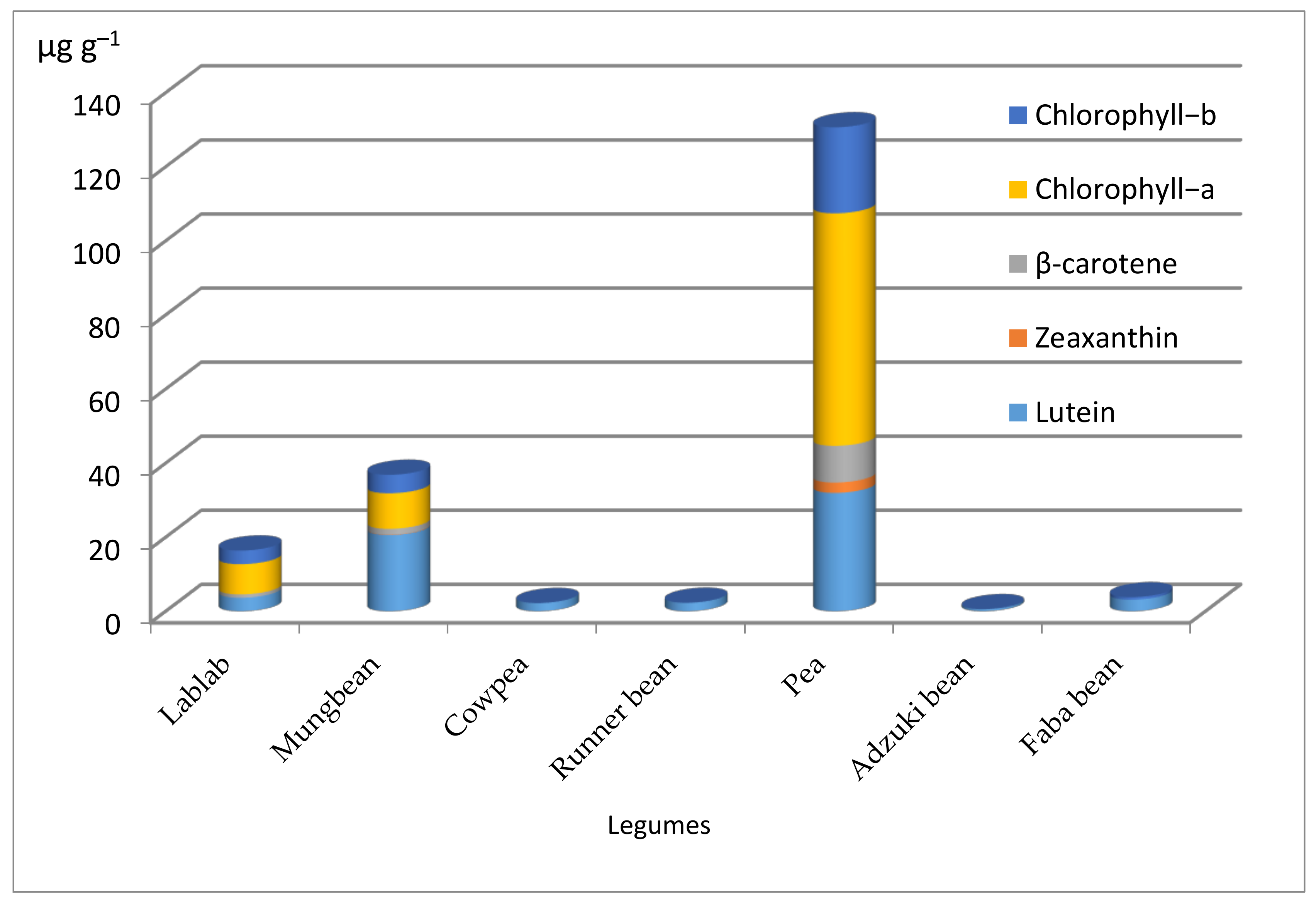
Some legumes, which are a potential dietary source of health-promoting compounds, were selected to confirm the wide applicability of the developed method. Notable differences in carotenoid and chlorophyll contents were observed among the seven selected legumes (Figure 4). Lutein, zeaxanthin, β–carotene, and chlorophyll–a and–b were the major pigments determined in the selected legumes. Lutein was found in all the legumes (Figure 4 and Figure 5), which was similar to the previously reported result [53]. In the present study, the lutein content ranged from 0.54 (Adzuki, Vigna angularis) to 31.94 μg g−1 (Pea, Pisum sativum), confirming that all selected legumes contained lutein. Zeaxanthin was not able to be detected in most of the legumes except for Mungbean (Vigna radiate, 0.09 μg g−1) and Pisum sativum (2.78 μg g−1). Similarly, β–carotene was only detected in three legumes, ranging from 0.92 (Lablab, Lablab purpureus) to 9.92 μg g−1 (Pisum sativum), revealing that Pisum sativum gave a statistically superior β–carotene concentration than others. The highest total carotenoid concentration (44.63 μg g−1) was observed in Pisum sativum, followed by Vigna radiate (22.23 μg g−1), while the lowest (0.54 μg g−1) was obtained from Vigna angularis. Lutein, zeaxanthin, and β–carotene are the major contributors to the total carotenoid content of foods [40]. Chlorophylls were detected in four legumes; the highest contents of chl–a and –b were in Pisum sativum with values of 62.77 and 23.21 μg g−1, respectively. Moreover, the highest total chlorophyll (85.91 μg g−1) was recorded in Pisum sativum, whereas the lowest (0.67 μg g−1) was produced in Faba bean (Vicia faba). In summary, Pisum sativum seed contained the major carotenoid components (lutein, zeaxanthin, and β–carotone) along with chlorophylls (chl–a and –b) in high levels compared to the other legumes; lutein was the dominant component found in all tested legumes. Other studies on the carotenoid content of legumes found carotenoid concentrations from some legumes, such as red kidney beans (8.29–20.95 μg g−1), lentils (4.53–21.34 μg g−1), black soybeans (4.41–6.09 μg g−1), cowpea (6.62–9.46 μg g−1), and white kidney beans (0.05–0.25 μg g−1) [51]. Fernández-marín et al. [54] analyzed carotenoids in 50 Fabaceae species and found lutein (4.80 μg g−1) in 98% of the species analyzed, zeaxanthin (0.80 μg g−1) in 80%, β–carotene (1.00 μg g−1) in 74% of the species analyzed, and a total carotenoid concentration of 8.10 μg g−1, confirming that the flour sample of all cultivars might not contain all carotenoid components. A study on six cultivars of green peas extracted using acetone (100%) and analyzed by the HPLC-C18 column gave chl–a (48.00–73.00 μg g−1), chl–b (21.00–28.00 μg g−1), lutein (12.00–19.00 μg g−1), and β–carotene (3.00–5.00 μg g−1); however, zeaxanthin was not detected by this method [30].

4. Conclusions

A reliable HPLC-UV-VIS method for simultaneous determination of carotenoids and chlorophylls was developed and validated using mobile phases of MTBE, MeOH-20mM ammonium acetate, and water delivered at a 0.90 mL min−1 flow rate through a C30-YMC Carotenoid (250 × 4.6 mm I.D., S-5 µm) column coupled with a UV-VIS detector set at 450 nm. It showed good linearity (R2, 0.9993–0.9999), sensitivity (LODs, 0.0051–0.0300 μg mL−1; LOQs, 0.0155–0.0909 μg mL−1), accurate recoveries (83.12–106.58%), and high precision (intra-day RSDs, 1.25–3.46%; inter-day RSDs, 2.80–4.20%). The method was applied on different legume samples and the sample preparation was simple as it consisted of a solid-liquid extraction procedure comprising acetone-ethanol (1:1) solvents. Results revealed that the developed method is appropriate for carotenoid and chlorophyll determination in legume seeds. In some reports, we observed difficulties in the separation of lutein and chlorophyll-b. Interestingly, this proposed method was able to separate lutein, zeaxanthin, β–cryptoxanthin, α–carotene, β–carotene, chlorophyll–a, and chlorophyll–b completely in a single run. Although the analysis of carotenoids was complicated by their diversity, complexity, instability, and the existence of cis-trans isomers with their diverse spectrum of polarities, the use of the HPLC C30 RP column can optimally separate a number of carotenoids, including isomerized components, with a simple sample preparation. Thus, this study provided a robust and rapid method using HPLC-UV-VIS coupled with a C30 column and an elution gradient for the determination of carotenoids and chlorophylls in legume seeds so as to accomplish selections of a large number of plant populations within a short period of time in a large-scale soybean quality breeding program.

Supplementary Materials

The following are available online at https://www.mdpi.com/article/10.3390/agronomy11040758/s1, Figure S1. Calibration curves of carotenoids and chlorophylls standards.

Author Contributions

B.S.G. conceived and planned the experiment; B.S.G. and J.S. carried out the experiment and formal analysis; Funding acquisition, B.L., J.S.; resources, J.Q., M.A., S.G., Y.F., Y.H., J.L.; Supervision, J.S.; Visualization, B.L.; Writing—original draft, B.S.G.; Writing—review and editing, S.Z., B.L., J.S. All authors made substantial contributions to the discussion of the results and agreed to the published version of the manuscript.

Funding

This work was supported by Ministry of Science and Technology of China (2016YFD0100201, 2016YFD0100504 and 2017YFD0101401), National Natural Science Foundation of China (31671716) and the Agricultural Science and Technology Innovation Program of CAAS (2060203-2).

Institutional Review Board Statement

Not applicable.

Informed Consent Statement

Not applicable.

Acknowledgments

The authors gratefully acknowledge Lijuan Qiu, Xuxiao Zong, Lixia Wang and Lanfen Wang, from the Institute of Crop Sciences, CAAS, for providing the legume cultivars employed in this study.

Conflicts of Interest

The authors declare no conflict of interest in publication of this manuscript.

References

  1. Rodriguez-Concepcion, M.; Avalos, J.; Bonet, M.L.; Boronat, A.; Gomez-gomez, L.; Hornero-Mendez, D.; Limon, M.C.; Meléndez-martínez, A.J.; Olmedilla-alonso, B.; Palou, A.; et al. A global perspective on carotenoids: Metabolism, biotechnology, and benefits for nutrition and health. Prog. Lipid Res. 2018, 70, 62–93. [Google Scholar] [CrossRef] [PubMed] [Green Version]
  2. Sachindra, N.M.; Sato, E.; Maeda, H.; Hosokawa, M.; Niwano, Y.; Kohno, M.; Miyashita, K. Radical scavenging and singlet oxygen quenching activity of marine carotenoid fucoxanthin and its metabolites. J. Agric. Food Chem. 2007, 55, 8516–8522. [Google Scholar] [CrossRef] [PubMed]
  3. Elvira-Torales, L.I.; García-Alonso, J.; Periago-Castón, M.J. Nutritional importance of carotenoids and their effect on liver health: A review. Antioxidants 2019, 8, 229. [Google Scholar] [CrossRef] [PubMed] [Green Version]
  4. Hrvolová, B.; Martínez-huélamo, M.; Colmán-martínez, M.; Hurtado-barroso, S.; Lamuela-raventós, R.M. Development of an advanced HPLC—MS/MS method for the determination of carotenoids and fat-soluble vitamins in human plasma. Int. J. Mol. Sci. 2016, 17, 1719. [Google Scholar] [CrossRef] [Green Version]
  5. DiPietro, N.; DiTomo, P.; Pandolf, A. Carotenoids in cardiovascular disease prevention. JSM Atheroscler. 2016, 1, 1002. [Google Scholar]
  6. Saini, R.K.; Keum, Y. Carotenoid extraction methods: A review of recent developments. Food Chem. 2018, 240, 90–103. [Google Scholar] [CrossRef]
  7. Mel´endez-Mart´ınez, A.J. An overview of carotenoids, apocarotenoids, and vitamin A in agro-food, nutrition, health, and disease. Mol. Nutr. Food Res. 2019, 63, 1801045. [Google Scholar] [CrossRef] [Green Version]
  8. Landrum, J.T.; Bone, R.A. Lutein, zeaxanthin, and the macular pigment. Arch. Biochem. Biophys. 2001, 385, 28–40. [Google Scholar] [CrossRef]
  9. Zhang, B.; Deng, Z.; Tang, Y.; Chen, P.; Liu, R.; Ramdath, D.D.; Liu, Q.; Hernandez, M.; Tsao, R. Fatty acid, carotenoid and tocopherol compositions of 20 Canadian lentil cultivars and synergistic contribution to antioxidant activities. Food Chem. 2014, 161, 296–304. [Google Scholar] [CrossRef] [PubMed]
  10. Eggersdorfer, M.; Wyss, A. Carotenoids in human nutrition and health. Arch. Biochem. Biophys. 2018, 652, 18–26. [Google Scholar] [CrossRef]
  11. Bhatnagar-Panwar, M.; Bhatnagar-Mathur, P.; Bhaaskarla, V.V.A.; Dumbala, S.R.; Sharma, K.K. Rapid, accurate and routine HPLC method for large-scale screening of pro-vitamin A carotenoids in oilseeds. J. Plant Biochem. Biotechnol. 2013, 24, 84–92. [Google Scholar] [CrossRef] [Green Version]
  12. Granado, F.; Olmedilla, B.; Gil-Martinez, E.; Blanco, I. A fast, reliable and low-cost saponification protocol for analysis of carotenoids in vegetables. J. Food Compos. Anal. 2001, 14, 479–489. [Google Scholar] [CrossRef]
  13. Slavin, M.; Yu, L.L. A single extraction and HPLC procedure for simultaneous analysis of phytosterols, tocopherols and lutein in soybeans. Food Chem. 2012, 135, 2789–2795. [Google Scholar] [CrossRef] [PubMed]
  14. Ferruzzi, M.G.; Blakeslee, J. Digestion, absorption, and cancer preventative activity of dietary chlorophyll derivatives. Nutr. Res. 2007, 27, 1–12. [Google Scholar] [CrossRef]
  15. Chen, J.P.; Tai, C.Y.; Chen, B.H. Improved liquid chromatographic method for determination of carotenoids in Taiwanese mango (Mangifera indica L.). J. Chromatogr. A 2004, 1054, 261–268. [Google Scholar] [CrossRef]
  16. Feltl, L.; Pacakova, V.; Stulik, K.; Volka, K. Reliability of carotenoid analyses: A review. Curr. Anal. Chem. 2005, 1, 93–102. [Google Scholar] [CrossRef]
  17. Inbaraj, B.S.; Chien, J.T.; Chen, B.H. Improved high performance liquid chromatographic method for determination of carotenoids in the microalga Chlorella pyrenoidosa. J. Chromatogr. A 2006, 1102, 193–199. [Google Scholar] [CrossRef] [PubMed]
  18. Puspitasari-nienaber, N.L.; Ferruzzi, M.G.; Schwartz, S.J. Simultaneous detection of tocopherols, carotenoids, and chlorophylls in vegetable oils by direct injection C30 RP-HPLC with coulometric electrochemical array detection. JAOCS 2002, 79, 633–640. [Google Scholar] [CrossRef]
  19. Oliver, J.; Palou, A. Chromatographic determination of carotenoids in foods. J. Chromatogr. A 2000, 881, 543–555. [Google Scholar] [CrossRef]
  20. Mangels, A.R.; Holde, J.M.; Beecrep, G.R.; Forman, M.R.; Lanz, E. Carotenoid content of fruits and vegetables: An evaluation of analytic data. J Am Diet Assoc 1993, 93, 284–296. [Google Scholar] [CrossRef]
  21. Ferruzzi, M.G.; Nguyen, M.L.; Sander, L.C.; Rock, C.L.; Schwartz, S.J. Analysis of lycopene geometrical isomers in biological microsamples by liquid chromatography with coulometric array detection. J. Chromatogr. B 2001, 760, 289–299. [Google Scholar] [CrossRef]
  22. Capello, C.; Fischer, U.; Hungerbu, K. What is a green solvent? A comprehensive framework for the environmental assessment of solvents. Green Chem. 2007, 9, 927–934. [Google Scholar] [CrossRef]
  23. Li, J.; Xie, J.J.; Yu, H.J.; Lv, J.; Zhang, F.J.; Wang, L.X.; Wang, C.; Tang, N.C.; Zhang, C.Y.; Dawuda, M.M.; et al. Reverse-phase high performance liquid chromatography for the quantification and optimization for extracting ten kinds of carotenoids in pepper ( Capsicum annuum L.) leaves. J. Agric. Food Chem. 2017, 65, 8475–8488. [Google Scholar] [CrossRef]
  24. ICH (International Conference on Harmonization). ICH Harmonised Tripartite Guideline, Validation of Analytical Procedures: Text and Methodology; ICH Q2; ICH: Geneva, Switzerland, 2005; pp. 4–12. [Google Scholar]
  25. Al-amri, K.A.; Mohsin, K.; Alanazi, F.K. Development and validation of a UPLC method for quantification of antiviral agent, Acyclovir in lipid-based formulations. Arab. J. Chem. 2014, 12, 1707–1714. [Google Scholar] [CrossRef] [Green Version]
  26. Gedawy, A.; Al-salami, H.; Dass, C.R. Development and validation of a new analytical HPLC method for simultaneous determination of the antidiabetic drugs, metformin and gliclazide. J. Food Drug Anal. 2018, 27, 315–322. [Google Scholar] [CrossRef] [PubMed] [Green Version]
  27. Guzman, I.; Yousef, G.G.; Brown, A.F. Simultaneous extraction and quantitation of carotenoids, chlorophylls, and tocopherols in Brassica vegetables. J. Agric. Food Chem. 2012, 60, 7238–7244. [Google Scholar] [CrossRef] [PubMed]
  28. Zeb, A.; Ullah, F. Reversed phase HPLC-DAD profiling of carotenoids, chlorophylls and phenolic compounds in Adiantum capillus-veneris leaves. Front. Chem. 2017, 5, 29. [Google Scholar] [CrossRef] [PubMed] [Green Version]
  29. SAS Institute Inc. Base SAS® 9.3 Procedures Guide: Statistical Procedures; SAS Institute Inc: Cary, NC, USA, 2011; 528p. [Google Scholar]
  30. Edelenbos, M.; Christensen, L.P.; Grevsen, K. HPLC determination of chlorophyll and carotenoid pigments in processed green pea cultivars (Pisum sativum L.). J. Agric. Food Chem. 2001, 49, 4768–4774. [Google Scholar] [CrossRef]
  31. Gandul-rojas, B.; Cepero, M.R.; Mínguez-mosquera, M.I. Use of chlorophyll and carotenoid pigment composition to determine authenticity of virgin olive oil. JAOCS 2000, 77, 853–858. [Google Scholar] [CrossRef]
  32. Kanamaru, K.; Wang, S.; Abe, J.; Yamada, T.; Kitamura, K. Identification and characterization of wild soybean (Glycine soja Sieb. et Zecc.) strains with high lutein content. Breed. Sci. 2006, 56, 231–234. [Google Scholar] [CrossRef] [Green Version]
  33. Kanamaru, K.; Wang, S.; Yamada, T.; Abe, J.; Kitamura, K. Genetic analysis and biochemical characterization of the high lutein trait of wild soybean (Glycine soja Sieb. et Zucc.). Breed. Sci. 2008, 58, 393–400. [Google Scholar] [CrossRef] [Green Version]
  34. Mendes-Pinto, M.M.; Ferreira, A.N.C.S.S.; Caris-Veyrat, C.; Pinho, P.G. De Carotenoid, chlorophyll, and chlorophyll-derived compounds in grapes and port wines. J. Agric. Food Chem. 2005, 53, 10034–10041. [Google Scholar] [CrossRef] [PubMed]
  35. Monma, M.; Ito, M.; Saito, M.; Chikuni, K. Carotenoid components in soybean seeds varying with seed color and maturation stage. Biosci. Biotech. Biochem 1994, 58, 926–930. [Google Scholar] [CrossRef]
  36. Stinco, C.M.; Benítez-gonzález, A.M.; Hernanz, D.; Vicario, I.M.; Meléndez-martínez, A.J. Development and validation of a rapid resolution liquid chromatography method for the screening of dietary plant isoprenoids: Carotenoids, tocopherols and chlorophylls. J. Chromatogr. A 2014, 1370, 162–170. [Google Scholar] [CrossRef] [PubMed]
  37. Fraser, P.D.; Pinto, M.E.S.; Holloway, D.E.; Bramley, P.M. Application of high-performance liquid chromatography with photodiode array detection to the metabolic profiling of plant isoprenoids. Plant J. 2000, 24, 551–558. [Google Scholar] [CrossRef]
  38. Dincel, D.; Tekkeli, S.E.K.; Önal, C.; Önal, A.; Sagirli, O. Liquid chromatographic analysis of carotenoids in foods. J. Chil. Chem. Soc. 2019, 64, 4492–4495. [Google Scholar] [CrossRef] [Green Version]
  39. Kamffer, Z.K.; Bindon, K.A.; Oberholster, A. Optimization of a method for the extraction and quantification of carotenoids and chlorophylls during ripening in grape berries (Vitis vinifera cv. Merlot). J. Agric. Food Chem. 2010, 58, 6578–6586. [Google Scholar] [CrossRef]
  40. Burns, J.; Fraser, P.D.; Bramley, P.M. Identification and quantification of carotenoids, tocopherols and chlorophylls in commonly consumed fruits and vegetables. Phytochemistry 2003, 62, 939–947. [Google Scholar] [CrossRef]
  41. Seguin, P.; Tremblay, G.; Pageau, D.; Liu, W.; Turcotte, P. Soybean lutein concentration: Impact of crop management and genotypes. Crop Sci. 2011, 51, 1151–1160. [Google Scholar] [CrossRef]
  42. Wang, S.; Kanamaru, K.; Li, W.; Abe, J.; Yamada, T.; Kitamura, K. Simultaneous accumulation of high contents of α -tocopherol and lutein is possible in seeds of soybean (Glycine max (L.) Merr.). Breed. Sci. 2007, 57, 297–304. [Google Scholar] [CrossRef] [Green Version]
  43. Sengul, U. Comparing determination methods of detection and quantification limits for aflatoxin analysis in hazelnut. J. Food Drug Anal. 2016, 24, 56–62. [Google Scholar] [CrossRef] [PubMed] [Green Version]
  44. Magnusson, B.; Örnemark, U. (Eds.) Eurachem Guide: The Fitness for Purpose of Analytical Methods—A Laboratory Guide to Method Validation and Related Topics, 2nd ed.; Eurachem: Teddington, UK, 2014; pp. 7–39. [Google Scholar]
  45. Karnjanawipagul, P.; Nittayanuntawech, W.; Rojsanga, P.; Suntornsuk, L. Analysis of beta-carotene in carrot by spectrophotometry. J. Pharm. Sci. 2010, 37, 8–16. [Google Scholar]
  46. Zeb, A. A simple, sensitive HPLC-DAD method for simultaneous determination of carotenoids, chlorophylls and α-tocopherol in leafy vegetables. Food Meas. 2017, 11, 979–986. [Google Scholar] [CrossRef]
  47. Petry, F.C.; Mercadante, A.Z. New method for carotenoid extraction and analysis by HPLC-DAD-MS/MS in freeze-dried citrus and mango pulps. J. Braz. Chem. Soc. 2018, 29, 205–215. [Google Scholar] [CrossRef]
  48. Lee, M.T.; Chen, B.H. Separation of lycopene and its cis isomers by liquid chromatography. Chromatographia 2001, 54, 613–617. [Google Scholar] [CrossRef]
  49. Sulistyowati, E.; Martono, S.; Riyanto, S.; Lukitaningsih, E. Development and validation for free aglycones daidzein and genistein in soybeans (Glycine max (L.) Merr.) using RP HPLC method. Int. J. Appl. Pharm. 2019, 11, 138–142. [Google Scholar] [CrossRef] [Green Version]
  50. Choudhury, H.; Gorain, B.; Karmakar, S.; Pal, T.K. Development and validation of RP-HPLC method: Scope of application in the determination of oil solubility of paclitaxel. J. Chromatogr. Sci. 2013, 52, 68–74. [Google Scholar] [CrossRef] [Green Version]
  51. Kan, L.; Nie, S.; Hu, J.; Wang, S.; Bai, Z.; Wang, J.; Zhou, Y.; Jiang, J.; Zeng, Q.; Song, K. Comparative study on the chemical composition, anthocyanins, tocopherols and carotenoids of selected legumes. Food Chem. 2018, 260, 317–326. [Google Scholar] [CrossRef]
  52. Ashokkumar, K.; Diapari, M.; Jha, A.B.; Tar’an, B.; Arganosa, G.; Warkentin, T.D. Genetic diversity of nutritionally important carotenoids in 94 pea and 121 chickpea accessions. J. Food Compos. Anal. 2015, 43, 49–60. [Google Scholar] [CrossRef]
  53. Tee, E.; Ah-Heng, G.; Swan-Choo, K. Carotenoid composition and content of legumes, tubers and starchy roots by HPLC. Mal. J. Nutr. 1995, 1, 63–74. [Google Scholar]
  54. Fernández-marín, B.; Míguez, F.; Méndez-fernández, L.; Agut, A.; Becerril, J.M.; García-Plazaola, J.I.; Kranner, I.; Colville, L. Seed carotenoid and tocochromanol composition of wild fabaceae species is shaped by phylogeny and ecological factors. Front. Plant Sci. 2017, 8, 1428. [Google Scholar] [CrossRef] [PubMed] [Green Version]
Figure 1. Chromatographic separation of carotenoids and chlorophylls for standard mixture and soybean seed sample by HPLC using C18 and C30 columns. (A) Standards by C18 column; (B) soybean seed sample by C18 column; (C) Standards by C30 column; (D) soybean seed sample by C30 column.
Agronomy 11 00758 g001
Figure 2. Effect of flour sample weight and extractant volume on the content of carotenoid and chlorophyll components. (A) ZDD20340; (B) Nanhuizao; (C) ZDD18672. V = volume; W = weight; V1 = 1.00 mL; V2 = 1.50 mL; W1 = 50.00 mg; W2 = 100.00 mg; W3 = 150.00 mg; Different lowercase letters at the top of each bar graph with the same color show statistically significant differences among treatments (p < 0.01).
Agronomy 11 00758 g002
Figure 3. Carotenoid and chlorophyll compositions and contents in soybean cultivars with different seed coat colors. Soybean cv. ZDD20340, Qingdou and Wilson five (green seed coat color); Nanhuizao (black seed coat color); ZDD18672, Zhonghuang13 and Zhonghuang 106 (yellow seed coat color).
Agronomy 11 00758 g003
Figure 4. Carotenoid and chlorophyll concentrations in different legume seeds. Lablab (Lablab purpureus), Mungbean (Vigna radiata), Cowpea (Vigna unguiculata), Runner bean (Phaseolus multiflorus), Pea (Pisum sativum), Adzuki bean (Vigna angularis), and Fababean (Vicia faba).
Agronomy 11 00758 g004
Figure 5. Chromatograms of different legumes. (A) Lablab (Lablab purpureus); (B) Mungbean (Vignia radiate); (C) Cowpea (Vigna unguiculata); (D) Runnerbean (Phaseolus multiflorus); (E) Pea (Pisum sativum); (F) Adzuki bean (Vigna angularis); (G) Fababean (Vicia faba).
Agronomy 11 00758 g005
Table 1. Retention time (min), limit of detection (μg mL−1), limit of quantification (μg mL−1), concentration range (μg mL−1), and regression relationships
AnalyteRt.LODLOQConc.RangeLinear RegressionR2
Lutein10.400.00960.02900.05–10.00Y = 111.14x + 0.930.9999
Zeaxanthin11.910.00650.01980.05–5.00Y = 125.56x + 4.400.9998
β-carotene28.580.01750.05290.10–10.00Y = 30.99x + 3.370.9993
α-carotene25.870.00630.01920.05–10.00Y = 131.65x + 7.440.9997
β-cryptoxanthin19.280.00510.01550.05–20.00Y = 91.02x + 10.960.9997
Chlorophyll-a13.420.03000.09090.10–30.00Y = 24.14x − 0.420.9999
Chlorophyll-b9.820.00810.02470.05–10.00Y = 77.43x + 0.090.9999
Rt: retention time; LOD: limit of detection; LOQ: limit of quantification; Conc. range: Concentration range; R2: coefficient of determination.
Table 2. Recovery of analytes obtained in samples by the HPLC-UV method
AnalyteConcentration
Added (μg mL−1)
Concentration
Found (μg mL−1)
Recovery (%)
Lutein0.002.00-
1.252.4095.28
2.502.88105.43
5.003.5794.27
Zeaxanthin0.000.03-
0.100.06106.58
0.500.20102.04
1.000.3597.19
β–carotene0.000.19-
2.500.9186.99
5.001.6286.02
7.502.2783.12
Chlorophyll–a0.006.01-
2.506.7488.51
5.007.6196.55
10.009.0290.34
Chlorophyll–b0.002.10-
1.002.4089.62
5.003.6088.06
10.005.4097.02
Values are mean of three replicates.
Table 3. Intra- and inter-day method and system precision obtained in samples by the HPLC-UV method
AnalytePrecision, RSD (%)
System PrecisionMethod Precision
RepeatabilityIntermediate
Intra-Sample (n = 1; i = 6)Mix-Standard (i = 6)Day-1
(n = 6)
Day-2
(n = 6)
Day-3
(n = 6)
(n = 6; k = 3)
Lutein0.320.232.192.442.393.33
Zeaxanthin0.810.263.462.652.384.20
β-carotene0.770.151.251.301.712.80
Chlorophyll-a0.560.742.382.312.243.44
Chlorophyll-b0.570.222.032.042.743.60
RSD: relative standard deviation, calculated as RSD = (Standard deviation)/Mean) × 100%; n: replicates; i = injection; k = days.
Publisher’s Note: MDPI stays neutral with regard to jurisdictional claims in published maps and institutional affiliations.