Article

In Vitro Pathogenesis Caused by Phytophthora cactorum and DNA Analysis of the Strawberry-Resistant Microplants with ISSR Markers

by
Wojciech Marecki
* and
Jadwiga Żebrowska
Department of Genetics and Horticultural Plant Breeding, Institute of Plant Genetics, Breeding and Biotechnology, Faculty of Agrobioengineering, University of Life Sciences in Lublin, Akademicka 15 Street, 20-950 Lublin, Poland
*
Author to whom correspondence should be addressed.
Agronomy 2021, 11(7), 1279; https://doi.org/10.3390/agronomy11071279
Submission received: 25 May 2021 / Revised: 21 June 2021 / Accepted: 22 June 2021 / Published: 24 June 2021
(This article belongs to the Section Crop Breeding and Genetics)

Abstract

:
The soil pathogenic fungus Phytophthora cactorum causes the most dangerous diseases occurring in strawberry plantations—strawberry crown rot and leather rot. Modern biotechnology methods, e.g., in vitro culture selection and molecular diagnostics can be utilized in the selection of cultivars that are less susceptible or resistant to Phytophthora diseases. In this study, in vitro selection of four strawberry microclones: ‘Elsanta’, ‘Feltar’, ‘Teresa’ and ‘Plena SVdT’ against Phytophthora cactorum (Lebert and Cohn) J. Schröt was carried out. Molecular analysis with inter simple sequence repeat (ISSR) markers was also used to evaluate genetic similarity of the selected resistant plants. None of the analyzed microclones showed complete resistance to the selection factor, but there were plants in all tested microclones that survived the pressure of the pathogen. Results showed that susceptibility to this pathogenic fungus was significantly differentiated and depended on the microclone. The ‘Feltar’ microclone had the significantly lowest susceptibility to Phytophthora disease, followed by the microclones ‘Elsanta’ and ‘Teresa’ with significantly higher susceptibility. The ‘Plena SVdT’ microclone showed the highest susceptibility to Phytophthora disease. This differentiation was linked to the genetic similarity observed at deoxyribonucleic acid (DNA) level between the resistant plants selected from microclones. Cluster analysis revealed that microclones with similar susceptibility to phytophthorosis, i.e., ‘Elsanta’, ‘Feltar’ and ‘Teresa’, appeared to be genetically similar. The microclone ‘Plena SVdT’ revealed a different course of phytophthorosis from the aforementioned microclones, being the least genetically similar to them.

1. Introduction

Breeding for conferring resistance is highly important in modern agriculture. The cultivation of cultivars that are less susceptible or even resistant to pathogens allows the restriction of fungicides that are harmful to humans and the environment. Production costs are thus reduced, and farmers obtain higher and more uniform yields of high-quality fruits [1].
The use of modern biotechnological methods, such as tissue cultures or molecular diagnostics can significantly accelerate the progress of breeding compared to conventional selection. Currently, one of many useful techniques in modern plant breeding is in vitro culture selection. Such selection is aimed at obtaining plants with new, valuable traits that would also be present in plants transferred to field conditions, and these traits would be inherited by the next generations. The tested plants can be selected for both biotic (resistance to fungal pathogens) and abiotic factors (drought, soil salinity, heavy metals, low temperature). In vitro culture selection can only be carried out on traits expressed at the cell, callus or seedling level [2,3,4]. The plants obtained in this method can be used for further breeding studies as a source of resistance genes. Sowik et al. [5] and Żebrowska [6,7] investigated the resistance of strawberry cultivars and clones via the selection of the pathogenic fungus Verticillium dahliae in an in vitro culture. Żebrowska [6] reported that the susceptibility of the tested strawberry cultivars in vitro was similar to the susceptibility of these cultivars under field conditions and that in vitro culture selection could be successfully applied in breeding programs.
Two of the most dangerous diseases occurring in strawberry plantations are strawberry crown rot and leather rot [8]. Both of these diseases are caused by the fungus of the genus Phytophthora and more precisely by its specialized form—Phytophthora cactorum.
Phytophthora cactorum is a common polyphagic organism in soils of the temperate climate zone. It causes diseases in 200 species of crops from 60 families [9]. This fungus mainly affects ornamental plants (such as rhododendron, pansy, pelargonium or begonia), fruit shrubs and trees (gooseberry, currant, peach, cherry, raspberry, apple tree and cherry) and forest trees (birch, black alder, Scots pine, common beech, common ash and spruce) causing a disease called phytophthorosis [8,10,11,12,13,14].
The source of P. cactorum on plantations may be the remains of infected plants left over from previous plantings, water drawn for irrigation from nearby ponds or rivers, irrigation systems, contaminated soil or infected seedlings [14]. Thick-walled spores of P. cactorum are called oospores and can survive in the soil for up to 6 years [14].
The first symptoms of strawberry crown rot are observed on the above-ground plant parts. Wilting of the youngest leaves in individual plants, which often have a blue-green color, is a characteristic symptom of the disease. The plant wilts and dies as a result of root infection. When removing infected strawberries from the ground, the upper part of the crown is often torn off, and the rest remains in the soil [15]. The newly attacked tissue is watery and light brown, and it turns an intensely brown-red color with disease development [15]. Tissue disintegration and necrosis are also observed. A day shorter than 13 h light per day is conducive to the disease. Plants are infected in nurseries (cryptic infection) and on production plantations [16].
Leather rot mainly occurs in years with high rainfall before and during harvest. Zoospores germinating on the surface of the fruit in the presence of water are responsible for the infection [15]. Fruits are infected at all stages of development, from fruit set to immature and fully developed ripe fruits. Light brown, later brown, dry, rotting spots develop on immature fruits that may cover the whole fruit. Light, grayish-yellow to pink-violet, discolored spots appear on ripening and ripe fruits [17]. The flesh of such fruits is firm, stringy, light brown in color with darker vascular bundles. Infected fruits are slightly softer than healthy ones, with a decidedly unpleasant smell and clearly bitter taste [18,19].
Ellis et al. [20] reported that the average losses due to this disease on strawberry plantations in the USA are 20–30%, and during a particularly severe epidemic in 1981 in Ohio, losses reached 50%. According to Bielenin [21], losses in strawberry seedlings caused by crown rot, originating from an infected plantation, reached 40% in 1994, while in the years 1995–1997, fruit losses ranged from 20% to 80% as a result of leather rot on the cultivars ‘Elsanta’, ‘Senga Sengana’ and ‘Sirius’.
Selection in order to obtain new strawberry cultivars resistant or tolerant to Phytophthora cactorum is currently one of the most important directions of breeding this species in Poland and in the world. So far, research on the susceptibility of strawberry cultivars to P. cactorum has been conducted mainly in field conditions. There are few reports on the use of biotechnological methods for resistance breeding, i.e., tissue cultures in conjunction with the molecular analysis of selected resistant forms.
Considering this breeding direction, the present study applied in vitro culture selection to assess the susceptibility of strawberry microclones to infection by the pathogen of the genus Phytophthora spp. in conjunction with molecular analysis of selected resistant forms.

2. Materials and Methods

The starting material for in vitro selection against Phytophthora cactorum (Lebert and Cohn) J. Schröt was obtained by in vitro propagating the ends of runners taken from the donor plants of strawberry cultivars: ‘Elsanta’, ‘Feltar’, ‘Plena’ and ‘Teresa’ derived from the collection of the Department of Genetics and Horticultural Plant Breeding of the University of Life Sciences in Lublin. The Dutch cultivar ‘Elsanta’ [‘Gorella’ × ‘Holiday’] is very susceptible to root system diseases (caused by Verticillium sp.). However, the susceptibility to soil pathogens, such as Phytophthora spp. of Polish cultivars ‘Feltar’ [(‘Senga Tigaiga’ × ‘Merton Dawn’) S1], ‘Plena’ [‘Senga Sengana’ × ‘Merton Dawn’] and ‘Teresa’ [‘Redgauntlet S1’ × ‘Senga Sengana S1’] selected at the University of Life Sciences in Lublin is not yet known.
As primary explants, 3–4 cm ends of young runners were collected together with the node containing the meristematic tissue (about 50–60 pieces from each cultivar) from the abovementioned donor plants.
Microplants of the microclone designated as ‘Plena SVdT’ were derived by micropropagation of plants that had positively passed the pressure of earlier in vitro selection against the fungus Verticillium dahliae Kleb. In this experiment, they were also selected for resistance to the pathogen Phytophthora cactorum.
The explants of cvs. ’Elsanta’, ‘Feltar’ and ‘Teresa’ were surface-disinfected with a solution of sodium hypochlorite with distilled water and the addition of a wetting agent for 15 min. Subsequently, the explants, rinsed three times in sterile distilled water, were placed under aseptic conditions under a laminar air flow chamber (POLON KL—21) on a modified Murashige and Skoog medium (MS medium) [22] supplemented with 1 mg × dm−3 IAA (indole-3-acetic acid), 1 mg × dm−3 BAP (6-benzylaminopurine) and 0.01 mg × dm−3 GA3 (gibberellic acid), recommended for strawberry micropropagation, with a fixed pH = 5.7. The culture was carried out for 6 weeks under controlled environmental conditions (growth chamber 20 °C, photoperiod 16 h day/8 h night, under white fluorescent lights at an intensity of 30 µmol m−2 s−1). This stage was completed after obtaining the sufficient number of aseptic plantlets for in vitro selection of all cultivars.
Soil phytopathogen, Phytophthora cactorum (Lebert and Cohn) J. Schröt, which infects strawberries, was used in the experiment. Pure Phytophthora cactorum culture, catalog number 1559, was obtained from the Plant Pathogen Bank in Poznań. The pathogen was grown on PDA medium (potato-dextrose agar, potato 300.0 g × dm3; agar 20.0 g × dm3; glucose 20.0 g × dm3) in Petri dishes. The liquid mycelium homogenate with conidia was prepared from a 3-week-old pathogen culture by pouring sterile distilled water over the culture surface. The resulting suspension was subsequently homogenized and diluted with sterile distilled water at a 1:10 v/v proportion to obtain the appropriate conidia density (105× mL−1).
Well-rooted microplants at the stage of at least 4 leaves served as the starting material for selection. Microclone plants were infected under aseptic conditions by immersing them in the inoculum for 1 min, after damaging the roots by cutting them with a scalpel to a length of about 1.5 cm. Next, 100 mL of the homogenate was used to inoculate 25 plants. Then the plants were placed on agar (0.6% w/v aqueous agar solution) in Petri dishes (5 plants per dish) and transferred to a growth chamber under controlled environmental conditions (growth chamber 20 °C, photoperiod 16 h day/8 h night, under white fluorescent lights at an intensity of 30 μmol m−2 s−1). The experiment was set up in triplicate for each microclone. One replicate consisted of 100 plants. A control sample was also prepared for each microclone variant. Control plants were immersed in sterile distilled water in the absence of pathogen according to the above procedure (mock inoculation). They were subsequently placed on the same agar medium as the infected microplants.
The development of disease symptoms (leaf chlorosis) was observed at the following five time points: 15, 30, 45, 60 and 75 days after inoculation. Chlorosis was assessed on a five-point evaluation scale in the range of 0 to 4. The same observations were made for the control plants. Susceptibility of the studied microclones to the selection factor was determined using the disease index for the extent and rate of infection development.
The extent of infection development (%) was determined based on the disease index according to McKinney [23], which was used to assess the course of pathogenesis and the degree of susceptibility of individual microclones to P. cactorum infection. The disease index was calculated according to the formula:
DI = (Σvn)/(NV) × 100
where:
DI—disease index (in %);
v—numerical value of infection class;
n—number of plants at a given observation time point in a given class;
N—total number of infected plants in a given sample;
V—numerical value of the highest class.
The extent of infection development (%) was expressed as the mean percentage of infected plants after the end of selection. The thus calculated value of the disease index indicated the susceptibility of a given microclone to Phytophthora.
The rate of infection development over time was determined using a disease index developed by Simmonds [24], which was calculated according to the following formula:
Disease index = r × I (1 − I)
where:
I—percentage of plants with symptoms of infection at a given observation time point in a given class;
r—rate of pathogen reproduction; it was assumed for susceptible cultivars that r = 1.
The values of this index, calculated at individual observation time points, provide information on the rate of infection development over time.
All data obtained in this experiment were statistically analyzed by analysis of variance (ANOVA), and the significance of differences between means was established using the Fisher LSD test at p ≤ 0.05 with Statistica 13.1 statistical software (Statistica 13.1. StatSoft.Polska. Available online: www.statsoft.pl, accessed on 15 May 2020).
After the observation was completed, DNA was isolated from 10 resistant plants of each microclone randomly chosen from those that survived the selection pressure caused by the pathogen. Plant DNA was isolated using the modified CTAB (hexadecyltrimethylammonium bromide) method described by Gawel and Jarret [25].
ISSR markers were used for the assessment of genetic similarity at the DNA level in respect to resistance to P. cactorum of the selected plant material. In the experiment, 16 ISSR markers supplied by Sigma-Aldrich were analyzed (sequences are listed in Table 1). The DNA amplifications were performed in a gradient thermal cycler (TProfessional Basic Gradient Biometra GmbH) at a final volume of 15 μL for each reaction, which contained 1.5 μL PCR buffer (Dream Taq Buffer, Thermo Scientific), 1.2 μL dNTP (10 mM dNTP MIX, Thermo Scientific), 0.7 μL oligonucleotide primer, 0.9 μL MgCl2 (25 mM, Thermo Scientific), 0.15 μL of Taq DNA polymerase (Dream Taq DNA polymerase 5 U/μL, Thermo Scientific) and 3 μL of template DNA. In the PCR step, the samples were initially subjected to 94 °C for 4 min and then 35 cycles of amplification. Each cycle involved the following steps: 94 °C for 45 s, 1 min at the primer annealing temperature, and 2 min at 72 °C of amplification. After the 35 cycles, the samples were kept at 72 °C for 7 min for a final extension step. The annealing temperature was adjusted according to the Tm of the primers used in the reaction. In order to check reproducibility, the primers used in this experiment were tested 2 times on the same sample. The resulting ISSR polymerase chain reaction (PCR) products were separated on a 1.5% agarose gel containing 0.1% ethidium bromide under 1X TBE buffer. The electrophoresis was carried out for 90 min at 100 V. After this time, the gel was illuminated with UV lamps in the visualization set. Gel images were taken using the Gene Snap Syngene software. Further processing and analysis of the images were performed using the Gene Tools Syngene software. The unweighted pair group method with arithmetic mean (UPGMA) dendrogram was generated on the basis of the obtained results using the Past3 software.

3. Results and Discussion

Analyzing the pathogenesis in microclones after P. cactorum infection, it could be concluded that the intensity of microplant death varied, and the extent and development rate of Phytophthora disease depended on the examined microclone. Gradual chlorosis of the leaves occurred on the plants inoculated in P. cactorum mycelium homogenate, leading to the death of the whole microplants. Chlorosis appeared first on the crown and the youngest leaves, over time covering the older leaves. Hetman [15] reported the appearance of symptoms in this order.
The symptoms of Phytophthora disease on the microplants developed gradually, becoming visible 15 days after inoculation. The emergence of leaf chlorosis occurred faster and more rapidly in all microclones inoculated in the homogenate of live Phytophthora cactorum mycelium (Table 2) than in the control plants (Table 3).
Table 2 legend:
  • 0—plants without infection symptoms;
  • 1—infection involving one leaf (25%);
  • 2—infection involving two leaves (50%);
  • 3—infection involving three leaves (75%);
  • 4—infection involving four or more leaves or entirely infected plants (100%).
Table 3 legend:
  • 0—no leaf chlorosis;
  • 1—chlorosis involving one leaf (25%);
  • 2—chlorosis involving two leaves (50%);
  • 3—chlorosis involving three leaves (75%);
  • 4—chlorosis involving four or more leaves (100%);
The first symptoms of Phytophthora disease were observed on the microplants of all tested microclones 15 days after infection (Table 2). The percentage of microplants with total chlorosis at the first observation time point was low in the case of the ‘Elsanta’ (1.67%), ‘Feltar’ (0.00%) and ‘Teresa’ (3.00%) microclones. The percentage of such microplants in the ‘Plena SVdT’ microclone, which had undergone prior selection for Verticillium disease, was higher and accounted for 13.67%. In this microclone, no disease-free microplants were observed. Seventy-five days after inoculation, a significant number of the microplants were highly infected and dying out. The highest percentage of such microplants was recorded for the ‘Plena SVdT’ microclone, while that of ‘Teresa’ and ‘Elsanta’ microclones was slightly lower, and the lowest percentage was for the ‘Feltar’ microclone.
In the ‘Plena SVdT’ microclone, compared to the remaining microclones, a rapid development of Phytophthora disease was observed, causing mass death of microplants in the first few days after inoculation.
Statistical analysis of the results showed significant differences between the mean values of the McKinney disease index for the experimental and control sample in all tested microclones (Table 4). The results suggested that P. cactorum was responsible for microplant deaths in the experimental sample.
The significance of differences in the extent of P. cactorum disease between the studied microclones was also analyzed. Significant differences were found in the extent expressed by the average McKinney disease index between all microclones (Table 5).
Analyzing the results presented in Table 5, it should be stated that the ‘Feltar’ microclone significantly had the lowest susceptibility to Phytophthora disease, followed by the significantly higher ‘Elsanta’ and ‘Teresa’ microclones. The ‘Plena SVdT’ microclone showed the highest susceptibility to Phytophthora disease in the tested plant material.
The rate of development of Phytophthora disease in each microclone was determined using the Simmonds disease index (Table 6).
Significant differences were found in the average rate of phytophthorosis development between the experimental and control samples in the ‘Elsanta’, ‘Teresa’ and ‘Plena SVdT’ microclones (Table 4). Significant differences in the rate of microplant deaths between the experimental and control samples were not found in the ‘Feltar’ microclone. Significant differences between the experimental and control samples in the majority of microclones could suggest that the infection caused by P. cactorum in the experimental sample had a significant influence on the acceleration of microplant deaths in the studied microclones.
Significant differences were also found for the rate of phytophthorosis development in all examined microclones (Table 7). Significant differences in the rate of infection development were found between the ‘Plena SVdT’ microclone and the ‘Elsanta’ and ‘Teresa’ microclones. The rate of disease development in the ‘Feltar’ microclone was not significantly different relative to the above microclones. Significant differences in the rate of Phytophthora disease development between the ‘Elsanta’ and ‘Teresa’ microclones and the ‘Plena SVdT’ microclone, in which a different disease course was observed, may indicate a different plant–pathogen reaction. In the latter microclone, infection developed most rapidly within the first 15 days after inoculation and gradually decreased over time. A different course of pathogenesis was observed in the remaining microclones, as the development of disease symptoms was gradual at subsequent observation time points.
Shokaeva et al. [26], assessing the susceptibility of several strawberry microclones to Botrytis cinerea, Phytophthora cactorum and salinity, found that there were differences in susceptibility to the selection factor among the studied microclones. Similar conclusions were reached by Eikemo et al. [27], who investigated the susceptibility of 26 strawberry cultivars to Phytophthora disease. The results of their work indicated that resistance to P. cactorum differed significantly between cultivars. Based on the above results, it can be concluded that there is no single mechanism of resistance to Phytophthora disease within the species Fragaria × ananassa, but it is a mechanism that depends mainly on cultivar origin. Eikemo et al. [27] and Schafleitner et al. [28] reported that P. cactorum resistance appeared to be polygenic, but not all factors influencing it were known.
Molecular analyses revealed that only eight of the 16 ISSR primers tested generated amplification products (Table 1). A single primer was used for the synthesis from five (ISSR 11) to 18 (ISSR 8) (an average of 5.9) polymorphic products. For eight analyzed primers, 52 DNA fragments were obtained, including 47 (89.58%) polymorphic ones. The size of the sequences for individual ISSR primers ranged from 300 to 3500 base pairs (Table 1). Primer 14 made it possible to distinguish all analyzed cultivars from each other.
The analysis of the UPGMA dendrogram, showing the genetic similarity of Phytophthora-resistant microclones (Figure 1), revealed the presence of two main clusters. The first group (I) with a genetic similarity of 35% was combined with the second group (II) that included the ‘Plena SVdT’ microclone. In the first group, the ‘Teresa’ microclone had 56% similarity to the other microclones. The ‘Feltar’ and ‘Elsanta’ microclones exhibited similarity of 64%.
Similar results of ISSR primer analysis of genetic similarity of over a dozen strawberry genotypes were obtained by Kaleybar et al. [29]. Of 25 tested ISSR primers, amplification products with a high polymorphism percentage of 96.5% were obtained for 12 of them. A single primer was involved in the synthesis from six to 18 (an average of 13.16) polymorphic products. Of 158 DNA fragments, 155 were polymorphic. Morales et al. [30] analyzed the genetic diversity between strawberry cultivars using ISSR and random amplified polymorphic DNA (RAPD) markers and estimated that the genetic similarity ranged from 30% to 88%. Genetic similarity estimated by Kaleybar et al. [29] ranged from 31% to 70%.
In the present study, it was possible to determine on the basis of the obtained results the genetic similarity at the DNA level between the tested strawberry microclones, taking into account their susceptibility to P. cactorum infection. Cluster analysis based on ISSR markers confirmed the results of the previous selection.
The microclones most genetically similar to each other in terms of resistance to Phytophthora disease were ‘Elsanta’, ‘Feltar’ and ‘Teresa’, which also demonstrated the highest similarity regarding their susceptibility to phytophthorosis. The microclone ‘Plena SVdT’ revealed a different course of phytophthorosis from the remaining microclones, being the least genetically similar to them, as was shown by the cluster analysis.
Molecular analysis of the selected resistant forms of each microclone revealed their genetic similarity at DNA level in respect to differences of susceptibility to pathogenic fungus. Our study expanded the previous knowledge on the resistance to P. cactorum of strawberry genotypes analyzed. The less susceptible plants selected in vitro can be used for further breeding as a source of resistance genes to pathogenic fungus. The conducted research confirmed the usefulness of biotechnological methods based on in vitro selection and molecular analyses in resistance breeding of Fragaria × ananassa Duch. These techniques constitute a favorable alternative to the current conventional breeding of strawberries aimed at obtaining resistance to Phytophthora disease.

Author Contributions

Conceptualization, W.M. and J.Ż.; Methodology, data collection and original data analysis, W.M. and J.Ż. Data presentation, writing, reviewing and editing, W.M. and J.Ż. All authors have read and agreed to the published version of the manuscript.

Funding

This research received no external funding.

Institutional Review Board Statement

Not applicable.

Informed Consent Statement

Not applicable.

Data Availability Statement

Not applicable.

Conflicts of Interest

The authors declare no conflict of interest.

References

  1. Michalik, B.; Spiss, L. Plant breeding directions. In Plant Breeding with Elements of Genetics and Biotechnology; PWRiL: Poznań, Poland, 2009; pp. 153–168. [Google Scholar]
  2. Woźny, A.; Przybył, K. Plant Cells under Stress. Vol. II. In Vitro Cells; UAM: Poznań, Poland, 2007. [Google Scholar]
  3. Michalik, B. Application of in vitro cultures. In Plant Breeding with Elements of Genetics and Biotechnology; PWRiL: Poznań, Poland, 2009; pp. 298–311. [Google Scholar]
  4. Paczos-Grzęda, E.; Olek, A. Use of in vitro culture in plant breeding. In Agrobiotechnology; Kowalczyk, K., Ed.; Publishing House of the University of Life Sciences: Lublin, Poland, 2013; pp. 58–69. [Google Scholar]
  5. Sowik, I.; Michalczuk, L.; Wójcik, D. A method for in vitro testing strawberry susceptibility to Verticillium wilt. J. Fruit Ornam. Plant. Res. 2008, 16, 111–121. [Google Scholar]
  6. Żebrowska, J.I. In vitro selection in resistance breeding of strawberry (Fragaria × ananassa duch.). Commun. Agric. Appl. Biol. Sci. 2010, 75, 699–704. [Google Scholar] [PubMed]
  7. Żebrowska, J.I. Efficacy of resistance selection to verticillium wilt in strawberry (Fragaria × ananassa Duch.) tissue culture. Acta Agrobot. 2011, 64, 3–12. [Google Scholar] [CrossRef]
  8. Hantula, J.; Lilja, A.; Nuorteva, H.; Parikka, P.; Werres, S. Pathogenicity, morphology and genetic variation of Phytophthora cactorum from strawberry, apple, rhododendron, and silver birch. Mycol. Res. 2000, 104, 1062–1068. [Google Scholar] [CrossRef]
  9. Garrido, C.; González-Rodríguez, V.E.; Carbú, M.; Husaini, A.M.; Cantoral, J.M. Fungal diseases of strawberry and their diagnosis. In Strawberry: Growth, Development and Diseases; Husaini, A.M., Neri, D., Eds.; CAB International: Wallingford, UK, 2016; pp. 157–195. [Google Scholar] [CrossRef]
  10. Ribeiro, O.K. A Source Book of the Genus Phytophthora; Strauss & Cramer GmbH: Bramsche, Germany, 1978. [Google Scholar]
  11. Bielenin, A. Fungi of the Genus Phytophthora in Fruit Crops: Occurrence, Harmfulness and Control, 1st ed.; Uniwersytet Przyrodniczy w Poznaniu: Skierniewice, Poland, 2002; pp. 1–78. [Google Scholar]
  12. Horst, R.K. Westcott’s Plant. Disease Handbook, 7th ed.; Springer-Verlag Berlin Heidelberg: New York, NY, USA, 2008. [Google Scholar] [CrossRef]
  13. Orlikowski, L.B.; Ptaszek, M.; Trzewik, A.; Orlikowska, T.; Szkuta, G.; Meszka, B.; Skrzypczak, C. Risk of horticultural plants by Phytophthora species. Prog. Plant. Prot. 2012, 52, 2–100. [Google Scholar]
  14. Orlikowski, L.B.; Meszka, B.; Ptaszek, M.; Łazecka, U.; Krawiec, P. Causes of soil dieback of raspberries: The most dangerous pathogens, similarities and differences in disease symptoms and the possibility of preventing their occurrence. In Proceedings of the Fruit Conference in Kraśnik “Jagodowe Trendy 2017”, Kraśnik, Poland, 1 March 2017; pp. 80–83. [Google Scholar]
  15. Hetman, B. Caution Phytophthora! Jagodnik 2015, 3, 20–23. [Google Scholar]
  16. Szkuta, G. Disease symptoms. In Phytophthora Cactorum (Leb. & Cohn) Schröeter—The Perpetrator Crown Rot of Strawberry; Państwowa Inspekcja Ochrony Roślin i Nasiennictwa Główny Inspektorat, PIORiN: Warsaw, Poland, 2005. [Google Scholar]
  17. Bielenin, A.; Cieślińska, M.; Łabanowska, B.H. Atlas of Diseases and Pests of Strawberry, 1st ed.; APS Press: St. Paul, MN, USA, 1998; pp. 1–60. [Google Scholar]
  18. Paulus, A.O. Fungal diseases of strawberry. Hort. Sci. 1990, 25, 885–889. [Google Scholar] [CrossRef] [Green Version]
  19. Sadowska, K.; Rataj-Guranowska, M. Phytophthora cactorum (Lebert & Cohn) J. Schröt. In A compendium of Symptoms of Plant Diseases and the Morphology of their Perpetrators. Phytophthora, Issue 8; Institute of Plant Protection–National Research Institute Bank of Plant Pathogens and Research on Their Biodiversity: Poznań, Poland, 2010; pp. 38–50. [Google Scholar]
  20. Ellis, M.A.; Wilcox, W.F.; Madden, L.V. Efficacy of metalaxyl, fosetyl-aluminum, and straw mulch for control of strawberry leather rot caused by Phytophthora cactorum. Plant. Dis. 1998, 82, 329–332. [Google Scholar] [CrossRef] [PubMed] [Green Version]
  21. Bielenin, A. Strawberry crown rot-a new disease in Polish conditions. Prog. Plant. Prot. 1999, 39, 332–335. [Google Scholar]
  22. Murashige, T.; Skoog, F. A revised medium for rapid growth and bioassays with tobacco tissue cultures. Physiol. Plant. 1962, 15, 473–497. [Google Scholar] [CrossRef]
  23. McKinney, H.H. Influence of soil temperature and moisture on infection of wheat seedlings by Helminthosporium sativum. J. Agr. Res. 1923, 26, 195–217. [Google Scholar]
  24. Simmonds, N.W. Fundamentals of Plant Breeding; PWRiL: Warsaw, Poland, 1987. [Google Scholar]
  25. Gawel, N.J.; Jarret, R.L. A modified CTAB DNA extraction procedure for Musa and Ipomoea. Plant. Mol. Biol. Rep. 1991, 9, 262–266. [Google Scholar] [CrossRef]
  26. Shokaeva, D.B.; Solovykh, N.V.; Skovorodnikov, D.N. In vitro selection and strawberry plant regeneration for developing resistance to Botrytis cinerea Pers., Phytophthora cactorum Leb. et Cohn (Schroet) and salinity stress. In Genomics, Transgenics, Molecular Breeding and Biotechnology of Strawberry; Husaini, A.M., Mercado, J.A., Eds.; Global Science Books Ltd., UK: Ikenobe, Kagawa ken, Japan, 2011; pp. 115–125. [Google Scholar]
  27. Eikemo, H.; Stensvand, A.; Davik, J.; Tronsmo, A.M. Resistance to crown rot (Phytophthora cactorum) in strawberry cultivars and in offspring from crosses between cultivars differing in susceptibility to the disease. Ann. Appl. Biol. 2003, 142, 83–89. [Google Scholar] [CrossRef]
  28. Schafleitner, S.; Bonneta, A.; Pedeprata, N.; Roccaa, D.; Chartierc, P.; Denoyes, B. Genetic variation of resistance of the cultivated strawberry to crown rot caused by Phytophthora cactorum. J. Berry Res. 2013, 3, 79–91. [Google Scholar] [CrossRef] [Green Version]
  29. Kaleybar, B.S.; Nematzadeh, G.A.; Ghasemi, Y.; Hamidreza, S.; Petroudi, H. Assessment of genetic diversity and fingerprinting of strawberry genotypes using inter simple sequence repeat marker. Horticult. Int. J. 2018, 2, 264–269. [Google Scholar] [CrossRef] [Green Version]
  30. Morales, R.; Resende, J.; Faria, M.; Andrade, M.; Resende, L.; Delatorre, C.; Silva, P. Genetic similarity among strawberry cultivars assessed by RAPD and ISSR markers. Sci. Agric. 2011, 68, 665–670. [Google Scholar] [CrossRef] [Green Version]
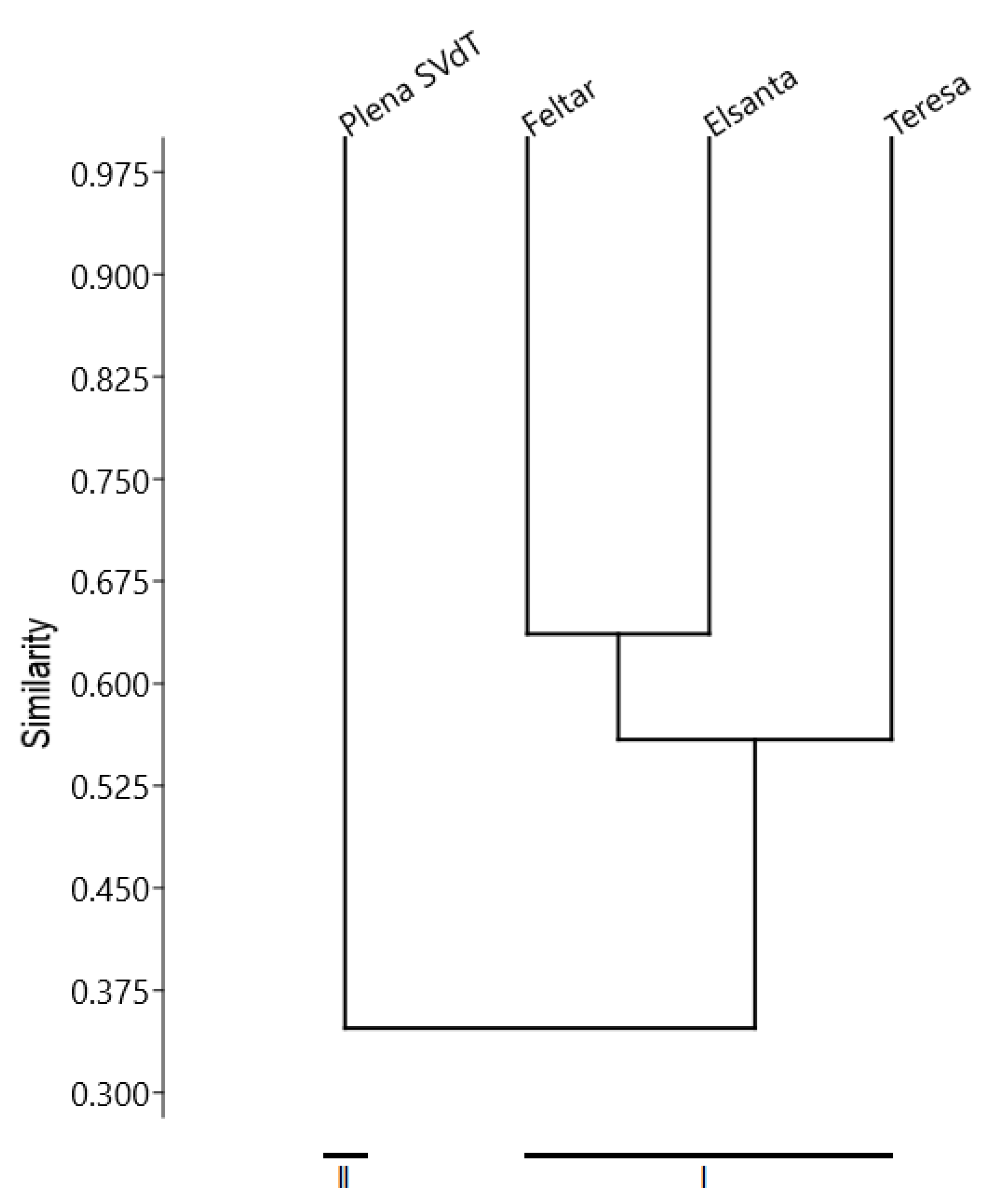
Figure 1. UPGMA dendrogram showing genetic similarity between the analyzed strawberry microclones resistant to Phytophthora disease.
Agronomy 11 01279 g001
Table 1. DNA polymorphism of the studied strawberry genomes using ISSR primers.
StarterSequence (5′–3′) (a)Number of Loci
TotalPolymorphic%P (b)Size Range (bp)
1VBVACACACACACACAC77100.0400–2000
2BDBCACACACACACACANo products---
3HBHCTCTCTCTCTCTCTNo products---
4GCVTCTCTCTCTCTCTCNo products---
5VCGTCTCTCTCTCTCTCNo products---
6BDVAGAGAGAGAGAGAGNo products---
7HVHTGTTGTTGTTGTTGT6583.3600–3000
8BDBCACCACCACCACCAC88100.0300–1500
9BDVCAGCAGCAGCAGCAGNo products---
10GAAGAAGAAGAAGAAGAA55100.01000–1500
11ATGATGATGATGATGATG6350.0300–3000
12TGGTGGTGGTGGTGGTGGNo products---
13GATAGATAGATAGATAGATA77100.0700–3500
14GACAGACAGACAGACAGACA6583.3300–2500
15CTAGCTAGCTAGCTAGNo products---
16AGTGAGTGAGTGAGTG77100.0300–2500
Mean6.55.9--
Totality524789.58300–3500
(a) explanation of symbols: H = A + T + C, B = G + T + C, D = G + A + T, V = G + A + C. (b) percentage of polymorphism.
Table 2. Percentage of microplants with symptoms of Phytophthora disease among strawberry microclones at subsequent observation time points.
MicrocloneEvaluation Scale (0–4)Observation Time Points
IIIIIIIVV
‘Elsanta’054.0030.6722.6716.009.33
132.6738.3337.6729.6721.00
27.6719.0022.6730.0032.33
34.007.6710.0010.3314.67
41.674.337.0014.0022.67
‘Feltar’080.0062.0043.3332.3324.67
118.0028.3335.6736.0036.00
21.677.3312.0015.3318.33
30.332.336.678.676.67
40.000.002.337.6714.33
‘Plena SVdT’00.000.000.000.000.00
13.330.000.000.000.00
236.671.671.671.671.33
346.3349.0031.005.673.33
413.6749.3367.3392.6795.33
‘Teresa’059.6729.6716.338.006.33
124.6731.3327.0018.0013.67
29.3314.0022.0026.3316.33
33.3311.0011.6713.0014.00
43.0014.0023.0034.6749.67
Table 3. Percentage of microplants with chlorosis among control strawberry microclones at subsequent observation time points.
MicrocloneEvaluation Scale
(0–4)
Observation Time Points
IIIIIIIVV
‘Elsanta’096.0095.0095.0095.0093.00
14.004.003.002.002.00
20.001.002.002.002.00
30.000.000.001.001.00
40.000.000.000.002.00
‘Feltar’096.0096.0096.0093.0092.00
13.002.001.002.002.00
21.002.003.003.002.00
30.000.000.002.002.00
40.000.000.000.002.00
‘Plena SVdT’093.0092.0092.0092.0092.00
14.005.004.004.003.00
22.002.003.001.001.00
31.001.001.002.002.00
40.000.000.001.002.00
‘Teresa’096.0095.0091.0091.0090.00
13.002.006.005.004.00
21.003.003.003.002.00
30.000.000.001.003.00
40.000.000.000.001.00
Table 4. Differences between the mean extent (%) and mean rate of Phytophthora disease, and the control sample in the studied microclones. Means marked with the same letter do not differ significantly at p ≤ 0.05.
Microclone‘Elsanta’‘Elsanta’ Control‘Feltar’‘Feltar’ Control‘Plena SVdT’‘Plena SVdT’ Control‘Teresa’‘Teresa’
Control
Mean extent (%)31.57a5.65b19.50a6.50b84.74a9.40b43.53a7.60b
Mean rate0.1399a0.0200b0.1128a0.0941a0.0710a0.0295b0.1414a0.0279b
Table 5. Differences in the extent of Phytophthora disease development expressed by the mean McKinney disease index (%). Means marked with the same letter do not differ significantly at p ≤ 0.05.
Microclone‘Plena SVdT’‘Teresa’‘Elsanta’‘Feltar’
Mean McKinney disease index (%)84.74a43.53b31.57c19.50d
Table 6. Simmonds disease index at subsequent observation time points for P. cactorum—infected microclones and the control sample.
MicrocloneEvaluation Scale
(0–4)
Observation Time Points
IIIIIIIVV
‘Elsanta’00.23550.20430.16700.12910.0803
10.21980.23600.23410.20620.1646
20.06800.15370.17450.20910.2148
30.03590.06720.08750.08970.1231
40.01580.03980.06330.11530.1633
‘Elsanta’ control00.03840.04750.04750.04750.0651
10.03840.03840.02910.01960.0196
20.00000.00990.01960.01960.0196
30.00000.00000.00000.00990.0099
40.00000.00000.00000.00000.0196
‘Feltar’00.15670.21750.20890.19490.1585
10.14500.18380.21500.22430.2291
20.01610.06780.09830.12410.1432
30.00330.02270.06140.07670.0616
40.00000.00000.02260.06910.1190
‘Feltar’
control
00.13220.17020.16520.15690.1334
10.12400.15000.16880.17400.1766
20.01580.06320.08870.10870.1227
30.00330.02220.05760.07080.0578
40.00000.00000.02210.06430.1049
‘Plena SVdT’00.00000.00000.00000.00000.0000
10.03190.00000.00000.00000.0000
20.22060.01640.01640.01640.0131
30.23620.24320.20860.05300.0321
40.11740.24390.21530.06730.0442
‘Plena SVdT’ control00.06510.07360.07360.07360.0736
10.03840.04750.03840.03840.0291
20.01960.01960.02910.00990.0099
30.00990.00990.00990.01960.0196
40.00000.00000.00000.00990.0196
‘Teresa’00.24060.20860.13560.07180.0582
10.18420.21460.19530.14260.1158
20.08450.12020.17140.19390.1352
30.03190.09610.10300.11230.1202
40.02780.11860.17630.22640.2486
‘Teresa’
control
00.03840.04750.08190.08190.0900
10.02910.01960.05640.04750.0384
20.00990.02910.02910.02910.0196
30.00000.00000.00000.00990.0291
40.00000.00000.00000.00000.0099
Table 7. Differences in the rate of Phytophthora disease development expressed by the mean Simmonds disease index. Means marked with the same letter do not differ significantly at p ≤ 0.05.
Microclone‘Teresa’‘Elsanta’‘Feltar’‘Plena SVdT’
Mean Simmonds disease index0.1414a0.1399a0.1128ab0.0710b
Publisher’s Note: MDPI stays neutral with regard to jurisdictional claims in published maps and institutional affiliations.