Abstract
This review examines the progress of electrostatic spore-trapping research and the potential for the practical application of electrostatic apparatuses in powdery mildew control. These apparatuses produce an electric field by charging an insulated conductor wire (ICW). Airborne pathogen spores are subjected to an attractive force in the electric field and are drawn to the charged ICW as a result of dielectrophoretic movement. The strength of the attractive force is commensurate with the field strength (determined by the magnitude of the voltage applied to the ICW). Single-charged monopolar electric field screens (SM screens) are constructed by arraying negatively charged cylindrical ICWs in parallel at a specific interval. The connected electric fields of these ICWs form a gap-free air-shielding barrier. Wind-dispersed spores are precipitated by this barrier to create spore-free air. Oppositely charged SM screens have been combined to develop double-charged dipolar electric field screens, which generate a stronger spore attraction force under lower voltage application. Thus, electric field screens represent a promising physical method for creating spore-free spaces in cropping facilities, where plants can be cultivated without risk of infection by airborne fungal pathogens.
1. Introduction
Powdery mildew was first detected in our greenhouse tomatoes in 2001 [1]. The isolated strain (KTP-01) was identified as Oidium neolycopersici L. Kiss (syn. Pseudoidium neolycopersici L. Kiss) [2], which was identical to isolates collected in various regions worldwide [3]. KTP-01 was found to be highly infectious to all tested commercial tomato cultivars [1,2], as well as a breed line resistant to a European tomato powdery mildew isolate [4]. Although conventional fungicides for powdery mildew pathogens are effective for its control, non-chemical control measures were adopted to reduce the risk of inducing fungicide-resistant strains of the pathogen [5,6]. The initial approach was to screen wild-type tomato plants resistant to new isolates [7,8,9] and identify resistance genes in these plants [10,11,12]. A new tomato line bred through interspecific hybridization was found to be highly resistant to the target pathogen isolate until a new pathogenic strain appeared [4,8]. Another biological approach was to use plant resistance-inducing bacteria or pathogen-antagonistic microbes to control powdery mildew. Yamamoto et al. [13] reported that the application of Bacillus amyloliquefaciens to soil induced systemic resistance in tomato plants against powdery mildew and bacterial wilt caused by Ralstonia solanacearum. Németh et al. [14] inoculated Ampelomyces strains into powdery mildew colonies on leaves and reported their mycoparasitic activity against colonial mycelia. Despite much interesting work, there has been little practical progress in this field because these protective effects are easily attenuated, and due to problems with agent preparation, limited application targets, high susceptibility to environmental conditions, and high cost.
By contrast, physical pathogen control methods are largely unaffected by biological and environmental conditions. With electrostatic methods, which are promising for physical pathogen control, an electric field is generated by an electric charge on a conductor as follows: a voltage generator picks up a negative charge from the ground and supplies it to a linked conductor; negative charge accumulates on the surface of the conductor; and an electric field is formed in the space surrounding the charged conductor. If another grounded conductor is placed within the electric field and the applied voltage exceeds a certain limit, the negative charge on the conductor moves to the ground via the grounded conductor (i.e., discharge between two conductors) [15]. By exploiting this discharge phenomenon, Nonomura et al. [16] devised a portable pen-shaped corona discharge generator, in which a powdery mildew colony on a leaf touched by a ground line is directly exposed to a plasma jet stream from the pointed tip of the generator. This exposure treatment destroyed all conidia and conidiophores in the colony instantaneously. However, due to its labor-intensive nature, this treatment is effective for only limited numbers of colonies at the initial stage of disease expansion.
Negative charge on an insulated conductor causes different phenomena within an electric field. If the applied voltage does not exceed the limit, the insulating coating of the conductor prevents discharge (i.e., charge movement) from the charged conductor. Charge remaining on the conductor negatively electrifies the outer surface and positively electrifies the inner surface of the coating through dielectric polarization [17]. An electric field forms around the insulated conductor due to surface charge on the insulator. Importantly, this surface charge imparts a perceivable force to any other charge entering the electric field. Matsuda et al. [18] utilized this force to design a spore trap based on an electric field screen, in which cylindrical insulated conductors were arrayed in parallel at a specific interval, thereby creating an electric field between the charged insulated conductors. Eventually, this spore trap device was further developed as an electrostatic air-shielding barrier to create spore-free spaces for plant cultivation [19].
In this review, we discuss the progress of electrostatic spore trapping research, from its initial experiments to the development of electric field screens targeting fungal spores subject to long-distance dispersal by wind. Most fungal spores are plant pathogens, which have varying spore production rates; powdery mildew is relatively prolific in the pre-harvest stage [20], whereas green molds are common post-harvest [21]. These fungi are often used as model biological materials for capture experiments. Based on results obtained using these species, we offer new insights into fungicide-independent control methods for airborne fungal pathogen spores, based on major contributions made by our joint researchers.
2. Construction of Electrostatic Spore Collection Probes
The powdery mildew pathogen infects tomato plants and typically produces white colonies (pustules) on leaves (Figure 1A). As shown in Figure 1B, numerous conidiophores (rod-shaped structures emerging from superficial hyphae) are produced in the pustules, and spores (conidia) develop at the tips of conidiophores [22]. Using a glass needle held by the micromanipulator of a high-fidelity digital microscope (Figure 1C), Matsuda et al. [23] collected mature conidia from the tips of conidiophores under the microscope. Conidia jumped toward the glass needle before coming into contact with it (Figure 1D). This phenomenon suggested electrostatic attraction between the conidia and needle. A glass needle was fabricated by heating, expanding, and cutting a glass tube (Figure S1). Frictional electrification occurred between the glass surface and surrounding air as the glass tube expanded [24]. Frictional electricity accumulated on the tip of the glass needle, producing an electrostatic field in the surrounding space (Figure S1). The spores were attracted to the needle as a result of an attractive force created by the electrostatic field.
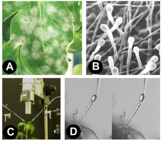
Figure 1.
(A) Fungal colonies on tomato leaves; (B) electron micrograph of conidiophores within a colony; (C) a high-fidelity digital microscope equipped with two micromanipulators; and (D) tomato powdery mildew spore collected with a glass needle held under a high-fidelity digital microscope.
To demonstrate the involvement of an electrostatic force in spore attraction, an electrostatic field was created using another electrostatic technique. In this experiment, Nonomura et al. [22] used an electrostatic spore collection probe (i.e., a pointed ebonite rod) (Figure 2A). Mature conidia on the conidiophore were collected using the attractive force created by the electrostatic field produced at the pointed tip of the electrostatic spore collection probe (Figure 2B). Mature conidia that had detached from the conidiophore were attracted to the probe tip, without requiring physical contact, demonstrating that our the hypothesis of electrostatic involvement was correct. The electrostatic spore collection probe was created by touching the flat end of the pointed ebonite rod to a negatively charged metal cap. The ebonite became electrified via dielectric polarization [17], thus producing an electrostatic field around the pointed tip. In the area occupied by an electrostatic field, a charged body will experience an electrostatic force directly proportional to the applied voltage.
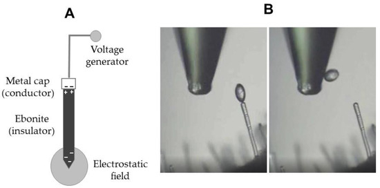
Figure 2.
(A) Structure of an electrostatic spore collection probe; and (B) attraction of a mature spore (conidium) on a tomato powdery mildew conidiophore by an electrostatic spore collection probe.
Under a digital microscope, the electrostatic spore collection probe was brought close to a mature conidium on a conidiophore. Mature conidia were easily distinguished due to the clear constriction between a conidium and the conidiophore tip end. A conidium was attracted to the probe tip without direct contact (Video S1A), providing clear evidence of the involvement of electrostatic force in spore attraction. These results laid the groundwork for our further electrostatic engineering research and electric field screen development.
The electrostatic spore collection probe was used for microscopic analysis of conidiogenesis by barley powdery mildew (Blumeria graminis f. sp. hordei) [25] and Cucurbitaceae powdery mildew (Podosphaera xanthii) [26]. The probe was modified to fabricate a time-controlled electrostatic spore attraction plate (Video S1B), to collect all conidia produced by these powdery mildew pathogens throughout their lifetime [27,28].
3. Dielectrophoretic Movement of Spores in an Electrostatic Field
3.1. Construction of an Electrostatic Field
The application of negative or positive voltage to an insulated conductor allows negative or positive charge to accumulate on the conductor surface, in turn producing an electric field in the surrounding space. This field induces surface charges in the insulating coating of the conductor. For example, when the insulator is an acrylic cylinder, opposite charges are created on the inner and outer surfaces of the cylinder through dielectric polarization (Figure S2) [17]. The outer surface charge of the monopolar cylinder produces an electrostatic field in the surrounding space (Figure S2), such that there is no current flow in the space, only charge.
With either negative or positive voltage application, a voltage applied at the same magnitude will produce the same field intensity, as the potential difference with respect to the ground remains the same. The same field intensity in turn creates the same magnitude of electrostatic force. Field intensity is strongest in the region closest to the charged/electrified body (in this case, the insulator). The electric field intensity gradient is used to capture objects that enter the field.
3.2. Airborne Spore Capture by an Electrified Insulated Conductor
Dielectrophoresis is a phenomenon by which a force is exerted on a dielectric (oppositely polarized) particle in an electric field [29]. This force does not require the particle to be charged, because all particles exhibit dielectrophoretic activity in the presence of the electric field. According to dielectrophoresis theory, the relative polarizability of the particles changes along the electric field strength gradient. Field strength, i.e., the force intensity at a given point in the electric field, becomes stronger as a point moves closer to the charged pole. Thus, particles in an electric field are attracted to a charged pole that produces an electric field.
In a negatively charged acrylic cylinder, airborne spores reaching the electrostatic field are positively polarized on the cylinder side and negatively polarized on the opposite side of the spore (Figure 3A). An attractive force is generated between the positive charge of the spore and the negative charge of the cylinder, and the spore is drawn to the cylinder according to its dielectrophoretic movement [18].
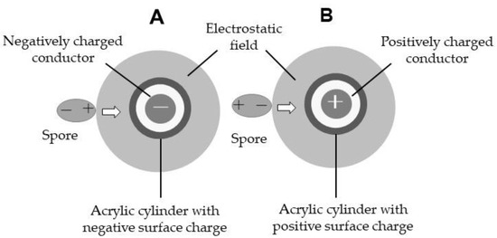
Figure 3.
Schematic representation of attraction of spores entering the electrostatic field of an insulator: (A) negatively or (B) positively electrified by dielectrophoresis.
In a positively charged acrylic cylinder (Figure 3B), spore polarization is oriented in the opposite direction. The spore is polarized negatively on the cylinder side and positively on the opposite side of the spore through dielectrophoresis [30]. If the applied voltage is the same in both insulated conductor wires (ICWs), an attractive force of the same strength (magnitude) is generated between the spore and cylinder. This spore attraction phenomenon prompted additional investigations into the use of electric field screens for capture applications.
In real-life spore capture applications, the targets are wind-dispersed spores. The movement of these spores in an electrostatic field is determined by the wind velocity vector and dielectrophoretic attractive force [18]. Naturally, an attractive force surpassing the force associated with the wind velocity is necessary to capture the spores.
3.3. Fabrication of a Single-Charged Monopolar Electric Field Screen
3.3.1. Air-Shielding Barrier Formed by Connected Electrostatic Fields
An insulator covering a charged conductor generates an electrostatic field in the surrounding space via electrification through dielectric polarization. A cylindrical insulator produces the electrostatic field concentrically (Figure 3). Thus, an air shield can be created by placing several electrostatically charged cylinders in close proximity to other cylinders in a screen-like configuration, allowing interaction between the associated electrostatic fields of individual cylinders (Figure 4A). Wind-dispersed spores that reach the field are uniformly attracted to the nearest cylinder (Figure 4B), and do not pass through the electrostatic barrier [18,30].
Figure 4.
(A) Vertically arrayed insulated conductor wires (ICWs); (B) air-shielding barrier of connected electrostatic fields comprising vertically arrayed ICWs (cross-sectional view); and (C) single-charged monopolar electric field screen.
One apparatus that utilizes this phenomenon is the electric field screen (Figure 4C), which is an air shielding device constructed using electrostatic engineering techniques. The electrostatic field created by the parallel array of insulated conducting wires allows air to permeate freely through the barrier while capturing wind-dispersed spores by establishing a strong attractive force. The electric field screen is air-permeable, allowing use in conjunction with other ventilation equipment. For example, in a greenhouse containing windows furnished with electric field screens, wind-dispersed spores precipitate from the ventilated air to the screen, while air passes freely through the screen. As a result, spore-free air is supplied to plants cultivated in the greenhouse [19]; this allows the plants to avoid infection by airborne pathogens, for non-agrochemical control of pathogens during crop cultivation.
There is a positive relationship between applied voltage and radial expansion of an electrostatic field (Figure S3A). A higher voltage produces a wider electrostatic field. In an electric field screen, the interval between the ICWs necessary for capture applications increases at twice the rate of the applied voltage (Figure S3B). As the necessary separation interval between the ICWs of the screen increases with the application of higher voltages, the screen becomes more permeable to air. However, higher voltages increase the likelihood of discharge at the electric connection points. In practical applications, 5-kV charging (separation interval, 10 mm) is often selected to minimize discharge events while promoting satisfactory air permeability.
3.3.2. Powdery Mildew Pathogen Control by a Single-Charged Monopolar Electric Field Screen
A single-charged monopolar electric field screen (SM screen) was designed to protect greenhouse tomatoes from infection by airborne conidia of the tomato powdery mildew pathogen. Insulated cylindrical conductor wires were installed on the roof and four lateral faces of a box-shaped frame to create an electric field screen box (Figure 5A). This box was used to cover a hydroponic trough used for growing tomato seedlings.

Figure 5.
(A) Rectangular box furnished with single-charged monopolar electric field screens to cover a hydroponic trough for culturing plant seedlings: (B) schematic representation of a method for inoculating powdery mildew pathogen conidia into tomato seedlings in screen-guarded and unguarded hydroponic troughs; (C) heavily infected tomato seedlings in the unguarded hydroponic trough (left) and healthy non-infected tomato seedlings in the screen-guarded hydroponic trough (right) at 1 month after inoculation; and (D) digital micrographs of conidia captured by the insulated conductor wire of the electric field screen box used to cover the hydroponic trough.
Powdery mildew pathogen conidia are dispersed by wind to reach host plant leaves, where they germinate and initiate infection. After infection has been established, the pathogen produces pustules (colonies) on the leaf surface, in which numerous conidia are produced and dispersed by wind to neighboring host plants, thereby spreading the infection.
The occurrence of powdery mildew disease in greenhouse tomatoes indicates that an outside source is responsible for the infection. The origin of the conidia remains unclear; however, several studies have suggested that these pathogens can travel several kilometers [31,32,33]. Once the pathogen invades a greenhouse, the disease spreads quickly after the first infection is established. Therefore, preventing the initial entry of conidia into the greenhouse is vital.
In an inoculation assay, conidia were mechanically blown onto tomato plants under the assumption that the conidia are wind-dispersed. Three hydroponic troughs culturing tomato seedlings were used for the experiment; the center trough was covered with an electric field screen box (Figure 5B). Inoculated tomato plants producing abundant conidia were used as spore (inoculum) sources. Air was blown continuously, at a rate of 3 m/s, toward the hydroponic tomato seedlings by an electric fan for one month. As conidia were produced daily on the inoculum plants [22], the test plants were continuously infected by the pathogen; thus, particularly severe infection conditions were established as an exacting test of the electric field screen system. This degree of infection is rarely encountered under natural conditions.
The spacing between the ICWs in the electric field screen box was 60 mm (30 kV charge; Figure 5C) to facilitate observation of the tomato seedlings. Photographs showed the external appearance of leaves of guarded and unguarded tomato seedlings after one month. Powdery mildew colonies expanded over the leaf surfaces of unguarded seedlings (Figure 5C, left), indicating severe infection. By contrast, tomato seedlings within the screen box remained uninfected throughout the experimental period (Figure 5C, right), confirming that healthy and successful growth could be attained under severe inoculation conditions.
At the end of the experiment, all ICWs were detached from the box to observe their surfaces using a high-fidelity digital microscope. Many conidia were detected on the conductor surfaces (Figure 5D). These results demonstrate that the electric field screen was able to capture all conidia blown at a rate of 3 m/s toward the screen box, thereby fully protecting tomato seedlings from the powdery mildew pathogen.
4. Spore Trapping by Two- and Three-Layer Double-Charged Dipolar Electric Field Screens
4.1. Negative and Positive Voltage Generators for Double Charging
The ICW is electrified by connecting a grounded negative or positive voltage generator. A negative voltage generator draws free electrons from the ground, which is an infinite source or sink of electrons, to the conductor wire (Figure S4A). Negative electricity accumulates on the surface of the wire conductor. In turn, negative charges are induced on the outer surface of the insulating wire coating, thereby negatively electrifying the insulator through dielectric polarization. A positive voltage generator pushes free electrons to the ground to positively charge the conductor wire (Figure S4B). The surface of the conducting wire becomes positively charged due to electrostatic induction [34]. A positive charge is induced on the outer surface of the insulating coating surrounding the conducting wire, and the coating becomes positively charged through dielectric polarization.
The amount of electricity required for electrification is proportional to the voltage applied by the voltage generator. The applied voltage corresponds to the potential difference with respect to the ground; a larger potential difference enhances electrostatic phenomena, as a larger attractive or repulsive force is generated. This allows the capture of spores.
Both voltage generators can be operated by a 12-V storage battery. A voltage generator is used to boost the initial voltage (12 V) to the designated voltage (up to 30 kV) using a transformer (coil) and Cockcroft circuit integrated into an electric circuit in the voltage generator [35]. The difference between the negative and positive voltage generators is that the Cockcroft circuit is set in reverse, such that negative electricity moves in the opposite direction (Figure S4). The greatest advantage of this voltage generator is that it can be operated using a 12-V direct current (DC) source. The electric power consumption (5 W) approaches that of a small lightbulb, such that the screen can operate for long periods of time using a regular storage battery. This is useful for practical implementation of the electric field screen.
4.2. Soft Polyvinyl Chloride Tube for Insulating Conductors
Acrylic resin has strong insulative properties, such that high voltages can be applied to a conductor insulated with this resin. However, acrylic resin is very difficult to process, which complicates the development of new types of electric field screens and limits real-world applications. To solve this problem, we employed polyvinyl chloride resin, which is commonly used to insulate metal materials. As we had no equipment in our laboratory capable of producing or coating materials with this resin, we used a commercially available soft vinyl chloride (Toalon) tube. The most serious drawback of this material is that it has extremely low volume resistivity, which limited our ability to charge it. Unexpectedly, this limitation motivated us to develop a new method for achieving the necessary capture capabilities at lower voltages.
The major factors that should be considered when designing an electric field screen are the quality of the insulation material, pole distance, range of applied voltage, and presence or absence of discharge, particularly arc discharge. All these factors are closely related. For example, for the same applied voltage, if the pole distance is shortened, discharge occurs more readily between the opposite poles. Therefore, we modified several screen parameters such as the applied voltage, while fixing other factors. The fixed parameters included the use of soft vinyl chloride tube to insulate the conductors and a 5-mm separation distance between the adjacent insulated conductors used to create the screen. The ICW was prepared by passing a copper or iron wire through the soft polyvinyl chloride tube and arranging these wires in parallel at a constant 5-mm interval as a skeletal structure for subsequent electric field screens.
4.3. Fabrication of the Double-Charged Dipolar Electric Field Screen
Conductor wires insulated with soft polyvinyl chloride tubes were arrayed in parallel at a specific interval (5 mm), linked to each other (and to a negative or positive voltage generator), and fixed with a polypropylene frame to construct the SM screen (Figure 6A). A double-charged dipolar electric field screen (DD screen) was constructed by pairing two SM screens linked to negative and positive voltage generators, respectively, to realize a two-layer DD screen (Figure 6B). A three-layer DD screen was constructed by placing the negatively charged SM screen beside a positively charged two-layer SM screen (Figure 6C). Two- and three-layer DD screens have oppositely charged ICWs in an offset configuration, where the electric field is created in the space between the oppositely charged ICWs (dipole) (Figure 6D,E). The insulating coating of the charged conductor prevents charges on the conductor surface from moving to the electric field (discharge of the charge conductor). The electric field formed between the oppositely charged poles can result in an electric discharge if the applied voltage exceeds a certain limit. Thus, this type of electric field is distinguished by the presence or absence of discharge, where the non-discharging electric field can be described as a static electric field [36]. Within the voltage range of the DD screen, a static electric field between the oppositely charged ICWs forms a zigzag pattern in two-layer screens (Figure 6D) and an x-shaped pattern in three-layer screens (Figure 6E).
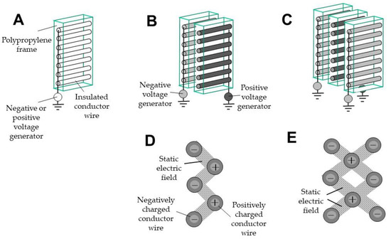
Figure 6.
(A) Single-charged monopolar electric field screen; (B, C) double-charged dipolar electric field screens (DD screens) constructed with (B) two and (C) three layers; (D,E) static electric fields formed between oppositely charged conductor wires of (D) two-layer and (E) three-layer screens (cross-sectional view).
4.4. Spore-Capturing Ability of Two- and Three-Layer DD Screens
In this experiment, lemon fruit was inoculated with green mold (P. digitatum) and the abundant spores that formed were used for inoculation (Figure S5A,B) [37]. Spores from the inoculated fruit were dusted onto a parchment paper-covered tray by gently tapping the fruit. The spore density was fixed at 104–105 spores/cm2/tray by counting the spores in several randomly selected areas of the tray using a high-fidelity digital microscope. The collected spores were placed in a pressure bottle and blown by compressed air from the outlet nozzle toward the DD screen (Figure S5C). The spore capture rate was determined by counting the spores trapped by the ICWs of the DD screen and the electrostatic spore attraction plate [27] placed over the screen (Figure S5D). Two- and three-layer DD screens were used for the assay to determine their ability to capture wind-dispersed spores, as these screen types had shown good results in preliminary experiments.
To determine the total number of microscopic spores used in this experiment, the surfaces of the ICWs were scanned with a high-fidelity digital microscope to ensure that all trapped spores were counted. Because spores that escaped the trap by passing through the screen remained uncounted in preliminary experiments, an electrostatic spore attraction plate was installed as an additional trapping apparatus on the opposite side of the screen [27]. Despite this precaution, we were unable to guarantee that all escaped spores had been trapped for counting. Nevertheless, the number of spores trapped by the electrostatic spore attraction plate at lower applied voltages declined gradually as the voltage increased, indicating enhancement of the spore capturing rate. The spore capturing rate was determined by calculating the numbers of spores trapped by the ICWs of the DD screen and electrostatic spore attraction plate.
The three-layer screen prevented all spores from passing through the apparatus at a charge of −0.9 kV (Table 1). Two- and three-layer screens use the same mechanism to trap spores; therefore, the two-layer screen was expected to prevent passage of the spores. However, the two-layer screen failed to capture even a small number of spores, as they appeared to escape via a ‘jumping’ phenomenon.

Table 1.
Numbers of Penicillium digitatum spores passing through electric field screens with double or triple layers of oppositely charged insulated conductor wires [37].
4.5. Spore Jumping Caused by Creeping Discharges
Within the electric field, minute particles such as spores are attracted to the nearest charged pole due to dielectrophoresis. However, when the spores reached the surface of the negatively charged pole, they were rarely electrified by negative discharge. When multiple spores were within the closed area, spores contiguous to other spores jumped away as a result of the charge between them. Thus, when a second spore was attracted to a negatively charged insulator close to a precedent, positively electrified spore, the first spore jumped away, instead of the second spore. This phenomenon can be explained in terms of creeping discharge between proximate spores.
When a second spore is captured at a site adjacent to a previously captured spore, both spores attain opposite charges (Figure 7A); this promotes the transfer of electricity (free electrons) between them. This flow of electricity occurs on the surface of the insulator as creeping discharge [38]. Eventually, the second spore becomes positively charged, and the first spore attains a negative charge. The first spore is subjected to a repulsive force from the same-charged insulator, which induces the jumping effect. For the green mold spores (diameter, 5 μm) used in this experiment, spore jumping was observed when two spores coexisted within a circle with a 20-μm radius [37]. If the separation of the two spores exceeded this distance, no creeping discharge (i.e., no spore jumping) occurred.
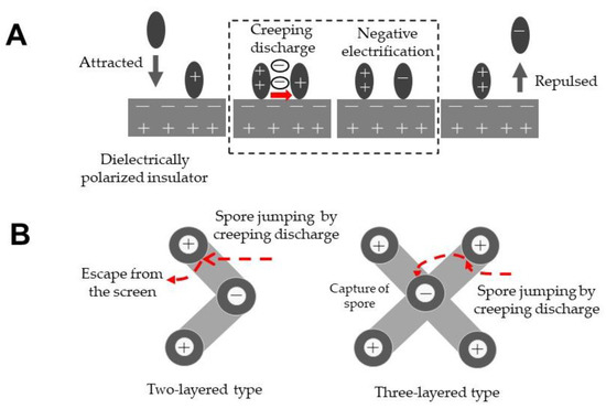
Figure 7.
(A) Schematic representation of creeping discharge between two spores and spore jumping on the electrified insulator covering a charged conductor; and (B) schematic representation of the escape (left) and recapture (right) of a jumping spore in two- and three-layer electric field screens, respectively.
Spore jumping occurs more frequently as spore density increases, as creeping discharge between the spores is generated more easily. From an electrostatic perspective, creeping discharge occurs more frequently when an insulator with lower surface resistivity is used or when the applied voltage is higher. The three-layer screen was effective in capturing spores (Table 1); therefore, to determine whether there is a difference in spore-capturing functionality between two- and three-layer screens, Takikawa et al. [37] re-examined the settings of the experiment. Large numbers of spores were blown onto the electric field screen, which readily induced creeping discharge (and therefore spore jumping). Microscopic observation indicated that spore jumping occurred when subsequent spores entered the area at an inter-spore distance of 20 μm. As shown in Figure 7B, spores that jumped were more easily trapped by the additional electric field in the three-layer screen.
The density of spores in air is very low; thus, the likelihood of creeping discharge between captured spores is also very low. From this perspective, the jumping of captured spores seldom occurs under ordinary circumstances. Therefore, two-layer screens can be applied for spore capture in most situations, as they exhibit the same capture ability as three-layers screens and are more cost-effective.
5. Practical Control of Powdery Mildew Conidia by Electric Field Screens
A three-layer DD screen with an ungrounded circuit (Figure 8A) was used to construct an electrostatic shelter (Figure 8B) to raise healthy plant seedlings in a pathogen-free space [39]. In the ungrounded circuit, free electrons in the conductor wires were supplied directly to other conductor wires according to the voltage produced by generators (Figure S6). An electric field screen with such a circuit requires no ground line. Based on this reasoning, the electric field screen may be placed arbitrarily and effectively acts as a portable device. This equipment is designed to be used in a greenhouse without an installed electric field screen. Three-layer screens are appropriate for such usage because they prevent entry by both pests and pathogen spores [39]. Because their structure is very simple, their size may be freely altered according to the scale of seedling cultivation. To enhance air permeation, the screens were installed on opposite faces of the shelter, along with a small axis fan.
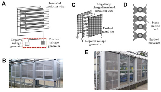
Figure 8.
(A,B) Schematic representation of (A) a three-layer DD screen with a non-grounded circuit and (B) an electrostatic seedling shelter furnished with three-layer DD screens; (C,D) schematic representation of (C) a single-charged dipolar electric field screen, consisting of grounded metal nets on either side of a layer of negatively charged insulated conductor wires (ICWs); and (D) static electric fields formed between the negatively charged ICWs and grounded metal nets; and (E) installation of single-charged dipolar electric field screens in the lateral windows of a greenhouse.
Matsuda et al. [40] devised a single-charged dipolar electric field screen (SD screen) to trap insect pests. This screen was constructed by placing two grounded metal nets on either side of the SM screen (Figure 8C); static electric fields were created in the space between the negatively charged conductor wires and grounded net (Figure 8D). Insects blown into the electric field were captured by the negatively charged conductor wire [40]. Nonomura et al. [41] reported that the single-charged dipolar screen repelled insects that reached the grounded metal net of the screen, as they instinctively avoided entering the static electric field. More importantly, Kakutani et al. [42] found that the negatively charged ICW of the SD screen trapped airborne powdery mildew conidia; these screens were applied to greenhouse windows to create a conidium- and pest-free greenhouse environment (Figure 8E).
The ICW is the heart of the electric field screen. These wires are easily constructed in a laboratory by passing a metal wire through a soft polyvinyl chloride tube. However, in outdoor, longer-term experiments, the tubes are susceptible to severe deterioration such as cracking, deformation, and discoloration due to changes in temperature, humidity, and ultraviolet irradiation levels. These issues limit practical implementation of the electric field screen.
Coating of a metal wire with weatherproof polyvinyl chloride resin is advisable to prolong screen operation in outdoor environments with minimal deterioration. In the preliminary experiments, a soft polyvinyl chloride tube showed optimal volume resistivity (108–109 Ωcm) for electric field screen functionality. The volume resistivity of polyvinyl chloride (1015 Ωcm) can be adjusted to 1014–108 Ωcm through the addition of plasticizers or ultraviolet absorbents, thereby enhancing weather resistance [43]. Polyvinyl chloride materials mixed with various substances are sold commercially as soft polyvinyl chloride; these materials have distinct qualities with different volume resistivity values. A remaining problem in electric field screen research is the selection of coating materials that meet the requirements for practical implementation and weather resistance of ICWs.
6. Conclusions
Electrostatic spore trapping techniques are based on the dielectrical polarization of an insulative coating by a charged conductor, and the creation of an electric field in the space surrounding the insulated charged conductor. Spores are captured within the electric field created by the insulated charged conductor. Unique arrangements of cylindrical charged ICWs were used to generate various types of electric field screen, which act as air-shielding barriers comprising combined electric fields to precipitate wind-dispersed pathogen spores from the air. This review provides an experimental basis for the development of physical strategies for the control of plant fungal pathogens.
Supplementary Materials
The following supporting information can be downloaded at: https://www.mdpi.com/article/10.3390/agronomy12102443/s1, Figure S1: Formation of an electrostatic field by frictional electricity accumulated on the tip of a glass needle, Figure S2: Schematic representation of an electrostatic field produced by a (left) negatively or (right) positively electrified insulator covering the negatively or positively charged conductor, Figure S3: (A) Expansion of an electrostatic field produced by insulated conductor wires (ICWs) applied with different voltages. (B) Different intervals between ICWs charged with different voltages, Figure S4: Schematic representation of the structure and function of (A) negative and (B) positive voltage generators, Figure S5: (A) Superficial colony formed over a lemon inoculated with green mold and (B) electron micrograph of conidia on conidiophores. (C) Instrument set used for a spore blowing assay. Conidia were collected and placed in a pressure bottle, and then blown toward a double-charged dipolar electric field screen (DD) screen by compressed air. (D) Diagram of a test box furnished with a DD screen and an axial-flow fan on the opposite side, Figure S6: Schematic representations of (A) grounded and (B) ungrounded circuits integrated into a DD screen, Video S1: (A) Trapping of a mature conidium by an electrostatic spore collection probe observed under a high-fidelity digital microscope. (B) Trapping of barley powdery mildew conidia with an electrostatic spore attraction plate.
Author Contributions
Conceptualization, H.T. and T.N.; methodology, T.N.; software, T.N.; validation, H.T. and T.N.; formal analysis, H.T.; investigation, T.N.; resources, T.N.; data curation, H.T.; writing—original draft preparation, H.T.; writing—review and editing, T.N.; visualization, T.N.; supervision, H.T.; project administration, T.N. All authors have read and agreed to the published version of the manuscript.
Funding
This research received no external funding.
Data Availability Statement
Not applicable.
Conflicts of Interest
The authors declare no conflict of interest.
References
- Matsuda, Y.; Kashimoto, K.; Takikawa, Y.; Aikami, R.; Nonomura, T.; Toyoda, H. Occurrence of new powdery mildew on greenhouse tomato cultivars. J. Gen. Plant Pathol. 2001, 67, 294–298. [Google Scholar] [CrossRef]
- Kashimoto, K.; Matsuda, Y.; Matsutani, K.; Sameshima, T.; Kakutani, K.; Nonomura, T.; Okada, K.; Kusakari, S.; Nakata, K.; Takamatsu, S.; et al. Morphological and molecular characterization for a Japanese isolate of tomato powdery mildew Oidium neolycopersici and its host range. J. Gen. Plant Pathol. 2003, 69, 176–185. [Google Scholar]
- Kiss, L.; Cook, R.T.A.; Saenz, G.S.; Cunnington, J.H.; Takamatsu, S.; Pascoe, I.; Bardin, M.; Nicot, P.C.; Sato, Y.; Rossman, A.Y. Identification of two powdery mildew fungi, Oidium neolycopersici sp. nov. and O. lycopersici, infecting tomato in different parts of the world. Mycol. Res. 2001, 105, 684–697. [Google Scholar] [CrossRef]
- Kashimoto, K.; Sameshima, T.; Matsuda, Y.; Nonomura, T.; Oichi, W.; Kakutani, K.; Nakata, K.; Kusakari, S.; Toyoda, H. Infectivity of a Japanese isolate of Oidium neolycopersici KTP-01 to a European tomato cultivar resistant to O. lycopersici. J. Gen. Plant Pathol. 2003, 69, 406–408. [Google Scholar] [CrossRef]
- Van den Bosch, F.; Gilligan, C.A. Models of fungicide resistance dynamics. Annu. Rev. Phytopathol. 2008, 46, 123–147. [Google Scholar] [CrossRef] [PubMed]
- Lucas, J.A.; Hawkins, N.J.; Fraaije, B.A. The evolution of fungicide resistance. Adv. Appl. Microbiol. 2015, 90, 29–92. [Google Scholar]
- Lindhout, P.; Pet, G.; van der Beek, H. Screening wild Lycopersicon species for resistance to powdery mildew (Oidium lycopersicum). Euphytica 1994, 72, 43–49. [Google Scholar] [CrossRef]
- Matsuda, Y.; Mori, Y.; Nishida, M.; Sakano, S.; Tarumoto, K.; Nonomura, T.; Nishimura, H.; Kusakari, S.; Toyoda, H. Screening of wild Lycopersicon species for resistance to Japanese isolate of tomato powdery mildew Oidium neolycopersici. Breed. Sci. 2005, 55, 355–360. [Google Scholar] [CrossRef][Green Version]
- Mieslerová, B.; Lebeda, A.; Chetelat, R.T. Variation in response of wild Lycopersicon and Solanum spp. against tomato powdery mildew (Oidium lycopersici). J. Phytopathol. 2000, 148, 303–311. [Google Scholar] [CrossRef]
- Li, C.; Bonnema, G.; Che, D.; Dong, L.; Lindhout, P.; Visser, R.; Bai, Y. Biochemical and molecular mechanisms involved in monogenic resistance responses to tomato powdery mildew. Mol. Plant Microbe Interact. 2007, 9, 1161–1172. [Google Scholar] [CrossRef] [PubMed][Green Version]
- Seifi, A.; Nonomura, T.; Matsuda, Y.; Toyoda, H.; Bai, Y. An avirulent tomato powdery mildew isolate induces localized acquired resistance to a virulent isolate in a spatiotemporal manner. Mol. Plant Microbe Ineract. 2012, 25, 372–378. [Google Scholar] [CrossRef][Green Version]
- Li, C.; Faino, L.; Dongi, L.; Fan, J.; Kiss, L.; Giovanni, C.; Lebeda, A.; Scott, J.; Matsuda, Y.; Toyoda, H.; et al. Characterization of polygenic resistance to powdery mildew in tomato at cytological, biochemical and gene expression level. Mol. Plant Pathol. 2012, 13, 148–159. [Google Scholar] [CrossRef]
- Yamamoto, S.; Shiraishi, S.; Kawagoe, Y.; Mochizuki, M.; Suzuki, S. Impact of Bacillus amyloliquefaciens S13-3 on control of bacterial wilt and powdery mildew in tomato. Pest Manag. Sci. 2015, 71, 722–727. [Google Scholar] [CrossRef]
- Németh, M.N.; Mizuno, Y.; Kobayashi, H.; Seress, D.; Shishido, N.; Kimura, Y.; Takamatsu, S.; Suzuki, T.; Takikawa, Y.; Kakutani, K.; et al. Ampelomyces strains isolated from diverse powdery mildew hosts in Japan: Their phylogeny and mycoparasitic activity, including timing and quantifying mycoparasitism of Pseudoidium neolycopersici on tomato. PLoS ONE 2021, 16, e0251444. [Google Scholar]
- Kaiser, K.L. Air breakdown. In Electrostatic Discharge; Kaiser, K.L., Ed.; Taylor & Francis: New York, NY, USA, 2006; pp. 1–93. [Google Scholar]
- Nonomura, T.; Matsuda, Y.; Kakutani, K.; Takikawa, Y.; Toyoda, H. Physical control of powdery mildew (Oidium neolycopersici) on tomato leaves by exposure to corona discharge. Can. J. Plant Pathol. 2008, 30, 517–524. [Google Scholar] [CrossRef]
- Halliday, D.; Resnick, R.; Walker, J. Electric discharge and electric fields. In Fundamentals of Physics; Johnson, S., Ford, E., Eds.; John Wiley & Sons: New York, NY, USA, 2005; pp. 561–604. [Google Scholar]
- Matsuda, Y.; Ikeda, H.; Moriura, N.; Tanaka, N.; Shimizu, K.; Oichi, W.; Nonomura, T.; Kakutani, K.; Kusakari, S.; Higashi, K.; et al. A new spore precipitator with polarized dielectric insulators for physical control of tomato powdery mildew. Phytopathology 2006, 96, 967–974. [Google Scholar] [CrossRef]
- Toyoda, H.; Matsuda, Y. Basic concept for constructing an electric field screen. In Electric Field Screen; Principles and applications; Toyoda, H., Ed.; Nobunkyo Production: Tokyo, Japan, 2015; pp. 3–17. [Google Scholar]
- Whipps, J.M.; Budge, S.P.; Fenlon, J.S. Characteristics and host range of tomato powdery mildew. Plant Pathol. 1998, 47, 36–48. [Google Scholar] [CrossRef]
- Kanetis, L.; Forster, H.; Adaskaveg, J.E. Determination of natural resistance frequencies in Penecillium digitatum using a new air-sampling method and characterization of fluodioxonil- and pyrimethanil-resistant isolates. Phytopathology 2010, 100, 738–746. [Google Scholar] [CrossRef]
- Nonomura, T.; Matsuda, Y.; Xu, L.; Kakutani, K.; Takikawa, Y.; Toyoda, H. Collection of highly germinative pseudochain conidia of Oidium neolycopersici from conidiophores by electrostatic attraction. Mycol. Res. 2009, 113, 364–372. [Google Scholar] [CrossRef]
- Matsuda, Y.; Sameshima, T.; Moriura, N.; Inoue, K.; Nonomura, T.; Kakutani, K.; Nishimura, H.; Kusakari, S.; Takamatsu, S.; Toyoda, H. Identification of individual powdery mildew fungi infecting leaves and direct detection of gene expression by single conidium PCR. Phytopathology 2005, 95, 1137–1143. [Google Scholar] [CrossRef]
- Moore, A.D. Frictional electricity. In Electrostatics, Exploring, Controlling, and Using Static Electricity, 2nd ed.; Laplacian Press: San Diego, CA, USA, 1997; pp. 22–27. [Google Scholar]
- Moriura, N.; Matsuda, Y.; Oichi, W.; Nakashima, S.; Hirai, T.; Sameshima, T.; Nonomura, T.; Kakutani, K.; Kusakari, S.; Higashi, K.; et al. Consecutive monitoring of lifelong production of conidia by individual conidiophores of Blumeria graminis f. sp. hordei on barley leaves by digital microscopic techniques with electrostatic micro-manipulation. Mycol. Res. 2006, 110, 18–27. [Google Scholar]
- Takikawa, Y.; Nonomura, T.; Miyamoto, S.; Okamoto, N.; Murakami, T.; Matsuda, Y.; Kakutani, K.; Kusakari, S.; Toyoda, H. Digital microscopic analysis of conidiogenesis of powdery mildew pathogens isolated from melon leaves. Phytoparasitica 2015, 43, 517–530. [Google Scholar] [CrossRef]
- Moriura, N.; Matsuda, Y.; Oichi, W.; Nakashima, S.; Hirai, T.; Nonomura, T.; Kakutani, K.; Kusakari, S.; Higashi, K.; Toyoda, H. An apparatus for collecting total conidia of Blumeria graminis f. sp. hordei from leaf colonies using electrostatic attraction. Plant Pathol. 2006, 55, 367–374. [Google Scholar] [CrossRef]
- Suzuki, T.; Nakamura, N.; Takagi, N.; Takikawa, Y.; Kakutani, K.; Matsuda, Y.; Matsui, K.; Nonomura, T. Quantitative analysis of the lifelong production of conidia released from single colonies of Podosphaera xanthii on melon leaves using electrostatic techniques. Aust. Plant Pathol. 2019, 48, 297–307. [Google Scholar] [CrossRef]
- Cross, J.A. Dielectrophoresis. In Electrostatics: Principles, Problems and Applications; De Barr, A.E., Ed.; Adam Hilger: Bristol, UK, 1987; pp. 269–276. [Google Scholar]
- Shimizu, K.; Matsuda, Y.; Nonomura, T.; Ikeda, H.; Tamura, N.; Kusakari, S.; Kimbara, J.; Toyoda, H. Dual protection of hydroponic tomatoes from rhizosphere pathogens Ralstonia solanacearum and Fusarium oxysporum f. sp. radicis-lycopersici and airborne conidia of Oidium neolycopersici with an ozone-generative electrostatic spore precipitator. Plant Pathol. 2007, 56, 987–997. [Google Scholar] [CrossRef]
- Aylor, D.E. The role of intermittent wind in the dispersal of fungal pathogens. Annu. Rev. Phytopathol. 1990, 28, 73–92. [Google Scholar] [CrossRef]
- Brown, J.K.M.; Hovmøller, M.S. Aerial dispersal of pathogens on the global and continental scales and its impact on plant disease. Science 2002, 297, 537–541. [Google Scholar] [CrossRef] [PubMed]
- Nonomura, T.; Matsuda, Y.; Yamashita, S.; Akahoshi, H.; Takikawa, Y.; Kakutani, K.; Toyoda, H. Natural woody plant, Mallotus japonicus, as an ecological partner to transfer different pathotypic conidia of Oidium neolycopersici to greenhouse tomatoes. Plant Protect. Sci. 2013, 49, S33–S40. [Google Scholar] [CrossRef]
- Griffith, W.T. Electrostatic phenomena. In The Physics of Everyday Phenomena, a Conceptual Introduction to Physics; Bruflodt, D., Loehr, B.S., Eds.; McGraw-Hill: New York, NY, USA, 2004; pp. 232–252. [Google Scholar]
- Wegner, H.E. Electrical charging generators. In McGraw-Hill Encyclopedia of Science and Technology, 9th ed.; Geller, E., Moore, K., Well, J., Blumet, D., Felsenfeld, S., Martin, T., Rappaport, A., Wagner, C., Lai, B., Taylor, R., Eds.; The Lakeside Press: New York, NY, USA, 2002; pp. 42–43. [Google Scholar]
- Toyoda, H.; Kusakari, S.; Matsuda, Y.; Kakutani, K.; Xu, L.; Nonomura, T.; Takikawa, Y. Electric field screen structures. In An Illustrated Manual of Electric Field Screens: Their Structures and Functions; Toyoda, H., Ed.; RAEFSS Publishing Department: Nara, Japan, 2019; pp. 9–16. [Google Scholar]
- Takikawa, Y.; Matsuda, Y.; Nonomura, T.; Kakutani, K.; Kimbara, J.; Osamura, K.; Kusakari, S.; Toyoda, H. Electrostatic guarding of bookshelves from mould-free preservation of valuable library books. Aerobiologia 2014, 30, 435–444. [Google Scholar] [CrossRef]
- Kebbabi, L.; Beroual, A. Fractal analysis of creeping discharging patterns propagating at solid/liquid interfaces: Influence of the nature and geometry of solid insulators. J. Physics. D Appl. Phys. 2006, 39, 177–183. [Google Scholar] [CrossRef]
- Takikawa, Y.; Matsuda, Y.; Nonomura, T.; Kakutani, K.; Kusakari, S.; Okada, K.; Toyoda, H. An electrostatic nursery shelter for raising pest and pathogen free tomato seedlings in an open-window greenhouse environment. J. Agric. Sci. 2016, 8, 13–25. [Google Scholar] [CrossRef][Green Version]
- Matsuda, Y.; Nonomura, T.; Kakutani, K.; Takikawa, Y.; Kimbara, J.; Kasaishi, Y.; Kusakari, S.; Toyoda, H. A newly devised electric field screen for avoidance and capture of cigarette beetles and vinegar flies. Crop. Prot. 2011, 30, 155–162. [Google Scholar] [CrossRef]
- Nonomura, T.; Matsuda, Y.; Kakutani, K.; Kimbara, J.; Osamura, K.; Kusakari, S.; Toyoda, H. An electric field strongly deters whiteflies from entering window-open greenhouses in an electrostatic insect exclusion strategy. Eur. J. Plant Pathol. 2012, 134, 661–670. [Google Scholar] [CrossRef]
- Kakutani, K.; Matsuda, Y.; Nonomura, T.; Kimbara, J.; Kusakari, S.; Toyoda, H. Practical application of an electric field screen to an exclusion of flying insect pests and airborne conidia from greenhouses with a good air penetration. J. Agric. Sci. 2012, 4, 51–60. [Google Scholar] [CrossRef]
- Toyoda, H.; Kusakari, S.; Matsuda, Y.; Kakutani, K.; Xu, L.; Nonomura, T.; Takikawa, Y. Basic knowledge in electrostatics. In An Illustrated Manual of Electric Field Screens: Their Structures and Functions; Toyoda, H., Ed.; RAEFSS Publishing Department: Nara, Japan, 2019; pp. 1–7. [Google Scholar]


Publisher’s Note: MDPI stays neutral with regard to jurisdictional claims in published maps and institutional affiliations. |
© 2022 by the authors. Licensee MDPI, Basel, Switzerland. This article is an open access article distributed under the terms and conditions of the Creative Commons Attribution (CC BY) license (https://creativecommons.org/licenses/by/4.0/).