Review

Citrus Canker—Distribution, Taxonomy, Epidemiology, Disease Cycle, Pathogen Biology, Detection, and Management: A Critical Review and Future Research Agenda

by
Syed Atif Hasan Naqvi
1,†,
Jie Wang
2,*,†,
Muhammad Tariq Malik
3,
Ummad-Ud-Din Umar
1,†,
Ateeq-Ur-Rehman
1,
Ammarah Hasnain
4,
Muhammad Aamir Sohail
5,
Muhammad Taimoor Shakeel
6,
Muhammad Nauman
1,
Hafeez-ur-Rehman
1,
Muhammad Zeeshan Hassan
1,
Maheen Fatima
1 and
Rahul Datta
7,*
1
Department of Plant Pathology, Faculty of Agricultural Sciences and Technology, Bahauddin Zakariya University, Main Campus, Bosan Road, Multan 60800, Pakistan
2
Key Laboratory of Tobacco Pest Monitoring Controlling Integrated Management, Tobacco Research Institute of Chinese Academy of Agricultural Sciences, Qingdao 266101, China
3
Mango Research Institute, Old-Shuja-Abad-Road, Multan 60000, Pakistan
4
Institute of Molecular Biology and Biotechnology, The University of Lahore, Lahore 54000, Pakistan
5
Hubei Key Laboratory of Plant Pathology, Huazhong Agricultural University, Wuhan 430070, China
6
Department of Plant Pathology, University College of Agriculture and Environmental Sciences, The Islamia University, Bahawalpur 63100, Pakistan
7
Department of Agrochemistry, Soil Science, Microbiology and Plant Nutrition, Faculty of Agrisciences, Mendel University in Brno, Zemedelska 1, 61300 Brno, Czech Republic
*
Authors to whom correspondence should be addressed.
These authors contributed equally to this work.
Agronomy 2022, 12(5), 1075; https://doi.org/10.3390/agronomy12051075
Submission received: 4 March 2022 / Revised: 22 April 2022 / Accepted: 23 April 2022 / Published: 29 April 2022

Abstract

:
Xanthomonas citri subsp. citri, a causative agent of the citrus canker (CC) disease, belongs to one of the essential groups of the bacterial phytopathogen family, Xanthomonadaceae. It has been a potential threat to the globally significant citrus fruit crop, which has remained under investigation for disease management and epidemiology since the 1980s. In Pakistan, the average yield of citrus is 11 t/ha, which is lower than other countries, including China, Brazil, and India, having average productions of 27, 26, and 22 tons/hectare, respectively. Citrus canker is one of the most devastating diseases, posing a significant threat to crop yield and fruit quality. To date, five distinct types (or forms) of the citrus canker have been recognized; the Asiatic (Canker A) form is most destructive and affects most citrus cultivars. Severe infection outcomes include dieback, defoliation, severely blemished fruit, premature fruit drop, and reduced fruit quality. The infection increases under humid, warm, cloudy climate, wind, and heavy rainfall. The analysis of plasmid and chromosomal DNA of X. citri subsp. citri depicted an evolutionary relationship among pathovars of Xanthomonas. The extensive study on the genome of X. citri subsp. citri has contributed to the current knowledge of plant host recognition of pathogens, host specificities, dissemination, and propagation. Regulatory programs, i.e., quarantine or exclusion, continued to be practiced, prohibiting infected citrus plant material into the existing stock. Other measures include removal of inoculums sources, resistant hosts, protective copper-containing sprays, and windbreak systems. In this review, we explored the latest trends in the areas of epidemiology, pathogenome, detection, host–pathogen interaction, biofilm formation, and management of X. citri subsp. citri.

1. Introduction

Citrus is one of the world’s major fruit crops (second to bananas), with more than 200,400 hectares cultivated and an annual production of 158 million tons. Globally, China produces the most citrus fruits [1]. As of 2020, citrus fruit production in China amounted to 44.6 million tons, accounting for 28.21% of the world’s citrus fruit production, with Brazil, India, Mexico, and the USA rounding up the top 5 countries (accounting for 59.45% of citrus fruit production) [2]. Pakistan is one of the largest citrus-producing countries, ranking 13th in citrus fruit production; Pakistan’s total citrus fruit production (primarily Kinnow) is approximately 2.0 million metric tons annually. Although there is no remarkable increases in citrus production which has increased 30.8% since 1991–1992 [1]. In 1991–1992, Pakistan produced 1.62 million tons of citrus, which increased to 2.1 million tons in 2008–2009 and 2.4 million tons in 2014–2015. In 2020, citrus fruit yield for Pakistan was 115,554 hg per ha; though Pakistan citrus fruit yield fluctuated substantially in recent years, it increased from 1971 to 2020, with 115,554 hg per ha in 2020 [1]. Pakistan’s citrus fruit production increased from 1993 to 1994; however, it started to decline in 1999 [3]. Citrus fruit crop requires a critically low temperature for its ripening, which, if not achieved, may lead to a decline in the production of fruit [3]. Therefore, one of the reasons for varied citrus fruit production might be the temperature variations in the citrus-growing areas of Pakistan [3]. Such an excellent temperature variation was recorded in 2006–2007 in citrus-producing regions, due to which, citrus production dropped from 2.4 to 1.4 million tons; however, the area under citrus fruit orchards remained the same [3]. In Pakistan, the average yield of citrus is 11 t/ha, which is lower than other countries, including China, Brazil, India, Mexico, and the USA, having average productions of 27, 26, 22, 21, and 20 tons/hectare, respectively [3]. Pakistan exports about 533,000 tons of citrus annually to Saudi Arabia, Kuwait, Dubai, Bahrain, Qatar, Netherlands, Oman, UK, Indonesia, Malaysia, Singapore, and Russia [3]. New citrus hybrids are being developed to produce delectable, juicy, and seedless fruits. Citrus fruits and juices contain carbohydrates, fiber, vitamin C, low fats, potassium, calcium, folate, thiamine, vitaminB6, niacin, vitamin A, magnesium, phosphorus, copper, flavonoids, riboflavin, limonoids, lignin, polysaccharides, fiber, carotenoids, and phenolic compounds [4]. These substances contribute to different pharmacological effects, e.g., anti-microbial, anti-oxidant, anti-cancer, cardiovascular, central nervous system, anti-inflammatory, anti-diabetic, reproductive, gastrointestinal, immune-logical, respiratory, obesity, and many others [4]. Pakistan has a very short product lifespan for citrus plants, 20–30 years compared to 50 years in other countries [3]. Lack of information regarding management practices, such as low doses of fertilizers and inter-cropping with wheat, maize, fodders, and other crops in orchards are responsible for low production and short lifespans [3]. In Pakistan, citrus fruit is predominantly cultivated in four provinces, namely: Punjab, Khyber Pakhtunkhwa (KPK), Sindh, and Baluchistan, where the Punjab province, according to the Pakistan Horticulture Development and Export Company [5], produces more than 90% of the total Kinnow production; KPK mainly produces oranges among all citrus fruits in the country [6]. Sargodha, Toba Tek Singh, and Mandi Bahauddin are three districts known for their citrus production in the Punjab province. Mandarins (Feutrell’s Early and Kinnow) and sweet orange (Mausami or Musambi and Red Blood) are important among all the citrus varieties cultivated in Pakistan [3,6] (Figure 1).
Citrus production is constrained due to numerous diseases, insect pests, nutritional imbalances, improper cultural practices, and sudden climatic changes [7]. Numerous diseases, e.g., gummosis, citrus canker, citrus tristeza virus, citrus greening, and citrus tree decline, attack citrus plants, hampering their production, causing heavy economic losses, and socially impacting growers, consumers, and the industry [3,7]. Citrus canker is a bacterial disease that affects all commercial citrus cultivars; there is no cure to minimize pathogen spread in the field. Control strategies are limited to the application of copper-based compounds and the removal of diseased trees [8]. Several plants from the cultivated family, Rutaceae [9], particularly the Citrus spp., i.e., Fortunella and Poncirus spp., are infected by Xanthomonas citri subsp. citri [10]. The aerobic bacterium requires maximum growth temperatures of 35–39 °C and optimal temperatures of 28–30 °C [11,12]. A wide range of virulence factors is included in CC development, such as structures for surface attachment, enzymes for degradation of the cell wall, a few secretion systems and their effectors, and the diffusible signal factor (DSF), which mediates the quorum sensing (QS) system [13]. Xanthomonas citri subsp. citri, a member of Xanthomonadaceae family, is one of the largest and most important groups of bacterial phytopathogens; it has been used as a model organism for pathogenesis and the phylogeny study, and is the causative agent of citrus canker (CC) disease, which has been the subject of extensive research in terms of epidemiology and management [3,7]. There is controversy over the geographical origin of citrus canker, and it is assumed Fortunella hindsii may have been a wild host plant in southern China [14]. Yet, some scientists reported that citrus canker originated in India; citrus canker was found in the oldest citrus herbarium of Herbaria of the Royal Botanical Gardens, England [15]. It is assumed that the disease originated first in tropical regions, such as South China, Indonesia, and India. In 1910, in Florida, the disease was first identified and transported through infected nursery stock imported from Japan in the nineteenth century, and spread throughout the southeastern US [16,17]. The disease also occurred in South America [18], South Africa [19], and Australia [20] earlier this century. In southern Iran, an atypical strain, XAC, was discovered, which showed extreme virulence on Mexican limes, grape fruits, and sweet oranges [21]. In Taiwan, citrus bacterial canker was first reported in 1932 [22]. Citrus canker is prevalent in over thirty citrus-growing countries in Asia, the Pacific, Indian Oceans, South America, and in the southeastern US [23]. The transportation of fruit from an infested zone to a production area free of disease imposes trade restrictions under regulations [24]. The causal agent is considered a quarantine organism in citrus-producing areas of Europe, where canker has not been reported so far. Exclusion or quarantine practices for X. citri subsp. citri are still being refined wherever citrus is grown worldwide, while new methods and tools for managing and eradicating CC are being developed [25]. The current review presents recent developments in the research of X. citri subsp. citri and CC, including taxonomy, distribution, epidemiology, disease cycle, pathogen biology, detection, and management.

2. Taxonomy of Citrus Canker Bacterium

Citrus canker, also known as Asiatic CC, was initially reported on in the United States of America in the early 1900s following an outbreak in numerous southeastern states [26]. In 1914, Hasse received samples from Florida, Texas, and Mississippi, and was able to isolate the bacterium [27]. After completing characterization and pathogenicity tests, Hasse called the bacterium Pseudomonas citri [27]. Since then, the bacterium has been classified into several genera, including Bacterium, Phytomonas, and, finally, Xanthomonas citri in 1939 [8,28,29]. Xanthomonas genus consists of 27 phytopathogens that cause critical diseases in ornamental plants and other crops [8]. The genus has a broad range of 68 host families, over 240 genera and 140 different pathovars [30]. The genus Xanthomonas can infect more than 350 species, including 268 dicots and 124 monocots, including grains, fruits, nuts, and plants belonging to Brassicaceae and Solanaceae families [8]. The strains of Xanthomonas citri have been assigned to the A strain within this species to show that they are linked to Asiatic CC [8]. Two more CC-producing Xanthomonads were discovered in the 1970s and were first classified as group C strains, which induce canker lesions solely in key lime (Citrus aurantifolia), and group B strains, which have a broader host range [31,32]. CC bacterium continued as X. citri until 1978; in the same year, 1978, Dye placed X. citri in X. campestris pv. citri to uphold citri at the infra subspecific level [33]. CC bacterium was again reassigned the title of X. citri by [34], while the B and C strains were placed in X. campestris pv. aurantifolii. Reference [35] disagreed with previous research and argued that more work was needed to place CC strains in X. citri and suggested A, B, and C strains continued as X. campestris pv. citri; then [36] performed research using DNA–DNA hybridization (DDH) based on renaturation rates with a diverse array of Xanthomonas strains, recommending strain A to X. axonopodis pv. citri and B and C to X. axonopodis pv. aurantifolii, respectively [7,8]. The research on CC bacterium taxonomy continued and, Ref. [37] again, using the S1 nuclease DNA–DNA hybridization technique, the researchers recommended X. axonopodis pv. citri strains in X. smithii subsp. smithii and the X. axonopodis pv. aurantifolii strains in X. fuscans subsp. aurantifolii, although the placement of strains in X. smithii after due deliberations was later considered illegitimate and was agreed upon by the previous legitimate proposal by [34]. Hence, the authors of reference [38] published an erratum “Emended classification of Xanthomonad pathogens on citrus” in systematic and microbiology and recommended the placement of strains in X. citri. Finally, the authors of reference [39] formally validated X. citri. as X. citri. subsp. citri. Reference [40] proposed important modifications to the taxonomy of Xanthomonads, including within X. citri, recommending the addition of several pathovars within X. axonopodis, as well as the placement of members of X. fuscans, into X. citri, using a polyphasic approach that included a multilocus sequence analysis (MLSA), a DDH calculation of whole-genome average nucleotide identity values, and phenotypic analyses. As a result, it has been suggested that X. fuscans subsp. aurantifolii be transferred to X. citri as X. citri pv. aurantifolii [40]. The authors submitted their recommendations for these adjustments to the International Journal of Systematic and Evolutionary Microbiology, which were accepted; from now on, the prokaryotic names X. citri subsp. citri (XCC) and X. fuscans subsp. aurantifolii (XFA) will be used in nomenclature for bacteria that cause CC [40]. The bacterium was gram-negative, rod-shaped, and polar flagella. In contrast, colonies of bacterium showed yellow colors on petri plates due to the presence of a carotenoid pigment called Xanthomonadin. Because of exopolysaccharide (EPS), it is known as xanthan, showing a glossy appearance, invitro [40]. The classification of bacterium consists of kingdom: Prokaryote, phylum: proteo-bacteria, class: Gamma-proteobacteria, order: Xanthomonadales, family: Xanthomonadaceae, genus: Xanthomonas, specie: citri, and subsp.: citri [7] (Table 1).

3. Phylogenetically Distinct Groups of CC

X. citri subsp. citri and X. citri subsp. aurantifolii have been further divided into sub-groups based on their significant differences in the host range, which is also a reason for true pathogenic variants [34,45]. It was reported that the division of these groups based on citrus host type and symptoms was made on bacterial isolation on various nutrient media [31].

3.1. Asiatic Citrus Canker Strains

The most important and widespread pathovar is the Asiatic-canker (also named cancrosis-A or true-canker), X. citri subsp. citri A strain is the most virulent and has a wide host range, including cultivars of citrus [46]. South-West Asian strains X. citri subsp. citri A are relatively less widespread [7]. Most recently, in Florida, at one location, a third pathogenic strain was found, which was designated Aw, apparently of Asiatic origin [47].

3.2. South-American Canker Strains
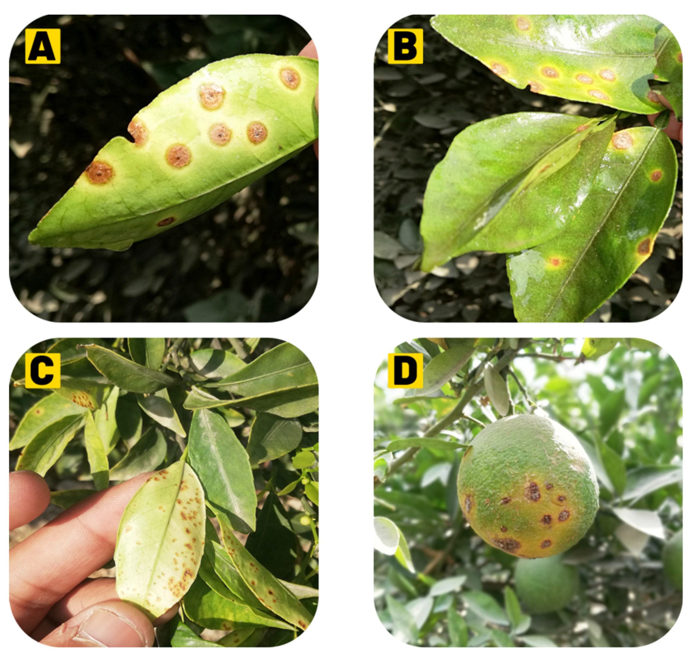
There are two types of South American canker strains which causes the same symptoms on the susceptible host as those produced by X. citri subsp. citri A strains, but all strains of South America have narrow host ranges [47]. X. citri subsp. aurantifolii B strain, also referred to as false canker or cancrosis B, has a more restricted host range and is found to primarily infect lemons and limes [32]. The Bstrain (XAUB as an acronym, XAC pathotype B; XAC-B) first appeared in Argentina in 1923 and it eventually extended to nearby Uruguay and Paraguay [32,33]. The B strain generally causes severe infections on lemon fruits (Citrus limon), limes (C. aurantiifolia), sour oranges (C. aurantium), but seldom on sweet oranges (C. sinensis) and pummelo fruit (C. maxima). Moreover, this strain does not infect grapefruit (C. paradisi) [30,32]. Hence, the Bstrain is not present in nature longer. Mexican lime cancrosis, CBC-C (XACtype C; XAC-C) or the Cstrain (XAUC as an acronym) was discovered in 1963 and present only in São Paulo, Brazil, where it just infects the Mexican lime [48]. The B and C strains are currently classified as X. axonopodis subsp. aurantifolii and produce many similar symptoms on the host as produced by the canker A strain [49,50]. The strains XAC, XAUB, and XAUC were compared and analyzed phenotypically and phylogenetically; all three strains were shown to have polar flagella with noticeable motility when cultured in semi-solid media [51]. In the presence of maltose and aspartic acid, only XAC can grow and hydrolyze pectate and gel [52]. Polyclonal antisera were prepared against XAC, but XAUB and XAUC showed little or no affinity to antisera. In contrast, XAC is susceptible to CP1 and CP2 bacteriophages, and XAUB and XAUC are not affected by these bacteriophages [53]. It was observed in culture media that XAUB has fastidious growth; XAC and XAUC both grow well in nutrient-agar (NA) and tryptophan–sucrose agar media. Moreover, these three strains show good growth in glutamic-acid rich media. A molecular analysis confirmed that XAUB and XAUC are more interlinked with one another than XAC [54,55,56]. In contrast, data obtained from physiological tests, i.e., phage-typing [57] total protein profiles after SDS-PAGE, DNA–DNA solution hybridizations [56,58], plasmid–DNA fingerprints [59], plasmid-based hybridization probes [59], PCR assay [60], DNA fragment sequence of gene hrp, restriction enzymes to analyze DNA fragments [61] confirm these conclusions. Furthermore, a gene required to cause CBC symptoms is the pthA gene, which was obtained from the XAC-A strain [62,63]. Total DNA hybridization with a pthA fragment revealed several profiles among XAC-A, XAUB, and XAUC; no hybridization with strains of X. axonopodis pv. citrumelo was observed [10].
Provisionally, two more CC strains were classified, named D and E strains, but now they do not exist or are categorized differently [64]. The D strain, which is also referred to as bacteriosis, induces disease in limes in Colima (Mexico), but its etiology is not confirmed yet [65,66]. This disease causes typical leaves and twig lesions, but no symptoms are observed on key Mexican lime fruit [67]. In this area, the suspected citrus pathogen no longer exists. It is now believed that the disease was caused by Alternaria limicola [68,69]. The second pathogen was the E strain, previously identified as a citrus canker in a Florida nursery. The disease is ‘called’ a bacterial spot of citrus produced by X. axonopodis subsp. citrumelo [34,50,70]. It can be distinguished based on these studies that three groups of X. axonopodis strains, i.e., A strain, B strain (including C and D strains), cause citrus cankers [50] and, notably, these strains have controversial taxonomy [71]. This interpretation was confirmed when the Xanthomonas genus was reclassified based on DNA–DNA hybridization and metabolic activity studies [7,8,10]. Moreover, Xanthomonas, causing diseases on citrus, was transferred from X. campestris to X. axonopodisspecies. Perhaps now, pathotype A, pathotypes B and C, and the CBS strains of X. axonopodis, are named X. axonopodis subsp. citri, X. axonopodis subsp. aurantifolii, and X axonopodis subsp. citrumelo, respectively, but the subcommittee on the taxonomy of plant pathogenic bacteria did not support this proposal [7,8,71].

4. Symptomatology

Canker symptoms are observed in all aerial parts of the plant [72]. They are characterized mainly by the formation of erumpent, corky, and raised pustules on the surface of leaves, fruits, and twigs, which serve as sources of bacterial inoculums [72]. Defoliation and fruit drop are also observed as plant responses to the infection [73,74]. Notably, Xanthomonas citri can survive in such plant debris for two months [75]. Severe symptoms are produced on trifoliate oranges, grapefruit, Mexican lime, and some sweet oranges; however, the actual host range depends primarily on a strain of citrus canker [74]. Generally, the susceptibility of young tissues to the citrus canker is more than mature tissues as there is a period of vulnerability in each flush around three times a year [76] (Figure 2).

4.1. Leaf Lesions

CC bacterium naturally penetrates the host tissues through stomata [77], hydathodes, lenticels, or wounds [78]. Citrus canker disease symptoms first appear as tan, brown, or grey-oily circular lesions, 2 to 10 mm in size, depending on the susceptibility of the host, the number of cycles of the infection, and optimal environmental conditions, i.e., the presence of water film and 20 to 30 °C temperature; canker protrudes from both surfaces of leaf tissue around 4–7 days after inoculation [78]. Symptoms might appear after more than 60 days under optimum conditions [79,80]. As the disease advances, host cell expansion (hypertrophy) and cell division (hyperplasia) occur, due to which the lesions become visible from small water-soaked spots and are surrounded by a yellow halo, which turns into slightly raised blister-like lesions and can be viewed with transmitted light [74,80]. The hyperplastic mesophyll tissue is an essential diagnostic symptom of the disease characterized by the formation of the canker due to rupturing the epidermis [78] and it releases abundant X. citri subsp. citri on the leaves. These lesions are elevated, are ‘corky’ in leaves, stems, and fruits, and then become dark and thick into the distinctive citrus canker under dry conditions [73]. A wound on the leaves or fruits or an injury by the Asiatic citrus leaf miner (Phyllocnistis citrella) significantly increases symptom severity [81,82,83].

4.2. Fruit Lesions

Fruits are susceptible for 90–120 days when they grow between 2.0 and 6.0 mm in diameter, depending on citrus species [77]. The lesions in the early stages look similar to large oily glands on the peel and become progressively dark and corky in texture, usually circular, and may occur individually or in groups, leading to premature fruit fall [84].

4.3. Twig Lesions

Twig lesions generally occur when leaves and fruits pass through one or more cycles of infection. Similar symptoms are produced on both twigs and fruits; twig lesions are not surrounded by chlorosis (but fruit lesions do) [24]. Citrus canker is endemic, the inoculum spreads by twig lesions on young shoots, and X. axonopodis subsp. citri. survival is prolonged in these areas; lesions with raised corky patches may persist for many years until girdling infections do not kill the twigs [75]. The highest susceptibility of citrus to X. axonopodis subsp. citri infection is during the last half of the growth development phase in all of the above ground citrus tissues [85]. Lesion incidence is seasonal, but sometimes severe precipitation and high temperatures coincide with periods of flush growth [84]. As leaves, stems, and fruits are fully grown, they become resistant to infection; once leaves are expanded between 50 and 80%, they become most susceptible [86]. New flushes, tender leaves, and stems are more likely to be vulnerable to citrus cankers than fully grown citrus [85]. When a pathogen severely attacks the host, it leads to defoliation, dieback, early fruit drop, and tree decline; hence, infected fruit is less valuable or unmarketable [87,88].

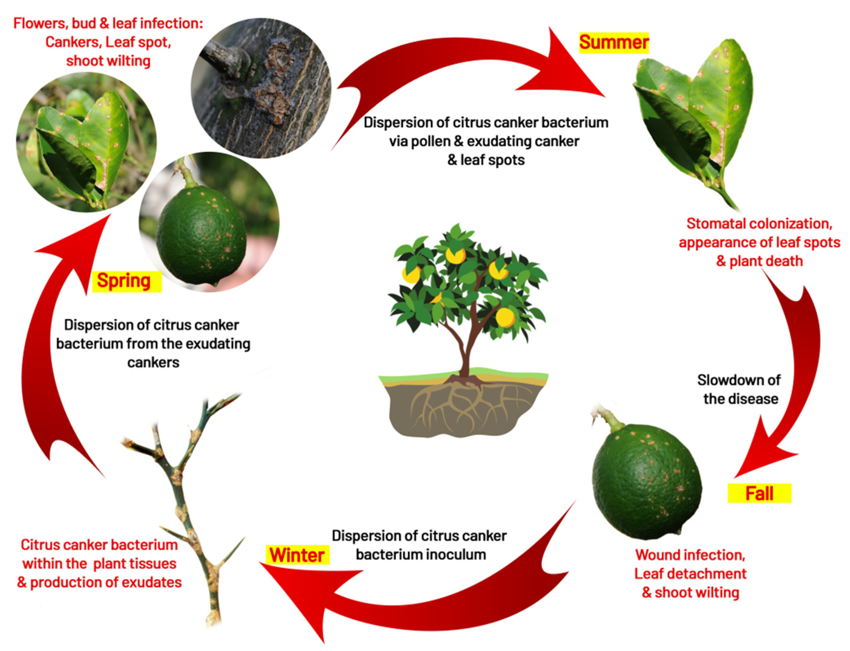
5. Disease Cycle and Epidemiology
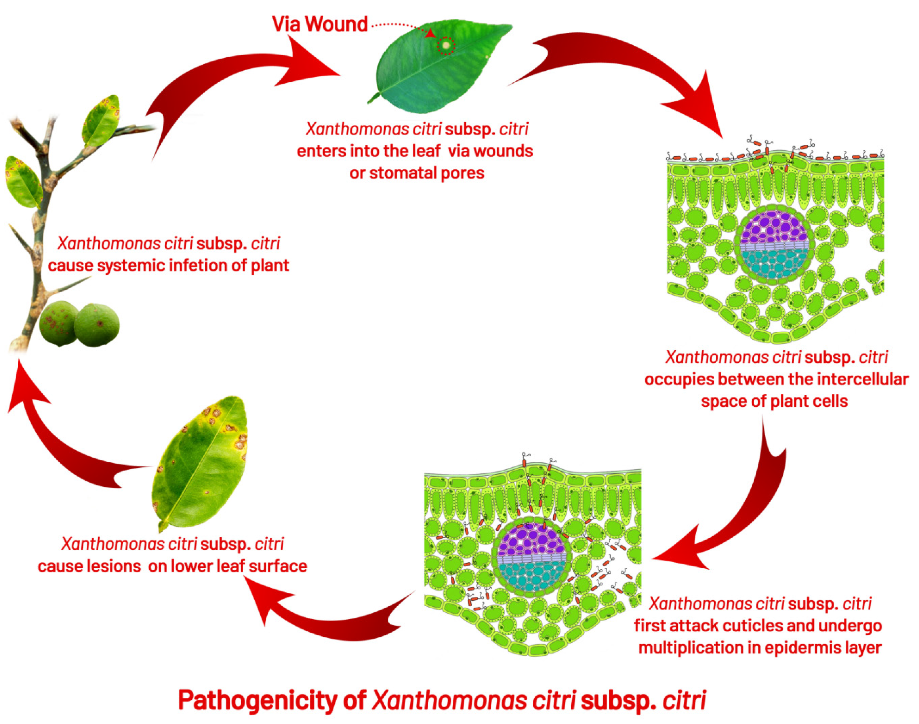
5.1. Infection

The bacteria penetrate the host by disrupting the leaf epidermis, inducing cell hyperplasia, and colonizing the apoplast [89]. Under optimum conditions, the pathogen multiplies 3 to 4 log units per lesion; for further disease development, bacterial cells may emerge from stomata openings to provide inoculum within five days [75]. For successful infection and lesion formation, free moisture for 20 min is required for the bacterial cells to ooze out from the lesion. As a result of water congestion, one to two bacterial cells are released from stomatal openings during inoculation [77,90]. After the initiation of growth of the host, almost all infections take place on stems and leaves within the first six weeks, while the first 90 days after petals falling is the most crucial time period for fruit infection [90]. Small and unnoticeable pustules are formed due to infections after this time period [10]. It has been reported that fruits are more susceptible to disease than leaves; hence, observations have been made that lesions of different sizes can be found on the rind of the same fruit during the infection of the bacterium [91] (Figure 3).

5.2. Survival

The main inoculum sources are branches, leaves, and twigs infected with cankers [84]. The disease is primarily carried in cankers on twigs and branches from one season to another, serving as a primary inoculum [10]. In leaf and fruit lesions, the bacteria remain alive until they fall; because the affected leaves fall off early, they may not act as the primary inoculum source [75]. Still, it was reported that, in infected leaves, the bacterium survived up to six months [92]. Reference [93] found that the bacterium survived for over 6months in the infected leaves, for 52 days in sterilized soil, and only 9 days in unsterilized soils, respectively. It was also observed that the organism could survive for 11–12 days under desiccation at 30 °C [93]. On citrus hosts, the bacterium survives epiphytically with a lower population without developing the symptoms, combined with non-citrus weeds, grass host, and soil [94,95,96]. However, in the absence of plant tissue or debris, the saprophytic existence of soil pathogens has not been observed [93,95]. The survival capability of the pathogen in subtropical soil is very limited [97], and bacterial inoculum dies within 24 to 72 h on different inert surfaces, such as cloths, metals, plastics, and processed wood in both sunlight and shade [91]. Due to antagonisms and competition with saprophytic microorganisms, the bacterial population decreases to an undetectable level 1–2 months after leaves or fruits fall to the ground [91]. In Japan and Brazil, it has been reported that X. axonopodis subsp. citri may survive on non-host plant material and in the root zone of certain weeds under eradicated diseased trees for a few weeks [98] (Figure 4).

5.3. Dispersal

Under natural conditions, rainfall splashes and rainfall associated with wind are reasons for the short-distance spread of disease. Still, dissemination to long-distances between geographical regions is mainly expected to occur through infected plant material [24]. During severe storms, such as tornadoes, disease spread takes up to 10 to 15 km [90]. Dispersal of diseases have been further explored with model-based data on wind direction and threshold parameters, which showed wind speed eight m/s and rainfall of nearly 0.32 cm/h helped the insects, such as P. citrella, and blowing sand penetrates the bacteria through stomatal pores or injuries caused by thorns [99]. The major reason for the dispersal of bacteria to average distance is the wind-driven rain. In Argentina, the wind-blown rain dispersed the bacteria from infected trees up to a distance of 32 m [100]. A drop of rainwater may contain up to 105 to 108 cfu/mL of bacteria [80,100]. Under globalization, the transmission of bacteria from one region to another through frequent communication and transportation increases the risk of infecting citrus farming in free areas of disease [101] (Figure 5).

5.4. Role of Insect (Leaf Miner Interaction)

The leaf miner plays an important role in the spread of citrus canker, but it has not yet been reported as a disease vector [81]. In earlier 1994, the distribution of citrus leaf miners was restricted to southeast and southwest Asia, while it spread after the mid-1990s to most of the world’s major citrus areas [81]. It was first reported in Florida and Brazil in 1993 and 1996 [102]. The feeding activities of the citrus leaf miner provide bacterial infections to the host in three ways: (1) wind-blown rain disseminates the bacteria, contacts the surface of the leaf; the leaf miner tears the cuticle and opens the mesophyll of the leaf, providing direct bacterial infection; (2) the leaf miner injuries are cured more slowly than mechanical injuries, which allow for longer exposure to bacterial infections; (3) leaf miner larvae may become contaminated with bacteria and carry it to the feeding galleries, where feeding activities lead to an increase in mesophyll cells infection [99,103]. Trees with leaf miner injuries remain susceptible for 7–14 days, compared with only 24 h for wind, thorns, or pruning injuries [104]. The prevalence of citrus canker increases in Brazil and Florida due to leaf miner injuries [74,90,104]. Still, it is believed that the host can tolerate some loss of leaf area without yield being affected because of leaf miner damage (up to 10%) [105]; there are reports that loss of 16–23% of leaf areas can lead to significant yield loss [106].

6. Detection and Identification of Citrus Bacterial Canker

The diagnosis of CC can be made using various methods and in most circumstances; however, when no official confirmation is required, the disease diagnosis can be made by recognizing common symptoms [8,107]. It is also possible to confirm the causal agent by isolating XCC from lesions on a solid medium and looking for xanthomonad-like colonies, which are yellow, convex, circular, semi-translucent, and have regular edges [52]. Infiltration of a bacterial suspension adjusted to 108 colony-forming units (CFU)/mL into the leaf mesophyll, followed by observation of water soaking and raised margins in the infiltrated portion of the leaf 2–4 days after inoculation, can be used to test pathogenicity in susceptible citrus species [76,107]. DNA-based assays and serological testing are routinely utilized methods for CC diagnosis when symptoms are atypical or an official confirmation is required for quarantine purposes [107]. Although molecular approaches can identify the presence of XCC in infected plant tissue before canker lesions occur, serology-based assays are usually sufficient for detecting XCC in symptomatic tissue [8]. Several primers based on rDNA sequences, plasmid-borne genes, and pathogenicity regulatory factors have been devised for polymerase chain reaction (PCR) detection of XCC [54,108,109,110,111]. In recent years, the introduction of real-time PCR and loop-mediated isothermal amplification procedures has improved the accuracy of diagnostic testing for XCC [11,112,113,114,115]. All existing conventional PCR methods need gel-visualization or primers, but all strains are not detected [54,90,116]. PCR primers are very effective for X. citri subsp. citri ‘A type’ detection, but these primers do not show consistency in X. citri subsp. Aurantifolii ‘B’ or ‘C’ strain detections [60,117]. New PCR primers based on the gene-sequence of pthA did not even detect one canker strain [54]. This technique, based on rep-PCR with BOX and ERIC primers, was developed to separate and distinguish the CBC pathotypes worldwide and the subgroups of pathotypes of citrus associated within specific geographical areas around the world [54,118]. Rapid, sensitive, and reliable real-time PCR assays were developed along with designed primers to detect all citrus–canker strains, which are important for both specificity and sensitivity [75,110]. Real-time PCR is easier to perform, less labor-intensive (no need for agarose gels), and much faster than conventional PCR [110]. If the sampling method is performed accurately, exact results can be obtained within 1 h. For plant pathogen detection, real-time PCR is becoming increasingly useful, i.e., for fungi [111,119,120,121,122,123,124,125,126,127,128,129,130,131,132,133,134,135,136,137,138,139,140,141,142,143,144,145,146,147,148,149,150,151,152,153,154,155,156,157,158,159,160,161,162,163,164,165,166,167,168,169,170,171,172,173,174,175,176,177,178,179,180,181,182,183,184,185,186,187,188,189,190,191,192,193,194,195,196,197,198,199,200,201,202,203,204,205,206,207,208,209,210,211,212,213,214,215,216,217,218,219,220], bacteria [122,123,124], and viruses [125,126]. A reliable, sensitive, SYBR Green real-time PCR assay in which primers are used to amplify conserved regions of a desired gene of pathogenicity to detect all known strains of XAC is based on sampling techniques conducted in the field samples [127]. The detection of the bacterium through PCR is based on an internal standard to make sure the quality of the DNA template for the reaction and ratio of PCR products is used to evaluate the early concentration of bacteria in citrus leaf tissues with lesions using internal standards and target pathogens [118]. Detection and comparison of X. axonopodis subsp. citri (XAC) from imported citrus fruits was based on an integrated approach involving isolation of bacteria from three conventional protocols viz., PCR, real-time PCR with SYBR-green, or a TaqMan-probe in canker lesions and LAMP [8,116,117]. The real-time PCR for fresh fruit samples with a TaqMan probe is the fastest screening method for the detection of bacteria [111]. Enzyme-linked immunosorbent assay (ELISA), or serological tests, have also proved to be useful for rapid detection of XCC, which are based on the ability of an antibody to recognize and bind to a specific antigen [111]. These tests are usually performed in the laboratory. Still, they are also available as strip-based kits that are easy to use in the field where the disease is suspected; these kits do not require special equipment or training, and the results are obtained within a few minutes [111,128]. Other older techniques for the detection of XCC have been developed, including physiological characterization, fatty acid profile analyses, protein profiling, hybridization, restriction fragment length polymorphism analysis, and comparison of plasmid DNA patterns [8,10,111,128] (Table 2).

7. Genome

The sequence of the genome of X. axonopodis subsp. citri (306-strain) has been completed [132]. The bacteria have 5,175,554 bp and two plasmids that are pXAC33 (33,699 bp) and pXAC64 (64,920 bp) with a rounded chromosome [132]. The comparison shows a high degree of resemblance (about 80%) between X. campestris pv. campestris (pathogens of crucifers) genome and X. axonopodis subsp. citri genome, where both contain many genes specific to their sequenced strains [132]. The specificity of the host and variation in pathogenesis processes can be explained through these genes [7,132]. Genomics studies of CC bacterium have significantly opened new avenues for better understanding of XAC pathogenicity and virulence [7]. Several molecular methods for analyzing the population structure of a few plant pathogens have been utilized, e.g., RAPD, Rep, Eric-PCR, RFLP, plasmid profile analysis, PCR amplification, SDS-PAGE, 16S rDNA sequence [16,133,134,136,137,138,139,140]. There are 4314 projected open reading frames (ORFs) on the single circular chromosome, which has a G1C content of 64.7% [141]. The G1C content of the two plasmids, pXAC64 (64,920 bp) and pXAC33 (33,699 bp), is 61.4 and 61.9%, respectively [132,141]. Pathogenicity, virulence, and ecological adaptation are involved in about 7% of XCC genes [141]. The XCC-A genome contains a large number of cell wall-degrading enzymes (CWDEs), proteases, iron receptors, genes related to energy metabolism pathways, the type 2 secretion system (T2SS), type 3 secretion system (T3SS) genes for flagella structural units, chemotactic protein genes, the xanthomonadin, and xanthan gum synthesis gene cluster (gumB to gumM), which are important in the epiphytic phase of the life cycle [142]. The 23-kb hrp (for hypersensitive reaction and pathogenicity) region has six operons, designated as hrpA to hrpF [143]. The hrp cluster is part of a pathogenicity island in the XCC genome and encodes the T3SS. The genes hrpG and hrpX are involved in the regulation of amino acid biosynthesis, oxidative phosphorylation, pentose–phosphate pathway, phenolic catabolism, and transport of sugar, iron, and potassium in response to exposure to the host environment [144], while an additional 124 and 90 unknown genes are regulated by hrpG and hrpX, respectively [144]. HrpG induces the expression of 11 proteins secreted by the T2SS and is a regulator of the T3SS [145,146]. In 2010, several XCC-A, XCC-A* (produces canker lesions in Mexican lime but not in grapefruit; Ref [147], XCC-Aw and X. citri pv. Bilvae, which causes CC-like symptoms in key lime) strains were characterized by amplified fragment length polymorphism (AFLP) and MLSA based on four partial housekeeping gene sequences (atpD, dnaK, efp, and gyrB) [147]. The study was performed on 157 XCC strains from Brazil, which were compared for their T3SS effector profiles using a qualitative PCR Southern blot technique [148]. Low genetic variability was observed for strains isolated in the northern part of the country, but more diversity was present in the strains isolated in the southern part [148]. The host plant genes, where products recognize pathogen effectors, are known as R genes, and the pathogen pathogenicity(pth) genes, which encode these recognizable effectors, are also referred to as avirulence (avr) genes [148]. The products of R genes, either directly or indirectly, interact with the products of avr genes; interaction between products of Rand avr genes is termed gene-for-gene resistance [149] whereas if no R genes correspond to XCC, then it leads imparting resistance to XCC host plant, which has been identified in citrus and citrus-related species [7,8,45].

8. Virulence

8.1. Type III Secretion System (T3SS)

Hypersensitive response and pathogenicity (hrp) of CC have a cluster of 24 genes on locus arranged in six operons from hrpA to hrpF, which are regulated by hrpGhrpX genes and codes T3SS in X. citri subsp. citri [144,150]. It was observed that XAC remained unsuccessful at inducing disease and HR in the cotton plant by the deletion of hrpB, hrpD, and hrpF operons [151]. This system was presumed to secrete the effector proteins [152,153], and pthA, the member of the avrBs3 gene family, targets the host susceptible gene lateral boundaries of organ 1 [154]. XAC uses T3SS against the host by injecting virulence proteins into host plant cells required for canker development in susceptible citrus plants and resistant plants, developing a hypersensitive response (HR) [151,155]. Effectors (virulence protein) are transported through specialized T3SS and use a hollow exterior (Hrp-pilus) that crosses the plant cell wall, delivering effectors across the plant plasma membrane through a translocon [156]. Xanthomonas strains usually harbor about 30 various effectors but their molecular activity is still unknown in most cases [156]. Transcription-activator-like (TAL) effectors form a large and important family of effectors found almost exclusively in Xanthomonas [157]. They act as transcription factors for plant genes and few induce expression of sugar exporters [13].

8.2. Citrus Specific pthA and Its Requirement for Canker Development

The first member of the gene family avrBs3/pthA is the pthA gene necessary for pathogenicity and was cloned through screening for pathogenicity [62,63,98]. Genes of avrBs3 are broadly spread in the genus Xanthomonas, but are not present in all Xanthomonads [63]; there are a minimum of 27 cloned members of the avrBs3 family [158]. Without evidence of the pth function, mostly avrBs3 members are isolated as avr genes for the first time while all genes pthA, pthB, pthC, and pthW of this family induce the citrus canker, and two genes, pthN and pthN2, are involved in the induction of cotton blight [159]. Intensive studies have been reported on the molecular virulence mechanism of the pathogen for th citrus canker [45,160]. Gene pthA, an effector of the type III secretion system (T3SS), is a determinant of cancer pathogenicity and is commonly found in Xanthomonas spp., which causes citrus canker [54,63,98]. The pthA gene’s exogenous insertion into X. axonopodis subsp. citrumelocauses bacterial citrus spot disease without causing erumpent lesions [62]. The transient expression of pthA produces citrus canker symptoms, causing cell hyperplasia, hypertrophy, and eventually, the plant dies [89]. In addition, pthB and pthC are functionally homologous genes that were cloned from X. citri subsp. aurantifolii B and C, and are important in inducing citrus canker [161]. Therefore, all three genes are functionally exchangeable and can be transmitted horizontally between strains of X. citri and X. campestris subsp. aurantifolii on plasmids [162]. Members of the pthA gene family are more than 3.8 kb in length and possess a high level of identity with DNA sequences (more than 90%) over their total length [138]. It seems that functionally homologous genes based on DNA hybridizations were found in all canker-causing strains and have not been found in isolated citrus strains that do not induce cankers, e.g., X. campestris subsp. citrumelo [55,161]; therefore, the single common gene was needed to induce citrus canker by Xanthomonas [45], while the XAC genome possessed several genes coding putative effectors [62,162]. The pthA is the most important effector that induces canker-like symptoms even in the absence of a pathogen when expressed transiently in plants [89,98]. Transient expression of pthA in host plant cells was sufficient at inducing symptoms of citrus canker in 10–14 days and pthA’s deletion eliminates the pathogen to cause citrus canker [154]. In all citrus species, pthA induces canker symptoms, but in other plant species, it changes the plant to immune; hence, this characteristic makes the XAC specific toward the citrus species [53,62,63]. The pthA mutation prevents XAC from inducing hyperplasia, hypertrophy, water soaking symptoms, and pathogen losses of the ability to grow within plants [62,63]. All strains of X. citri subsp. citri group A were examined, which has three pthA alleles in addition to pthA; two of them are slightly functional to produce cankers in citrus, and one seems non-functional [63,163]. In the case of host recognition or avoidance, multiple homologs seem to provide rapid development of new genes of pathogenicity through recombination [164]. X. axonopodis subsp. citri have three homologs of avrBs3/pthA while only ap11 homologue was reported to participate in virulence to induce canker formation, while the functions of other homologous were insignificant or not assessable [163]. The pthA is transported through T3SS, which consists of hrp gene cluster products, and the transcription of hrp genes is induced and regulated by the hrpG and hrpX regulators [144,155,165,166]. The hrpG or hrpX gene mutations in X. citrus subsp. citri led to a loss of pathogenicity in citrus [144,167]. Other virulence-related genes are required for X. citri subsp. citri, in addition to the pthA and hrp genes, to cause disease [45], e.g., it was reported that opsX gene plays an important role in the production of lipopolysaccharides (LPS) and extracellular polysaccharides (EPS), growth, and virulence in planta [168].

8.3. Adhesion and Extracellular Polysaccharides (EPSs)

Extracellular polysaccharides (EPSs) and lipopolysaccharides (LPSs) defend the bacterium from unfavorable environmental conditions [169]. Xanthomonas spp. Produce characteristic EPS and xanthan, which results in bacterial colonies being mucoid; it is known as xanthan gum [170]. Xanthan is a polymer of repetitive units of pentasaccharide, having a backbone of side chains of cellulose and trisaccharide, used in the nutritional and pharmaceutical industries commercially as a thickening agent [171,172]. The xanthan production is managed by various genetic loci, including the 12 gum gene clusters from Gum B to Gum M that are highly maintained among Xanthomonas spp. [172,173]. Xanthan production in Xanthomonas is regulated hierarchically by the gene cluster regulation of pathogenicity factors (rpf) [174]. Host plants wilted due to infections caused by vascular pathogens that obstructed the flow of water in xylem vessels due to xanthan production [175,176]. A plethora of research depicted various gum genes of Xanthomonas spp. e.g., X. axonopodis subsp. citri, X. axonopodis subsp. manihotis, X. campestris subsp. campestris, and X. oryzae pv. oryzae has been involved in epiphytic survival and the development of bacteria in plants to induce disease symptoms [172,177,178,179,180,181]. It is interesting that gum genes from X. axonopodis subsp. citri are not essential in Citrus sinensis for disease development and growth of bacteria. Still, in Citrus limon, these gum genes play an important role in bacterial virulence, showing that xanthan virulence depends on the host plant and environmental conditions [177,178]. The rpf protein has been involved in DSF synthesis to regulate the genes and determine the synthesis of extracellular polysaccharides [183]. In many xanthomonads, the gum gene cluster is involved in EPS biosynthesis. The Gum B mutant showed defective EPS production and biofilms formation, hence, decreased infection in lemon [13,184].
XAC produces abundant extracellular polysaccharides (EPS) [184]. Bacterial cells are incorporated in a dense matrix of EPS of canker lesions and are disseminated with EPS through rain [13]. The EPS molecules effectively protect the bacteria in water from the ‘dilution effect’ and desiccation in air, hence playing an important role in bacterial ecology [185]. Bacteria enter the cell through stomata or wounds and remain stick to the host’s cell wall through an interaction between EPS and citrus agglutinins [186]. Experimental evidence suggests that basal plant defense responses are always suppressed by xanthan, e.g., deposition of callose in the plant cell wall. The chelating divalent calcium ions in the apoplast of the plant are required to activate plant defense responses [187,188]. Further, xanthan plays an important role in biofilms formation in X. axonopodis subsp. citri, and X. campestris subsp. campestris [177,189]. Microbes create a biofilm matrix, made up of proteins, extracellular DNA, and polysaccharides, essential for the establishment of bacterial colonies while polysaccharide overproduction has been shown to alter colony shape and help to identify certain species [190]. The matrix’s polysaccharide component can give a variety of benefits to the biofilm’s cells, including adhesion, protection, and structure, and aggregative polysaccharides operate as molecular glue, allowing bacteria to stick to both biotic and abiotic surfaces by allowing them to withstand physical stresses, such as fluid movement that might detach them from a nutrition source [191,192,193].

8.4. Lipopolysaccharides (LPSs)

In plant pathogenic bacteria, the major virulence factor responsible for host infectivity is lipopolysaccharides and it is increasingly recognized as a major plant pathogen-associated molecular pattern (PAMP) [98,194]. LPSs are major components and characteristic structures of the outer membrane of Gram-negative bacteria [194]. LPS molecules are usually composed of hydrophilic heteropolysaccharides formed by three major sub-structures, the O-specific polysaccharide (O-antigen), a repetitive sugar sub-unit; the core oligosaccharide region, which is covalently connected to the lipid A of the glycolipid moiety, and the lipid ‘A’ attached to the external plasma membrane [195,196].
LPSs have been recognized as a virulence factor during plant pathogenic interactions and involved in bacterial pathogenicity [197]. LPSs are present in bacteria’s outer membrane, which protects the pathogen from hostile medium found within plant tissues, reduces the membrane permeability, and allows the bacteria to grow under unfavorable environmental conditions [197]. LPSs can prevent hypersensitive response (HR) in plants by avirulent bacteria that have been widely studied in plant cells [198]. In various Xanthomonas spp., insilico analyses have been carried out to identify and characterize the genes involved in LPS biosynthesis, showing wxacO and rfbC genes involved in LPS biosynthesis that reduces the pathogen motility, lack of resistance to stress, and virulence in grapefruit [199,200]. Further, the two-component regulatory system (TCRS) ColR/ColS has been reported to play multiple roles in LPSs and catalase production, biofilm formation, resistance to stress, transcription of hrpD6 and hpaF genes, and knockout of either colR and ColS, causing loss of pathogenicity in grapefruit [13].

8.5. Quorum Sensing

Virulence factors involve pathogen’s ability to express their genetic, biochemical, and structural features to the host [13]. Quorum sensing (QS) is a mechanism that facilitates communication among bacterial cells through production and detection of signal molecules [13,201,202]. Xanthomonas has QS regulatory systems facilitated by molecules of the diffusible signal factor (DSF) family, which regulates the QS pathway of Xanthomonas spp. comprising three major QS regulons: RpfF, RpfC, and RpfG [203,204,205]. Furthermore, transcriptome analysis characterized RpfF, RpfC, and RpfG regulons, which showed RpfF controls the unique genes responsible for the QS-pathway complexity and other sensory mechanisms involved in canker development [204]. RpfC and RpfG control individual genes that play a wider role in gene regulation and their involvement in chemotaxis, motility and flagellate biosynthesis, extracellular enzymes production, adhesion, stress tolerance and regulations, transport, and transport detoxification [206]. The QS process depends on the production, release, and detection of small signaling molecules known as autoinducers (AIs) [206]. The MJ Daniels Research Group, for the first time, reported the detection of activity of DSF molecules as autoinducers [174]. The synthesis of DSF in X. axonopodis subsp. citri is based on genes rpfF and rpfB, and it is an autoinducer in bacteria and regulates quorum sensing [203]. The Rpf/DSF system involves the initial attachment of XAC to the host and controls a range of virulence-related characteristics, such as the synthesis of extracellular enzymes (proteases, endoglucanases), extracellular polysaccharides, EPSs biosynthesis, and biofilm formation in several strains of pathogens [13].

9. Nutrition

Bacteria can take nutrients from their hosts through enzyme secretions that degrade the host’s cell wall and bacteria utilize the cell wall’s breakdown products as nutrition sources [62]. In XAC genome pectin esterases are not present, but bacteria possess three pectate lyases, six cellulases, five xylanases, and an endoglucanase, while in cellulose, it contains endoglucanaseBcsZ (gi|22001634),which hydrolyzes 1,4-β-D-glucosidic linkages [62]; moreover, permease, through which degraded pectin products are imported into the bacterial cells in a hydrogen-transport coupled fashion [62]. The pthA is also necessary for optimal growth of the bacteria in the host, which is probably directly injected into the host cell [62]. X. citri subsp. citri produces less enzymes as compared to X. campestris subsp. campestris, which degrades the cell wall; because of this, the two pathogens may cause their hosts to suffer from different symptoms [132].

10. Integrated Management Programs

IDM has been introduced to control the occurrence of citrus bacterial canker disease (CBCD) in new seedlings [9]. This program recommends that only citrus bacterial canker disease-resistant cultivars may be planted [107]. For commercial cultivation, it is recommended to plant sweet oranges, such as Tahiti lime, Pera pre-immunized, FolhaMurcha, Valencia, and Navelina, mandarins, such as Ponkan, Dancy, Loose Jacket, Satsuma, Batangas, and Willowleaf accessions [9]. Many studies have been carried out to control CC disease through cultural, chemical, or biological management strategies but have shown limited effects [207,208]. When leaf miners attack citrus varieties, or under changing weather conditions, the development of disease becomes far more complex and harder to control the eradication of diseased trees is the only way to control the CC disease [7,9,11]. Resistance genotypes in citruses and relative genera have long been researched around the world, and several types of citrus germplasms with certain resistance levels have been reported, e.g., calamondin (C. mitis) and kumquats (Fortunella spp.) are highly resistant, and mandarins (C. reticulata) were also reported to be resistant [78,162,209]. Suppose plants are inoculated artificially with the bacterium or planted in combination with sweet oranges. In that case, all plants show characteristic symptoms of citrus cankers without any complete or active resistant citrus genotypes [7,9]. As no resistant varieties were identified, breeding efforts have made little progress in the production of resistant cultivars, and few experiments in molecular breeding have shown transformants of some resistance through the transfer of antibacterial genes to citrus fruits. Still, only a lower disease incidence has been achieved without complete resistance [8,9]. The molecular mechanism of pathogenesis remains unclear, and there are no resistance genes isolated; it is very tough to obtain resistant genotypes through breeding programs [210,211]. The resistant mandarin varieties are grown in Southeast Asia, where the climate is most favorable for epidemics; the citrus canker was not a major issue until more vulnerable sweet oranges were brought into the disease regions of China and Japan [78]. Since the 1950s, eradication/control programs have been established in São Paulo, Brazil, to prevent the pathogen spread in the production area of sweet oranges [9]. Contrarily, the nearby zones of Paraná State, Brazil, Misiones, Corrientes, and Argentina have adopted integrated program strategies to efficiently prevent and control citrus cankers in sweet oranges [212,213,214]. The program is mainly concerned with shifting of citrus plantations to the disease-free area, having resistant citrus varieties, and in these regions, regulations are there, not only to deal with more resistant varieties, but also to produce disease-free nursery trees, as well as other means to exclude the XAC from citrus plantations [212]. The regulations for the management of CC disease include (i) nurseries must be situated in disease-free areas; (ii) the design of citrus production areas must be managed in order to decrease the danger of an epidemic of CC by constructing windbreaks, applying preventive copper sprays, building fences to avoid the entrance of bacteria to the citrus plantation; (iii) the planting and harvesting tools should be disinfected; workers should also disinfect their clothes, shoes, and gloves; (iv) fresh fruits should be strictly inspected for domestic and export markets to prevent the fruits in citrus groves from citrus cankers; workers should also disinfect the storage and packaging houses; (v) infected summer and autumn shoots should be pruned; (vi) disease management forecasts should be considered; (vii) control of citrus leaf miners; (viii) use of chemical inducers, which induce systemic acquired resistance (SAR) in plants [215,216].

10.1. Quarantines

Federal quarantine barriers are regulatory responses to diseases, which could be found in almost any country. Still, the exact locations of such barriers are difficult issues, for biological and political reasons [9,24]. These barriers are usually placed two or more two miles away from any known infestation [24]. The distribution of host plant materials is limited within quarantine areas, affecting both the citrus agriculture sector and homeowners with citrus trees [9]. In commercial production, it is recommended to disinfect the fruits in packing houses and disinfect the harvesting and transport equipment [24]. Fresh fruits are often restricted in market distributions from regulated areas [7]. Commercial citrus planting needs sanitization stations at plantation doors, a caution that has become a national demand, even outside quarantine areas [7]. Citrus replanting in commercial or residential areas that have undergone eradication efforts is against the law before the disease is declared eliminated [7,9]. People are informed in residential areas—that it is illegal to transport fruits to neighbors and family; decontamination of all equipment that is moved between properties during lawn and garden services is required [7,9]. These measures are publicized through intensive media reporting and expertise in community relations [24].

10.2. Cultural Control

Eradication of any disease is the method used to manage that specific problem if it has not been endemic in a region [7]. Quarantine and eradication are key measures to control the entry and dissemination of pathogens in many countries [9]. Eradication measures involve destruction by cutting and burning citrus species [24]. Sometimes, herbicides are used instead of cutting and burning to kill citrus plants [18]. The infested property is quarantined, followed by the eradication procedure for at least a year without planting or propagation of citrus fruits, with inspection at least twice a year [24].
Data from Argentina showed that the pathogen could disseminate in rainfall with wind up to 32 m (105 ft), which provided the scientific basis for eradicating this disease [50]. This has been translated in the U.S. and many other countries into regulating policies, allowing survey teams to locate diseased citrus trees, to remove and kill the trees, as well as exposed trees within a radius of 38.1 m (125 ft) of a diseased tree [76,217]. Now, Brazil uses a distance of 30 m (98 ft) to remove exposed infected trees. If infections of the Brazilian plants are 0.50% or lower, all trees will be removed within a radius of 30 m of the infected plantation. When the infection exceeds 0.5%, the whole block will be removed [217]. New canker infections occur in known source trees, at about 1900 ft (579 m) [17]. In January 2000, a new regulation, “the 1900 ft rule”, was set up. In March 2000, it was implemented, which involved the eradication of infected citrus trees along with healthy trees within 1900-ft of an infected tree [17,218]. Each 1900 ft radius circle has a surface area of 1.06 km2 (0.41 miles), leading to the removal of dooryard citrus in infected areas by implementing the 1900 ft rule [9]. Pruning of infected twigs, along with application of a 1% Bordeaux mixture at regular intervals, before the onset of monsoon, also proved to be very effective in the management of disease [219,220,221,222,223].

10.3. Chemical Control

For the management of CC, it has been reported that every year, from November to December, pruning of infected twigs with three to four sprays of a 1% Bordeaux mixture could be used to reduce disease [224]. Control of disease by applying four sprays of 5000 ppm copper oxychloride or a 1% Bordeaux mixture and two prunings gave excellent results [225,226]. Chemicals such as Perenox, Ultrasulphur, and a mixture of Blitox + nickel chloride, sodium arsenate + copper sulphate, were used against citrus cankers [227,228,229,230]. The application of 1% glycerin spray and 500–1000 ppm of streptomycin–sulphate was found useful in controlling disease on acid lime [231]. Acid lime canker was reduced by six sprays of 1000 ppm streptomycin with two prunings [232]. Streptocycline, in combination with Bordeaux mixture, and Agrimycin, are effective antibiotics against CC [233]. For field tests with different chemicals, Paushamycin + Blitox and Bordeaux mixture showed the best control of CC [234]. In nurseries, treatments of young plants have been reported by applying neem cake solution on the leaves [235,236]. The application of streptocycline + copper oxychloride (0.1%), preferably at intervals of seven days or fifteen days, has been found very effective against CC [236]. Integrated application of copper oxychloride (0.3%), streptocycline (100 ppm), and suspension of neem cake on pruned infected twigs has shown to be very useful to control the disease [237].
In field experiments in Argentina, copper ammonium carbonate with 8% metallic copper was consistently found better than other products to control CC; regarding field trials on trees of ripened grapefruits, three applications of copper ammonium carbonate (CAC) or copper hydroxide + maneb per season were examined and reduced the number of lesions found on fruits but not on leaves [238]. The recommendation to add mancozeb to copper spray was effective for copper resistance [239]. Sanitary procedures have been explained for persons or tools that encounter citrus in quarantine regions, e.g., by applying sprayable ammonium detergent disinfectants [23].

10.4. Biological Control

There has been a surge in the hunt for more environmentally-friendly plant disease treatment methods [7]. Researchers are looking for more ecological approaches to manage phytopathogens in the field due to chemical residues in soils and water bodies and increased consumer concerns [7]. Some recent research used the antagonistic activity of microorganisms and plant-derived compounds to suppress citrus cankers [7]. Studies on biological control are still in the initial phases, to control CC [239,240]. Some bacterial strains, including Pseudomonas syringae, Erwinia herbicola, Bacillus subtilis, and Pseudomonas fluorescence isolated from citrus phylloplane have been found to be antagonistic to the citrus canker pathogen, invitro [133,241,242,243,244]. However, it appears hard to find antagonistic bacteria that stabilize on mature citrus tree leaves [245]. For example, P. aeruginosa LV produces a bioactive combination of secondary metabolites, the most important of which is an organocopper antibiotic that reduces the formation of citrus canker lesions in Valencia oranges by 90% [245]. The authors discovered a bacteriolytic impact on X. citri that was not accompanied by any signs of phytotoxicity [246]. At low micromolar concentrations, several secondary metabolites from P. aeruginosa also reduced canker formation [247,248]. However, because P. aeruginosa is an opportunistic human infection, its use as a biocontrol agent is fraught with dangers and must be strictly managed [9]. Bacillus spp. isolated from citrus rhizosphere and leaves have also been proposed as a citrus canker biocontrol agent because they inhibited X. citri growth in vitro and in field circumstances [76,240,249,250]. X. citri and several species of Pseudomonas and Bacillus suppressed growth and canker formation, and Citrobacter isolated from sweet orange phylloplane, by degrading the X. citri quorum sensing molecule DSF [251]. Other bacterial genera, such as Cronobacter and Enterobacter, similarly inhibited X. citri growth in vitro by producing bacteriocins; however, the protective effect of these bacterial species on X. citri-infected citrus trees was not assessed [252]. The sensitivity of bacteriophages (phages) can be utilized to identify intra-species sub-groups in bacteria [252]. Cp1 and Cp2 phages have long been employed to identify X. citri strains [253]. The employment of phages in biological control, on the other hand, poses significant difficulties [254]. Phages have a very short active life on the leaf surface and must be administered at high concentrations to be effective [255]. The authors of reference [256] found that the jumbo phage XacN1, originally isolated from an orange orchard soil in Japan, can infect a wide range of X. citri isolates, making it a promising choice for future field experiments. In greenhouse and field testing, a mixture of phages obtained from orange orchards combined with ASM successfully decreased canker symptoms [257]. Compared to copper–mancozeb alone, the combination of phages and copper–mancozeb did not improve citrus canker control [258]. Interestingly, filamentous integrative phages, such as XACF1, reduced the virulence of X. citri, suggesting that they could be used as citrus canker biocontrol agents [259].

10.5. Field Screening

Globally, field screening was carried out to assess the response of citrus varieties in local environmental conditions to CC [9,24]. Suppose very intensive control programs have been carried out. In that case, highly susceptible varieties, i.e., several early–mid-season sweet oranges, grapefruits, and Mexican limes (e.g., Navel, Hamlin), are not recommended for planting [212]. Mid and late season oranges, tangerines (tangerine, tangelo, and tangerine), and Tahiti limes have been identified with acceptable resistance to citrus cankers in screening programs [9]. These cultivars may be susceptible in the young stages and need to be sprayed to control the leaf miner to avoid damage to emerging flushes that predispose them to infection [9].

10.6. Induced Systemic Resistance

Induced systemic resistance (ISR) is an active resistance mechanism in plants activated through biotic or abiotic infection. The mechanism increases physical or chemical barriers of host plants against infection [260]. Different inducer compounds, e.g., salicylic acid, harpin protein, and benzothiadiazoles are used effectively to induce resistance in plants against diseases [261,262]. ISR mechanisms can simultaneously control the disease and decrease the risk of developing pathogen resistance [263]. ISR activity may be used early in the season to complement the protectant activity of Cu, which slows the growth of bacteria in rapidly growing leaves [263]. Chemicals sold for the treatment of citrus canker include ‘Actigard’, approved for acibenzolar-S-methyl, a benzothiadiazole in the USA, and ‘Bion’ in Europe and South-America (Syngenta Crop Protection); ‘Messenger’ (Eden Bioscience) is approved for the harpin protein (a hrp-gene product) [63]. In Florida, several ISR inducers (e.g., Messenger, Nutri-phite, Oxycom, and FNX-100) are evaluated for control of X. citri subsp. citri [9].

10.7. Leaf Miner Control

Leaf miners do not propagate cankers, but extensive bacterial invasion through leaf miner galleries increases inoculum levels significantly, making it difficult to control the disease [24]. Leaf miner control on the first summer flush can significantly reduce the pressure of the disease, but there is no effective control of leaf miners on late summer flushes. On spring flush, it causes no damage; therefore, control is required [63]. Applications of Agri-mek, petroleum oil, Assail, Micromite, or Spintor on time minimize leaf miner damage [24,63].

10.8. Control through Plant Extracts

To avoid or reduce the deleterious effects of synthetic pesticides on the ecosystem, it is necessary to find alternative approaches to manage plant pathogenic microorganisms [264,265]. As an alternative to synthetic pesticides, green plants have proved to be effective chemo-therapeutics and can be used as valuable sources of natural pesticides [266]. The use of several plant byproducts that possess antimicrobial properties on several pathogenic bacteria and fungi has been reported by many researchers [267,268,269,270,271,272]. In Pakistan, where the farms are small, and the economic conditions of farmers are also not good, standard antibiotics, on account of their high costs, are often beyond the reach of an average farmer [272]. Under such conditions, the use of plant extracts/diffusates to manage bacterial plant diseases appeared to have good potential [273]. Different plant extracts, e.g., Allium sativum L., Allium cepa L., Azadirachta indica, Calotropis gigantea, Dalbrgia sissoo, Eucalyptus camelduensis, Gardenia florida, and Melia azedarach have been used by farming communities to mitigate the multiplication of XCC [273]. Essential oil is a broad word that refers to any volatile aromatic molecule generated from plants [274]. Essential oils have long been known for their antibacterial properties against pathogenic and phytopathogenic microorganisms [275]. Many essential oils from Citrus aurantium, Citrus aurantifolia, and Fortunella sp. have been shown to kill X. citri [276]. Citral from C. aurantifolia inhibited X. citri growth, the most in disc diffusion experiments, while limonene, geranyl acetate, and trans-caryophyllene from Fortunella sp. had little effect [276]. Citral has a MIC of 0.5 mg/mL, indicating that substantial doses would be needed to control X. citri in the field [276]. Other plant-derived chemicals may be useful in the fight against citrus canker, e.g., water and acetone extracts from the leaf gallnuts of the Chinese “sumac” (Rhus chinensis) suppressed the development of X. citri at a dose of 1 mg/mL [277]. After further separation of the gallnut leaf extracts, the bioactive chemicals were methyl gallates and gallic acids [277]. Gallic acids (MIC 4 mg/mL) were active at significantly lower quantities (MIC 0.1 mg/mL) than methyl gallates (MIC 0.1 mg/mL) [277]. At low micromolar concentrations, synthetic gallates also reduced X. citri growth in vitro, but when administered to fully formed cankers, these chemicals prevented X. citri host colonization after artificial infiltration and reduced the bacterial population [278]. Alkyl gallate amphiphile structures exhibit enhanced chemical entry in target cells, similar to pyridinium-tailored molecules [279]; membrane permeabilization and the divisional septum have been identified as primary targets of these molecules in X. citri [280]. These compounds were found to be low in toxicity in human cells, and further molecule development led to more lipophilic and lethal monoacetylated alkyl gallates [280]. Reference [281] studied that C. coriaria is a potential candidate plant for managing phytopathogenic Xanthomonas. In forest trees, it has been reported that diffusates of Phyllanthus emblica, Acacia nilotica, Sapindus mukorossi, and Terminalia chebula (large, black type) were found to be most effective against XCC [282]. Methanolic leaf extracts of Psidium guajava L. could be developed as antibacterial agents to control plant pathogenic bacteria as they were able to inhibit the growth of Xanthomonas spp. with all concentrations [283].

10.9. Factors Affecting Successful Eradication of Citrus Canker

X. citri subsp. Citri possess some unique characteristics that make them desirable for eradication: (i) for long periods; the bacterium cannot live outside the host lesion; (ii) bacteria lack an effective vector; (iii) the typical elevated lesions are easily recognized and can be diagnosed quickly and accurately; (iv) the bacterial host range is limited to a highly valuable perennial fruit plant; (v) many commercial citrus species are moderately to highly susceptible. For that reason, disease control measures were moderately efficient and comparatively expensive; during previous campaigns in Florida, Australia, and South-Africa, it has been eliminated successfully [9].

11. Conclusions and Future Prospects

Citrus canker, as the most feared citrus disease worldwide, continues to be a potential threat to citriculture. Broad-spectrum pathogenicity and cultivation of susceptible varieties of citrus canker and the emergence of new strains are major threats to the world’s production of citrus. The new citrus canker strains are also evolving because of mutations in the genome. Various physiological, biochemical, serological, molecular, and pathogenic variations are found among these strains. Therefore, detailed biological and molecular characterizations of the pathogens and their genomes are crucial for their proper identification [84]. Recent conclusions have shown that the vascular systems of plants generally contain internal microbes (endophytes). There is a considerable chance that an antagonistic microbe found among these endophytes will help control citrus canker, biologically [84]. The risk of citrus canker must be prevented or reduced by establishing and using windbreaks, constructing barriers to prevent bacterial access to orchards, applying antibiotics, utilizing preventive copper-based sprays, biological control approaches, and developing genetic engineering-based canker resistant varieties.

Author Contributions

Conceptualization, S.A.H.N., J.W., M.T.M., U.-U.-D.U., A.-U.-R., M.F., and M.T.S.; methodology, H.-u.-R., A.H., M.Z.H., and M.A.S.; software, S.A.H.N. and U.-U.-D.U.; validation, J.W.; formal analysis, M.N.; investigation, S.A.H.N., M.T.M., and U.-U.-D.U.; resources, A.-U.-R.; data curation, S.A.H.N., M.F., and U.-U.-D.U.; writing—original draft preparation, S.A.H.N., J.W., M.F., U.-U.-D.U., A.-U.-R., A.H., M.T.S., and R.D.; writing—review and editing, S.A.H.N., J.W., M.F., U.-U.-D.U., A.-U.-R., A.H., and M.T.S.; supervision, J.W.; project administration, S.A.H.N., U.-U.-D.U., J.W., and R.D. All authors have read and agreed to the published version of the manuscript.

Funding

Science and Technology Project of Guizhou (201907) and Anhui (202034180004192).

Acknowledgments

Special thanks are extended to the two anonymous reviewers for providing critical comments and suggestions, which helped improve the review article.

Conflicts of Interest

The authors declare no conflict of interest.

References

  1. Ministry of National Food Security and Research. Agricultural-Statistics-of-Pakistan-2019–2020; Ministry of National Food Security and Research: Islamabad, Pakistan, 2020. [Google Scholar]
  2. FAO STAT, Statistics Division, Food and Agriculture Organization of the United Nations. Agriculture Statistics 2019–2020; Food and Agriculture Organization of the United Nations: Rome, Italy, 2020. [Google Scholar]
  3. Siddique, M.I.; Garnevska, E. Citrus Value Chain(s): A Survey of Pakistan Citrus Industry. In Agricultural Value Chain; IntechOpen: London, UK, 2017. [Google Scholar] [CrossRef] [Green Version]
  4. Al-Snafi, A.E. Nutritional value and pharmacological importance of citrus species grown in Iraq. IOSR J. Pharm. 2016, 6, 76–108. [Google Scholar] [CrossRef]
  5. Pakistan Horticulture Development Export Company (PHDECo). Citrus Marketing Strategy; Pakistan Horticulture Development Export Company: Lahore, Pakistan, 2018. [Google Scholar]
  6. Agriculture Marketing Information Service of Pakistan. District Wise Data of Citrus; Agriculture Marketing Information Service of Pakistan: Lahore, Pakistan, 2018. [Google Scholar]
  7. Martins, P.M.M.; de Oliveira Andrade, M.; Benedetti, C.E.; de Souza, A.A. Xanthomonas citri subsp. citri: Host interaction and control strategies. Trop. Plant Pathol. 2020, 45, 213–236. [Google Scholar] [CrossRef]
  8. Ference, C.M.; Gochez, A.M.; Behlau, F.; Wang, N.; Graham, J.H.; Jones, J.B. Recent advances in the understanding of Xanthomonas citri ssp. citri pathogenesis and citrus canker disease management. Mol. Plant Pathol. 2018, 19, 1302–1318. [Google Scholar] [CrossRef] [PubMed] [Green Version]
  9. Randsborg, K. Introduction. EPPO Bull. 1979, 9, 341–342. [Google Scholar] [CrossRef]
  10. da Gama, M.A.S.; de Lima Ramos Mariano, R.; da Silva J’unior, W.J.; de Farias, A.R.G.; Barbosa, M.A.G.; da Silva Velloso Ferreira, M.A.; J’unior, C.R.L.C.; Santos, L.A.; de Souza, E.B. Taxonomic Repositioning of Xanthomonas campestris pv. viticola (Nayudu 1972) Dye 1978 as Xanthomonas citri pv. viticola (Nayudu 1972) Dye 1978 comb. nov. and Emendation of the Description of Xanthomonas citri pv. anacardii to Include Pigmented Isolates Pathogenic to Cashew Plant. Phytopathology 2018, 108, 1143–1153. [Google Scholar] [PubMed] [Green Version]
  11. Mehrotra, R. Bacteria and Bacterial Diseases. In Plant Pathology; Tata McGraw-Hill Pub. Co. Ltd.: New Delhi, India, 1980; pp. 636–638. [Google Scholar]
  12. Whiteside, J.O.; Garnsey, S.M. Compendium of Citrus Diseases; APS Press: College Park, MD, USA, 1988. [Google Scholar]
  13. Li, L.; Li, J.; Zhang, Y.; Wang, N. Diffusible signal factor (DSF)-mediated quorum sensing modulates expression of diverse traits in Xanthomonas citri and responses of citrus plants to promote disease. BMC Genom. 2019, 20, 55. [Google Scholar] [CrossRef]
  14. Lee, H.A. Further data on the susceptibility of rutaceous plants to citrus-canker. J. Agric. Res. 1918, 15, 661–665. [Google Scholar]
  15. Fawcett, H.S.; Jenkins, A.E. Records of Citrus Canker from Herbarium Specimens of the Genus Citrus in England and the United States. Phytopathology 1933, 23, 820–824. [Google Scholar]
  16. Graham, J.; Hartung, J.; Stall, R.; Chase, A. Pathological, restriction-fragment length polymorphism, and fatty acid profile relationships between Xanthomonas campestris from citrus and noncitrus hosts. Phytopathology 1990, 80, 829–836. [Google Scholar] [CrossRef]
  17. Gottwald, T.R.; Hughes, G.; Graham, J.H.; Sun, X.; Riley, T. The citrus canker epidemic in Florida: The scientific basis of regulatory eradication policy for an invasive species. Phytopathology 2001, 91, 30–34. [Google Scholar] [CrossRef] [Green Version]
  18. Rossetti, V. Citrus canker in Latin America: A review. In Proceedings of the International Society of Citriculture, Orlando, FL, USA, 1–8 May 1977; pp. 918–924. [Google Scholar]
  19. Doidge, E.M. Citrus canker in South Africa. S. Afr. Fruit Grow. 1916, 3, 265–268. [Google Scholar]
  20. Garnsey, S.; Ducharme, E.; Lightfield, J.; Seymour, C.; Griffiths, J. Citrus canker. Citrus Ind. 1979, 60, 5. [Google Scholar]
  21. Bártová, V.; Bárta, J.; Jarošová, M. Antifungal and antimicrobial proteins and peptides of potato (Solanum tuberosum L.) tubers and their applications. Appl. Microbiol. Biotechnol. 2019, 103, 5533–5547. [Google Scholar] [CrossRef] [PubMed]
  22. Okabe, N. Bacterial Diseases of Plants Occurring in Formosa I. J. Soc. Trop. Agric. 1932, 4, 470–483. [Google Scholar]
  23. Del Campo, R.; Russi, P.; Mara, P.; Mara, H.; Peyrou, M.; De León, I.P.; Gaggero, C. Xanthomonas axonopodispv. citri enters the VBNC state after copper treatment and retains its virulence. FEMS Microbiol. Lett. 2009, 298, 143–148. [Google Scholar] [CrossRef] [Green Version]
  24. Schubert, T.S.; Rizvi, S.A.; Sun, X.; Gottwald, T.R.; Graham, J.H.; Dixon, W.N. Meeting the challenge of eradicating citrus canker in Florida—Again. Plant Dis. 2001, 85, 340–356. [Google Scholar] [CrossRef] [Green Version]
  25. Naseem, S.; Shah, H.A.; Ali, Z. First report on characterization of citrus disease causing bacteria and related phages isolated in Pakistan. Int. J. Agric. Biol. 2017, 19, 857–864. [Google Scholar] [CrossRef]
  26. Stevens, H.E. Citrus canker. A preliminary bulletin. Fla. Agric. Expt. Sta. Bull. 1914, 122, 113–118. [Google Scholar]
  27. Hasse, C.H. Pseudomonas citri, the cause of citrus canker—A preliminary report. J. Agric. Res. 1915, 4, 97–100. [Google Scholar]
  28. Doidge, E.M. The Origin and Cause of Citrus Canker in South Africa. Union So. Afr. Dept. Agric. Sei. Bul. 1916, 8, 20. [Google Scholar]
  29. Dowson, W.J. On the systematic position and generic names of the gram negative bacterial plant pathogens. Zentr. Bakteriol. Parasitenk. Abt. II 1939, 100, 177–193. [Google Scholar]
  30. Sena-Velez, M.; Redondo, C.; Graham, J.H.; Cubero, J. Presence of extracellular DNA during biofilm formation by Xanthomonas citri subsp citri strains with different host range. PLoS ONE 2016, 11, e0156695. [Google Scholar] [CrossRef] [PubMed]
  31. Bansal, K.; Kumar, S.; Patil, P.B. Phylogenomic insights into diversity and evolution of nonpathogenic Xanthomonas strains associated with citrus. mSphere 2020, 5, e00087-20. [Google Scholar] [CrossRef] [PubMed] [Green Version]
  32. Patané, J.S.L.; Martins, J.; Rangel, L.T.; Belasque, J.; Digiampietri, L.A.; Facincani, A.P.; Ferreira, R.M.; Jaciani, F.J.; Zhang, Y.; Varani, A.M.; et al. Origin and diversification of Xanthomonas citri subsp. citri pathotypes revealed by inclusive phylogenomic, dating, and biogeographic analyses. BMC Genom. 2019, 20, 700. [Google Scholar] [CrossRef] [PubMed] [Green Version]
  33. Young, J.M.; Dye, D.W.; Bradbury, J.F.; Panagopoulos, C.G.; Robbs, C.F. Proposed nomenclature and classification for plant pathogenic bacteria. N. Z. J. Agric. Res. 1978, 21, 153–177. [Google Scholar] [CrossRef]
  34. Gabriel, D.W.; Kingsley, M.T.; Hunter, J.E.; Gottwald, T. Reinstatement of Xanthomonas citri (ex Hasse) and Xanthomonas phaseoli (ex Smith) to species and reclassification of all Xanthomonas campestris pv citri strains. Int. J. Syst. Bacteriol. 1989, 39, 14–22. [Google Scholar] [CrossRef] [Green Version]
  35. Young, J.M.; Bradbury, J.F.; Gardan, L.; Gvozdyak, R.I.; Stead, D.E.; Takikawa, Y.; Vidaver, A.K. Comment on the reinstatement of Xanthomonas citri (Ex Hasse 1915) Gabriel et al. 1989 and X. phaseoli (Ex Smith 1897) Gabriel et al. 1989—Indication of the need for minimal standards for the genus Xanthomonas. Int. J. Syst. Bacteriol. 1991, 41, 172–177. [Google Scholar] [CrossRef]
  36. Vauterin, L.; Hoste, B.; Kersters, K.; Swings, J. Reclassification of Xanthomonas. Int. J. Syst. Bacteriol. 1995, 45, 472–489. [Google Scholar] [CrossRef]
  37. Schaad, N.W.; Postnikova, E.; Lacy, G.; Sechler, A.; Agarkova, I.; Stromberg, P.E.; Stromberg, V.K.; Vidaver, A.K. Reclassification of Xanthomonas campestris pv. citri (ex Hasse 1915) Dye 1978 forms A, B/C/D, and E as X. smithii subsp. citri (ex Hasse) sp. nov. nom. rev. comb. nov., X. fuscans subsp. aurantifolii (ex Gabriel 1989) sp. nov. nom. rev. comb. nov., and X. alfalfae subsp. citrumelo (ex Riker and Jones) Gabriel et al., 1989 sp. nov. nom. rev. comb. nov.; X. campestris pv. malvacearum (ex Smith 1901) Dye 1978 as X. smithii subsp. smithii nov. comb. nov. nom. nov.; X. campestris pv. alfalfae (ex Riker and Jones, 1935) Dye 1978 as X. alfalfae subsp. alfalfae (ex Riker et al., 1935) sp. nov. nom. rev.; and ‘‘var. fuscans’’ of X. campestris pv. phaseoli (ex Smith, 1987) Dye 1978 as X. fuscans subsp. fuscans sp. nov. Syst. Appl. Microbiol. 2005, 28, 494–518. [Google Scholar]
  38. Schaad, N.W.; Postnikova, E.; Lacy, G.; Sechler, A.; Agarkova, I.; Stromberg, P.E.; Stromberg, V.K.; Vidaver, A.K. Emended classification of Xanthomonad pathogens on citrus. Syst. Appl. Microbiol. 2006, 29, 690–695. [Google Scholar] [CrossRef] [Green Version]
  39. Euzeby, J. List of new names and new combinations previously effectively, but no validly, published, list. Int. J. Syst. Evol. Microbiol. 2007, 57, 893–897. [Google Scholar]
  40. Constantin, E.C.; Cleenwerck, I.; Maes, M.; Baeyen, S.; Van Malderghem, C.; De Vos, P.; Cottyn, B. Genetic characterization of strains named as Xanthomonas axonopodis pv. dieffenbachiae leads to a taxonomic revision of the X. axonopodis species complex. Plant Pathol. 2016, 65, 792–806. [Google Scholar] [CrossRef]
  41. Holland, D.F.V. The families and genera of the bacteria. V. Generic index of the commoner forms of bacteria. J. Bacteriol. 1920, 5, 191–229. [Google Scholar]
  42. Bergey, D.H.; Harrison, F.C.; Breed, R.S.; Hammer, B.W.; Huntoon, F.M. Bergey’s Manual of Determinative Bacteriology, 1st ed.; Williams Wilkins: Baltimore, MD, USA, 1923. [Google Scholar]
  43. Namekata, T.; Oliveira, A.D. Comparative serological studies between Xanthomonas citri and a bacterium causing canker on Mexican lime. In Proceedings of the Third International Conference on Plant Pathogenic Bacteria; Maas Geesteranus, H.P., Ed.; Centre of the Agricultural Publication and Documentation: Wageningen, The Netherlands, 1972; pp. 151–152. [Google Scholar]
  44. Dye, D.W.; Bradbury, J.F.; Goto, M.; Hayward, A.C.; Lelliott, R.A.; Schroth, M.N. International standards for naming pathovars of phytopathogenic bacteria and a list of pathovar names and pathotypes. Rev. Plant Pathol. 1980, 59, 153–168. [Google Scholar]
  45. Brunings, A.M.; Gabriel, D.W. Xanthomonas citri: Breaking the surface. Mol. Plant Pathol. 2003, 4, 141–157. [Google Scholar] [CrossRef]
  46. Maloy, O.; Baudoin, A. Disease control principles. In Enclyclopedia of Plant Pathology; Maloy, O.C., Murray, T.D., Eds.; Wiley: New York, NY, USA, 2001; pp. 330–332. [Google Scholar]
  47. Izadiyan, M.; Taghavi, S.M.; Farahbakhsh, F. Characterization of Xanthomonas citri subsp. CITRI isolated from grapefruit in Iran. J. Plant Pathol. 2018, 100, 257–267. [Google Scholar] [CrossRef]
  48. Civerolo, E. Bacterial canker disease of citrus [Xanthomonas campestris]. J. Rio Gd. Val. Hortic. Soc. 1984, 35, 811–818. [Google Scholar]
  49. Civerolo, E. Citrus bacterial canker disease in tropical regions. Colloques-Inra 1994, 66, 45. [Google Scholar]
  50. Stall, R.E.; Civerolo, E.L. Research relating to the recent outbreak of citrus canker in Florida. Annu. Rev. Phytopathol. 1991, 29, 399–420. [Google Scholar] [CrossRef]
  51. Humphries, J. Bacteriology; John Murray Albermack Street: London, UK, 1974; p. 452. [Google Scholar]
  52. Schaad, N.W.; Jones, J.B.; Chun, W. Laboratory Guide for the Identification of Plant Pathogenic Bacteria; American Phytopathological Society (APS Press): St. Paul, MN, USA, 2001. [Google Scholar]
  53. Vernière, C.; Hartung, J.S.; Pruvost, O.P.; Civerolo, E.L.; Alvarez, A.M.; Maestri, P.; Luisetti, J. Characterization of phenotypically distinct strains of Xanthomonas axonopodispv. citri from Southwest Asia. Eur. J. Plant Pathol. 1998, 104, 477–487. [Google Scholar] [CrossRef]
  54. Cubero, J.; Graham, J. Genetic relationship among worldwide strains of Xanthomonas causing canker in citrus species and design of new primers for their identification by PCR. Appl. Environ. Microbiol. 2002, 68, 1257–1264. [Google Scholar] [CrossRef] [PubMed] [Green Version]
  55. Leite, R.; Minsavage, G.V.; Bonas, U.; Stall, R.E. Detection and identification of phytopathogenic Xanthomonas strains by amplification of DNA sequences related to the hrp genes of Xanthomonas campestris pv. vesicatoria. Appl. Environ. Microbiol. 1994, 60, 1068–1077. [Google Scholar] [CrossRef] [PubMed] [Green Version]
  56. Vauterin, L.; Yang, P.; Hoste, B.; Vancanneyt, M.; Civerolo, E.; Swings, J.; Kersters, K. Differentiation of Xanthomonas campestris pv. citri strains by sodium dodecyl sulfate-polyacrylamide gel electrophoresis of proteins, fatty acid analysis, and DNA-DNA hybridization. Int. J. Syst. Evol. Microbiol. 1991, 41, 535–542. [Google Scholar] [CrossRef] [Green Version]
  57. Wu, M.K.; Gee, A.D.; Wesselink, P.; Moorer, W. Fluid transport and bacterial penetration along root canal fillings. Int. Endod. J. 1993, 26, 203–208. [Google Scholar] [CrossRef]
  58. Egel, D.; Graham, J.; Stall, R. Genomic relatedness of Xanthomonas campestris strains causing diseases of citrus. Appl. Environ. Microbiol. 1991, 57, 2724–2730. [Google Scholar] [CrossRef] [Green Version]
  59. Pruvost, O.; Hartung, J.; Civerolo, E.; Dubois, C.; Perrier, X. Plasmid DNA fingerprints distinguish pathotypes of Xanthomonas campestris pv. citri, the causal agent of citrus bacterial canker disease. Phytopathology 1992, 82, 485–490. [Google Scholar] [CrossRef]
  60. Hartung, J.; Daniel, J.-F.; Pruvost, O. Detection of Xanthomonas campestris pv. citri by the polymerase chain reaction method. Appl. Environ. Microbiol. 1993, 59, 1143–1148. [Google Scholar] [CrossRef] [Green Version]
  61. Zhang, M.; Meng, Q. Automatic citrus canker detection from leaf images captured in field. Pattern Recognit. Lett. 2011, 32, 2036–2046. [Google Scholar] [CrossRef] [Green Version]
  62. Swarup, S.; De Feyter, R.; Brlansky, R.H.; Gabriel, D.W. A pathogenicity locus from Xanthomonas citri enables strains from several pathovars of X. campestris to elicit cankerlike lesions on citrus. Phytopathology 1991, 81, 802–809. [Google Scholar] [CrossRef]
  63. Swarup, S.; Yang, Y.; Kingsley, M.T.; Gabriel, D.W. An Xanthomonas citri pathogenicity gene, pthA, pleiotropically encodes gratuitous avirulence on nonhosts. Mol. Plant Microbe. Interact. 1992, 5, 204–213. [Google Scholar] [CrossRef]
  64. Medina-Urrutia, V.M.; Stapleton, J.J. Control of Mexican lime bacteriosis with copper-based products. Proc. Fla. State Hortic. Soc. 1987, 99, 22–25. [Google Scholar]
  65. Stapleton, J.; Garza-Lopez, J. Epidemiology of a citrus leaf-spot disease in Colima, Mexico. Phytopathology 1988, 78, 440–443. [Google Scholar] [CrossRef]
  66. Urrutia, M. Isolation, pathogenicity, and partial host range of Alternaria limicola, causal agent of mancha foliar de los citricos in Mexico. Plant Dis. 1994, 78, 879. [Google Scholar]
  67. Graham, J.H.; Gottwald, T. Research perspectives on eradication of citrus bacterial diseases in Florida. Plant Dis. 1991, 75, 1193–1200. [Google Scholar] [CrossRef]
  68. International Standards for Phytosanitary Measures (ISPM) ISPM 27 Diagnostic Protocols, DP 6: Xanthomonas citri subsp. citri; IPPC, FAO: Rome, Italy, 2014.
  69. Timmer, L. Anthracnose diseases. In Compendium of Citrus Diseases, 2nd ed.; Timmer, L.W., Garnsey, S.M., Graham, J.H., Eds.; APS Press: St. Paul, MN, USA, 2000; pp. 21–22. [Google Scholar]
  70. Cernadas, R.A.; Benedetti, C.E. Role of auxin and gibberellin in citrus canker development and in the transcriptional control of cell-wall remodeling genes modulated by Xanthomonas axonopodis pv. citri. Plant Sci. 2009, 177, 190–195. [Google Scholar] [CrossRef]
  71. Swings, J.; Van den Mooter, M.; Vauterin, L.; Hoste, B.; Gillis, M.; Mew, T.; Kersters, K. Reclassification of the Causal Agents of Bacterial Blight (Xanthomonas campestris pv. oryzae) and Bacterial Leaf Streak (Xanthomonas campestris pv. oryzicola) of Rice as Pathovars of Xanthomonas oryzae (ex Ishiyama 1922) sp. nov., nom. rev. Int. J. Syst. Evol. Microbiol. 1990, 40, 309–311. [Google Scholar] [CrossRef] [Green Version]
  72. Mahaffee, W.F.; Kloepper, J.W. Temporal changes in the bacterial communities of soil, rhizosphere and endorhiza associated with field-grown cucumber (Cucumis sativus L.). Microb. Ecol. 1997, 34, 210–223. [Google Scholar] [CrossRef]
  73. Holt, J.G.; Krieg, N.R.; Sneath, P.H.A.; Staley, J.T.; Williams, S.T. Bergey’s Manual of Determinative Bacteriology, 9th ed.; Williams and Wilkins: Baltimore, MD, USA, 1994. [Google Scholar]
  74. Gottwald, T.R.; Sun, X.; Riley, T.; Graham, J.H.; Ferrandino, F.; Taylor, E.L. Geo-referenced spatiotemporal analysis of the urban citrus canker epidemic in Florida. Phytopathology 2002, 92, 361–377. [Google Scholar] [CrossRef] [Green Version]
  75. Graham, J.H.; Gottwald, T.R.; Cubero, J.; Achor, D.S. Xanthomonas axonopodispv. citri: Factors affecting successful eradication of citrus canker. Mol. Plant Pathol. 2004, 5, 1–15. [Google Scholar] [CrossRef]
  76. Daungfu, O.; Youpensuk, S.; Lumyong, S. Endophytic Bacteria Isolated from Citrus Plants for Biological Control of Citrus Canker in Lime Plants. Trop. Life Sci. Res. 2019, 30, 73–88. [Google Scholar] [CrossRef]
  77. Graham, J.; Gottwald, T.; Riley, T.; Achor, D. Penetration through leaf stomata and growth of strains of Xanthomonas campestris in citrus cultivars varying in susceptibility to bacterial diseases. Phytopathology 1992, 82, 1319–1325. [Google Scholar] [CrossRef]
  78. Koizumi, M. Citrus Canker: The World Situation. Citrus Canker: An International Perspective; Timmer, L.W., Ed.; University of Florida: Lake Alfred, FL, USA, 1985; pp. 2–7. [Google Scholar]
  79. Loucks, K.W. Citrus Canker and Its Eradication in Florida; Department of Agriculture, Division of Plant Industry: St. Gainesville, FL, USA, 1934. [Google Scholar]
  80. Goto, M. Citrus canker. Plant Dis. Int. Importance 1992, 3, 170–208. [Google Scholar]
  81. Chagas, M.; Parra, J.R.; Namekata, T.; Hartung, J.S.; Yamamoto, P.T. Phyllocnistiscitrella Stainton (Lepidoptera: Gracillariidae) and its relationship with the citrus canker bacterium Xanthomonas axonopodispvcitri in Brazil. Neotrop. Entomol. 2001, 30, 55–59. [Google Scholar] [CrossRef]
  82. Christiano, R.; Dalla Pria, M.; Jesus Junior, W.C.; Parra, J.R.P.; Amorim, L.; Bergamin Filho, A. Effect of citrus leaf-miner damage, mechanical damage and inoculum concentration on severity of symptoms of Asiatic citrus canker in Tahiti lime. Crop Prot. 2007, 26, 59–65. [Google Scholar] [CrossRef]
  83. Hall, D.G.; Gottwald, T.R.; Bock, C.H. Exacerbation of citrus canker by citrus leafminerPhyllocnistiscitrella in Florida. Fla. Entomol. 2010, 93, 558–566. [Google Scholar] [CrossRef]
  84. Das, A. Citrus canker—A review. J. Appl. Hortic. 2003, 5, 52–60. [Google Scholar] [CrossRef]
  85. Stall, R. Xanthomonas campestris pv. citri detection and identification by enzyme-linked immunosorbent assay. Plant Dis. 1982, 231, 231–236. [Google Scholar]
  86. Bergamin Filho, A.; Hughes, G. Citrus Canker Epidemiology-Methodologies and Approaches: A Moderated Discussion Session. In Proceedings of the International Citrus Canker Research Workshop, Ft. Pierce, FL, USA, 20–22 June 2022; pp. 24–25. [Google Scholar]
  87. Francis, M.; Redondo, A.; Burns, J.; Graham, J. Soil application of imidacloprid and related SAR-inducing compounds produces effective and persistent control of citrus canker. Eur. J. Plant Pathol. 2009, 124, 283–292. [Google Scholar] [CrossRef]
  88. Dewdney, M.; Graham, J. Florida Citrus Pest Management Guide: Citrus Canker; Institute of Food and Agricultural Sciences, University of Florida: Gainesville, FL, USA, 2012; p. 4. [Google Scholar]
  89. Duan, Y.P.; Castaneda, A.; Zhao, G.; Erdos, G.; Gabriel, D. Expression of a single, host-specific, bacterial pathogenicity gene in plant cells elicits division, enlargement, and cell death. Mol. Plant-Microbe Interact. 1999, 12, 556–560. [Google Scholar] [CrossRef] [Green Version]
  90. Gottwald, T.; Graham, J.; Schubert, T. An epidemiological analysis of the spread of citrus canker in urban Miami, Florida, and synergistic interaction with the Asian citrus leafminer. Fruits 1997, 6, 383–390. [Google Scholar]
  91. Graham, J.; Gottwald, T.; Riley, T.; Cubero, J.; Drouillard, D. Survival of Xanthomonas campestris pv. citri (Xcc) on various surfaces and chemical control of Asiatic Citrus Canker (ACC). In Proceedings of the International Citrus Canker Research Workshop, Ft. Pierce, FL, USA, 20–22 June 2000; p. 7. [Google Scholar]
  92. Rao, Y.; Hingorani, M. Survival of Xanthomonas citri (Hasse) Dowson in leaves and soil. Indian Phytopath 1963, 16, 362–364. [Google Scholar]
  93. Verniere, C.; Gottwald, T.; Pruvost, O. Disease development and symptom expression of Xanthomonas axonopodispv. citri in various citrus plant tissues. Phytopathology 2003, 93, 832–843. [Google Scholar] [CrossRef] [PubMed] [Green Version]
  94. Goto, M.; Serizawa, S.; Morita, M. Studies on Citrus Canker Disease. III. Survival of Xanthomonas Citri (Hasse) Dowson in Soils and on the Surface of Weeds; Bulletin of the Faculty of Agriculture, Shizuoka University: Shizuoka, Japan, 1970; Volume 20, pp. 21–29. [Google Scholar]
  95. Goto, M. Survival of Xanthomonas citri in the bark tissues of citrus trees. Can. J. Bot. 1972, 50, 2629–2635. [Google Scholar] [CrossRef]
  96. Leite, R., Jr.; Mohan, S. Evaluation of citrus cultivars for resistance to canker caused by Xanthomonas campestris pv. citri (Hasse) Dye in the State of Paraná, Brazil. Proc. Int. Soc. Citric. 1984, 1, 385–389. [Google Scholar]
  97. Graham, J.; Gottwald, T.; Civerolo, E.; McGuire, R. Population dynamics and survival of Xanthomonas campestris in soil in citrus nurseries in Maryland and Argentina. Plant Dis. 1989, 43, 423–427. [Google Scholar] [CrossRef]
  98. Teper, D.; Pandey, S.S.; Wang, N. The HrpG/HrpX Regulon of Xanthomonads—An Insight to the Complexity of Regulation of Virulence Traits in Phytopathogenic Bacteria. Microorganisms 2021, 9, 187. [Google Scholar] [CrossRef]
  99. Luthra, J.C.; Sattar, A. Citrus canker and its control in Punjab. Punjab Fruit J. 1942, 6, 179–182. [Google Scholar]
  100. Stall, R.E.; Miller, J.; Marco, G.M.; de Echenique, B.C. Population dynamics of Xanthomonas citri causing cancrosis of citrus in Argentina. In Proceedings of the Florida State Horticultural Society; Florida State Horticultural Society: Alexandria, VA, USA, 1980; pp. 10–14. [Google Scholar]
  101. Traoré, Y.N.; Ngoc, L.B.T.; Vernière, C.; Pruvost, O. First report of Xanthomonas citripv. citri causing citrus canker in Mali. Plant Dis. 2008, 92, 977. [Google Scholar] [CrossRef]
  102. Heppner, J.B. Citrus leafminer, Phyllocnistiscitrella, in Florida (Lepidoptera: Gracillariidae: Phyllocnistinae). Trop. Lepid. Res. 1993, 1, 49–64. [Google Scholar]
  103. Parsai, P.S. Citrus canker. In Proceedings of the Seminar on Diseases of Horticultural Plants, Simla, India, 10–15 June 1959; pp. 91–95. [Google Scholar]
  104. Bacon, C.W.; Hinton, D.M. Endophytic and biological control potential of Bacillus mojavensis and related species. Biol. Control 2002, 23, 274–284. [Google Scholar] [CrossRef] [Green Version]
  105. Knapp, J. Citrus Leafminer, Phyllocnistiscitrella Stainton: Current Status in Florida-1995; University of Florida: Gainesville, FL, USA, 1995. [Google Scholar]
  106. Peña, J.E.; Hunsberger, A.; Schaffer, B. Citrus leafminer (Lepidoptera: Gracillariidae) density: Effect on yield of ‘Tahiti’ lime. J. Econ. Entomol. 2000, 93, 374–379. [Google Scholar] [CrossRef] [PubMed] [Green Version]
  107. Behlau, F.; Belasque, J. Cancro Citrico—A Doenca e Seu Controle; Fundecitrus: Araraquara, Brazil, 2014. [Google Scholar]
  108. Coletta-Filho, H.D.; Takita, M.A.; Souza, A.A.; Neto, J.R.; Destefano, S.A.L.; Hartung, J.S.; Machado, M.A. Primers based on the rpf gene region provide improved detection of Xanthomonas axonopodis pv. citri in naturally and artificially infected citrus plants. J. Appl. Microbiol. 2006, 100, 279–285. [Google Scholar] [CrossRef] [PubMed]
  109. Hartung, J.S. Plasmid-based hybridization probes for detection and identification of Xanthomonas campestris pv citri. Plant Dis. 1992, 76, 889–893. [Google Scholar] [CrossRef]
  110. Mavrodieva, V.; Levy, L.; Gabriel, D.W. Improved sampling methods for real-time polymerase chain reaction diagnosis of citrus canker from field samples. Phytopathology 2004, 94, 61–68. [Google Scholar] [CrossRef] [PubMed] [Green Version]
  111. Sun, X.; Stall, R.E.; Jones, J.B.; Cubero, J.; Gottwald, T.R.; Graham, J.H.; Dixon, W.N.; Schubert, T.S.; Chaloux, P.H.; Stromberg, V.K.; et al. Detection and characterization of a new strain of citrus canker bacteria from key Mexican lime and alemow in South Florida. Plant Dis. 2004, 88, 1179–1188. [Google Scholar] [CrossRef] [PubMed] [Green Version]
  112. Cubero, J.; Graham, J.H. Quantitative real-time polymerase chain reaction for bacterial enumeration and allelic discrimination to differentiate Xanthomonas strains on citrus. Phytopathology 2005, 95, 1333–1340. [Google Scholar] [CrossRef] [PubMed] [Green Version]
  113. Golmohammadi, M.; Cubero, J.; Penalver, J.; Quesada, J.M.; Lopez, M.M.; Llop, P. Diagnosis of Xanthomonas axonopodis pv. citri, causal agent of citrus canker, in commercial fruits by isolation and PCR-based methods. J. Appl. Microbiol. 2007, 103, 2309–2315. [Google Scholar] [CrossRef]
  114. Park, D.S.; Wook Hyun, J.; Jin Park, Y.; Sun Kim, J.; Wan Kang, H.; Ho Hahn, J.; Joo Go, S. Sensitive and specific detection of Xanthomonas axonopodis pv. citri by PCR using pathovar specific primers based on hrpW gene sequences. Microbiol. Res. 2006, 161, 145–149. [Google Scholar] [CrossRef]
  115. Rigano, L.A.; Siciliano, F.; Enrique, R.; Sendín, L.; Filippone, P.; Torres, P.S.; Qüesta, J.; Dow, J.M.; Castagnaro, A.P.; Vojnov, A.A. Biofilm formation, epiphytic fitness, and canker development in Xant Hartung homonas axonopodispv. citri. Mol. Plant-Microbe Interact. 2007, 20, 1222–1230. [Google Scholar] [CrossRef] [Green Version]
  116. Hartung, J.S.; Pruvost, O.P.; Villemot, I.; Alvarez, A. Rapid and sensitive colorimetric detection of Xanthomonas axonopodispv. citri by immunocapture and a nested-polymerase chain reaction assay. Pathology 1996, 8695, 101. [Google Scholar]
  117. Gabriel, D.; Gottwald, T.R.; Lopes, S.A.; Wulff, N.A. Bacterial pathogens of citrus: Citrus canker, citrus variegated chlorosis and Huanglongbing. In The Genus Citrus; Woodhead Publishing: Sawston, UK, 2020; pp. 371–389. [Google Scholar]
  118. Cubero, J.; Graham, J.; Gottwald, T. Quantitative PCR method for diagnosis of citrus bacterial canker. Appl. Environ. Microbiol. 2001, 67, 2849–2852. [Google Scholar] [CrossRef] [PubMed] [Green Version]
  119. Qi, M.; Yang, Y. Quantification of Magnaporthegrisea during infection of rice plants using real-time polymerase chain reaction and northern blot/phosphoimaging analyses. Phytopathology 2002, 92, 870–876. [Google Scholar] [CrossRef] [PubMed] [Green Version]
  120. Winton, L.; Stone, J.; Watrud, L.; Hansen, E. Simultaneous one-tube quantification of host and pathogen DNA with real-time polymerase chain reaction. Phytopathology 2002, 92, 112–116. [Google Scholar] [CrossRef] [PubMed] [Green Version]
  121. Vandemark, G.; Barker, B.; Gritsenko, M. Quantifying Aphanomyces euteiches in alfalfa with a fluorescent polymerase chain reaction assay. Phytopathology 2002, 92, 265–272. [Google Scholar] [CrossRef] [Green Version]
  122. Schaad, N.; Berthier-Schaad, Y.; Sechler, A.; Knorr, D. Detection of Clavibactermichiganensis subsp. sepedonicus in potato tubers by BIO-PCR and an automated real-time fluorescence detection system. Plant Dis. 1999, 83, 1095–1100. [Google Scholar] [CrossRef] [Green Version]
  123. Schaad, N.W.; Frederick, R.D. Real-time PCR and its application for rapid plant disease diagnostics. Can. J. Plant Pathol. 2002, 24, 250–258. [Google Scholar] [CrossRef]
  124. Weller, S.; Elphinstone, J.; Smith, N.; Stead, D. Detection of Ralstonia solanacearum from potato tissue by post-enrichment TaqMan PCR. EPPO Bull. 2000, 30, 381–383. [Google Scholar] [CrossRef]
  125. Roberts, C.A.; Dietzgen, R.G.; Heelan, L.A.; Maclean, D.J. Real-time RT-PCR fluorescent detection of tomato spotted wilt virus. J. Virol. Methods 2000, 88, 1–8. [Google Scholar] [CrossRef]
  126. Mumford, R.; Walsh, K.; Boonham, N. A comparison of molecular methods for the routine detection of viroids. EPPO Bull. 2000, 30, 431–435. [Google Scholar] [CrossRef]
  127. Mackay, I.M.; Arden, K.E.; Nitsche, A. Real-time PCR in virology. Nucleic Acids Res. 2002, 30, 1292–1305. [Google Scholar] [CrossRef] [Green Version]
  128. Al-Saleh, M.A.; Widyawan, A.; Saleh, A.A.; Ibrahim, Y.E. Distribution and pathotype identification of Xanthomonas citri subsp. citri recovered from southwestern region of Saudi Arabia. Afr. J. Microbiol. Res. 2014, 8, 673–679. [Google Scholar]
  129. Adriko, J.; Aritua, V.; Mortensen, C.N.; Tushemereirwe, W.K.; Kubiriba, J.; Lund, O.S. Multiplex PCR for specific and robust detection of Xanthomonas campestris pv. musacearum in pure culture and infected plant material. Plant Pathol. 2012, 61, 489–497. [Google Scholar] [CrossRef]
  130. Weisburg, W.G.; Barns, S.M.; Pelletier, D.A.; Lane, D.J. 16S ribosomal DNA amplification for phylogenetic study. J. Bacteriol. 1991, 173, 697–703. [Google Scholar] [CrossRef] [PubMed] [Green Version]
  131. Adriko, J.; Mbega, E.R.; Mortensen, C.N.; Wulff, E.G.; Tushemereirwe, W.K.; Kubiriba, J.; Lund, O.S. Improved PCR for identification of members of the genus Xanthomonas. Eur. J. Plant Pathol. 2014, 138, 293–306. [Google Scholar] [CrossRef]
  132. da Silva, A.R.; Ferro, J.A.; Reinach, F.d.C.; Farah, C.S.; Furlan, L.R.; Quaggio, R.B.; Monteiro-Vitorello, C.B.; Van Sluys, M.-A.; Almeida, N.A.; Alves, L. Comparison of the genomes of two Xanthomonas pathogens with differing host specificities. Nature 2002, 417, 459–463. [Google Scholar] [CrossRef] [PubMed]
  133. Kalita, P.; Bora, L.C.; Bhagabati, K.N. Phylloplane microflora of citrus and their role in management of citrus canker. Indian Phytopathol. 1996, 49, 234–237. [Google Scholar]
  134. Jalan, N.; Kumar, D.; Andrade, M.O.; Yu, F.; Jones, F.B.; Graham, J.H.; White, F.F.; Setubal, J.C.; Wang, N. Comparative genomic and transcriptome analyses of pathotypes of Xanthomonas citri subsp. citri provide insights into mechanisms of bacterial virulence and host range. BMC Genom. 2013, 14, 551. [Google Scholar] [CrossRef] [Green Version]
  135. Hartung, J.S.; Civerolo, E. Genomic Fingerprints of Xanthomonas campestris pv. citri Strains. Phytopathology 1987, 77, 282–285. [Google Scholar] [CrossRef]
  136. Lazo, G.R.; Roffey, R.; Gabriel, D.W. Pathovars of Xanthomonas campestris are distinguishable by restriction fragment-length polymorphism. Int. J. Syst. Evol. Microbiol. 1987, 37, 214–221. [Google Scholar] [CrossRef] [Green Version]
  137. Cooksey, D.A.; Graham, J.H. Genomic fingerprinting of two pathovars of phytopathogenic bacteria by rare-cutting restriction enzymes and field inversion gel electrophoresis. Phytopathology 1989, 79, 745–750. [Google Scholar] [CrossRef]
  138. Leach, J.; White, F.; Rhoads, M.; Leung, H. A repetitive DNA sequence differentiates Xanthomonas campestris pv. oryzae from other pathovars of X. campestris. Mol. Plant-Microbe Interact. 1990, 3, 238–246. [Google Scholar] [CrossRef]
  139. Pooler, M.R.; Ritchie, D.F.; Hartung, J.S. Genetic relationships among strains of Xanthomonas fragariae based on random amplified polymorphic DNA PCR, repetitive extragenic palindromic PCR, and enterobacterial repetitive intergenic consensus PCR data and generation of multiplexed PCR primers useful for the identification of this phytopathogen. Appl. Environ. Microbiol. 1996, 62, 3121–3127. [Google Scholar] [PubMed]
  140. Hauben, L.; Vauterin, L.; Swings, J.; Moore, E. Comparison of 16S ribosomal DNA sequences of all Xanthomonas species. Int. J. Syst. Evol. Microbiol. 1997, 47, 328–335. [Google Scholar] [CrossRef] [PubMed] [Green Version]
  141. Van Sluys, M.A.; Monteiro-Vitorello, C.B.; Camargo, L.E.A.; Menck, C.F.M.; Da Silva, A.C.R.; Ferro, J.A.; Simpson, A.J. Comparative genomic analysis of plant-associated bacteria. Annu. Rev. Phytopathol. 2002, 40, 169–189. [Google Scholar] [CrossRef] [PubMed]
  142. Dunger, G.; Relling, V.M.; Tondo, M.L.; Barreras, M.; Ielpi, L.; Orellano, E.G.; Ottado, J. Xanthan is not essential for pathogenicity in citrus canker but contributes to Xanthomonas epiphytic survival. Arch. Microbiol. 2007, 188, 127–135. [Google Scholar] [CrossRef]
  143. Rossier, O.; Van den Ackerveken, G.; Bonas, U. HrpB2 and HrpF from Xanthomonas are type III-secreted proteins and essential for pathogenicity and recognition by the host plant. Mol. Microbiol. 2000, 38, 828–838. [Google Scholar] [CrossRef] [Green Version]
  144. Guo, Y.; Figueiredo, F.; Jones, J.; Wang, N. HrpG and HrpX play global roles in coordinating different virulence traits of Xanthomonas axonopodispv. citri. Mol. Plant-Microbe Interact. 2011, 24, 649–661. [Google Scholar] [CrossRef] [Green Version]
  145. Yamazaki, A.; Hirata, H.; Tsuyumu, S. HrpG regulates type II secretory proteins in Xanthomonas axonopodis pv. citri. J. Gen. Plant Pathol. 2008, 74, 138–150. [Google Scholar] [CrossRef]
  146. Yamazaki, A.; Hirata, H.; Tsuyumu, S. Type III regulators hrpG and hrpXct control synthesis of alpha-amylase, which is involved in in planta multiplication of Xanthomonas axonopodis pv. citri. J. Gen. Plant Pathol. 2008, 74, 254–257. [Google Scholar] [CrossRef]
  147. Sturz, A.V.; Christie, B.R.; Nowak, J. Bacterial endophytes: Potential role in developing sustainable systems of crop production. Crit. Rev. Plant Sci. 2000, 19, 1–30. [Google Scholar] [CrossRef]
  148. Jaciani, F.J.; Ferro, J.A.; Ferro, M.I.T.; Vernière, C.; Pruvost, O.; Belasque, J., Jr. Genetic diversity of a Brazilian strain collection of Xanthomonas citri subsp. citri based on the type III effector protein genes. Plant Dis. 2012, 96, 193–203. [Google Scholar] [CrossRef] [PubMed] [Green Version]
  149. Mysore, K.S.; Ryu, C.M. Nonhost resistance: How much do we know? Trends Plant Sci. 2004, 9, 97–104. [Google Scholar] [CrossRef] [PubMed]
  150. Weber, E.; Berger, C.; Bonas, U.; Koebnik, R. Refinement of the Xanthomonas campestris pv. vesicatoriahrpD and hrpE operon structure. Mol. Plant-Microbe Interact. 2007, 20, 559–567. [Google Scholar] [CrossRef] [PubMed] [Green Version]
  151. Dunger, G.; Arabolaza, A.; Gottig, N.; Orellano, E.; Ottado, J. Participation of Xanthomonas axonopodispv. citrihrp cluster in citrus canker and nonhost plant responses. Plant Pathol. 2005, 54, 781–788. [Google Scholar] [CrossRef]
  152. Dunger, G.; Garofalo, C.G.; Gottig, N.; Garavaglia, B.S.; Rosa, M.C.P.; Farah, C.S.; Orellano, E.G.; Ottado, J. Analysis of three Xanthomonas axonopodispv. citri effector proteins in pathogenicity and their interactions with host plant proteins. Mol. Plant Pathol. 2012, 13, 865–876. [Google Scholar] [CrossRef]
  153. Sgro, G.G.; Ficarra, F.A.; Dunger, G.; Scarpeci, T.E.; Valle, E.M.; Cortadi, A.; Orellano, E.G.; Gottig, N.; Ottado, J. Contribution of a harpin protein from X anthomonasaxonopodispv. citri to pathogen virulence. Mol. Plant Pathol. 2012, 13, 1047–1059. [Google Scholar] [CrossRef]
  154. Al-Saadi, A.; Reddy, J.D.; Duan, Y.P.; Brunings, A.M.; Yuan, Q.; Gabriel, D.W. All five host-range variants of Xanthomonas citri carry one pthA homolog with 17.5 repeats that determines pathogenicity on citrus, but none determine host-range variation. Mol. Plant-Microbe Interact. 2007, 20, 934–943. [Google Scholar] [CrossRef] [Green Version]
  155. Alegria, M.C.; Docena, C.; Khater, L.; Ramos, C.H.; Da Silva, A.C.; Farah, C.S. New protein-protein interactions identified for the regulatory and structural components and substrates of the type III Secretion system of the phytopathogen Xanthomonas axonopodis Pathovar citri. J. Bacteriol. 2004, 186, 6186–6197. [Google Scholar] [CrossRef] [Green Version]
  156. Büttner, D.; Bonas, U. Regulation and secretion of Xanthomonas virulence factors. FEMS Microbiol. Rev. 2010, 34, 107–133. [Google Scholar] [CrossRef] [Green Version]
  157. Boch, J.; Bonas, U. Xanthomonas AvrBs3 family-type III effectors: Discovery and function. Annu. Rev. Phytopathol. 2010, 48, 419–436. [Google Scholar] [CrossRef]
  158. Leach, J.E.; White, F.F. Bacterial avirulence genes. Annu. Rev. Phytopathol. 1996, 34, 153–179. [Google Scholar] [CrossRef] [PubMed] [Green Version]
  159. Chakrabarty, P.; Duan, Y.; Gabriel, D. Cloning and characterization of a member of the Xanthomonas avr/pth gene family that evades all commercially utilized cotton R genes in the United States. Phytopathology 1997, 87, 1160–1167. [Google Scholar] [CrossRef] [PubMed] [Green Version]
  160. Moreira, L.M.; Almeida, N.F.; Potnis, N.; Digiampietri, L.A.; Adi, S.S.; Bortolossi, J.C.; da Silva, A.C.; da Silva, A.M.; de Moraes, F.E.; de Oliveira, J.C. Novel insights into the genomic basis of citrus canker based on the genome sequences of two strains of Xanthomonas fuscans subsp. aurantifolii. BMC Genom. 2010, 11, 238. [Google Scholar] [CrossRef] [PubMed]
  161. Gabriel, D. Why do pathogens carry avirulence genes? Physiol. Mol. Plant Pathol. 1999, 55, 205–214. [Google Scholar] [CrossRef] [Green Version]
  162. Koizumi, M.; Kochinotsu, B. Relation of temperature to the development of citrus canker lesions in the spring. Proc. Int. Soc. Citric. 1977, 3, 924–928. [Google Scholar]
  163. Kanamori, H.; Tsuyumu, S. Comparison of nucleotide sequences of canker-forming and non-canker-forming pthA homologues in Xanthomonas campestris pv. citri. Jpn. J. Phytopathol. 1998, 64, 462–470. [Google Scholar] [CrossRef]
  164. Yang, B.; Zhu, W.; Johnson, L.B.; White, F.F. The virulence factor AvrXa7 of Xanthomonas oryzaepv. oryzae is a type III secretion pathway-dependent nuclear-localized double-stranded DNA-binding protein. Proc. Natl. Acad. Sci. USA 2000, 97, 9807–9812. [Google Scholar] [CrossRef] [Green Version]
  165. Wengelnik, K.; Bonas, U. HrpXv, an AraC-type regulator, activates expression of five of the six loci in the hrp cluster of Xanthomonas campestris pv. vesicatoria. J. Bacteriol. 1996, 178, 3462–3469. [Google Scholar] [CrossRef] [Green Version]
  166. Wengelnik, K.; Van den Ackerveken, G.; Bonas, U. HrpG, a key hrp regulatory protein of Xanthomonas campestris pv. vesicatoria ls homologous to two-component response regulators. Mol. Plant-Microbe Interact. 1996, 9, 704–712. [Google Scholar] [CrossRef]
  167. Laia, M.L.; Moreira, L.M.; Dezajacomo, J.; Brigati, J.B.; Ferreira, C.B.; Ferro, M.I.; Silva, A.C.; Ferro, J.A.; Oliveira, J.C. New genes of Xanthomonas citri subsp. citri involved in pathogenesis and adaptation revealed by a transposon-based mutant library. BMC Microbiol. 2009, 9, 12. [Google Scholar] [CrossRef]
  168. Kingsley, M.T.; Gabriel, D.W.; Marlow, G.C.; Roberts, P.D. The opsX locus of Xanthomonas campestris affects host range and biosynthesis of lipopolysaccharide and extracellular polysaccharide. J. Bacteriol. 1993, 175, 5839–5850. [Google Scholar] [CrossRef] [PubMed] [Green Version]
  169. Casabuono, A.; Petrocelli, S.; Ottado, J.; Orellano, E.G.; Couto, A.S. Structural analysis and involvement in plant innate immunity of Xanthomonas axonopodispv. citri lipopolysaccharide. J. Biol. Chem. 2011, 286, 25628–25643. [Google Scholar] [CrossRef] [PubMed] [Green Version]
  170. Zimaro, T.; Thomas, L.; Marondedze, C.; Sgro, G.G.; Garofalo, C.G.; Ficarra, F.A.; Gottig, N. The type III protein secretion system contributes to Xanthomonas citri subsp. citri biofilm formation. BMC Microbiol. 2014, 14, 1–15. [Google Scholar] [CrossRef] [Green Version]
  171. Jansson, P.-E.; Kenne, L.; Lindberg, B. Structure of the extracellular polysaccharide from Xanthomonas campestris. Carbohydr. Res. 1975, 45, 275–282. [Google Scholar] [CrossRef]
  172. Becker, A.; Katzen, F.; Pühler, A.; Ielpi, L. Xanthan gum biosynthesis and application: A biochemical/genetic perspective. Appl. Microbiol. Biotechnol. 1998, 50, 145–152. [Google Scholar] [CrossRef] [PubMed]
  173. Vojnov, A.A.; Zorreguieta, A.; Dow, J.M.; Daniels, M.J.; Dankert, M.A. Evidence for a role for the gumB and gumC gene products in the formation of xanthan from its pentasaccharide repeating unit by Xanthomonas campestris. Microbiology 1998, 144, 1487–1493. [Google Scholar] [CrossRef] [PubMed] [Green Version]
  174. Tang, J.-L.; Liu, Y.-N.; Barber, C.; Dow, J.; Wootton, J.; Daniels, M. Genetic and molecular analysis of a cluster of rpf genes involved in positive regulation of synthesis of extracellular enzymes and polysaccharide in Xanthomonas campestris pathovar campestris. Mol. Gen. Genet. MGG 1991, 226, 409–417. [Google Scholar] [CrossRef]
  175. Denny, T. Involvement of bacterial polysaccharides in plant pathogenesis. Annu. Rev. Phytopathol. 1995, 33, 173–197. [Google Scholar] [CrossRef]
  176. Chan, J.W.; Goodwin, P.H. The molecular genetics of virulence of Xanthomonas campestris. Biotechnol. Adv. 1999, 17, 489–508. [Google Scholar] [CrossRef]
  177. Hao, G.; Stover, E.; Gupta, G. Overexpression of a modified plant thionin enhances disease resistance to citrus canker and huanglongbing (HLB). Front. Plant Sci. 2016, 7, 1078. [Google Scholar] [CrossRef] [Green Version]
  178. Duan, S.; Jia, H.; Pang, Z.; Teper, D.; White, F.; Jones, J.; Zhou, C.; Wang, N. Functional characterization of the citrus canker susceptibility gene CsLOB1. Mol. Plant Pathol. 2018, 19, 1908–1916. [Google Scholar] [CrossRef] [PubMed] [Green Version]
  179. Chou, F.-L.; Chou, H.-C.; Lin, Y.-S.; Yang, B.-Y.; Lin, N.-T.; Weng, S.-F.; Tseng, Y.-H. TheXanthomonas campestris gumDGene Required for Synthesis of Xanthan Gum Is Involved in Normal Pigmentation and Virulence in Causing Black Rot. Biochem. Biophys. Res. Commun. 1997, 233, 265–269. [Google Scholar] [CrossRef] [PubMed]
  180. Dharmapuri, S.; Sonti, R.V. A transposon insertion in the gumG homologue of Xanthomonas oryzaepv. oryzae causes loss of extracellular polysaccharide production and virulence. FEMS Microbiol. Lett. 1999, 179, 53–59. [Google Scholar] [CrossRef] [PubMed] [Green Version]
  181. Kemp, B.P.; Horne, J.; Bryant, A.; Cooper, R.M. Xanthomonas axonopodispv. manihotisgumD gene is essential for EPS production and pathogenicity and enhances epiphytic survival on cassava (Manihot esculenta). Physiol. Mol. Plant Pathol. 2004, 64, 209–218. [Google Scholar] [CrossRef]
  182. Kim, J.-G.; Li, X.; Roden, J.A.; Taylor, K.W.; Aakre, C.D.; Su, B.; Lalonde, S.; Kirik, A.; Chen, Y.; Baranage, G. Xanthomonas T3S effector XopN suppresses PAMP-triggered immunity and interacts with a tomato atypical receptor-like kinase and TFT1. Plant Cell 2009, 21, 1305–1323. [Google Scholar] [CrossRef] [Green Version]
  183. Siciliano, F.; Torres, P.; Sendín, L.; Bermejo, C.; Filippone, P.; Vellice, G.; Ramallo, J.; Castagnaro, A.; Vojnov, A.; Marano, M.R. Analysis of the molecular basis of Xanthomonas axonopodispv. citri pathogenesis in Citrus limon. Electron. J. Biotechnol. 2006, 9, 3–13. [Google Scholar] [CrossRef] [Green Version]
  184. Huang, Q.; Wu, J.; Li, X.; Liu, M.; Kong, Y. Toxicity and biochemical action of amicarthiazol on citrus canker pathogen, Xanthomonas citri ex Hasse. Pesticide Biochem. Physiol. 2006, 84, 188–195. [Google Scholar] [CrossRef]
  185. Goto, M.; Hyodo, H. Role of extracellular polysaccharides of Xanthomonas campestris pv. citri in the early stage of infection. Jpn. J. Phytopathol. 1985, 51, 22–31. [Google Scholar] [CrossRef]
  186. Takahashi, T.; Doke, N. A role of extracellular polysaccharides of Xanthomonas campestris pv. citri in bacterial adhesion to citrus leaf tissues in preinfectious stage. Jpn. J. Phytopathol. 1984, 50, 565–573. [Google Scholar] [CrossRef]
  187. Yun, M.H.; Torres, P.S.; El Oirdi, M.; Rigano, L.A.; Gonzalez-Lamothe, R.; Marano, M.R.; Castagnaro, A.P.; Dankert, M.A.; Bouarab, K.; Vojnov, A.A. Xanthan induces plant susceptibility by suppressing callose deposition. Plant Physiol. 2006, 141, 178–187. [Google Scholar] [CrossRef] [Green Version]
  188. Aslam, S.N.; Newman, M.-A.; Erbs, G.; Morrissey, K.L.; Chinchilla, D.; Boller, T.; Jensen, T.T.; De Castro, C.; Ierano, T.; Molinaro, A. Bacterial polysaccharides suppress induced innate immunity by calcium chelation. Curr. Biol. 2008, 18, 1078–1083. [Google Scholar] [CrossRef] [PubMed]
  189. Dow, J.M.; Crossman, L.; Findlay, K.; He, Y.-Q.; Feng, J.-X.; Tang, J.-L. Biofilm dispersal in Xanthomonas campestris is controlled by cell–cell signaling and is required for full virulence to plants. Proc. Natl. Acad. Sci. USA 2003, 100, 10995–11000. [Google Scholar] [CrossRef] [PubMed] [Green Version]
  190. Limoli, D.H.; Jones, C.J.; Wozniak, D.J. Bacterial Extracellular Polysaccharides in Biofilm Formation and Function. Microbiol. Spectr. 2015, 26, 3. [Google Scholar] [CrossRef] [Green Version]
  191. Branda, S.S.; Vik, Å.; Friedman, L.; Kolter, R. Biofilms: The matrix revisited. Trends Microbiol. 2005, 13, 20–26. [Google Scholar] [CrossRef]
  192. Sutherland, I.W. Microbial polysaccharides from Gram-negative bacteria. Int. Dairy J. 2001, 11, 663–674. [Google Scholar] [CrossRef]
  193. Stoodley, P.; Sauer, K.; Davies, D.G.; Costerton, J.W. Biofilms as complex differentiated communities. Annu. Rev. Microbiol. 2002, 56, 187–209. [Google Scholar] [CrossRef] [PubMed] [Green Version]
  194. Hilario, E.; De Keyser, S.; Fan, L. Structural and biochemical characterization of a glutathione transferase from the citrus canker pathogen Xanthomonas. Acta Crystallogr. Sect. D Struct. Biol. 2020, 76, 778–789. [Google Scholar] [CrossRef]
  195. Omar, A.A.; Murata, M.M.; El-Shamy, H.A.; Graham, J.H.; Grosser, J.W. Enhanced resistance to citrus canker in transgenic mandarin expressing Xa21 from rice. Transgenic Res. 2018, 27, 179–191. [Google Scholar] [CrossRef]
  196. Vorhölter, F.-J.; Niehaus, K.; Pühler, A. Lipopolysaccharide biosynthesis in Xanthomonas campestris pv. campestris: A cluster of 15 genes is involved in the biosynthesis of the LPS O-antigen and the LPS core. Mol. Genet. Genom. 2001, 266, 79–95. [Google Scholar] [CrossRef]
  197. Dow, J.M.; Osbourn, A.E.; Wilson, T.G.; Daniels, M.J. A locus determining pathogenicity of Xanthomonas campestris is involved in lipopolysaccharide biosynthesis. MPMI-Mol. Plant Microbe Interact. 1995, 8, 768–777. [Google Scholar] [CrossRef]
  198. Petrocelli, S.; Tondo, M.L.; Daurelio, L.D.; Orellano, E.G. Modifications of Xanthomonas axonopodispv. citri lipopolysaccharide affect the basal response and the virulence process during citrus canker. PLoS ONE 2012, 7, e40051. [Google Scholar] [CrossRef] [PubMed] [Green Version]
  199. Patil, P.B.; Bogdanove, A.J.; Sonti, R.V. The role of horizontal transfer in the evolution of a highly variable lipopolysaccharide biosynthesis locus in xanthomonads that infect rice, citrus and crucifers. BMC Evol. Biol. 2007, 7, 243. [Google Scholar] [CrossRef] [PubMed] [Green Version]
  200. Li, J.; Wang, N. Genome-wide mutagenesis of Xanthomonas axonopodispv. citri reveals novel genetic determinants and regulation mechanisms of biofilm formation. PLoS ONE 2011, 6, e21804. [Google Scholar]
  201. Waters, C.M.; Bassler, B.L. Quorum sensing: Cell-to-cell communication in bacteria. Annu. Rev. Cell Dev. Biol. 2005, 21, 319–346. [Google Scholar] [CrossRef] [Green Version]
  202. Papenfort, K.; Bassler, B.L. Quorum sensing signal–response systems in Gram-negative bacteria. Nat. Rev. Microbiol. 2016, 14, 576. [Google Scholar] [CrossRef]
  203. Ryan, R.P.; Vorhölter, F.-J.; Potnis, N.; Jones, J.B.; Van Sluys, M.-A.; Bogdanove, A.J.; Dow, J.M. Pathogenomics of Xanthomonas: Understanding bacterium–plant interactions. Nat. Rev. Microbiol. 2011, 9, 344–355. [Google Scholar] [CrossRef]
  204. Ryan, R.P.; An, S.-Q.; Allan, J.H.; McCarthy, Y.; Dow, J.M. The DSF family of cell–cell signals: An expanding class of bacterial virulence regulators. PLoS Pathog. 2015, 11, e1004986. [Google Scholar] [CrossRef]
  205. Dow, J.M. Diffusible signal factor-dependent quorum sensing in pathogenic bacteria and its exploitation for disease control. J. Appl. Microbiol. 2017, 122, 2–11. [Google Scholar] [CrossRef]
  206. Guo, Y.; Zhang, Y.; Li, J.-L.; Wang, N. Diffusible signal factor-mediated quorum sensing plays a central role in coordinating gene expression of Xanthomonas citri subsp. citri. Mol. Plant-Microbe Interact. 2012, 25, 165–179. [Google Scholar] [CrossRef] [Green Version]
  207. Tan, X.; Huang, S.; Ren, J.; Yan, W.; Cen, Z. Study on a bacterial strain Bt8 for biocontrol against citrus bacterial canker. Wei Sheng Wuxue Bao Acta Microbiol. 2006, 46, 292–296. [Google Scholar]
  208. Balogh, B.; Canteros, B.I.; Stall, R.E.; Jones, J.B. Control of citrus canker and citrus bacterial spot with bacteriophages. Plant Dis. 2008, 92, 1048–1052. [Google Scholar] [CrossRef] [PubMed] [Green Version]
  209. McCollum, G.; Bowman, K.; Gottwald, T. (262) Screening Citrus Germplasm for Resistance to Xanthomonas axonopodispv. Citri. HortScience 2006, 41, 1048E–1049E. [Google Scholar] [CrossRef] [Green Version]
  210. Lahaye, T.; Bonas, U. Molecular secrets of bacterial type III effector proteins. Trends Plant Sci. 2001, 6, 479–485. [Google Scholar] [CrossRef]
  211. Cernadas, R.A.; Camillo, L.R.; Benedetti, C.E. Transcriptional analysis of the sweet orange interaction with the citrus canker pathogens Xanthomonas axonopodispv. citri and Xanthomonas axonopodispv. aurantifolii. Mol. Plant Pathol. 2008, 9, 609–631. [Google Scholar] [CrossRef] [PubMed]
  212. Leite, R., Jr.; Mohan, S. Integrated management of the citrus bacterial canker disease caused by Xanthomonas campestris pv. citri in the State of Paraná, Brazil. Crop Prot. 1990, 9, 3–7. [Google Scholar] [CrossRef]
  213. Nazaré, A.C.; Polaquini, C.R.; Cavalca, L.B.; Anselmo, D.B.; Saiki MD, F.C.; Monteiro, D.A.; Zielinska, A.; Rahal, P.; Gomes, E.; Scheffers, D.; et al. Design of antibacterial agents: Alkyl dihydroxybenzoates against Xanthomonas citri subsp. citri. Int. J. Mol. Sci. 2018, 19, 3050. [Google Scholar] [CrossRef] [Green Version]
  214. Stall, R.E.; Seymour, C.P. Canker, a threat to citrus in the Gulf-Coast states. Plant Dis. 1983, 67, 581–585. [Google Scholar] [CrossRef]
  215. Behlau, F.; Barelli, N.; Belasque, J., Jr. Lessons from a case of successful eradication of citrus canker in a citrus-producing farm in São Paulo State, Brazil. J. Plant Pathol. 2014, 96, 561–568. [Google Scholar]
  216. Stein, B.; Ramallo, J.; Foguet, L.; Graham, J.H. Citrus leafminer control and copper sprays for management of citrus canker on lemon in Tucuman, Argentina. Florida State Hortic. Soc. 2007, 120, 127–131. [Google Scholar]
  217. McGuire, R.G. Evaluation of bactericidal chemicals for control of Xanthomonas on citrus. Plant Dis. 1988, 72, 1016–1020. [Google Scholar] [CrossRef]
  218. Bock, C.H.; Graham, J.H.; Gottwald, T.R.; Cook, A.Z.; Parker, P.E. Wind speed effects on the quantity of Xanthomonas citri subsp. citri dispersed downwind from canopies of grapefruit trees infected with citrus canker. Plant Dis. 2010, 94, 725–736. [Google Scholar] [CrossRef] [PubMed] [Green Version]
  219. Talibi, I.; Boubaker, H.; Boudyach, E.H.; Ait Ben Aoumar, A. Alternative methods for the control of postharvest citrus diseases. J. Appl. Microbiol. 2014, 117, 1–17. [Google Scholar] [CrossRef] [PubMed]
  220. Cacciola, S.O.; Lio, G.M.D.S. Management of citrus diseases caused by Phytophthora spp. In Integrated Management of Diseases Caused by Fungi, Phytoplasma and Bacteria; Springer: Dordrecht, The Netherlands, 2008; pp. 61–84. [Google Scholar]
  221. Luiz, M.H.R.; Takahashi, L.T.; Bassanezi, R.C. Optimal control in citrus diseases. Comput. Appl. Math. 2021, 40, 1–13. [Google Scholar] [CrossRef]
  222. Govinda Rao, P. Citrus diseases and their control in Andhra State. Andhra Agric. J. 1954, 1, 187–192. [Google Scholar]
  223. Paracer, C. Some important diseases of fruit trees. Punjab Hort. J. 1961, 1, 45–47. [Google Scholar]
  224. Graham, J.H.; Johnson, E.G.; Myers, M.E.; Young, M.; Rajasekaran, P.; Das, S.; Santra, S. Potential of nano-formulated zinc oxide for control of citrus canker on grapefruit trees. Plant Dis. 2016, 100, 2442–2447. [Google Scholar] [CrossRef]
  225. Patel, R.; Desai, M. Control of Citrus Canker1. Indian J. Hortic. 1970, 27, 93–98. [Google Scholar]
  226. Kishun, R.; Chand, R. Studies on germplasm resistance and chemical control of citrus canker. Indian J. Hortic. 1987, 44, 126–132. [Google Scholar]
  227. Chowdhury, S. Citrus canker in Assam. Pl. Prot. Bull 1951, 3, 78–79. [Google Scholar]
  228. Nirvan, R. Citrus canker and its control. Hort. Adv. 1961, 5, 171–175. [Google Scholar]
  229. Patel, M.; Padhya, A. Sodium arsenite-Copper sulphate spray for the control of citrus canker. Curr. Sci. 1964, 33, 87–88. [Google Scholar]
  230. Ram, G.; Nirvan, R.; Saxena, M. Control of citrus canker. Prog. Hort. 1972, 12, 240–243. [Google Scholar]
  231. Rangaswami, G.; Rao, R.R.; Lakshaman, A. Studies on the control of citrus canker with Streptomycin. Phytopathology 1959, 49, 224–226. [Google Scholar]
  232. Balaraman, K.; Purushotman, R. Control of citrus canker on acid lime. South Indian Hortic. 1981, 29, 175–177. [Google Scholar]
  233. El-Goorani, M.A. The occurrence of citrus canker disease in United Arab Emirates (UAE). J. Phytopathol. 1989, 125, 257–264. [Google Scholar] [CrossRef]
  234. Kale, K.; Raut, J.; Ohekar, G. Efficacy of fungicides and antibiotics against acidlime (Citrus aurantifolia (Christm.) swingle) canker. Pesticides 1988, 22, 26–27. [Google Scholar]
  235. Reddy, G.; Rao, A. Control of canker in citrus nurseries. Agric. J. 1960, 7, 1–13. [Google Scholar]
  236. Dakshinamurthi, V.; Rao, D. Preliminary studies on the control of citrus Canker on acid lime. Andhra Agric. J. 1959, 6, 145–148. [Google Scholar]
  237. Kale, K.; Kolte, S.; Peshney, N. Economics of chemical control of citrus canker caused by Xanthomonas campestris pv. citri under field conditions. Indian Phytopathol. 1994, 47, 253–255. [Google Scholar]
  238. Kumar, A.; Sharma, N.; Ahmad, M.; Siddiqui, M.W. Climate change, food security, and livelihood opportunities in mountain agriculture. Clim. Dyn. Hortic. Sci. 2015, 28, 349–360. [Google Scholar]
  239. Timmer, L. Evaluation of bactericides for control of citrus canker in Argentina. In Proceedings of the Florida State Horticultural Society; Florida State Horticultural Society: Alexandria, VA, USA, 1988; pp. 6–9. [Google Scholar]
  240. Canteros, B.I.; Gochez, A.M.; Moschini, R.C. Management of citrus canker in Argentina, a success story. Plant Pathol. J. 2017, 33, 441. [Google Scholar] [CrossRef] [PubMed] [Green Version]
  241. Das, K.; Datta, K.; Karmakar, S.; Datta, S.K. Antimicrobial Peptides—Small but Mighty Weapons for Plants to Fight Phytopathogens. Protein Pept. Lett. 2019, 26, 720–742. [Google Scholar] [CrossRef] [PubMed]
  242. Das, R.M.B.; Mondal, P.; Khatua, D.; Mukherjee, N. Biological management of citrus canker on acid lime through Bacillus subtilis (S-12) in West Bengal, India. J. Biopestic. 2013, 7, 38–41. [Google Scholar]
  243. Ota, T. Interactions in vitro and in vivo between Xanthomonas campestris pv. citri and Antagonistic pseudomonas sp. Jpn. J. Phytopathol. 1983, 49, 308–315. [Google Scholar] [CrossRef]
  244. Goto, M.; Yaguchi, Y. Relationship between defoliation and disease severity in citrus canker. Jpn. J. Phytopathol. 1979, 45, 689–694. [Google Scholar] [CrossRef]
  245. Unnamalai, N.; Gnanamanickam, S. Pseudomonas fluorescens is an antagonist to Xanthomonas citri (Hasse) Dye, the incitant of citrus canker. Curr. Sci. 1984, 53, 703–704. [Google Scholar]
  246. Marco, G.M.; Stall, R.E. Control of bacterial spot of pepper initiated by strains of Xanthomonas campestris pv. vesicatoria that differ in sensitivity to copper. Plant Dis. 1983, 67, 779–781. [Google Scholar] [CrossRef] [Green Version]
  247. de Oliveira, A.G.; Spago, F.R.; Simionato, A.S.; Navarro, M.O.; da Silva, C.S.; Barazetti, A.R.; Andrade, G. Bioactive organocopper compound from Pseudomonas aeruginosa inhibits the growth of Xanthomonas citri subsp. citri. Front. Microbiol. 2016, 7, 113. [Google Scholar] [CrossRef]
  248. Murate, L.S.; de Oliveira, A.G.; Higashi, A.Y.; Barazetti, A.R.; Simionato, A.S.; da Silva, C.S.; Simões, G.C.; Santos, I.M.O.D.; Ferreira, M.R.; Cely, M.V.T.; et al. Activity of secondary bacterial metabolites in the control of citrus canker. In Embrapa Soja-Artigo em anais de congresso (ALICE). Agric. Sci. 2015, 6, 295–303. [Google Scholar]
  249. Spago, F.R.; Mauro, C.I.; Oliveira, A.G.; Beranger JP, O.; Cely MV, T.; Stanganelli, M.M.; Andrade, G. Pseudomonas aeruginosa produces secondary metabolites that have biological activity against plant pathogenic Xanthomonas species. Crop Prot. 2014, 62, 46–54. [Google Scholar] [CrossRef]
  250. Huang, T.P.; Tzeng, D.D.S.; Wong, A.C.L.; Chen, C.H.; Lu, K.M.; Lee, Y.H.; Huang, W.D.; Hwang, B.F.; Tzeng, K.C. DNA polymorphisms and biocontrol of Bacillus antagonistic to citrus bacterial canker with indication of the interference of phyllosphere biofilms. PLoS ONE 2012, 7, e42124. [Google Scholar] [CrossRef] [PubMed]
  251. Rabbee, M.F.; Ali, M.S.; Baek, K.-H. Endophyte Bacillus velezensis isolated from Citrus spp. Controls streptomycin-resistant Xanthomonas citri subsp. citri that causes citrus bacterial canker. Agronomy 2019, 9, 470. [Google Scholar] [CrossRef] [Green Version]
  252. Choi, J.; Moon, E. Identification of novel bioactive hexapeptides against phytopathogenic bacteria through rapid screening of a synthetic combinatorial library. J. Microbiol. Biotechnol. 2009, 19, 792–802. [Google Scholar] [CrossRef] [PubMed] [Green Version]
  253. Gholami, D.; Goodarzi, T.; Aminzadeh, S.; Alavi, S.M.; Kazemipour, N.; Farrokhi, N. Bacterial secretome analysis in hunt for novel bacteriocins with ability to control Xanthomonas citri subsp. citri. Iran J. Biotechnol. 2015, 13, 10–19. [Google Scholar] [CrossRef]
  254. Ahmad, A.A.; Askora, A.; Kawasaki, T.; Fujie, M.; Yamada, T. The filamentous phage XacF1 causes loss of virulence in Xanthomonas axonopodis pv. citri, the causative agent of citrus canker disease. Front. Microbiol. 2014, 5, 321. [Google Scholar] [CrossRef] [Green Version]
  255. Nilsson, A.S. Phage therapy-constraints and possibilities. Upsala J. Med. Sci. 2014, 119, 192–198. [Google Scholar] [CrossRef]
  256. Kering, K.K.; Kibii, B.J.; Wei, H. Biocontrol of phytobacteria with bacteriophage cocktails. Pest. Manag. Sci. 2019, 75, 1775–1781. [Google Scholar] [CrossRef]
  257. Yoshikawa, G.; Askora, A.; Blanc-Mathieu, R.; Kawasaki, T.; Li, Y.; Nakano, M.; Ogata, H.; Yamada, T. Xanthomonas citri jumbo phage XacN1 exhibits a wide host range and high complement of tRNA genes. Sci. Rep. 2018, 8, 4486. [Google Scholar] [CrossRef]
  258. Ibrahim, Y.E.; Saleh, A.A.; Al-Saleh, M.A. Management of Asiatic citrus canker under field conditions in Saudi Arabia using bacteriophages and acibenzolar-s-methyl. Plant Dis. 2017, 101, 761–765. [Google Scholar] [CrossRef] [Green Version]
  259. Buttimer, C.; McAuliffe, O.; Ross, R.P.; Hill, C.; O’Mahony, J.; Coffey, A. Bacteriophages and bacterial plant diseases. Front. Microbiol. 2017, 8, 34–41. [Google Scholar] [CrossRef] [Green Version]
  260. Lin, Y.; He, Z.; Rosskopf, E.N.; Conn, K.L.; Powell, C.A.; Lazarovits, G. A nylon membrane bag assay for determination of the effect of chemicals on soilborne plant pathogens in soil. Plant Dis. 2010, 94, 201–206. [Google Scholar] [CrossRef] [PubMed]
  261. Kessmann, H.; Staub, T.; Hofmann, C.; Maetzke, T.; Herzog, J.; Ward, E.; Uknes, S.; Ryals, J. Induction of systemic acquired disease resistance in plants by chemicals. Annu. Rev. Phytopathol. 1994, 32, 439–459. [Google Scholar] [CrossRef]
  262. Romero, A.; Kousik, C.; Ritchie, D. Resistance to bacterial spot in bell pepper induced by acibenzolar-S-methyl. Plant Dis. 2001, 85, 189–194. [Google Scholar] [CrossRef] [PubMed] [Green Version]
  263. Wei, Z.-M.; Laby, R.J.; Zumoff, C.H.; Bauer, D.W.; He, S.Y.; Collmer, A.; Beer, S.V. Harpin, elicitor of the hypersensitive response produced by the plant pathogen Erwinia amylovora. Science 1992, 257, 85–88. [Google Scholar] [CrossRef] [PubMed]
  264. Oostendorp, M.; Kunz, W.; Dietrich, B.; Staub, T. Induced disease resistance in plants by chemicals. Eur. J. Plant Pathol. 2001, 107, 19–28. [Google Scholar] [CrossRef]
  265. Hostettmann, K.; Wolfender, J.L. The search for biologically active secondary metabolites. Pestic. Sci. 1997, 51, 471–482. [Google Scholar] [CrossRef]
  266. Mahajan, A.; Das, S. Plants and microbes-Potential source of pesticide for future use. Pestic. Inf. 2003, 28, 33–38. [Google Scholar]
  267. Balandrin, M.F.; Klocke, J.A.; Wurtele, E.S.; Bollinger, W.H. Natural plant chemicals: Sources of industrial and medicinal materials. Science 1985, 228, 1154–1160. [Google Scholar] [CrossRef]
  268. Dorman, H.; Deans, S.G. Antimicrobial agents from plants: Antibacterial activity of plant volatile oils. J. Appl. Microbiol. 2000, 88, 308–316. [Google Scholar] [CrossRef]
  269. Parameswari, C.; Tulasi Latha, A. Antibacterial activity of Ricinus communis leaf extract. Indian Drugs 2001, 38, 587–588. [Google Scholar]
  270. Rath, C.; Dash, S.; Mishra, R. In vitro susceptibility of Japanese mint (Mentha arvensis L.) essential oil against five human pathogens. Indian Perfum. 2001, 45, 57–62. [Google Scholar]
  271. Britto, S.J.; Senthilkumar, S. Antibacterial activity of Solanum incanum L. leaf extracts. Asian J. Microbiol. Biotechnol. Environ. Sci. 2001, 3, 65–66. [Google Scholar]
  272. Bylka, W.; Szaufer-Hajdrych, M.; Matławska, I.; Goślińska, O. Antimicrobial activity of isocytisoside and extracts of Aquilegia vulgaris L. Lett. Appl. Microbiol. 2004, 39, 93–97. [Google Scholar] [CrossRef] [PubMed]
  273. Shimpi, S.; Bendre, R. Stability and antibacterial activity of aqueous extracts of Ocimumcanum leaves. Indian Perfum. 2005, 49, 225. [Google Scholar]
  274. Tahir, H.A.; Sahi, S.T.; Habib, A.; Haq, I.U.; Ahmad, A.; Ashraf, W. Evaluation of plant extracts as biocontrol agents against Xanthomonas axonopodis pv citri the cause of citrus canker. Pak. J. Phytopathol. 2016, 28, 35–43. [Google Scholar]
  275. Rios, J.L. Essential oils: What they are and how the terms are used and defined. In Essential Oils in Food Preservation, Flavor and Safety; Academic Press: London, UK, 2016; pp. 3–10. [Google Scholar]
  276. Pandey, A.K.; Kumar, P.; Singh, P.; Tripathi, N.N.; Bajpai, V.K. Essential oils: Sources of antimicrobials and food preservatives. Front. Microbiol. 2017, 7, 2161. [Google Scholar] [CrossRef] [Green Version]
  277. Mirzaei-Najafgholi, H.; Tarighi, S.; Golmohammadi, M.; Taheri, P. The effect of citrus essential oils and their constituents on growth of Xanthomonas citri subsp. citri. Molecules 2017, 22, 591. [Google Scholar] [CrossRef] [Green Version]
  278. Feng, C.T.; Su, H.J.; Chen, C.T.; Ho, W.C.; Tsou, Y.R.; Chern, L.L. Inhibitory effects of Chinese medicinal herbs on plant-pathogenic bacteria and identification of the active components from gallnuts of Chinese sumac. Plant Dis. 2012, 96, 1193–1197. [Google Scholar] [CrossRef] [Green Version]
  279. Silva, I.C.; Regasini, L.O.; Petrãnio, M.S.; Silva, D.H.S.; Bolzani, B.S.; Belasque, J., Jr.; Sacramento, L.V.S.; Ferreira, H. Antibacterial activity of alkyl gallates against Xanthomonas citri subsp. citri. J. Bacteriol. 2013, 195, 85–94. [Google Scholar] [CrossRef] [Green Version]
  280. Wang, P.Y.; Fang, H.S.; Shao, W.B.; Zhou, J.; Chen, Z.; Song, B.A.; Yang, S. Synthesis and biological evaluation of pyridinium-functionalized carbazole derivatives as promising antibacterial agents. Bioorg Med. Chem. Lett. 2017, 27, 4294–4297. [Google Scholar] [CrossRef]
  281. Król, E.; de Sousa Borges, A.; da Silva, I.L.; Polaquini, C.R.; Regasini, L.O.; Ferreira, H.; Scheffers, D.J. Antibacterial activity of alkyl gallates is a combination of direct targeting of FtsZ and permeabilization of bacterial membranes. Front. Microbiol. 2015, 6, 390. [Google Scholar] [CrossRef] [PubMed]
  282. Mohana, D.; Raveesha, K. Anti-bacterial activity of Caesalpiniacoriaria (Jacq.) Willd. against plant pathogenic Xanthomonas pathovars: An eco-friendly approach. J. Agric. Technol. 2006, 2, 317–327. [Google Scholar]
  283. Akhtar, M.A.; Rahber-Bhatti, M.; Aslam, M. Antibacterial activity of plant diffusate against Xanthomonas campestris pv. citri. Int. J. Pest Manag. 1997, 43, 149–153. [Google Scholar] [CrossRef]
Figure 1. (A) World citrus production areas and (B) Pakistan’s various districts participating in world citrus production. In Pakistan, citrus fruit is predominantly cultivated in four provinces.
Agronomy 12 01075 g001
Figure 2. (A) Raised, corky, and sunken lesions on the upper side of the leaf. (B) Lesions on the lower side of the leaf. (C) Initial lesions on the lower surface of the leaf. (D) Canker symptoms on the fruit.
Agronomy 12 01075 g002
Figure 3. Dispersion of citrus canker bacterium in orchards.
Agronomy 12 01075 g003
Figure 4. How canker bacterium initiates local infection into leaves, twigs, and fruits.
Agronomy 12 01075 g004
Figure 5. Cellular interaction of Xanthomonas citri subsp. citri with the host and how it expresses symptoms on host plant parts.
Agronomy 12 01075 g005
Table 1. Citrus canker bacterium A strain classification details, from the start of the studies.
Sr. No.GenusSpecie*f.sp./*pv/subsp.YearReference
1.Pseudomonascitrinot reported1915[27]
2.Xanthomonascitrinot reported1915[27]
3.Bacteriumcitrinot reported1916[28]
4.Bacilluscitrinot reported1920[41]
5.Phytomonascitrinot reported1923[42]
6.Xanthomonascitrinot reported1939[29]
7.Xanthomonascitriaurantifolia1972[43]
8.Xanthomonascampestrisaurantifolia1978[33]
9.Xanthomonascampestriscitri1980[44]
10.Xanthomonascitriaurantifolia1989[34]
11.Xanthomonasaxonopodiscitri1995[36]
12.Xanthomonassmithiicitri2005[37]
13.Xanthomonascitricitri2006[38]
14.Xanthomonascitrisubsp. citri2007[39]
15.Xanthomonascitrisubsp. citri2016[40]
*f.sp. stands for forma special and *pv for pathovar (classification of a pathogen beyond sub specie levels). Subsp.: Sub specie.
Table 2. List of various primers used for the detection of Xanthomonas citri pv. citri.
Sr. No.PrimerTarget RegionSequenceReference
1.P16SF1/P16SR216S rDNA5-AGAGTTTGATCCTGGCTCAG-3
5-ACGGCTACCTTGTTACGACTT-3
[129]
2.FD1/RP216S rDNA5-AGAGTTTGATCCTGGCTCAG-3
5-ACGGCTACCTTGTTACGACTT-3
[130]
3.X-ITS, F3/X-ITS R2Internal transcribed spacer5-GGCGGGGACTTCGAGTCCCTAA-3
5-CTGCAGGATACTGCCGAAGCA-3
[131]
4.X-fyuaF/X-fyuaRFyuA5-GCCGGTGGACTACGATTGGAATTA-3
5-GTCGCGGCGCCACTTCA-3
[131]
5.J-pth 1/J-pth 2Pathogenicity5-CTTCAACTCAAACGCCGGAC-3
5-CATCGCGCGCTGTTCGGGAG-3
[54]
6.DLH 1/DLH 2Pathogenicity5-TTGGTGTCGTCGCTTGTAT-3
5-CACGGGTGCAAAAAATCT-3
[60]
Publisher’s Note: MDPI stays neutral with regard to jurisdictional claims in published maps and institutional affiliations.

