Article

IAA Accumulation Promotes the Root Growth of Tea Plants under Aluminum

1
Tea Research Institute, Chinese Academy of Agricultural Sciences, Hangzhou 310008, China
2
Tea Research Institute, Qingdao Agricultural University, Qingdao 266109, China
3
Jianxing Hornors College, Zhejiang University of Technology, Hangzhou 310023, China
*
Authors to whom correspondence should be addressed.
These authors contributed equally to this work.
Agronomy 2022, 12(5), 1110; https://doi.org/10.3390/agronomy12051110
Submission received: 21 February 2022 / Revised: 24 April 2022 / Accepted: 30 April 2022 / Published: 2 May 2022
(This article belongs to the Special Issue Advances in Tea Agronomy: From Yield to Quality)

Abstract

:
A moderate supply of aluminum (Al) can promote the root growth of plant species, such as tea, that have adapted to acid soils. The formation and development of roots are regulated by phytohormones. However, the role that phytohormones play in facilitating the beneficial effects of Al on root growth is unclear. In the present study, tea plants cultured hydroponically with either 0 or 0.4 mmol/L Al were characterized and compared in terms of root growth, hormone concentrations, and the expression of related genes. The hormone content in tea roots was measured by UPLC–MS, and the expression levels of genes involved in phytohormone biosynthesis, transport, and response were analyzed by qRT-PCR. Our results showed that the indole acetic acid (IAA) concentrations in the root base were significantly higher in plants supplied with Al compared to control plants without Al during the experimental period from 6 h to 7 d. By using inhibitors of IAA synthesis and transport, we found that Al induces short-term IAA synthesis in the root tip and enhances IAA transport in other parts of the root over the longer term, thus promoting root growth in tea plants by limiting IAA accumulation. The expression levels of genes YUC8 and AMI1, involved in IAA accumulation, were consistently upregulated in the root tip, whereas expression of PIN1/2/8, involved in IAA transport, was induced in the root base under Al supply at 6 h. In addition, the expression of PIN2 and PIN5 was highly induced in root stem by Al after 7 d and probably contributed to alleviating IAA accumulation in the root tip and thereby promoted root growth in tea plants. Meanwhile, the XET and expansin activities were increased under Al supply at 6 h. Thus, we conclude that the mechanism of Al-induced root growth is mediated by auxin accumulation and transport in cooperation with cell wall loosening in tea plants.

1. Introduction

The tea plant is a typical perennial leaf-harvesting cash crop; it is suitable for cultivating in an acidic soil environment and has typical aluminum accumulation characteristics. Aluminum (Al) is the most abundant element in soil and makes up 8% of the Earth’s crust. In neutral or moderately acidic soils, Al is found mainly in the form of insoluble compounds. However, under acidic conditions (pH < 5.5), Al is prone to being solubilized into ionic Al (Al3+) and absorbed by roots, resulting in the inhibition of root growth in susceptible species [1]. Al toxicity has become a major factor limiting crop production in acidic soils [2,3]. It was estimated that about 50% of the arable lands are acidic worldwide [4]. In the tropics and subtropics, 60% of soils are acid and the acidification is a natural process, thereby Al toxicity is becoming one of the most widespread constraints causing yield losses of crops in acidic soils throughout the world [5,6].
In most plants, soluble Al3+ ions usually confer a characteristic spectrum of cellular damage symptoms, such as plasma membrane disintegration, cytoskeletal disruption, and DNA damage. In contrast, some plants are considered Al hyperaccumulators. For example, tea plants (Camellia sinensis) can accumulate Al up to 3000 mg/kg in old leaves without displaying any symptoms of toxicity [7]. In tea plants, root elongation is not inhibited but rather promoted by addition of an appropriate amount of Al [8,9,10]. Cheng et al. (2021) found that 0.3 mM Al-treated tea cuttings began to develop adventitious root (AR) primordia within 7 days, and ARs occurred after 21 days, which were significantly delayed in the control [11]. Understanding the mechanism of the detoxification and beneficial effect of Al in tea plants is important for increasing crop yield in acidic soil.
The root is the critical site of Al stress, where Al is deposited, and toxicity occurs [12]. Plant hormones are involved in the response of root growth to Al toxicity in many plants [13]. Among them, auxin is reported to play a central role in root growth regulation under Al stress [14,15,16]. Wang et al. (2016) reported that Al treatments increased the accumulation of endogenous auxin in soybean roots [17]. Increased indole-3-acetic acid (IAA) accumulation in root tips greatly alleviates Al3+ toxicity in plants [18]. In the plant kingdom, YUC8 and AMI1 are involved in conversion of IPyA to IAA and IAM-dependent IAA production, respectively [19]. Zhang et al. (2018) found that it was auxin polar transport that plays a critical role in Al-induced root growth regulation in maize [20]. Exogenous application of IAA markedly alleviated the Al3+-induced inhibition of root growth by increasing IAA accumulation and allowing recovery of the damaged cell structure in root tips [18]. Yang et al. (2014) reported that Al induces localized enhancement of auxin biosynthesis in the root apex [21]. In addition, the rooting abilities of plants depend on their ability to bind the corresponding inactive forms of auxin [22].
Meanwhile, the cell wall is the main site for Al accumulation when plants are under Al stress. Evidence indicates that the cell wall, especially the hemicellulose component, may impact Al resistance. Pectin methylation and modifications of xyloglucan oligosaccharides, which are the major components of cell wall hemicellulose in nonpoalean monocotyledons and dicotyledons, are recognized as two vital strategies affecting the cell wall’s Al-binding capacity. The highly methylesterified pectin undergoes demethylesterification by pectin methylesterase (PME), resulting in the release of negatively charged carboxylic groups with a particularly high affinity for Al3+. Increased PME activity would further aggravate toxicity, since more Al accumulates in demethylesterificated pectin [23]. It is widely accepted that deposition of a large amount of Al in the cell wall causes its properties to be altered. Liu et al. (2020) found that AtHB7 and AtHB12, two HD-Zip Ⅰ transcription factors, oppositely regulate Al resistance by affecting Al accumulation in root cell walls [24]. In addition, the results of our preliminary study showed that XTH31 could affect Al sensitivity by modulating cell wall xyloglucan content and Al binding capacity [25]. Further research reported that the auxin signal induced by Al could modify the cell wall structure or components [15]. Among them, XET and expansin played a key role in the modification of cell wall structure in response to Al [25,26]. In tea plants, XET1 expression increases rapidly and continuously due to Al treatment [27]. However, for tea plants, as an Al hyperaccumulator, the appropriate concentration of Al could promote the growth of tea roots. Whether a different synergistic mechanism between auxin and the cell wall is involved in this process remains unknown.
In the present study, we hypothesized that Al could affect the distribution of IAA, which would subsequently modulate cell wall extensibility to improve root growth. Based on this hypothesis, plant hormones, especially of IAA, and the expression of genes involved in IAA biosynthesis and transport in tea roots exposed to Al were analyzed in the short-term and long-term. Additionally, the activity of two key enzymes involved in cell wall loosening and the expression of their encoding genes were determined. The results will elucidate the role of endogenous IAA on the growth of tea roots exposed to beneficial levels of Al and highlight the auxin transport genes contributing to alleviating IAA accumulation in root tip and thereby promoting root growth in tea plants. Uncovering the mechanisms of Al-promoted root growth could provide candidate genes for genetic engineering in tea plants and be beneficial in crop breeding for adaptation to acid soils.

2. Materials and Methods

2.1. Plant Growth and Growth Conditions

Tea (Camellia sinensis (L.) O. Kuntze) seeds of cultivar ‘Longjing 43′ were germinated in quartz sand. Uniform seedlings were transplanted into 0.2 mmol/L CaSO4 for 1 week and then transferred to 1/4 nutrient solution as previously described by Ruan et al. (2010). For Al treatment, seedlings were supplied with 0.4 mmol/L, Al2(SO4)3·18H2O in culture. The control (CK) was a group of tea plants similarly cultured but without Al. The solution pH was adjusted to 4.5 and the solution refreshed every 2 days. The plants were cultivated in a greenhouse under 200 µmol m−2 s−1 fluorescent white light. Relative humidity and temperature were maintained around 80% and 25 °C.
To elucidate the effects of Al on the synthesis and transportation of IAA in tea roots, the interactive treatment of an IAA synthesis inhibitor (10 µmol/L AVG) and an IAA transport inhibitor (10 µmol/L NPA) in combination with Al at 0 and 0.4 mmol/L were set up with four replicates per condition. The culture conditions were the same as above. The root tip samples (0-1 cm, transparent zone) were collected for each treatment, and the samples of the root stem samples (1-2 cm above the root tip, mainly including the elongation zone) and the root base samples (2-8 cm above the root stem, the lateral root zone) were collected at 0 h, 6 h, 24 h, and 7 d after beginning treatment.

2.2. Phytohormone Quantification

The procedures for the extraction and quantification of the various phytohormones for liquid chromatography tandem mass spectrometry (LC–MS/MS) analysis were performed according to the methods described in Glauser et al. (2013) [28] and Yang et al. (2019) [29] with minor modifications. Briefly, approximately 200 mg of frozen roots were ground into a fine powder in liquid nitrogen. Then, 1.5 mL of isopropanol–formic acid (99.5:0.5, v/v) as well as 5–10 glass beads (2 mm diameter) were added to each tube, and the mixture was homogenized for 3 min at 30 Hz using a bead mill (Retsch MM301, Haan, Germany). Samples were centrifuged (3 min, 14,000× g), and the supernatant was recovered and evaporated to dryness using a Centrivap centrifugal evaporator (Labconco, Kansas City, Missouri, USA). The residue was resuspended in 0.7 mL of MeOH–water (85:15, v/v) and passed through a solid phase extraction (SPE) cartridge (Sep-Pak C18 cartridge, Waters, Milford, Massachusetts, USA) conditioned beforehand with 1 mL of MeOH and 1 mL of MeOH 85%. Analytes were eluted in 1 mL of MeOH 85%, and the eluent was evaporated to dryness by centrifugal evaporation. The residue was finally dissolved in 400 μL of MeOH–water (80:20, v/v) for the control and 400 μL of the standard mixture for the spiked samples. Then, 10 μL of this solution was loaded into the LC–MS/MS 8030 system (Shimadzu, Kyoto of Japan). Liquid chromatography was performed using a 2.0 mm × 75 mm Shim-pack XR-ODS Ⅱ column (2.2 μm, Shimadzu) with a column temperature of 40 °C. The contents of phytohormones were quantified on the basis of the ratio of ion intensity with their respective internal standards.
In the IAA inhibitor experiments, the IAA content was quantified using the following method for high performance liquid chromatography (HPLC). IAA was extracted and purified according to the method described by Zhang et al. (2013) [30] with modifications. First, 0.1 g of a fresh root sample was ground in liquid nitrogen; then, 1 mL of pre-cooled (4 °C) methanol–water–acetic acid (80:20:1, v/v) was added to the sample. After overnight extraction at 4 °C, the solids were separated by centrifugation (8000× g, 10 min) and re-extracted for 2 h in an additional 0.5 mL of the same extraction solution. The supernatants were then concentrated to 2.0 mL and were passed through a Sep-Pak Plus C18 cartridge (SepPak Plus, Waters, Milford, MA, USA). After washing with 3 mL 20% methanol containing 1% (v/v) acetic acid, the cartridges were eluted with 0.5 mL pure methanol for HPLC measurement. The determination was repeated at least three times. Analyses were carried out on the HPLC system (Agilent 1100, Palo Alto, California, USA) equipped with an RF-20A Fluorescence detector (Shimadzu, Kyoto, Japan).
All experiments were repeated six times to calculate the mean ± SEM.

2.3. XET and Expansin Enzyme Activity Measurement

For a highly sensitive assay of XET activity, a standard radiochemical method based on Fry et al. (1992) [31] was used. Protein was purified with ammonium sulfate. We took 3.5 mL 1.0% (w/w) 450 kBq xyloglucan solution as the substrate and added the same volume of 200 mmol/L sodium succinate buffer containing 10 mmol/L CaCl2 for adjustment to 64 kBq/mL [1-3H]XXXGol; we then distributed the substrate into 96-well plates and maintained the volume of each well at 50 µL (3.2 kBq per well). We thawed the proteinase samples to be tested followed by shaking to achieve resuspension. We then added 20 µL proteinase into each well followed by mixing, and this was replaced by distilled water for the blank well. The final volume of each well was 70 µL, which contained 0.33%(w/w) 2.3 kBq xyloglucan. After allowing the reaction to proceed for 75 min, 16 µL of the reaction mixture was loaded onto 4 cm × 4 cm Whatman 3 mm paper and dried under a fume hood at room temperature; the paper was then washed under flowing tap water, dried, and processed for 3H counting as an indication of XET enzyme activity.
For the extraction of expansin proteins from the roots of tea plants, we followed the methods of Cosgrove (2000) [32]. We took 10 g of new root tips from 12 tea plants that were subjected to the same treatment, which were homogenized in 2 mL of buffer A (15 mmol/L sodium phosphate, pH 6.0). The homogenate was centrifuged (6000× g) in an Eppendorf centrifuge for 5 min to generate a cell wall pellet, and the supernatant was discarded. The pellet was washed four times by resuspending and centrifuging in buffer A. The final wall pellet was resuspended in 0.3 to 0.5 mL of buffer B (50 mmol/L 4-hydroxyethyl (Hepes), pH 7.0, 1 mol/L NaCl, 2 mmol/L EDTA, and 2 mmol/L sodium metabisulfite) for at least 1 h. After centrifugation, the supernatant was desalted and concentrated using a microconcentrator in preparation for its use in assays. An enzyme linked immunosorbent assay (ELISA) kit was then used to assay the expansin activity in tea roots.

2.4. RNA Isolation and Gene Expression Analysis

Total RNA was isolated using an RNAprep Pure Plant Kit (Tiangen, Beijing, China) following the manufacturer’s instructions and reverse transcribed using a PrimeScript™ 1st Strand cDNA Synthesis Kit (Takara, Japan). Quantitative real-time PCR (RT-qPCR) was performed on an Applied Biosystems 7300 machine (Carlsbad, CA, USA). Three biological and two technical replicates were analyzed. The glyceraldehyde 3-phosphate dehydrogenase (CsGAPDH) gene was used as a reference gene, and the primer sequences are listed in Table S1. The relative expression was quantified using the 2−ΔΔCt method.

3. Results

3.1. Effect of Al on Tea Root Growth

To study the effect of Al on tea root growth, the length and number of LRs were analyzed. The results showed no LR formation under 0 mmol/L Al, while numerous LRs were induced under 0.4 mmol/L Al after 7 days (Figure 1A,B). The length of the main roots cultured in Al were not significantly different to those of the CK group, whereas the total length of LRs was greater in the Al-treated plants than in the CK group (Figure 1C). This suggests that 0.4 mmol/L Al promoted the formation and elongation of LRs in tea plants.

3.2. Effect of Al on Phytohormone Accumulation in Tea Root

Hormones play an important role in root growth. We speculated this phenotypic difference in tea roots due to Al may result from its effect on the accumulation and distribution of hormones. To test this idea, the concentrations of hormones were determined in tea roots. First, six kinds of hormones in the main root bases were analyzed using targeted metabolomics. The results show that most of the analyzed hormones had obviously changed in content in response to Al treatment (Figure 2). There were three kinds of hormone whose contents significantly differed between the 0.4 mmol/L Al and CK groups. Among them, only the root base concentration of IAA was significantly higher under 0.4 mmol/L Al treatment than in CK. In addition, the JA and ABA contents were significantly reduced under 0.4 mmol/L Al treatment.
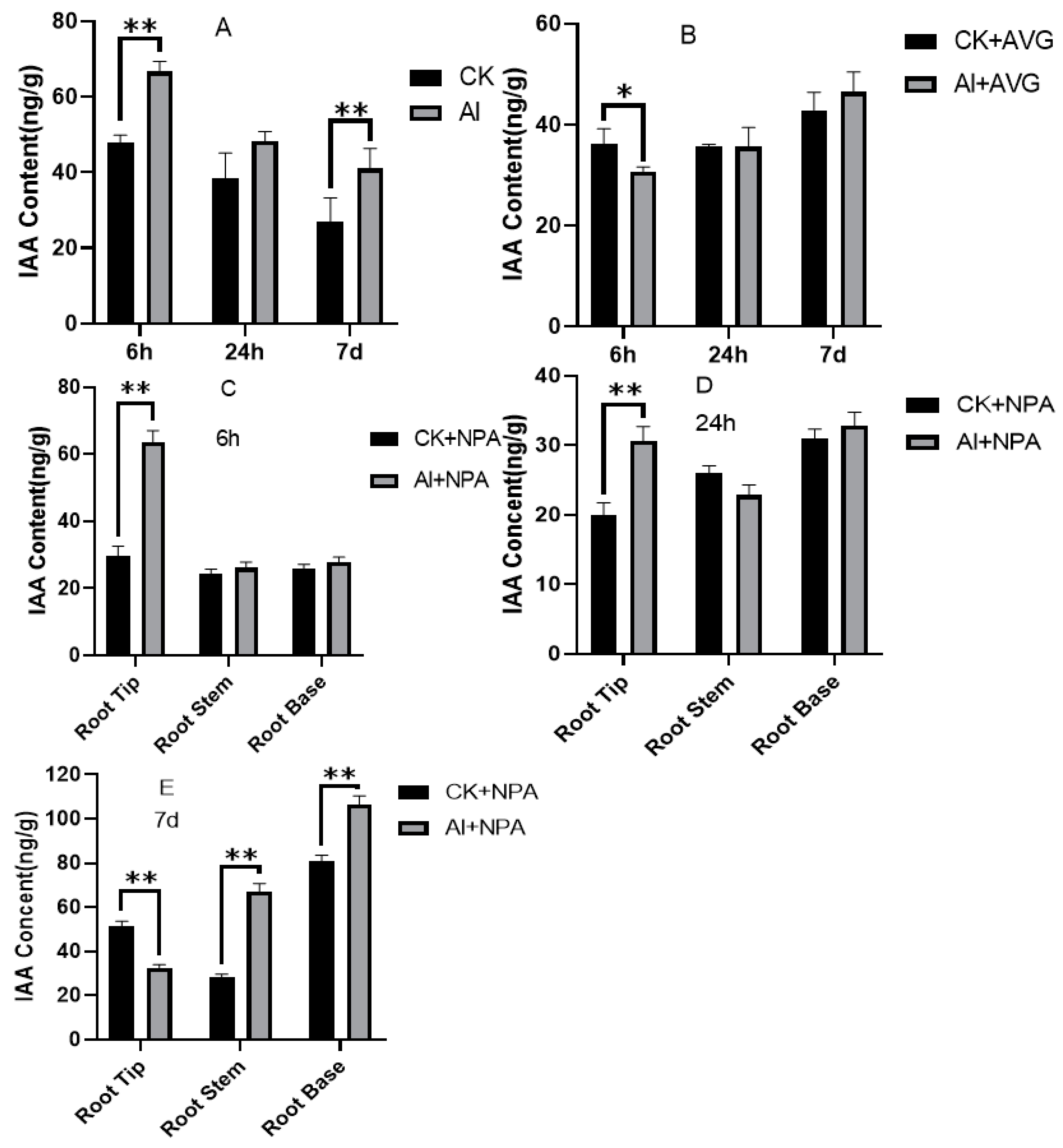
To determine whether the promotion of root growth under Al was related to IAA distribution, root bases were collected to examine the endogenous IAA levels using HPLC. The IAA concentration reached 66.62 ng/g after 6 h, which was significantly higher than that of the control plants (Figure 3A). After 7 d, the IAA concentration decreased in Al-exposed plants but was still significantly higher than in control plants. This indicates that Al may promote the accumulation of IAA in roots to accelerate the growth of LRs in tea plants.
Two experiments were carried out to further verify whether the promotion of root growth under Al was related to IAA distribution. The IAA concentration in the root tips in the Al group was significantly lower than that of the CK group after the roots were treated for 6 h by the IAA synthesis inhibitor AVG, and there was no difference when the tea plants were treated for 24 h and 7 d (Figure 3B). When the roots of tea plants were treated for 6 h by the IAA transport inhibitor, the IAA concentration in the root tips of the Al group was significantly higher than that of the CK group, but it remained at a relatively low level in the root stem and root base (Figure 3C). When the incubation time reached 24 h, the IAA content began to decrease in the root tips, but the IAA concentration in the root tips of the Al group was still significantly higher than that of the CK group, and the IAA content in the root base began to increase (Figure 3D). When the incubation time reached 7 d, the IAA concentration in the root tips of the Al-treated group was significantly lower compared with the CK group, whereas the IAA concentration in the root stem and root base was significantly higher (Figure 3E). Hence, the concentration of Al may affect the synthesis and transportation of IAA and lead to IAA accumulation in the roots of tea plants to stimulate LR growth.

3.3. Expression of Genes Involved in IAA Accumulation, Distribution, and Response

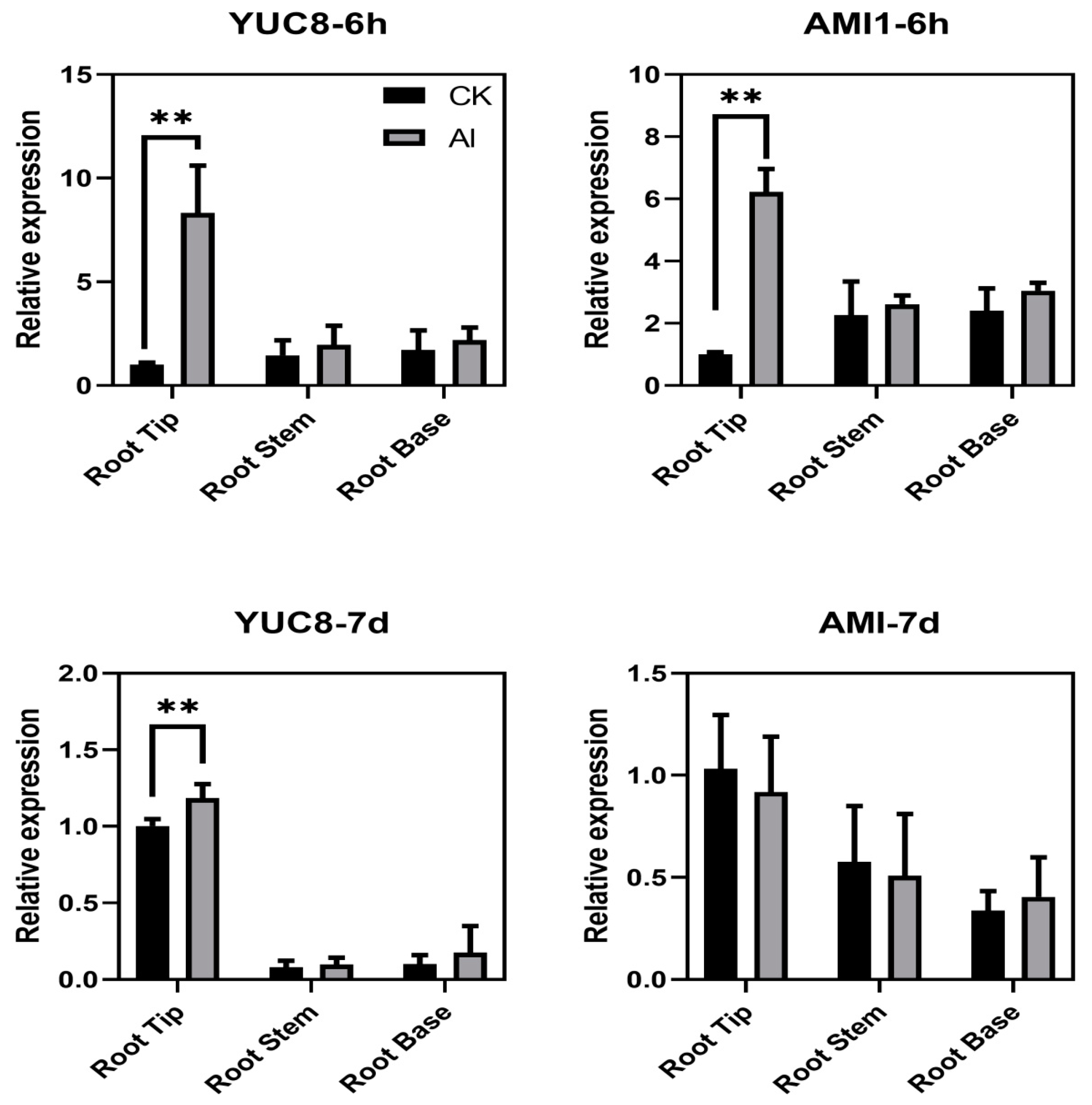
To study how IAA regulated tea root growth under Al treatment, the expression profiles of genes related to IAA accumulation, transport, and response were analyzed in three different root segments. The results show that Al influences gene expression in a tissue-specific manner. YUC8 and AMI1 are two key genes involved in IAA biosynthesis. Expression of these two genes in the root tips was significantly upregulated (more than ten-fold) after 6 h (Figure 4), while there were no significant changes in the other two segments. After 7 days, only YUC8 levels were slightly increased in root tips.
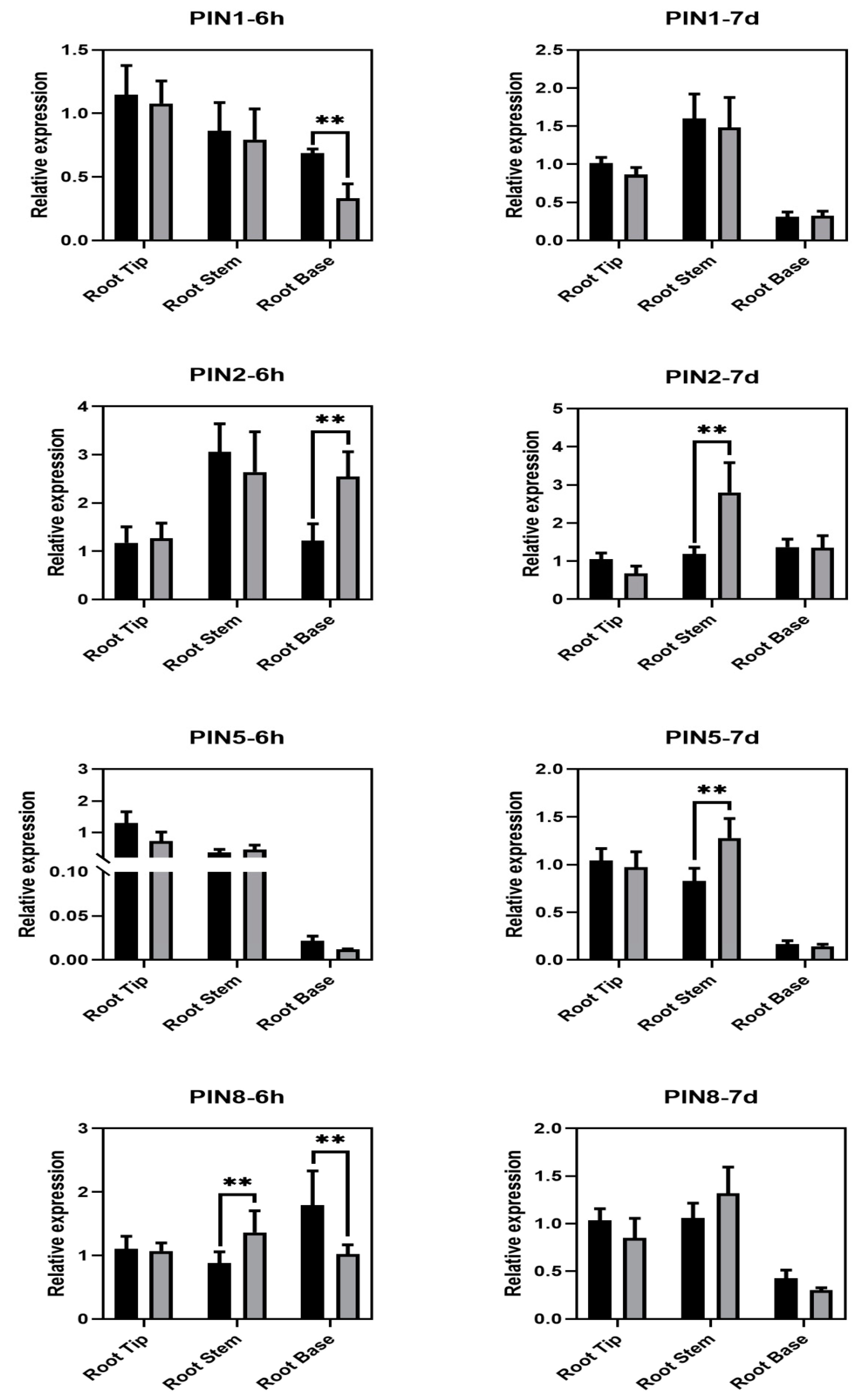
The PIN-FORMED (PIN) gene family is comprised of members considered to function as auxin efflux carriers and play a pivotal role in polar auxin transport. The expression profiles of four PIN genes identified from the tea genome were analyzed. An effect of Al on the expression of PIN genes was found mainly in the root stem and base (Figure 5), with no obvious change in the root tip. Specifically, the expression of PIN2 increased rapidly in the root base after Al treatment for 6 h, while it returned to a normal level after 7 days. After 7 days, the expression of PIN2 and PIN5 were upregulated only in the root stems. Al enhanced the expression of PIN8 in the root stem, while it inhibited it in the root base.
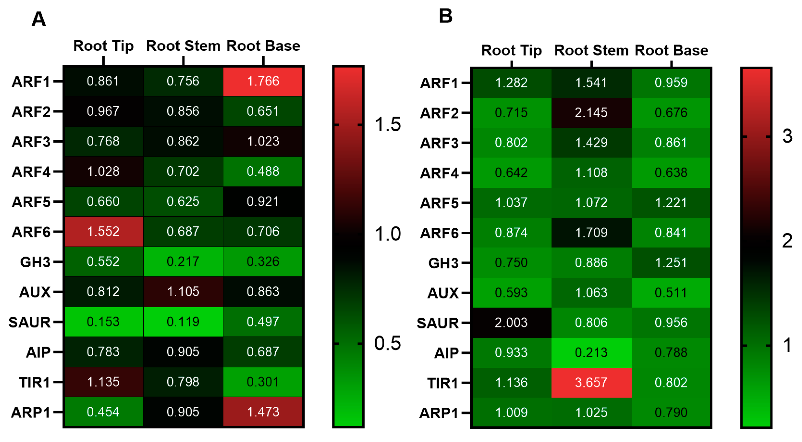
Finally, the expression profiles of 12 auxin response genes were studied. The expression of most genes was repressed when treated for 6 h with Al, and only ARF1, ARF6, and ARP1 genes were upregulated by Al treatment. The expression of ARF6 was induced only in the root tip, and ARF1 and ARP1 were induced in the root base. The expression of GH3 and SAUR was downregulated in all three root parts (Figure 6A). After 7 days, more genes were induced by Al treatment. Most analyzed genes (ARF1, ARF2, ARF3, ARF6, and TIR1) were upregulated in the root stem. The expression of TIR1 increased more than 3.5-fold in the Al group compared to the CK group. The expression of SAUR in the root tip was upregulated more than twofold. In addition, the expression of ARF5 and GH3 was slightly increased in the root base (Figure 6B).

3.4. Effect of Al on Activity and Gene Expression of XET and Expansin

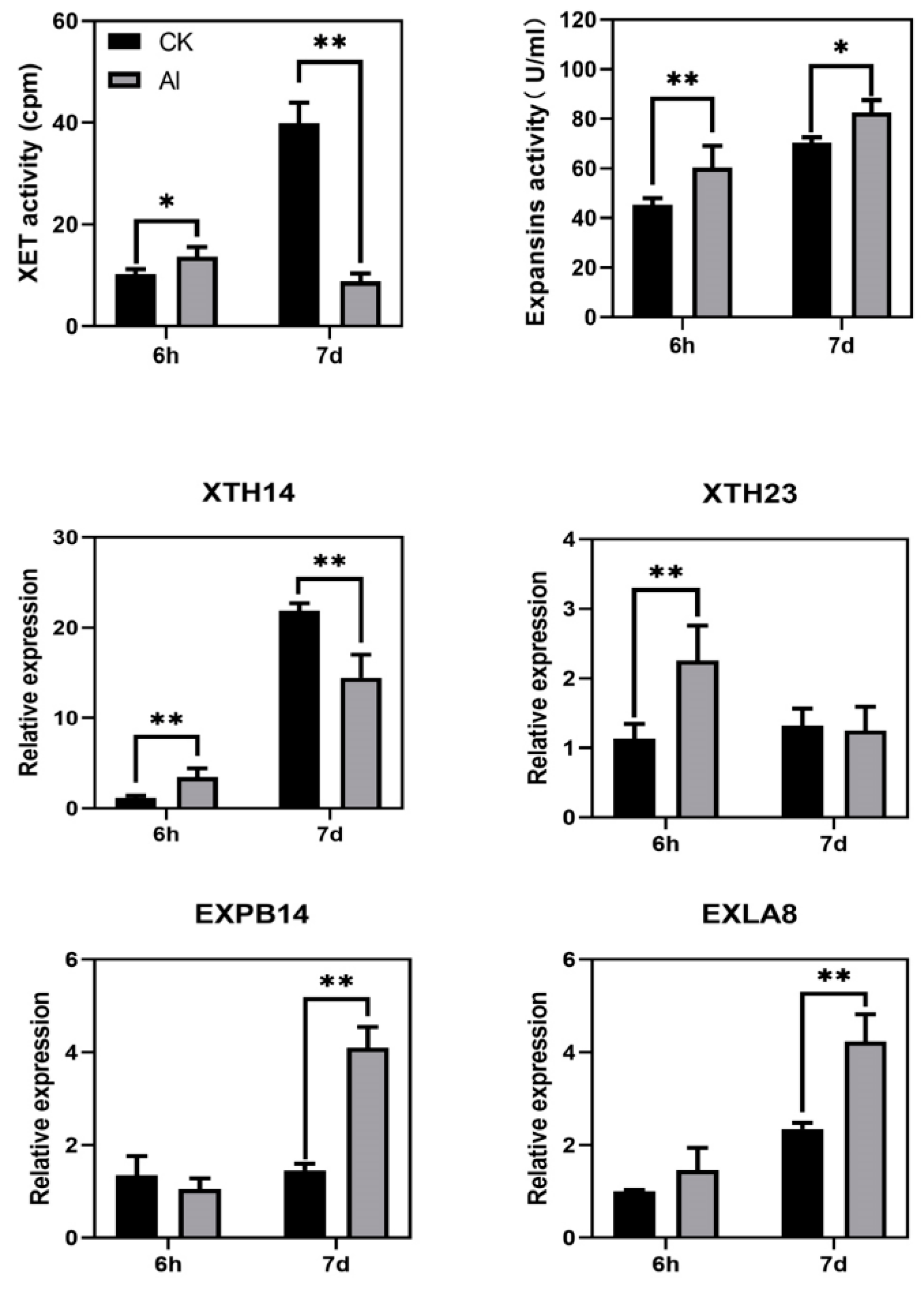
To study whether Al regulates IAA resulting in cell wall loosening, the activity of XET and expansin enzymes, involved in cell wall loosening, were determined. XET activity was slightly enhanced by Al after 6 h, whereas it was repressed by 200% in the control at 7 days. In contrast, expansin activity significantly increased at 6 h and 7 d (Figure 7). Consistently, XTH14 and XTH23 were upregulated at 6 h. After 7 days, expression of XTH14 was reversed, showing a dramatic decrease. The two genes encoding expansin, EXPB14 and EXLA8, showed the same change tendency, with no change at 6 h but an increase of more than twofold at 7 d.

4. Discussion

Al can be taken up by plant roots and cause phytotoxicity; however, plants display a large variation in Al tolerance among species and cultivars [22]. As an Al hyperaccumulator, tea plants can accumulate abundant Al without any toxic symptoms. Appropriate amounts of Al supply have even been thought necessary for root growth in tea plants [9,10]. In our study, 0.4 mmol/L Al dramatically increased the number of lateral roots, suggesting that Al is beneficial for enhancing root formation. Plant hormones play a key role in adventitious root formation [21]. Here, we focused on how Al regulates hormone biosynthesis, transport, and response to influence root growth.

4.1. Effect of Al on Phytohormone Accumulation in Tea Roots

Previous studies indicate that phytohormone levels change appreciably in different plant tissues upon exposure to Al [13,33]. In the present study, among the six assayed phytohormones, IAA was found to be significantly increased in the root base when supplied with 0.4 mmol/L Al (Figure 2). It is well known that IAA plays a central role in regulating root development and root architecture, and there is increasing evidence regarding the importance of auxin biosynthesis at the root tip for its regulation [34]. Plant hormones are involved in plant adaptation to diverse environmental stresses, including Al toxicity. Wang et al. (2016) found that Al could interrupt auxin transport and accumulation in the roots of alfalfa, which was sensitive to Al toxicity [18]. In Arabidopsis, auxin treatment alters the expression of Alsensitivel (ALS1), thereby influencing the pattern of Al distribution in the cell [35]. Al stress induces local auxin biosynthesis and enhances auxin signaling response in the root transition zone (TZ), which eventually results in the inhibition of root growth [20,36]. In the Al-tolerant tea plant, root growth is regulated by Al in a different way compared to Al-sensitive plants. In the present study, the IAA contents in the roots of tea plants were found to be significantly increased under Al, thus increasing the number and length of lateral roots remarkably, suggesting that Al might improve tea root growth by regulating IAA accumulation.
In the present study, we found the levels of JA and ABA in roots supplied with 0.4 mmol/L Al were lower than in the CK group. In Arabidopsis and tomato, JA enhances Al-induced root growth [37,38]. This indicates that JA plays different roles in Al tolerance in different species. ABA is involved in various plant biological processes. In response to abiotic stresses, plants quickly accumulate ABA to cope with environmental changes [39,40]. However, overaccumulation of ABA can also induce senescence [41]. Whether it is beneficial or harmful depends on the dynamics of the seasonal regiment of ABA and the crop stress profile [42]. In our study, JA and ABA accumulation might be reduced in tea plants, thus resulting in improved root growth. However, further research is necessary to reveal the roles of JA and ABA played in Al-induced root growth of tea plants

4.2. Effect of Al on IAA Biosynthesis, Transport, and Response in Tea Roots

In plants, changes in IAA content are closely correlated with root growth. In the present study, the effect of exogenous AVG (IAA synthesis inhibitor) and NPA (IAA transport inhibitor) on the IAA distribution in roots suggests that Al could function through regulation of IAA synthesis and transport (Figure 3A–F). The expression of genes involved in IAA biosynthesis, transport, and response was studied. YUC was recently defined as an enzyme having activity for conversion of IPyA to IAA based on genetic, biochemical, and metabolite evidence [43]. The YUCCA genes appear to be spatiotemporally regulated by Al and are involved in the local production of IAA [44]. In our results, one member of YUCCA, YUC8, was significantly upregulated in root tips under Al treatment, which is in accordance with the results obtained from Arabidopsis, where it was reported that Al upregulates YUC3/5/7/8/9 in the root-apex TZ and overexpression of YUC8 could regulate root growth [36]. Another gene, AMI1, is a member of the isogene family that converts IAM into IAA [45]. Suppression of the NtAMI1 gene by RNAi caused severe growth inhibition in a medium containing IAM [46]. The transcription of this gene was found to be upregulated in the Al-treated roots at 6 h and subsequently returned to the same level as the CK group at 7 d. This indicates that the expression of the AMI1 gene is responsive to Al for a short period and then is restored to normal after a longer period.
The PIN-FORMED (PIN) gene family of transmembrane proteins is thought to comprise auxin efflux carriers that play a pivotal role in polar auxin transport, mediating the growth of different plant tissues [47]. In the present study, four members of the PIN gene family in tea plants were investigated. The expression profiles of these members showed spatiotemporal variation. PIN2 was upregulated in the Al-supplied roots at 6 h and 7 d, which is in accordance with reports in alfalfa, where the PIN2 gene was upregulated in root tips exposed to Al [18]. However, in pea (Pisum sativum), Al inhibits the PIN2-based polar auxin transport and increases the cytoplasmic free IAA of the root transition zone, thus resulting in acidification of the cell wall in this region and exacerbating the effects of Al toxicity [48]. All the above results confirm that the PIN2 gene regulates IAA transport both instantaneously and persistently to improve root formation. Furthermore, the expression levels of PIN5 and PIN8 were also increased in the root stem but at different response time points. PIN8 was only upregulated at 6 h, while PIN5 was only upregulated at 7 d. This indicates that PIN5 and PIN8 regulate IAA in a time-dependent manner. Similarly, the expression of PIN5 was also observed at a higher level in Al-treated node cuttings of tea plant than that in the control condition during the later stages (days 7−43) [11]. Taken together, over relatively long term, the expression of PIN2 and PIN5 was accordant with the enhanced transport of accumulated IAA from root tip to root base by Al and might contribute to the mechanism of Al-induced root growth in tea plants.
A previous study demonstrated that ARF6 and ARF8 are positive regulators of adventitious root initiation [49]. In our study, the expression of several auxin response genes was also investigated. After 6 h with Al treatment, only ARF1, ARF6, and ARP1 were upregulated. This indicates the possible role of ARF1 and ARF6 in promoting adventitious root formation by Al in tea plants. In addition, Lakehal et al. (2019) reported that ARF could interact with AUX/IAA, forming coreceptor complexes with TIR1 proteins. After 7 d of Al treatment, TIR1 was increased 2.5-fold more in the root base of Al-treated plants than the CK group. This suggests that TIR1 might interact with ARF and be involved in root formation in response to Al [50].

4.3. Effect of Al on the Enzyme Activity and Gene Expression of XET and Expansin

The cell wall is the main site of Al perception and plays a major role in root elongation. In citrus, the difference between two species (Citrus sinensis and Citrus grandis) in Al tolerance were found to be associated with Al translocation efficiency from roots to shoots and the structural modification of the root cell wall [51]. In tea plants, biochemical modification of the cell wall reduces the site of Al binding to roots and triggers the activity of loosening agents to promote root growth [27,52]. Auxin could promote cell elongation by inducing proton extrusion, whereby the cell wall is acidified for elongation [13]. The XET catalyzes the splitting of xyloglucan chains and the linking of a newly generated reducing ends to a nonreducing ends of another XyG chain, thereby loosening the cell wall [25,53]. Previous studies have shown that Al inhibits the activity and expression of XET in most plants [54]. Cui et al. (2005) found that XET, regulated by a gradient of auxin and GA, was involved in the hormone-mediated differential growth of the upper and lower halves of horizontally orientated leaf sheaths during gravitropic bending in rice seedlings [55]. However, in the present study, we found that 0.4 mmol/L Al enhanced the activity of XET at 6 h but inhibited XET activity at 7 d. The expression of XET14 and XTH23 genes were upregulated at 6 h. This is in accordance with the results from Safari et al. (2018) [27], which reported that XET activity was upregulated two-fold in tea roots supplied with 0.4 mmol/L Al compared with the control at 24 h but recovered at 96 and 192 h. Collectively, the results confirm that appropriate Al concentrations can increase XET activity, though this promoting effect was time dependent. Previous studies have shown that the activities of both XET and expansins have a positive role in promoting the root growth of tea plants by promoting the expression of expansin and XTH genes [52]. Therefore, the expansin related to the cell wall extension was also studied. Our results revealed that expansin activity was enhanced by Al addition, and EXPB14 and EXLB4 expression were upregulated two-fold more than in the control, suggesting Al might upregulate expansin gene expression and thus, enzyme activity to enhance root growth.

5. Conclusions

An appropriate concentration of Al (0.4 mmol/L) was observed to increase the number of LRs and the IAA content in roots, thus promoting the root growth of tea plants. The effect of exogenous AVG (IAA synthesis inhibitor) and NPA (IAA transport inhibitor) on IAA distribution in roots suggests that Al could exert these effects via regulation of IAA synthesis and transport. The genes YUC8 and AMI1, for IAA accumulation, were upregulated by Al in root tips, whereas expression of PIN1/2/8 for IAA transport was induced in the root base with Al treatment. Meanwhile, the activities of XET and expansin were increased in response to Al treatment for 6 h. Taken together, the results show that Al regulates the distribution of IAA in roots by affecting the synthesis and transportation of IAA in cooperation with cell wall loosening to promote root growth in tea plants. However, whether this mechanism is retained extensively in the plant kingdom and how it has evolved during plant adaptation to environments remain to be revealed in the future.

Supplementary Materials

The following supporting information can be downloaded at: https://www.mdpi.com/article/10.3390/agronomy12051110/s1, Table S1. List of primers used in this study.

Author Contributions

Y.S. and K.F. designed the research; M.W., Y.G. and L.Y. performed the experiments; Y.S. (Yifan Shi), L.Y. and J.H. helped to analyze the samples and data; K.F. and Y.S. (Yuanzhi Shi) revised the article. All authors have read and agreed to the published version of the manuscript.

Funding

This work was financially supported by the National Natural Science Foundation of China (no. 31572199); the China Agriculture Research System of MOF and MARA (CARS-19); and the Special Fund for Scientific Research of the Tea Research Institute of the Chinese Academy of Agricultural Sciences (grant no.1610212021001).

Data Availability Statement

The data presented in this study are available on request from the corresponding author.

Conflicts of Interest

The authors declare no conflict of interest.

Abbreviations

Al: aluminum; AR: adventitious root; AVG: IAA synthesis inhibitor; CK: control; ELISA: enzyme linked immunosorbent assay; HPLC: high performance liquid chromatography; IAA: indole-3-acetic acid; LR: lateral root; LC–MS/MS: liquid chromatography tandem mass spectrometry; MR: main root; NPA: IAA transport inhibitor; PIN: PIN-FORMED; PME: pectin methylesterase; SA: salicylic acid; SPE: solid phase extraction; TZ: transition zone.

References

  1. Ma, J.F.; Chen, Z.C.; Shen, R.F. Molecular mechanisms of Al tolerance in gramineous plants. Plant Soil 2014, 381, 1–12. [Google Scholar] [CrossRef]
  2. Kochian, L.V. Cellular mechanisms of aluminum toxicity and resistance in plants. Annu. Rev. Plant Physiol. Plant Mol. Biol. 1995, 46, 237–260. [Google Scholar] [CrossRef]
  3. Kochian, L.V.; Hoekenga, O.A.; Piñeros, M.A. How do crop plants tolerate acid soils? Mechanisms of aluminum tolerance and phosphorous efficiency. Annu. Rev. Plant Biol. 2004, 55, 459–493. [Google Scholar] [CrossRef] [PubMed]
  4. Jaskowiak, J.; Kwasniewska, J.; Milewska-Hendel, A.; Kurczynska, E.U.; Szurman-Zubrzycka, M.; Szarejko, I. Aluminum Alters the Histology and Pectin Cell Wall Composition of Barley Roots. Int. J. Mol. Sci. 2019, 20, 3039. [Google Scholar] [CrossRef] [PubMed] [Green Version]
  5. Rahman, R.; Upadhyaya, H. Aluminium Toxicity and Its Tolerance in Plant: A Review. J. Plant Biol. 2020, 64, 101–121. [Google Scholar] [CrossRef]
  6. Kochian, L.V.; Piñeros, M.A.; Liu, J.; Magalhaes, J.V. Plant Adaptation to Acid Soils: The Molecular Basis for Crop Aluminum Resistance. Annu. Rev. Plant Biol. 2015, 66, 571–598. [Google Scholar] [CrossRef]
  7. Ruan, J.; Wong, M.H. Aluminium absorption by intact roots of the Al-accumulating plant Camellia sinensis L. Agronomie 2004, 24, 137–142. [Google Scholar] [CrossRef] [Green Version]
  8. Hajiboland, R.; Barceló, J.; Poschenrieder, C.; Tolrà, R. Amelioration of iron toxicity: A mechanism for alumi-num-induced growth stimulation in tea plants. J. Inorg. Biochem. 2013, 128, 183–187. [Google Scholar] [CrossRef]
  9. Fan, K.; Wang, M.; Gao, Y.; Ning, Q.; Shi, Y. Transcriptomic and ionomic analysis provides new insight into the beneficial effect of Al on tea roots’ growth and nutrient uptake. Plant Cell Rep. 2019, 38, 715–729. [Google Scholar] [CrossRef]
  10. Sun, L.; Zhang, M.; Liu, X.; Mao, Q.; Shi, C.; Kochian, L.V.; Liao, H. Aluminium is essential for root growth and development of tea plants (Camellia sinensis). J. Integr. Plant Biol. 2020, 62, 984–997. [Google Scholar] [CrossRef]
  11. Cheng, L.; Liu, H.; Zhao, J.; Dong, Y.; Xu, Q.; Yu, Y. Hormone Orchestrates a Hierarchical Transcriptional Cascade That Regulates Al-Induced De Novo Root Regeneration in Tea Nodal Cutting. J. Agric. Food Chem. 2021, 69, 5858–5870. [Google Scholar] [CrossRef] [PubMed]
  12. Čiamporová, M. Morphological and Structural Responses of Plant Roots to Aluminium at Organ, Tissue, and Cellular Levels. Biol. Plant 2002, 45, 161–171. [Google Scholar] [CrossRef]
  13. Kopittke, P.M. Role of phytohormones in aluminium rhizotoxicity. Plant Cell Environ. 2016, 39, 2319–2328. [Google Scholar] [CrossRef] [PubMed]
  14. Zhou, P.; Yang, F.; Ren, X.; Huang, B.; An, Y. Phytotoxicity of aluminum on root growth and indole-3-acetic acid ac-cumulation and transport in alfalfa roots. Environ. Exp. Bot. 2014, 104, 1–8. [Google Scholar] [CrossRef]
  15. Wang, S.; Yuan, S.; Su, L.; Lv, A.; Zhou, P.; An, Y. Aluminum toxicity in alfalfa (Medicago sativa) is alleviated by exogenous foliar IAA inducing reduction of Al accumulation in cell wall. Environ. Exp. Bot. 2017, 139, 1–13. [Google Scholar] [CrossRef]
  16. Liu, N.; You, J.; Shi, W.; Liu, W.; Yang, Z. Salicylic acid involved in the process of aluminum induced citrate exudation in Glycine max L. Plant Soil 2011, 352, 85–97. [Google Scholar] [CrossRef]
  17. Wang, P.; Yu, W.Q.; Zhang, J.R.; Rengel, Z.; Xu, J.; Han, Q.Q.; Chen, L.M.; Li, K.Z. Auxin enhances aluminium-induced citrate exudation through upregulation of GmMATE and activation of the plasma membrane H+-ATPase in soybean roots. Ann. Bot. 2016, 118, 933–940. [Google Scholar] [CrossRef] [Green Version]
  18. Wang, S.; Ren, X.; Huang, B.; Wang, G.; Zhou, P.; An, Y. Aluminium-induced reduction of plant growth in alfalfa (Medicago sativa) is mediated by interrupting auxin transport and accumulation in roots. Sci. Rep. 2016, 6, 30079. [Google Scholar] [CrossRef] [Green Version]
  19. Sánchez-Parra, B.; Frerigmann, H.; Alonso, M.-M.P.; Loba, V.C.; Jost, R.; Hentrich, M.; Pollmann, S. Characterization of Four Bifunctional Plant IAM/PAM-Amidohydrolases Capable of Contributing to Auxin Biosynthesis. Plants 2014, 3, 324–347. [Google Scholar] [CrossRef] [Green Version]
  20. Zhang, M.; Lu, X.; Li, C.; Zhang, B.; Zhang, C.; Zhang, X.-S.; Ding, Z. Auxin Efflux Carrier ZmPGP1 Mediates Root Growth Inhibition under Aluminum Stress. Plant Physiol. 2018, 177, 819–832. [Google Scholar] [CrossRef] [Green Version]
  21. Yang, Z.B.; Geng, X.Y.; He, C.M.; Zhang, F.; Wang, R.; Horst, W.J.; Ding, Z.J. TAA1-Regulated Local Auxin Biosynthesis in the Root-Apex Transition Zone Mediates the Aluminum-Induced Inhibition of Root Growth in Arabidopsis. Plant Cell 2014, 26, 2889–2904. [Google Scholar] [CrossRef] [PubMed] [Green Version]
  22. Wei, K.; Ruan, L.; Wang, L.; Cheng, H. Auxin-Induced Adventitious Root Formation in Nodal Cuttings of Camellia sinensis. Int. J. Mol. Sci. 2019, 20, 4817. [Google Scholar] [CrossRef] [PubMed] [Green Version]
  23. Rangel, A.F.; Rao, I.M.; Horst, W.J. Intracellular distribution and binding state of aluminum in root apices of two common bean (Phaseolus vulgaris) genotypes in relation to Al toxicity. Physiol. Plant 2009, 135, 162–173. [Google Scholar] [CrossRef] [PubMed]
  24. Liu, Y.; Xu, J.; Guo, S.; Yuan, X.; Zhao, S.; Tian, H.; Dai, S.; Kong, X.; Ding, Z. AtHB7/12 Regulate Root Growth in Response to Aluminum Stress. Int. J. Mol. Sci. 2020, 21, 4080. [Google Scholar] [CrossRef]
  25. Zhu, X.F.; Shi, Y.Z.; Lei, G.J.; Fry, S.C.; Zhang, B.C.; Zhou, Y.H.; Braam, J.; Jiang, T.; Xu, X.Y.; Mao, C.Z.; et al. XTH31, Encoding an In Vitro XEH/XET-Active Enzyme, Regulates Aluminum Sensitivity by Modulating In Vivo XET Action, Cell Wall Xyloglucan Content, and Aluminum Binding Capacity in Arabidopsis. Plant Cell 2012, 24, 4731–4747. [Google Scholar] [CrossRef] [Green Version]
  26. Che, J.; Yamaji, N.; Shen, R.F.; Ma, J.F. An Al-inducible expansin gene, OsEXPA10 is involved in root cell elongation of rice. Plant J. 2016, 88, 132–142. [Google Scholar] [CrossRef]
  27. Safari, M.; Ghanati, F.; Safarnejad, M.R.; Chashmi, N.A. The contribution of cell wall composition in the expansion of Camellia sinensis seedlings roots in response to aluminum. Planta 2017, 247, 381–392. [Google Scholar] [CrossRef]
  28. Glauser, G.; Veyrat, N.; Rochat, B.; Wolfender, J.L.; Turlings, T.C.J. Ultra-high pressure liquid chromatography-mass spectrometry for plant meabolomics: A systematic comparison of high-resolution quadrupole-time-of-flight and single stage Orbitrap mass spectrometers. J. Chromatogr. A 2013, 1292, 151–159. [Google Scholar] [CrossRef]
  29. Yang, H.; Wang, Y.; Li, L.; Li, F.; He, Y.; Wu, J.; Wei, C. Transcriptomic and Phytochemical Analyses Reveal Root-Mediated Resource-Based Defense Response to Leaf Herbivory by Ectropis oblique in Tea Plant (Camellia sinensis). J. Agric. Food Chem. 2019, 67, 5465–5476. [Google Scholar] [CrossRef]
  30. Zhang, Y.; Zhong, Y.; Gao, C.; Dong, Z.; Chen, N.; Wang, M. Determination of five endogenous hormones in wheat by high performance liquid chromatography. Chin. J. Chromatogr. 2013, 31, 800–803. [Google Scholar] [CrossRef]
  31. Fry, S.C.; Smith, R.C.; Renwick, K.F.; Martin, D.J.; Hodge, S.K.; Matthews, K.J. Xyloglucan endotransglycosylase, a new wall-loosening enzyme activity from plants. Biochem. J. 1992, 282, 821–828. [Google Scholar] [CrossRef] [PubMed] [Green Version]
  32. Cosgrove, D.J. Loosening of plant cell walls by expansins. Nature 2000, 407, 321–326. [Google Scholar] [CrossRef]
  33. Sun, P.; Tian, Q.Y.; Chen, J.; Zhang, W.H. Aluminum-induced inhibition of root elongation in Arabidopsis is mediated by ethylene and auxin. J. Exp. Bot. 2010, 61, 347–356. [Google Scholar] [CrossRef] [PubMed] [Green Version]
  34. Overvoorde, P.; Fukaki, H.; Beeckman, T. Auxin control of root development. Cold Spring Harb. Perspect. Biol. 2010, 2, a001537. [Google Scholar] [CrossRef] [PubMed] [Green Version]
  35. Zhu, X.F.; Lei, G.J.; Wang, Z.W.; Shi, Y.Z.; Braam, J.; Li, G.X.; Zheng, S.J. Coordination between Apoplastic and Symplastic Detoxification Confers Plant Aluminum Resistance. Plant Physiol. 2013, 162, 1947–1955. [Google Scholar] [CrossRef] [Green Version]
  36. Liu, G.C.; Gao, S.; Tian, H.Y.; Wu, W.W.; Robert, H.S.; Ding, Z.J. Local Transcriptional Control of YUCCA Regulates Auxin Promoted Root-Growth Inhibition in Response to Aluminum Stress in Arabidopsis. PLoS Genet. 2016, 12, e1006360. [Google Scholar] [CrossRef]
  37. Wang, Z.; Liu, L.; Su, H.; Guo, L.; Zhang, J.; Li, Y.; Xu, J.; Zhang, X.; Guo, Y.-D.; Zhang, N. Jasmonate and aluminum crosstalk in tomato: Identification and expression analysis of WRKYs and ALMTs during JA/Al-regulated root growth. Plant Physiol. Biochem. 2020, 154, 409–418. [Google Scholar] [CrossRef]
  38. Yang, Z.-B.; He, C.; Ma, Y.; Herde, M.; Ding, Z. Jasmonic Acid Enhances Al-Induced Root Growth Inhibition. Plant Physiol. 2016, 173, 1420–1433. [Google Scholar] [CrossRef] [Green Version]
  39. Zhu, J.-K. Abiotic Stress Signaling and Responses in Plants. Cell 2016, 167, 313–324. [Google Scholar] [CrossRef] [Green Version]
  40. Xie, Y.; Bao, C.; Chen, P.; Cao, F.; Liu, X.; Geng, D.; Li, Z.; Li, X.; Hou, N.; Zhi, F.; et al. Abscisic acid homeostasis is mediated by feedback regulation of MdMYB88 and MdMYB124. J. Exp. Bot. 2020, 72, 592–607. [Google Scholar] [CrossRef]
  41. Becker, W.; Apel, K. Differences in gene expression between natural and artificially induced leaf senescence. Planta 1993, 189, 74–79. [Google Scholar] [CrossRef]
  42. Blum, A. Towards a conceptual ABA ideotype in plant breeding for water limited environments. Funct. Plant Biol. 2015, 42, 502–513. [Google Scholar] [CrossRef]
  43. Won, C.; Shen, X.; Mashiguchi, K.; Zheng, Z.; Dai, X.; Cheng, Y.; Kasahara, H.; Kamiya, Y.; Chory, J.; Zhao, Y. Conversion of tryptophan to indole-3-acetic acid by TRYPTOPHAN AMINOTRANSFERASES OF ARABIDOPSIS and YUCCAs in Ara-bidopsis. Proc. Natl. Acad. Sci. USA 2011, 108, 18518–18523. [Google Scholar] [CrossRef] [PubMed] [Green Version]
  44. Yue, J.; Hu, X.; Huang, J. Origin of plant auxin biosynthesis. Trends Plant Sci. 2014, 19, 764–770. [Google Scholar] [CrossRef] [PubMed]
  45. Zhao, Y. Auxin Biosynthesis: A Simple Two-Step Pathway Converts Tryptophan to Indole-3-Acetic Acid in Plants. Mol. Plant 2012, 5, 334–338. [Google Scholar] [CrossRef] [PubMed] [Green Version]
  46. Nemoto, K.; Hara, M.; Suzuki, M.; Seki, H.; Muranaka, T.; Mano, Y. The NtAMI1 gene functions in cell division of tobacco BY-2 cells in the presence of indole-3-acetamide. FEBS Lett. 2009, 583, 487–492. [Google Scholar] [CrossRef] [Green Version]
  47. Křeček, P.; Skůpa, P.; Libus, J.; Naramoto, S.; Tejos, R.; Friml, J.; Zazimalova, E. The PIN-FORMED (PIN) protein family of auxin transporters. Genome Biol. 2009, 10, 249. [Google Scholar] [CrossRef] [Green Version]
  48. Li, X.; Li, Y.; Mai, J.; Tao, L.; Qu, M.; Liu, J.; Shen, R.; Xu, G.; Feng, Y.; Xiao, H.; et al. Boron Alleviates Aluminum Toxicity by Promoting Root Alkalization in Transition Zone via Polar Auxin Transport. Plant Physiol. 2018, 177, 1254–1266. [Google Scholar] [CrossRef] [Green Version]
  49. Lin, Y.; Lai, Z.; Lin, L.; Lai, R.; Tian, Q.; Ye, W.; Zhang, D.; Yang, M.; Chen, Y.; Zhang, Z. Endogenous target mimics, microRNA167, and its targets ARF6 and ARF8 during somatic embryo development in Dimocarpus longan Lour. Mol. Breed. 2015, 35, 1–15. [Google Scholar] [CrossRef]
  50. Lakehal, A.; Chaabouni, S.; Cavel, E.; Le Hir, R.; Ranjan, A.; Raneshan, Z.; Novak, O.; Pacurar, D.I.; Perrone, I.; Jobert, F.; et al. A Molecular Framework for the Control of Adventitious Rooting by TIR1/AFB2-Aux/IAA-Dependent Auxin Signaling in Arabidopsis. Mol. Plant 2019, 12, 1499–1514. [Google Scholar] [CrossRef]
  51. Zhang, H.; Li, X.Y.; Lin, M.L.; Hu, P.P.; Lai, N.W.; Huang, Z.R.; Chen, L.S. The aluminum distribution and translocation in two citrus species differing in aluminum tolerance. BMC Plant Biol. 2022, 22, 93. [Google Scholar] [CrossRef] [PubMed]
  52. Ning, Q.Y.; Fan, K.; Wang, M.; Shi, Y.Z. Responses of expansins and XTHs to different aluminum concentration in roots of tea plant [Camellia sinensis (L.) O. Kuntze]. Acta Agric. Zhejiangensis 2018, 30, 961–969. [Google Scholar]
  53. Zhu, X.F.; Wan, J.X.; Sun, Y.; Shi, Y.Z.; Braam, J.; Li, G.X.; Zheng, S.J. Xyloglucan Endotransglucosylase-Hydrolase 17 Interacts with Xyloglucan Endotransglucosylase-Hydrolase31 to Confer Xyloglucan Endotransglucosylase Action and Affect Aluminum Sensitivity in Arabidopsis. Plant Physiol. 2014, 165, 1566–1574. [Google Scholar] [CrossRef] [PubMed]
  54. Zhang, M.; Ma, Y.; Horst, W.J.; Yang, Z.-B. Spatial–temporal analysis of polyethylene glycol-reduced aluminium accumulation and xyloglucan endotransglucosylase action in root tips of common bean (Phaseolus vulgaris). Ann. Bot. 2016, 118, 1–9. [Google Scholar] [CrossRef] [PubMed] [Green Version]
  55. Cui, D.; Neill, S.J.; Tang, Z.; Cai, W. Gibberellin-regulated XET is differentially induced by auxin in rice leaf sheath bases during gravitropic bending. J. Exp. Bot. 2005, 56, 1327–1334. [Google Scholar] [CrossRef] [PubMed] [Green Version]
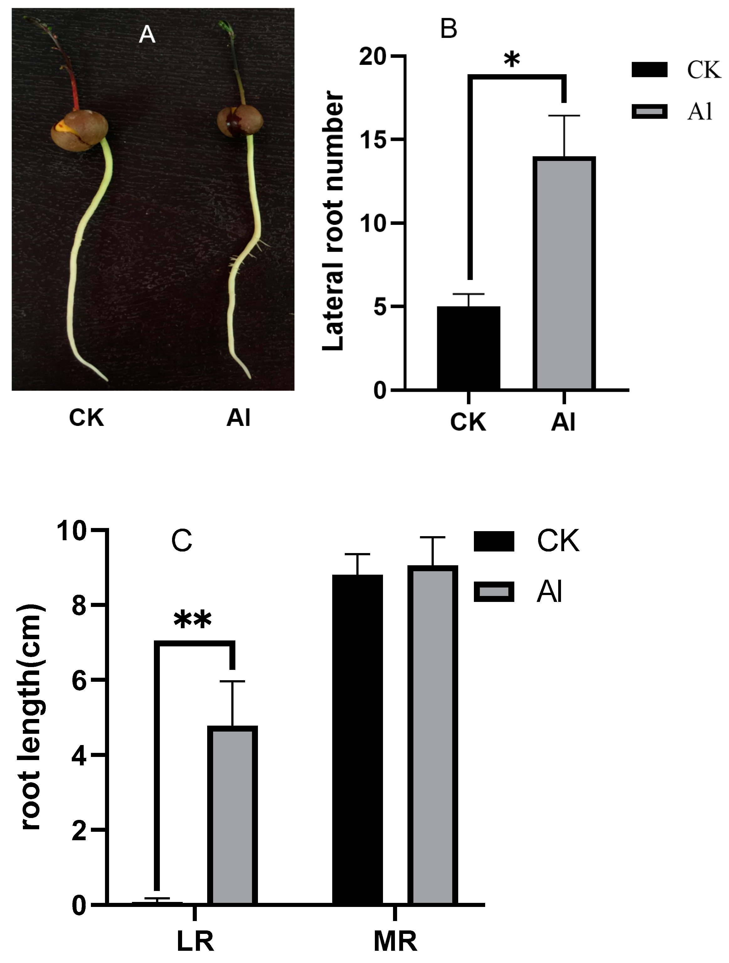
Figure 1. Analysis of lateral roots (LRs) and main root (MR)of tea plants for the CK (without Al) and 0.4 mmol/L Al (with Al) groups after 7 d. (A) visible phenotypic differences in LR formation; (B) differences in the LR number; (C) difference in the total root length of LRs and MR (* indicates significance at 0.05 probability level, ** indicates significance at 0.01 probability level).
Agronomy 12 01110 g001
Figure 2. The difference in the content (ng/g) of six hormones (JA, ABA, SA, tzR, ACC, and IAA) in tea root base for the CK and 0.4 mmol/L Al groups (** indicates significance at 0.01 probability level).
Agronomy 12 01110 g002
Figure 3. A comparison of changes in IAA content in the tea roots with and without Al treatment and with IAA synthesis and transport inhibitors. (A) Time course of the difference in IAA content in the root tips with and without Al treatment; (B) Time course of the difference in IAA content in the root tips with the IAA synthesis inhibitor AVG, with and without Al treatment; (C) The difference in IAA content in the root tips, root stems, and root base after 6 h with the IAA transport inhibitor NPA, with and without Al treatment; (D) The difference in IAA content in the root tips, root stems, and root base after 24 h with the IAA transport inhibitor NPA, with and without Al treatment; (E) The difference in IAA content in the root tips, root stems, and root base after 7 d with the IAA transport inhibitor NPA, with and without Al treatment (*, ** indicate significance at 0.05 and 0.01 probability levels, respectively).
Agronomy 12 01110 g003
Figure 4. Changes in YUC8 and AMI1 expression in tea roots with and without Al treatment after incubation for 6 h and 7 d (** indicates significance at 0.01 probability level).
Agronomy 12 01110 g004
Figure 5. Change in the expression of PIN gene family members in tea roots with and without Al treatment after incubation for 6 h and 7 d (** indicates significance at 0.01 probability level).
Agronomy 12 01110 g005
Figure 6. The expression profiles of 12 auxin response genes in tea roots after treatment with Al for (A) 6 h and (B) 7 d compared with the CK group.
Agronomy 12 01110 g006
Figure 7. Change in XET and expansin activity and the expression of related genes in the root base after treatment for 6 h and 7 d with Al compared with the CK group (*, ** indicate significance at 0.05 and 0.01 probability levels, respectively).
Agronomy 12 01110 g007
Publisher’s Note: MDPI stays neutral with regard to jurisdictional claims in published maps and institutional affiliations.