Article

Comparative Study between the Photosynthetic Parameters of Two Avocado (Persea americana) Cultivars Reveals Natural Variation in Light Reactions in Response to Frost Stress

1
Northern Agriculture R&D, MIGAL—Galilee Research Institute, P.O. Box 831, Kiryat Shemona 11016, Israel
2
Morris Kahn Marine Research Station, Department of Marine Biology, Leon H. Charney School of Marine Sciences, University of Haifa, Mt. Carmel, Haifa 3498838, Israel
3
Group of Agrophysics Studies, Department of Plant Sciences, MIGAL—Galilee Research Institute, Kiryat Shemona 11016, Israel
*
Author to whom correspondence should be addressed.
Agronomy 2022, 12(5), 1129; https://doi.org/10.3390/agronomy12051129
Submission received: 11 April 2022 / Revised: 4 May 2022 / Accepted: 5 May 2022 / Published: 7 May 2022
(This article belongs to the Topic Biophysics of Photosynthesis: From Molecules to the Field)

Abstract

:
Avocado is a commercially important fruit tree which is sold worldwide. Originating in subtropical regions of the South America, this species is now grown worldwide and is sometimes exposed to cold temperatures. Specifically, frost stress harms the crop yield and its quality. While it is known in general that the photosynthetic apparatus changes in response to cold conditions, there is still not much information regarding the photosynthetic apparatus response to sporadic frost stress. In this study, we tracked the photosynthetic apparatus’ light reaction of ‘Hass’ and ‘Ettinger’ avocado cultivars to frost stress, with Ettinger being known to be more resilient to cold than Hass. We found that in avocado trees, the photosynthetic apparatus’ response to frost occurs at the level of photosystem II (PSII) itself, rather than a photoprotective response to a stress. The Hass apparatus incorrectly interprets the reduction in electron transport rate activity and by that increases its light harvesting complex size at the expense of its reaction centers which then increases the apparatus’ probability to generate reactive oxygen species. The results of this study open opportunities to further research the process which regulates the feedback mechanism that controls the photosynthetic unit’s size in Hass when compared to the Ettinger cultivar, and whether it is part of a feedback regulation from the carbon assimilation step or indirectly from a stomatal limitation which arises in these subtropical species. While corroborating past studies performed on avocados, this study suggests using advanced chlorophyll a fluorescence protocols when researching natural variation in crops.

1. Introduction

Avocado (Persea americana Mill.) is a commercially important and extremely popular fruit tree in the Lauraceae family due to its highly beneficial nutritional values [1]. Avocado cultivars originated through hybridization of the South American species, each maintaining its unique properties, inter alia, different levels of tolerances to cold stress [2]. Hass, which composes 85% of all the avocados sold worldwide, is cold sensitive [2]; while Ettinger, a less popular cultivar that serves in Israel mainly as a pollinator, is considered cold-stress tolerant [3]. Avocado orchards face colder weather conditions due to global climate change, where it is expected that the frequency and magnitude of cold weather waves will be elevated [4]. In Israel, cold stress occurs sporadically during late autumn and through the winter, mainly during nights when the temperatures can reach below zero values in extreme events [5].
Frost stress in plants affect mostly the membranes within various tissues [6]. Chloroplasts start swelling because of loss of water due to crystallization of ice granules, causing a decrease in the carbon assimilation rate, followed by a reduction in size of starch granules. The thylakoid membranes become distorted and the resulting phenomenon is decoupling of the photosynthetic machinery which results in photoinhibition. In photoinhibition, the homeostasis between reactive oxygen species (ROS) generated by the photosynthetic apparatus and the scavenging of the ROS is interrupted [7], because PSII becomes reduced for prolonged time under a low electron transport rate. This condition promotes the excited chlorophyll Chl* to interact with molecular oxygen and, by that process, promote ROS synthesis in excess. Eventually, metabolic activity is reduced and toxic waste accumulates, and necrosis patches appear on the leaves [6].
In subtropical crop species, it was shown that there is a decrease in carbon assimilation rates during frost stress, which occurs either directly due to the photoinhibition of the reaction centers or indirectly due to decreased stomatal conductance which responds to water dehydration due to the creation of ice granules which lower tissue’s water potential [8]. More thorough studies that researched the stomatal conductance mechanism showed that stomatal limitation originated in a sensitivity of the guard cells to the cold [9]. Subtropical species tend to show increased photodamage after sunny days following cold nights [10]. In avocado, previous studies show that—during the illumination period after cold stress in the dark—there is a high correlation between carbon assimilation and the ROS scavenging mechanism, and that stomatal conductance decreases together with carbon assimilation [2,11]. Specifically, it was found that the frost tolerant Ettinger obtains a higher ROS scavenging mechanism when compared to Hass [3]. In summary, better understanding of the mechanism that lies behind frost stress sensitivity in avocado in general, and Hass in particular, is required for improved agronomic solutions and practices that may alleviate financial losses to growers due to extreme cold weather events [12]. The aim of this study is to elaborate on the differences between stress tolerant and stress sensitive cultivars of avocado (Ettinger and Hass, respectively) during the first hours after frost stress, specifically studying its light utilization capabilities and photoprotective mechanisms response to the stress, as these are the first to be damaged during the frost stress.

2. Materials and Methods

2.1. Plant Material

The experiments were performed during late summer of 2017 on intact leaves of two avocado cultivars, Ettinger and Hass, from a commercial orchard located in Kibbutz Snir in northeastern Israel (33°14′40.0″ N, 35°40′17.1″ E). Part of the experiments were performed on detached branches that were transferred to the laboratory and other experiments conducted on intact leaves on the trees in natural conditions in the orchard.

2.2. Frost Stress Treatment

Six branches from mature Ettinger and Hass avocado trees were cut and placed in 10 L buckets containing water to a depth of 20 cm and transported immediately to a dark room for 30 min of dark adaptation. Next, the branches were moved to a refrigerator room with −3 °C for 6 h. Then, the buckets were transferred and re-acclimated back to outdoor conditions for 2 h under a light intensity of ~400 (µmol photons m−2 s−1) (light intensity was documented from a nearby meteorological station).

2.3. Chlorophyll a Fluorescence Measurement

The photosynthetic response to frost stress was documented for both Hass and Ettinger at four instances: immediately before the stress in the dark (‘Before’), immediately after the stress in the dark (‘After’), and at two other time instances after the stress in the light, 1 h (‘1 h’) and 2 h (‘2 h’) in outdoor conditions. Additional information regarding the natural photosynthetic behavior of the two cultivars was acquired out in the orchard from random trees on two separate occasions during the summer. Various pre-set protocols were performed with a pulsed amplitude modulation handheld fluorometer with its own artificial illumination, Fluoropen—Max 100 (FP100 max, PSI, Drasov, Czech Republic), as performed by Tadmor et al. [13]:
a. Light energy utilization distribution was calculated during illumination protocols as suggested by Kramer et al. [14]. This technique calculates the division of absorbed light energy between the three main processes of light utilization—photochemistry, photoprotective measures, and passive dissipation—which can lead to an increase in ROS generation;
b. Relaxation kinetics were calculated from the saturating pulses during the relaxation period after a controlled illumination period, as suggested by Lichtenthaler et al. [15]. This technique calculates the magnitude of the three main processes within the photoprotection mechanism—energy dependent (qE—Xanthophyll cycle), state transition dependent (qT—Transition of the light harvesting complexes from PSII to PSI and vice versa), and photoinhibition dependent (qI—regeneration of damaged D1 subunit within PSII) quenching of fluorescence;
c. The fluorescence transient analysis (also known as the JIP-Test) was performed according to the theory of energy fluxes by Strasser et al. [16]. This technique reports on the operation of PSII units at the initial moment of illumination. Data that can be extracted include, but are not limited to, connectivity between units of PSII and the ability to transfer electrons to the quinones downstream, and then to PSI within the electron transport chain;
d. The light response curve and statistical fit was performed as suggested by Eilers and Peeters [17]. This technique reports on the characteristics of the PSII units after their activation. The outputs the ratio of electrons excited to the number of photons absorbed (initial slope), maximal production (or CO2 assimilation) rate (Pm), relative strength of photoinhibition (W), and characteristic light intensity (Ik).

2.4. Statistical Analysis
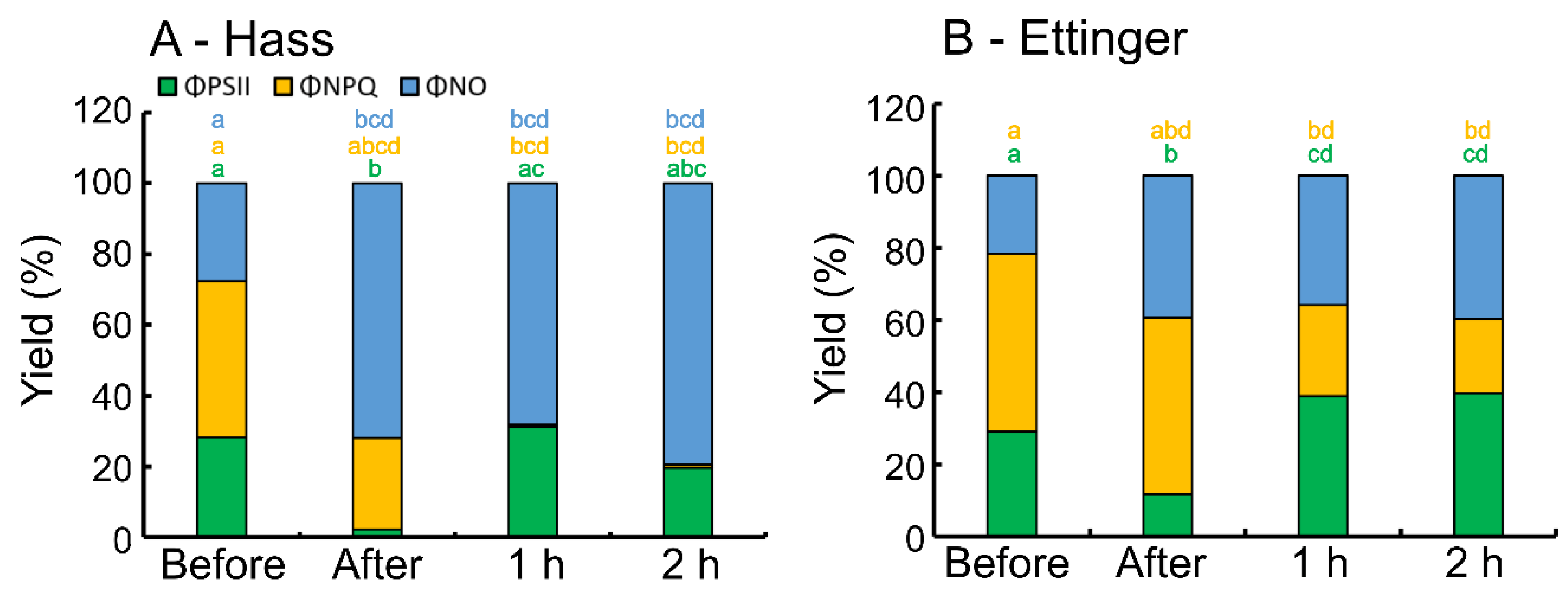
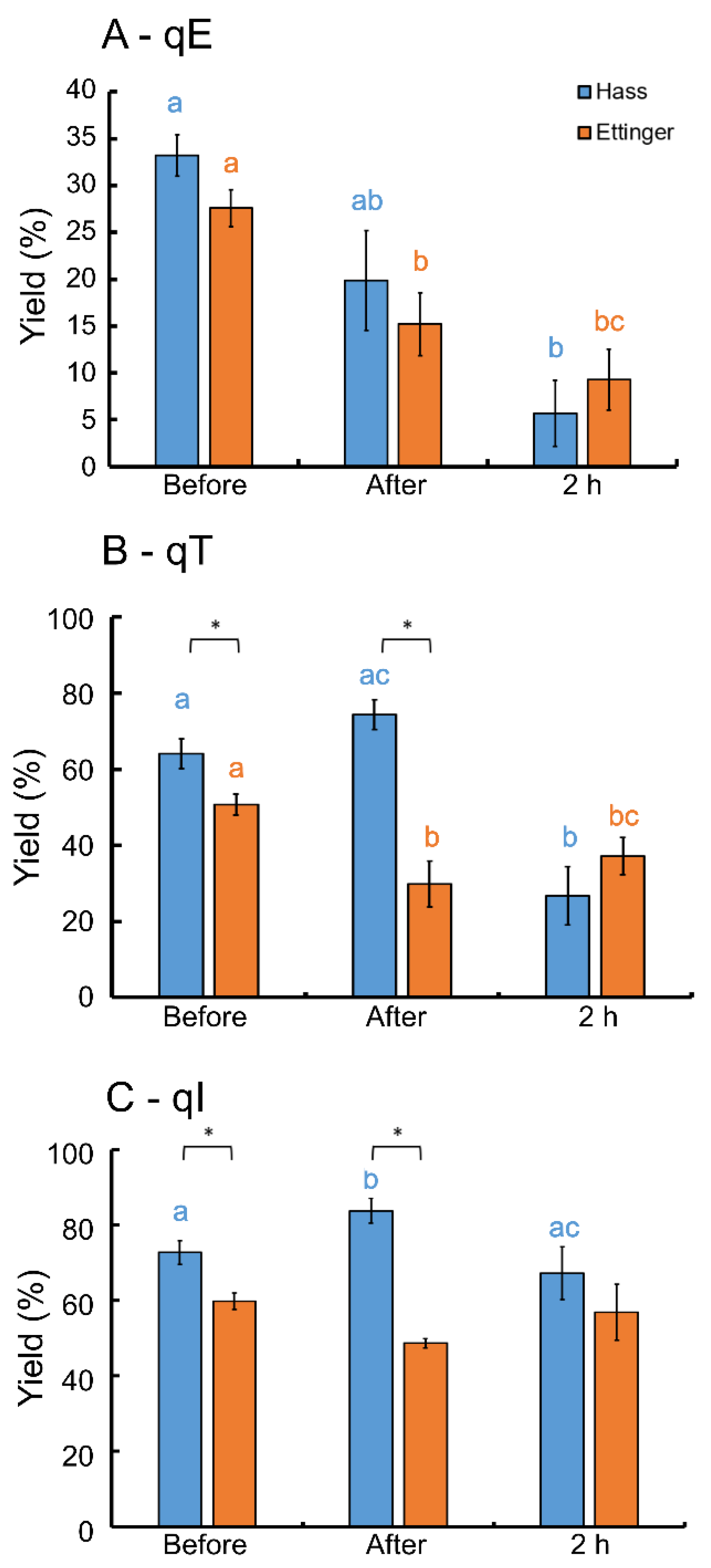
Each group included 4–6 biological repeats coming from branches in the laboratory, or that were measured on intact leaves in the field. Each group was checked for outliers that were replaced with a median value of that group if found. In case only two groups were compared, a t-test was used once both the Shapiro–Wilk’s test for normality and Levene tests for homogeneity of variances were met. In case one of the tests was violated, a Wilcoxon U test was used instead. In case more groups were to be compared, a one-way ANOVA was used if both Shapiro–Wilk and Levene’s tests were satisfied. In case Levene test was violated, ANOVA was analyzed with Welch’s correction. In case a Shapiro–Wilk test was violated, Kruskal–Wallis non-parametric test was selected. Post-hoc multiple comparisons were performed with Tukey’s honest significant difference or a Dunn–Bonferroni test in parametric or non-parametric cases, respectively. Differences in repeated measurements were checked with a repeat-measures ANOVA or Friedman’s test for parametric or non-parametric cases, respectively. Post-hoc multiple comparisons were performed with a Bonferroni adjustment or Wilcoxon U test in the case of parametric or non-parametric cases, respectively. Statistically significant differences were always determined at p < 0.05. Statistical calculations were performed in R language [18] with R studio [19].

3. Results

Two cultivars of avocado were exposed to frost stress under laboratory conditions (Figure 1). The experiment appreciates the effect that frost stress has on the photosynthetic apparatus light utilization capabilities. Before the frost stress, 40% of the total absorbed energy was diverted to photoprotective measures in leaves of Hass, about 30% went to photochemistry, and the rest were passively dissipated (Figure 1A).
In Hass, six hours later, after frost stress in the dark, energy that was diverted towards photochemistry was substantially reduced to a minimum while energy diverted toward regulated dissipation decreased by half (Figure 1A, second bar from the left). About 70% of the absorbed energy was passively dissipated. Then, at both the 1 h and 2 h mark under light conditions, energy diverted to photochemistry returned to its original level before the frost stress (Figure 1A ‘1 h’ and ‘2 h’ bars, green fraction), but energy diverted towards the regulated dissipation process was completely minimized, and the majority of the energy was passively dissipated (Figure 1A ‘1 h’ and ‘2 h’, blue fraction). The distribution of the absorbed energy was similar in the two cultivars (Figure 1B, ‘Before’). However, in Ettinger following the frost stress, the amount of energy diverted towards photochemistry reduced to 30% (Figure 1B, ‘After’). The photoprotective activity did not change considerably (Figure 1B, yellow fraction compares ‘Before’ and ‘After’ columns). The non-regulated energy dissipation in Ettinger multiplied (Figure 1B, blue fraction compares ‘Before’ and ‘After’ columns). Along the light period outdoors, energy diverted towards photochemistry in Ettinger increased beyond the starting level (Figure 1B, ‘1 h’ and ‘2 h’, green fractions). However, in contrast to Hass, energy was still diverted via regulated dissipation processes, even if it was reduced by half (Figure 1B, ‘1 h’ and ‘2 h’, yellow fractions).
We were interested in elucidating the extent that each of the three main processes of photoprotection were involved before and after the frost stress in the two cultivars, given that in Hass the energy diverted to the photoprotective mechanisms was minimized after the frost stress. There was no significant difference between the two cultivars at each time point with regard to the energy-dependent quenching (Figure 2A). State transitions and photoinhibition already differ between the two cultivars at the beginning of the experiment, prior to the frost stress treatment (Figure 2B,C, ‘Before’ time instance). After frost stress and during the light period, the energy dependent process for both cultivars decreased proportionally (Figure 2, Panel A, qE). The energy dependent process was shut down for the Hass concomitant with the energy that was not diverted to it (compare the 2 h point in Figure 2A to the 2 h in Figure 1A). In the case of the Ettinger, there was no difference between the time point right after the stress and 2 h in the light, exactly as expected.
The state transition energy quenching process, which was significantly higher in Hass than Ettinger before the frost stress, remained much more activated for Hass than Ettinger right after the stress (Figure 2B, ‘After’ time instance). As seen for the energy-dependent quenching, the activity of this process in Hass was minimized after two hours (compare Figure 2B blue bars to the minimum fraction of ΦNPQ for Hass in Figure 1A at the 2 h mark). The main differences between the cultivars, state transition wise, was that the response to the frost stress occurred right before and right after the frost stress (Figure 2B, asterisks). Photoinhibition-dependent quenching, which is related to the regeneration of the D1 peptide within PSII, increased in Hass right after the frost stress, but did not change for Ettinger (Figure 2C). It therefore seems that the decision to divert energy to the photoprotective mechanism or not, was made during the dark period in the frost in both cultivars. Eventually, later on—during the light period—we observed a minimal difference between the cultivars between the rates of the photoprotective mechanisms’ relaxation, despite the fact that the Hass apparatus did not divert energy to the regulated processes as seen in Ettinger. This implies that the main difference between the two cultivars’ response to frost stress lies at a regulation at the PSII level and is not merely a response of the photoprotective mechanisms to the inflicted stress.
We therefore focused on analyzing the variable fluorescence of the Kautzky effect [20]. This test records the rise of fluorescence at the very beginning of the illumination period at very short time intervals. This enables us to document the reduction in factors downstream of PSII, starting at the reduction in the Qa. By this, we limit the analysis strictly to the frost stress itself—i.e., what happens before and after exposure with the intact photosynthetic apparatus of avocado leaves to frost stress in the dark (Figure 3). The two transients already differ substantially before the frost stress. The first inflection point, which reports on the saturation of Qa recorded at ~103 µs for both cultivars was 30% higher in Ettinger when compared to that of Hass (Figure 3A,B, green curves). Furthermore, the second inflection point, which relates to the saturation of the plastoquinone pool, was obtained faster in Ettinger than in Hass (Figure 3A,B, around 104 µs). Right after the frost stress, the transient for Ettinger changes considerably at the first inflection point, while the transient for Hass showed almost no difference between the two states (Figure 3A,B, red curves). Finally, the decline towards the end of the recording is much steeper in Hass than in Ettinger (Figure 3A,B, red curves around 106 µs).
Parameterization of the fluorescence transient curves revealed that Hass obtained more PSII units with smaller antenna size than Ettinger before the frost stress (Table 1, Density of Reducing PSIIs). However, this does not necessarily lead to a higher linear electron transport rate towards PhotoSystem I (PSI), the contrary is true. There were significantly fewer electrons transferred toward the acceptor side of PSI in case of Hass than in Ettinger prior to the frost stress (Table 1, Electron transfer to the PSI acceptor side). After experiencing frost stress, both cultivars responded differently, where Hass increased its antenna size, while not changing the density of the PSIIs (Table 1, apparent antenna size). This resulted in increased electron transfer toward the plastoquinone pool and also increased the linear electron flow towards PSI. The Ettinger decreased its antenna size while the density of its PSIIs also did not change significantly.
The marked difference between the two cultivars under frost stress was found in the density of ‘closed’ PSIIs, which dissipate excess energy instead of performing photochemistry (Table 1, Density of energy-dissipating PSIIs). Ettinger had almost four-fold higher dissipating PSIIs than Hass, although this difference was not statistically significant between the two cultivars. This occurred since the closed PSIIs variance in Ettinger was much higher than that of the Hass. This implies a larger range of plasticity in Ettinger to frost stress than in Hass.
Based on this misinterpretation of frost stress by Hass as observed in the photosystem’s functional organization (Table 1), we postulated that the Hass cultivar general response to photosynthetic stress is less regulated than Ettinger. Thus, we suspected that the differences in variability between the two cultivars may also manifest under natural (i.e., non-stressed) conditions. Therefore, we performed a light response curve in the orchard early in the morning immediately before sunrise on both cultivars (Figure 4). At this point, the photosynthetic apparatus is completely oxidized and regenerated. Maximum photosynthetic activity of Hass was four-times smaller than that of Ettinger. While Ettinger reached a plateau in high light intensities, Hass photosynthetic activity declined after reaching maximum activity at one-third of the intensity of that of Ettinger (Hass asymptotes at 300 µmol photons m−2 s−1 while Ettinger asymptotes at 1000 µmol photons m−2 s−1), implying photoinhibition of the Hass reaction centers.
By parameterizing the light response curve with a statistical fit, several characteristics are extracted (Table 2). Light use efficiency (LUE) is represented by the initial slope of the light response curve and reports on the number of photons needed to excite one electron within the PSII complexes (Table 2, Slope). The maximum photosynthetic activity was almost twice as high in Ettinger compared with Hass (Table 2, Pm), and the light intensity at which the maximum activity was reached was almost 230% higher in Ettinger than in Hass (Table 2, Im), which explains its tendency to generate ROS. Finally, the size of the antenna, or the time at which the PSII antenna reached saturation and energy started to be dissipated by photoprotection, happened much faster in Hass than Ettinger, where the antenna size was almost 2.5-times smaller in Hass than Ettinger (Table 2, Ik).

4. Discussion

This study describes the response of the photosynthetic apparatus of two avocado cultivars to frost stress at various time points before, during, and right after the stress. The Ettinger response to frost stress agrees with the common dogma [21]—a compromised photosystem will divert most of the absorbed energy towards photoprotective mechanisms (Figure 1A). Havaux et al. (2000) [22] showed that during chilling stress, carotenoid and flavonoid levels increased over time while the NPQ portion increased with the process. This is also true at both high and low irradiances, where the NPQ process takes on a constant high activity upon infliction of stress to the plant [23]. On the other hand, the response of Hass to frost stress was attenuated considerably. First, right after the frost stress, photochemistry was reduced to a minimum (Figure 1A, ‘After’), and later, during the light, absorbed energy that was diverted towards the photoprotective mechanisms was minimized as well. Quantum yield significantly decreased in corn (Z. mays) [24], Arabidopsis (A. thaliana) [25], and potato (S. toberosum) leaves [26] in response to water stress, silencing of two membranal ion transporters, and sap sucked by insects, respectively. In the case of potato, upon interaction with the insect, necrotic patches appeared in the leaf tissues. Therefore, each of the responses of subtropical species to frost that were documented in the past—for dehydration, increase in ROS, and disruption in ions due to membrane malfunction—can be the cause for the abrupt silencing in the quantum yield of PSII. We suggest further investigating this point in order to understand what caused this phenomenon. Next, photoprotective-related energy was minimized in Hass but not in Ettinger in the light period after the frost stress (Figure 1A). This finding is corroborated by other documented knowledge, showing that—in general—freezing stress will deteriorate the organization of the thylakoid membranes [21]. This will decrease the stabilization of the photosystems, and by that will reduce the electron transport rate and carbon assimilation [2]. The effect of minimizing the energy diverted towards regulated photoprotection was shown in other studies, for example, through the use of artificial inhibitors that prevented electron transfer between the donor side of PSII, Qa, and the plastoquinone pool [27]. Similar findings were reported by the co-authors for apple trees inhibited by atrazinone group which is used as a natural fruitlet thinner [13]. This implies that the regulation of frost response in avocado is found at PSII level [28], rather than the accompanying photoprotective mechanism. To our best knowledge, this is the first time that a minimization of energy diversion towards regulated photoprotective mechanisms are documented for plants and crops in natural conditions, i.e., without the use of artificial inhibitors. Absorbed light energy diversion towards the photoprotective mechanism is minimized in Hass, and its photoprotective mechanism activity also decreased in the light after the frost stress (Figure 2). If during frost stress the principal damage occurs in the thylakoid membranes, it is reasonable that the photoprotective mechanisms that relate to the photosynthetic membranes will also be damaged (Figure 2A,B). Lootens P. et al. [29] report that in response to chilling stress in industrial chicory (C. intybus), the NPQ mechanism declines in the three components of relaxation time coefficients, but more severe decline occurs in the qT and qI relaxation coefficients. Frankart et al. [30] show that the application of artificial herbicides to common duckweed (Lemna minor) resulted in a decrease in the magnitude of relaxation constant coefficients, which corroborate our results. If there is damage to the thylakoid membrane, then the manifestation will be a decrease in the processes that stem out of it. The fact that there was no difference in the qI between Hass and Ettinger (Figure 2C) was probably due to the fact that we examined only the very first hours after the stress, and not over days. The main differences between the two cultivars occurred even before the frost stress, which implies a different strategy of photoprotection, and corroborates our hypothesis that the difference between the two cultivars is regulated at PSII level rather than the photoprotective mechanism (Figure 2B,C).
If indeed the natural sensitivity towards frost stress in Hass is higher than in Ettinger, it should be visible at the photosystems level (Figure 3 and Table 1). The Hass photosynthetic apparatus seemed to interpret the frost stress incorrectly. Jedmowsky et al. [31] reports on a decline in the first phase of the transient of wild barley under severe drought stress, i.e., limitation of water to the tissues. This corroborates the general recurring theme in this study that, already after 6 h in freezing temperature in the dark, dehydration of the tissues may occur which leads to water limitation. In the case of Hass, it increased its antenna complex size and the rate of linear electron flow (Table 1) which may push its apparatus to increase ROS generation. This fact is corroborated by the study of Weil et al. [3], showing that Hass has a less ROS scavenging activity when compared to Ettinger following frost stress. Increase in the apparent antenna size due to stress conditions was documented by Srivastava et al. [32]. The authors found that the PSII increases its apparent antenna size when pea (Pisum sativum) experiences hot and dry weather, implying again the relation of water limitation to the frost stress. In their study, the authors noted that additional local maximum is then present in the fluorescence transient plot (the so-called K point within the fluorescence transient); however, this was not visible in our study. This misinterpretation of environmental conditions should manifest in the general behavior of the two cultivars (Figure 4 and Table 2). Hass maximum photosynthetic activity was almost four times lower than that of Ettinger under non-stressed conditions. In fact, the sensitivity of Hass resulted in a photoinhibition profile of moderate to high light intensities (Figure 4, the decline in maximum activity at 1000 µmol photons m−2 s−1).
The main limitation of this study is the fact that some of the experiments were performed on detached branches under laboratory conditions. We suggest that this experiment should be repeated under field conditions in order to corroborate the above findings. Another suggestion would be to include, in future studies, additional avocado cultivars in order to elaborate on the natural sensitivity of avocado to frost stress. We foresee the implementation of the advanced chlorophyll a fluorescence methodology in frost stress detection and characterization in the field of agronomy sciences. Chlorophyll a fluorescence has reached a point where the methodology is sound, easy-to-use with diverse documentations and examples [33,34]. We therefore wish that more end-users will use these methodologies to unravel sub-optimal photosynthetic performances in the future. This step will turn the light reactions of the photosynthetic apparatus a into a target for natural variation selection for better crops which are more resilient to stress in their natural environment.

5. Conclusions

This study articulates the use of chlorophyll a fluorescence technique as a way to detect natural variations in crops. By comparing the response of two avocado cultivars to frost stress, we were able to determine—at the level of light utilization within their photosynthetic apparatus—what are the kinetic differences, chlorophyll fluorescence wise, between them. We found that in the case of the frost sensitive Hass, the cultivar misinterprets the decrease in photosynthesis as a need in more light harvesting complexes. This results in an opposite outcome to the desired one: an increase in the probability to accumulate ROS when the apparatus is found in a compromised state. This misinterpretation was found to exist for Hass not only during frost stress, but rather under natural conditions as well, which manifests in a reduced ability to perform photosynthesis. This study opens various future research routes, especially in the most popular avocado cultivar, Hass. Therefore, understanding how to increase its resilience via the photosynthetic apparatus may aid in increasing its fruit yield in the field. Additionally, the use of techniques that are very simple and easy to operate opens new opportunities for agriculture experts and photosynthesis specialists that will be able to expand their understanding of the response of crops to environmental conditions via a deeper understanding of the response of the photosynthetic apparatus to stress in general, a technique that was kept mostly in academic studies to date.

Author Contributions

Conceptualization, L.R. and O.L.; Formal analysis, O.L.; Funding acquisition, L.R. and O.L.; Investigation, A.W.; Methodology, O.L.; Project administration, O.L.; Resources, L.R.; Supervision, L.R.; Validation, A.W., D.T. and O.L.; Visualization, O.L.; Writing—original draft, A.W., L.R., D.T. and O.L.; Writing—review and editing, A.W., L.R., D.T. and O.L. All authors have read and agreed to the published version of the manuscript.

Funding

This study received no external funding.

Institutional Review Board Statement

Not applicable.

Informed Consent Statement

Not applicable.

Data Availability Statement

Data is available upon request from the corresponding author.

Conflicts of Interest

The authors declare no conflict of interest.

References

  1. Rendón-Anaya, M.; Ibarra-Laclette, E.; Méndez-Bravo, A.; Lan, T.; Zheng, C.; Carretero-Paulet, L.; Perez-Torres, C.A.; Chacón-López, A.; Hernandez-Guzmán, G.; Chang, T.-H. The Avocado Genome Informs Deep Angiosperm Phylogeny, Highlights Introgressive Hybridization, and Reveals Pathogen-Influenced Gene Space Adaptation. Proc. Natl. Acad. Sci. USA 2019, 116, 17081–17089. [Google Scholar] [CrossRef] [Green Version]
  2. Joshi, N.C.; Yadav, D.; Ratner, K.; Kamara, I.; Aviv-Sharon, E.; Irihimovitch, V.; Charuvi, D. Sodium Hydrosulfide Priming Improves the Response of Photosynthesis to Overnight Frost and Day High Light in Avocado (Persea Americana Mill, Cv. ‘Hass’). Physiol. Plant. 2020, 168, 394–405. [Google Scholar] [CrossRef] [Green Version]
  3. Weil, A.; Sofer-Arad, C.; Bar-Noy, Y.; Liran, O.; Rubinovich, L. Comparative Study of Leaf Antioxidant Activity as a Possible Mechanism for Frost Tolerance in ‘Hass’ and ‘Ettinger’Avocado Cultivars. J. Agric. Sci. 2019, 157, 342–349. [Google Scholar] [CrossRef]
  4. Rivero, R.M.; Mittler, R.; Blumwald, E.; Zandalinas, S.I. Developing Climate-Resilient Crops: Improving Plant Tolerance to Stress Combination. Plant J. 2022, 109, 373–389. [Google Scholar] [CrossRef] [PubMed]
  5. Bar-Noy, Y.; Sofer-Arad, C.; Perel, M.; Cohen, H.; Senesh, N.; Noy, M.; Rubinovich, L. Frost protection efficiency evaluation in avocado with a horizontal wind machine. Fruits Int. J. Trop. Subtrop. Hortic. 2019, 74, 124–129. [Google Scholar] [CrossRef]
  6. Larcher, W. Climatic Constraints Drive the Evolution of Low Temperature Resistance in Woody Plants. J. Agric. Meteorol. 2005, 61, 189–202. [Google Scholar] [CrossRef] [Green Version]
  7. Asada, K. Production and Scavenging of Reactive Oxygen Species in Chloroplasts and Their Functions. Plant Physiol. 2006, 141, 391–396. [Google Scholar] [CrossRef] [PubMed] [Green Version]
  8. Powles, S.B.; Berry, J.A.; Björkman, O. Interaction between Light and Chilling Temperature on the Inhibition of Photosynthesis in Chilling-Sensitive Plants*. Plant Cell Environ. 1983, 6, 117–123. [Google Scholar] [CrossRef]
  9. Allen, D.J.; Ratner, K.; Giller, Y.E.; Gussakovsky, E.E.; Shahak, Y.; Ort, D.R. An Overnight Chill Induces a Delayed Inhibition of Photosynthesis at Midday in Mango (Mangifera indica L.). J. Exp. Bot. 2000, 51, 1893–1902. [Google Scholar] [CrossRef] [PubMed] [Green Version]
  10. Bauer, H.; Wierer, R.; Hatheway, W.H.; Larcher, W. Photosynthesis of Coffea arabica after chilling. Physiol. Plant. 1985, 64, 449–454. [Google Scholar] [CrossRef]
  11. Zaro, G.C.; Caramori, P.H.; Gaspar de Oliveira, C.M.; Nagashima, G.T.; Rosisca, J.R.; Cavenaghi Prete, C.E. Assessment of cold stress in avocado cultivars based on visual, physiological and biochemical criteria. Aust. J. Crop Sci. 2019, 13, 881–888. [Google Scholar] [CrossRef]
  12. Woolf, A.B.; Cox, K.A.; White, A.; Ferguson, I.B. Low Temperature Conditioning Treatments Reduce External Chilling Injury of ‘Hass’ Avocados. Postharvest Biol. Technol. 2003, 28, 113–122. [Google Scholar] [CrossRef] [Green Version]
  13. Tadmor, Y.; Raz, A.; Reikin-Barak, S.; Ambastha, V.; Shemesh, E.; Leshem, Y.; Crane, O.; Stern, R.A.; Goldway, M.; Tchernov, D.; et al. Metamitron, a Photosynthetic Electron Transport Chain Inhibitor, Modulates the Photoprotective Mechanism of Apple Trees. Plants 2021, 10, 2803. [Google Scholar] [CrossRef]
  14. Kramer, D.M.; Johnson, G.; Kiirats, O.; Edwards, G.E. New Fluorescence Parameters for the Determination of QA Redox State and Excitation Energy Fluxes. Photosynth. Res. 2004, 79, 209–218. [Google Scholar] [CrossRef]
  15. Lichtenthaler, H.K.; Buschmann, C.; Knapp, M. How to Correctly Determine the Different Chlorophyll Fluorescence Parameters and the Chlorophyll Fluorescence Decrease Ratio RFd of Leaves with the PAM Fluorometer. Photosynthetica 2005, 43, 379–393. [Google Scholar] [CrossRef]
  16. Strasser, R.J.; Tsimilli-Michael, M.; Srivastava, A. Analysis of the Chlorophyll a Fluorescence Transient. In Chlorophyll a Fluorescence: A Signature of Photosynthesis; Papageorgiou, G.C., Govindjee, Eds.; Springer: Dordrecht, The Netherlands, 2004; pp. 321–362. ISBN 978-1-4020-3218-9. [Google Scholar]
  17. Eilers, P.H.C.; Peeters, J.C.H. A Model for the Relationship between Light Intensity and the Rate of Photosynthesis in Phytoplankton. Ecol. Model. 1988, 42, 199–215. [Google Scholar] [CrossRef]
  18. R Core Team. R: A Language and Environment for Statistical Computing; R Foundation for Statistical Computing: Vienna, Austria, 2021. [Google Scholar]
  19. RStudio Team. RStudio: Integrated Development Environment for R; RStudio, PBC: Boston, MA, USA, 2022. [Google Scholar]
  20. Govindjee. Sixty-Three Years since Kautsky: Chlorophyll a Fluorescence. Aust. J. Plant Physiol. 1995, 22, 131–160. [Google Scholar]
  21. Allen, D.J.; Ort, D.R. Impacts of Chilling Temperatures on Photosynthesis in Warm-Climate Plants. Trends Plant Sci. 2001, 6, 36–42. [Google Scholar] [CrossRef]
  22. Havaux, M.; Kloppstech, K. The Protective Functions of Carotenoid and Flavonoid Pigments against Excess Visible Radiation at Chilling Temperature Investigated in Arabidopsis Npq and Tt Mutants. Planta 2001, 213, 953–966. [Google Scholar] [CrossRef]
  23. Li, X.-G.; Duan, W.; Meng, Q.-W.; Zou, Q.; Zhao, S.-J. The Function of Chloroplastic NAD (P) H Dehydrogenase in Tobacco during Chilling Stress under Low Irradiance. Plant Cell Physiol. 2004, 45, 103–108. [Google Scholar] [CrossRef] [Green Version]
  24. DeTar, R.A.; Höhner, R.; Manavski, N.; Blackholm, M.; Meurer, J.; Kunz, H.H. Loss of Salt Overly Sensitive 1 prevents virescence in chloroplast K+/H+ EFFLUX ANTIPORTER-deficient mutants. Plant Physiol. 2022, kiac142. [Google Scholar] [CrossRef] [PubMed]
  25. Chen, Z.; Liu, Z.; Han, S.; Jiang, H.; Xu, S.; Zhao, H.; Ren, S. Using the diurnal variation characteristics of effective quantum yield of PSII photochemistry for drought stress detection in maize. Ecol. Indic. 2022, 138, 108842. [Google Scholar] [CrossRef]
  26. Sperdouli, I.; Andreadis, S.S.; Adamakis, I.D.S.; Moustaka, J.; Koutsogeorgiou, E.I.; Moustakas, M. Reactive Oxygen Species Initiate Defence Responses of Potato Photosystem II to Sap-Sucking Insect Feeding. Insects 2022, 13, 409. [Google Scholar] [CrossRef]
  27. Klughammer, C.; Schreiber, U. Complementary PS II Quantum Yields Calculated from Simple Fluorescence Parameters Measured by PAM Fluorometry and the Saturation Pulse Method. PAM Appl. Notes 2008, 1, 27–35. [Google Scholar]
  28. Krüger, G.H.; Tsimilli-Michael, M.; Strasser, R.J. Light stress provokes plastic and elastic modifications in structure and function of photosystem II in camellia leaves. Physiol. Plant. 1997, 101, 265–277. [Google Scholar] [CrossRef]
  29. Lootens, P.; Devacht, S.; Baert, J.; Van Waes, J.; Van Bockstaele, E.; Roldàn-Ruiz, I. Evaluation of cold stress of young industrial chicory (Cichorium intybus L.) by chlorophyll a fluorescence imaging. II. Dark relaxation kinetics. Photosynthetica 2011, 49, 185–194. [Google Scholar] [CrossRef]
  30. Frankart, C.; Eullaffroy, P.; Vernet, G. Comparative effects of four herbicides on non-photochemical fluorescence quenching in Lemna minor. Environ. Exp. Bot. 2003, 49, 159–168. [Google Scholar] [CrossRef]
  31. Jedmowski, C.; Brüggemann, W. Imaging of fast chlorophyll fluorescence induction curve (OJIP) parameters, applied in a screening study with wild barley (Hordeum spontaneum) genotypes under heat stress. J. Photochem. Photobiol. B Biol. 2015, 151, 153–160. [Google Scholar] [CrossRef]
  32. Srivastava, A.; Guissé, B.; Greppin, H.; Strasser, R.J. Regulation of antenna structure and electron transport in photosystem II of Pisum sativum under elevated temperature probed by the fast polyphasic chlorophyll a fluorescence transient: OKJIP. Biochim. Et Biophys. Acta BBA-Bioenerg. 1997, 1320, 95–106. [Google Scholar] [CrossRef] [Green Version]
  33. Baker, N.R. Chlorophyll Fluorescence: A Probe of Photosynthesis in Vivo. Annu. Rev. Plant Biol. 2008, 59, 89–113. [Google Scholar] [CrossRef] [Green Version]
  34. Kalaji, H.M.; Schansker, G.; Ladle, R.J.; Goltsev, V.; Bosa, K.; Allakhverdiev, S.I.; Brestic, M.; Bussotti, F.; Calatayud, A.; Dąbrowski, P. Frequently Asked Questions about in Vivo Chlorophyll Fluorescence: Practical Issues. Photosynth. Res. 2014, 122, 121–158. [Google Scholar] [CrossRef] [PubMed] [Green Version]
Figure 1. Distribution of absorbed light energy in avocado cultivars Hass (A) and Ettinger (B) (P. americana Mill) before and after frost stress. Each bar is divided into: green—yield of photochemistry of PSII (ΦPSII); yellow—yield of regulated non-photochemical quenching of PSII (ΦNPQ); and blue—yield of the non-regulated energy dissipation of PSII (ΦNO), respectively. N = 6, letter annotation refers to statistically significant differences between time instances at p < 0.05.
Agronomy 12 01129 g001
Figure 2. Quantum yield of the photoprotective mechanisms of two cultivars of avocado during cold stress. Panels (AC) represent energy-dependent quenching (qE), State transition-dependent quenching (qT) and photoinhibition-dependent quenching (qI), respectively. The colors blue and orange represent the two cultivars Hass and Ettinger, respectively. N = 6, error bars represent standard error of the mean. Brackets pointing down with asterisks represent a statistically significant difference between the two cultivars on the respected time instance. Colored letters with annotation present statistically significant difference between time points for each cultivar. Statistical significance was determined at p < 0.05.
Agronomy 12 01129 g002
Figure 3. Double normalized fluorescence transient profiles for two cultivars of avocado (P. Americana Mill) before and after frost stress. Panels (A,B) represent data for cultivars Hass and Ettinger, respectively. Each panel includes two curves which are average of six biological repeats (N = 6). The colors green and dark red represent information gathered right before (‘Before’) and right after (‘After’) cold stress inflicted on the samples.
Agronomy 12 01129 g003
Figure 4. Response of the relative electron transport rate (rETR) of Hass (blue) and Ettinger (dark red) avocado (P. americana Mill.) cultivars to light at non-stressed conditions. The connecting dashed lines are the averaged statistical fit on each of the respected set of points (R2 > 0.98). Error bars represent standard error of the mean.
Agronomy 12 01129 g004
Table 1. Analyzed parameters of the fluorescence transient curve for two cultivars of avocado right before and right after frost stress. N = 6, error represents standard error of the mean.
StressApparent Antenna SizeDensity of Reducing PSIIsDensity of Energy Dissipating PSIIsElectron Transfer Qa to QbElectron Transfer to PSI Acceptor Side
HassBefore1.68 ± 0.08 * ’0.60 ± 0.02 *--0.90 ± 0.02 ’0.22 ± 0.02 * ’
After1.89 ± 0.08 * ’0.52 + 0.020.16 ± 0.031.03 ± 0.08 ’0.29 ± 0.01 ’
EttingerBefore1.99 ± 0.08 * ’0.51 ± 0.02 *--0.84 ± 0.060.25 ± 0.01 *
After1.86 ± 0.03 * ’0.54 ± 0.010.78 ± 0.311.2 ± 0.010.28 ± 0.01
* Statistically significant difference between the two cultivars. ’ Statistically significant difference between two time points for each cultivar.
Table 2. Light response curve parameters for two avocado cultivars measured at room temperature conditions. Slope—linear slope at the beginning of each curve; Im—light intensity at the maximum photosynthetic activity; Ik—Light intensity at the transfer from photosynthetic efficient to photoprotective efficient activity; Pm—maximum photosynthetic activity; w—photoinhibition magnitude. N =6, error represents standard error of the mean. Statistical significance difference between the two cultivars was determined at p < 0.05.
CultivarSlope
(e Photons−1)
Im
(µmol Photons m−2 s−1)
Ik
(µmol Photons m−2 s−1)
Pm
(µmol e m−2 s−1)
w
(R.U.)
Hass0.22 ± 0.02295.99 ± 42.28 **74.23 ± 11.17 **21.62 ± 6.15 **2.02 ± 0.40
Ettinger0.27 ± 0.00705.09 ± 68.67 **169.33 ± 30.37 **45.93 ± 7.95 **2.59 ± 0.64
** Statistically significant difference between the two cultivars.
Publisher’s Note: MDPI stays neutral with regard to jurisdictional claims in published maps and institutional affiliations.

