Article

Functional Analysis of BcSNX3 in Regulating Resistance to Turnip Mosaic Virus (TuMV) by Autophagy in Pak-choi (Brassica campestris ssp. chinensis)

1
State Key Laboratory of Crop Genetics & Germplasm Enhancement, Key Laboratory of Biology and Genetic Improvement of Horticultural Crops (East China), Engineering Research Center of Germplasm Enhancement and Utilization of Horticultural Crops, Nanjing Agricultural University, Nanjing 210095, China
2
Nanjing Suman Plasma Engineering Research Institute, Nanjing 210095, China
3
Key Laboratory of Biology and Genetic Improvement of Horticultural Crops, Ministry of Agriculture, Institute of Vegetables and Flowers, Chinese Academy of Agricultural Sciences, Beijing 100081, China
4
Horticulture Research Institute, Shanghai Academy of Agricultural Sciences, Shanghai 201403, China
*
Author to whom correspondence should be addressed.
Agronomy 2022, 12(8), 1757; https://doi.org/10.3390/agronomy12081757
Submission received: 25 May 2022 / Revised: 24 July 2022 / Accepted: 24 July 2022 / Published: 26 July 2022
(This article belongs to the Special Issue Vegetables Breeding for Stress Tolerance and Quality Improvement)

Abstract

:
Sorting nexin protein is a class of highly conserved eukaryotic proteins containing the PX domain. Recent studies related to SNX in plants have focused on the regulation of abiotic stress processes, and there are few studies on the involvement of SNX in biological stress processes in plants. In this paper, a YTH assay and BiFC experiments were conducted twice to show that BcSNX3 (Brassica campestris Sorting nexin 3) interacted with CP and VPg of TuMV, and the interaction between BcATG8h (Brassica campestris autophagy-related gene 8h) and BcSNX3 was also found by YTH and BiFC. The colocalization of BcSNX3 and BcATG8b (Brassica campestris autophagy-related gene 8b) revealed BcSNX3 and autophagosome at the same place in the cell. QRT-PCR analysis showed that TuMV infection promotes the expression of BcSNX3, and the overexpression of this gene hinders the expression of autophagy-related genes and facilitates TuMV infection. VIGS was used to repress the expression level of the BcSNX3 gene in pak-choi to further study the function of BcSNX3 in the infection process of TuMV. After inoculation with TuMV, it was found that the accumulation of viral RNA in BcSNX3-gene-silenced plants was significantly less than in control plants. The accumulation of TuMV virus in the Arabidopsis snx3 knockout mutant was also less than in the wild type after TuMV inoculation. These results suggest that TuMV infection facilitates the expression of BcSNX3, and this gene may promote virus infection by inhibiting autophagy degradation of the virus and interacting with the CP and VPg of the virus. These results lay the foundation for the TuMV resistance breeding of pak-choi.

1. Introduction

Autophagy is a highly conserved substance recycling process prevalent in eukaryotes [1]. In plants, autophagy maintains intracellular homeostasis by breaking down and recovering intracellular substances through a rigorous regulatory pathway and then participates in the plant’s response to various developmental and environmental signals [2,3]. Recent studies have suggested that autophagy is involved in plant response to abiotic stress, for example, under drought and salt stress [4,5,6]. To maintain normal life activities, AtATG18a (Arabidopsis thaliana autophagy-related gene 18a) expression level increases during salt and drought stresses, and AtATG18a is also required for salt- and drought-induced autophagy. These induced AtATG18a-gene-silencing plants are sensitive to salt and drought stress [7]. At the same time, autophagy is also involved in the process of plant response to biological stress [8,9]. For example, NbATG8C1 (Nicotiana benthamiana autophagy-related gene 8 C1) and NbATG8i ((Nicotiana benthamiana autophagy-related gene 8 C1)), the isoforms of ATG8, interacted with citrus leaf blotch virus (CLBV) movement protein (MP) to limit the intercellular spread of the virus in plants [10]. Moreover, a new selective autophagy cargo receptor, OsP3IP, mediated the degradation of rice stripe virus (RSV) P3 and interacted with OsATG8b and P3. The transgenic overexpression of NbP3IP in Nicotiana benthamiana conferred resistance to RSV infection [11]. A virus-induced overexpression of small peptide 1 (VISP1) induced selective autophagy and compromised antiviral immunity by inhibiting SGS3/RDR6-dependent viral siRNA amplification, whereas visp1 mutants exhibited opposite effects [12]. Autophagy also plays an important role in inhibiting cell death and the defense response to the biotrophic pathogens, such as those causing powdery mildews. Under nutritive conditions, Arabidopsis atg2, atg5, atg7, and atg10 mutants showed early senescence and cell death at the late growth stage. These mutants also showed powdery mildew resistance and mold-induced cell death [13]. The AtATG6 (Arabidopsis thaliana autophagy-related gene 6) gene can inhibit programmed cell death (PCD) and improve plant disease resistance [14].
Intracellular protein sorting is essential for maintaining the normal life activities of organisms. SNX is an important regulatory factor in the regulation of disease occurrence in mammals, and it participates in the regulation of important signaling pathways related to tumors and other diseases [15]. SNXs consisted of a series of proteins that are involved in identifying and sorting cargoes during retrograde transport from the endosome to the Golgi or plasma membrane [16,17]. At present, 33 types of SNXs are known in mammals and another six in plants, some of which can bind to retromer [18]. All the SNX proteins contain a phox homology (PX) domain encoded by a sequence of 100–130 amino acids, and they are involved in the endocytosis, sorting, and degradation of loaded proteins to maintain cell signal homeostasis and equilibrium [17,19,20]. Sorting nexins provide diversity for retromer-dependent trafficking events, retromer complexes regulate endosomal sorting, and endosomal sorting plays a central role in development and normal tissue homeostasis [21]. SNX3 regulates the intracellular circulation of the secretory Wnt receptor Wntless. Moreover, the reverse transport pathway of the Wnt sorting receptor Wntless (Wls) also depends on the regulation of SNX3, which is a necessary condition for Wnt secretion and cell proliferation [22,23]. The activation of the Wnt signaling pathway can regulate tumorigenesis and Wls can promote the proliferation of breast cancer cells through the Wnt signaling pathway [24]. The Wnt signaling pathway also plays an important role in the regulation of the cell cycle, metastasis, and apoptosis in lung cancer [25]. As a tumor suppressor, KCTD11 can bind β-catenin to regulate the activity of the Wnt pathway and inhibit the occurrence of lung cancer [26].
The roles of SNX in response to abiotic stress in plants and to disease in mammals have also been studied [27,28], but there are few studies on how SNX responds to biological stress in plants. Here, we describe the involvement of a sorting nexin SNX3 in the plant defense against turnip mosaic virus (TuMV). Overall, we observed that BcSNX3 affected TuMV infection and viral protein accumulation in pak-choi. These results provide a reference for a study of the resistance of TuMV in pak-choi.

2. Materials and Methods

2.1. Plant Materials and Growth Conditions

The pak-choi variety 49CX was used as the experimental material for TuMV infection and turnip yellow mosaic virus (PTY)-induced BcSNX3 gene silencing. 49CX seeds were scattered in Petri dishes covered with moist filter paper and placed at room temperature for 2–3 days. After germination, the seeds were transferred to 32-hole plates containing substrate (roseite: peat soil = [1:2]). Growth conditions: 2 °C/20 °C, light 16 h/ dark 8 h, relative humidity 65–75%. The TuMV infection and VIGS experiments were carried out when the plant was grown to 3–4 leaves.
Nicotiana benthamiana was used for the transient expression assay and TuMV inoculation when tobacco was grown to 4–5 leaves. The tobacco seeds were distributed evenly in a pot containing nutritious soil. When the two cotyledons were fully unfolded, seedlings were transplanted into new square pots containing substrate (roseite: peat soil [1:2]) for them to continue to grow under the same conditions as pak-choi.
Arabidopsis snx3 mutants were purchased from AraShare (a non-profit Arabidopsis Share Center, http://www.arashare.cn, accessed on 20 July 2021). The Col-0 (wild type) and snx3 mutant seeds were placed in a 2 mL centrifuge tube, sterilized 3 times with 75% alcohol, and then washed 3 times with sterile water. The seeds were seeded on solid MS medium and placed upside down in a 4 °C refrigerator for 2 days and then transferred to the light incubator for culture under controlled conditions (25 °C 8-h darkness/16-h light, 40% relative humidity). When two true leaves were formed, the plants were translated into the growth substrate (roseite: peat soil = 2:1), and they grew continually under the same conditions. TuMV was inoculated when the rosettes were open.

2.2. Gene Cloning and Plasmid Construction

Coding sequences of BcSNX3, BcATG8b, and BcATG8h from pak-choi were amplified from pak-choi cDNA with PCR using PrimeSTAR Max DNA polymerase (Takara, Dalian, China). Gateway technology (Invitrogen, Waltham, MA, USA), except otherwise stated, was used to generate the clones reported in this study. These cloned genes were recombined into pDONR221 (Invitrogen) using BP clonase II (Invitrogen) following the manufacturer’s protocol. The insert was then transferred using a recombination to the indicated binary destination vector using LR clonase II (Invitrogen) following the standard conditions and procedure recommended by the supplier. All constructs were confirmed with DNA sequencing.

2.3. DUAL Membrane Yeast-Two-Hybrid (mYTH) Assay

mYTH tests were performed using the DUALmembrane system (Dualsystems Biotech AG, Schlieren, Switzerland) according to the manufacturer’s protocols (Clontech, Mountain View, CA, USA). The full-length CDS of BcSNX3 was recombined into the pPR3N vector and TuMV proteins were recombined into the pBT3 vector; the primers are shown in Table S1. Briefly, Saccharomyces cerevisiae (strain NMY51) were co-transformed with different combinations and then grown on the selection medium lacking leucine and tryptophan (SD/−Leu/−Trp) (Clontech) at 30 °C for up to 4 days [29]. After yeast monoclonal colonies were grown, the monoclones were selected and resuspended in normal saline (0.9% NaCl solution) and diluted 10, 100, and 1000 times successively. A total of 6 μL of the prepared bacteria solution was placed on SD/-Leu/-Trp and SD/-Ade/-His/-Leu/-Trp +25 mM 3-amino-1, 2, 4-triazole (3-AT) (Sigma-Aldrich, St. Louis, MO, USA) solid medium to identify the interactions.

2.4. Transient Protein Expression

Agrobacterium-mediated transient protein expression was followed as previously described [30]. For BiFC, the CP and VPg of TuMV were transferred into the modified pEarleyGate202-yellow fluorescence protein (YFP) vector and the BcSNX3 was transferred into pEar-leyGate201-nYFP. The OD600 was adjusted to 0.6–0.8 for each gene construct. For the subcellular localization assay, the OD600 was adjusted to 0.3–0.4. For colocalization, the OD600 of 104-BcSNX3 and 102-BcATG8b was adjusted to 0.6–0.8, and then these two kinds of bacteria were mixed in equal volume. Three-to-four-week-old N. benthamiana plants were used for agroinfiltration.

2.5. Virus-Induced Gene Silencing (VIGS) Experiment

To acquire pak-choi silencing plants, VIGS was implemented according to a previous report [31]. The 80 bp BcSNX3-specific palindromic DNA fragment (5′–TTAGACAAGATGTCACAAAGGAAAACTGAGACCCTTTCTCGAGAAAGGGTCTCAGTTTTCCTTTGTGACATCTTGTCTAA–3′) was primarily designed and synthesized by the GeneScript company (Nanjing, China). The sequence was inserted into the SnaBI site of the PTY vector, which was linearized, and the PTY-BcSNX3 construct was generated. Thereafter, PTY and PTY-BcSNX3 plasmids were wrapped in gold particles and bombarded into two-week-old seedlings of pak-choi using gene gun-mediated transformation (1300 psi, PDS-1000/He, Bio-Rad, Hercules, CA, USA). Four to five plants were bombarded for one plasmid, and three replicates were carried out. After two weeks, the virus symptoms on leaves became increasingly obvious with the initiation and expansion of new leaves, and this phenomenon was deemed the characterization for the initial screening of positive silencing plants. QRT-PCR was conducted to identify the efficiency of the BcSNX3 gene silenced, while the PTY plants were used as controls.

2.6. TuMV Inoculation

The procedure of TuMV infiltration was conducted according to [32]. For the OD600 of pCambiaTuMV, GFP was adjusted to 0.3–0.4 and then N. benthamiana was infected via Agrobacterium infection when it formed four-to-five true leaves. Two weeks later, whether the plants were successfully infected could be identified by their symptoms, and then the leaves with signs of disease were selected and ground in the phosphate buffer into slurry. The slurry was filtered using gauze and infiltrated into 49CX and Arabidopsis.

2.7. RNA Extraction and qRT-PCR

The leaves of the pak-choi were collected 30 days after inoculation with TuMV, the leaves of N. benthamiana were collected at 3 and 7 days after inoculation with TuMV, and the leaves of Arabidopsis were collected 15 days after inoculation with TuMV. Moreover, total RNA was isolated with a RNAprep Pure Plant Total RNA Isolation Kit (TIANGEN, Beijing, China), after which 1 μg of total RNA was treated with 5× gDNA Clean Reaction Mix (AG, Changsha, China) and reverse transcribed following the protocol provided by the manufacturer using a 5× Evo M-MLV RT Reaction Mix (AG, Changsha, China). An RT-qPCR was accomplished to identify the gene expression levels with a 2× Hieff® qPCR SYBR Green Master Mix (YEASEN, Shanghai, China), with BcActin serving as the internal standard, as we previously described [30]. The primers used in this study are listed in Table S1.

2.8. Statistical Analysis

The experiments reported in this study were repeated at least three times. The data are presented as means SD from one representative experiment, and statistical significance was analyzed using Student’s t-test. Significance values with p < 0.05 were denoted.

3. Results

3.1. TuMV Infection Accelerates the Expression Level of SNX3

To determine the effect of TuMV infection on SNX3, the mRNA levels of SNX3 in mock and TuMV-infected pak-choi and N. benthamiana were analyzed. The results showed that the expression level of BcSNX3 was significantly increased in the plants inoculated with TuMV (Figure 1A). The latter expression levels of the NbSNX3a (Nicotiana benthamiana sorting nexin 3a) and the NbSNX3b (Nicotiana benthamiana sorting nexin 3b) also increased markedly when N. benthamiana was infected by TuMV 7 days post-infiltration (dpi) (Figure 1B,C). Therefore, SNX3 may respond to TuMV infection.

3.2. Subcellular Localization of BcSNX3

An online bioinformatics analysis (https://wolfpsort.hgc.jp/, accessed on 20 June 2021) indicated that BcSNX3 might be located in the nucleus, chloroplast, cytoplasm and peroxysome. To verify this, we generated a construct (35S: YFP-BcSNX3) by fusing BcSNX3 to 35S: YFP, while the vector 35S: YFP was used as a control (Figure 2A). The two constructs were transiently expressed in tobacco (N. benthamiana) epidermal cells using Agrobacterium tumefaciens mediated transformation. We observed that the YFP signal was distributed evenly in the cell membrane and nucleus of epidermal cells when the control vector was used. However, the fluorescence signal was detected in the nucleus and membrane of cells expressing the fusion protein YFP-BcSNX3 (Figure 2B), indicating that BcSNX3 localized to the nucleus.

3.3. BcSNX3 Interacts with CP and VPg

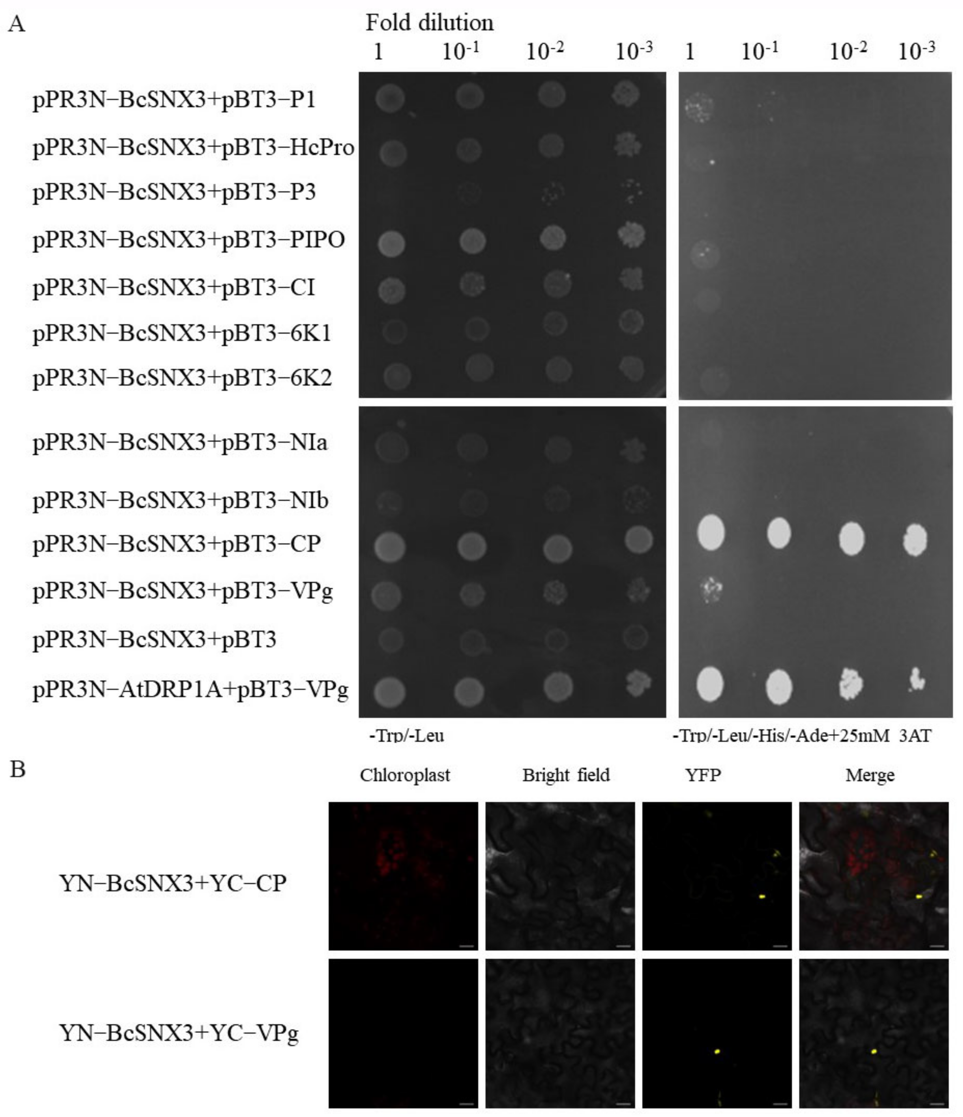
The SNX3 transcriptional level was increased when the pak-choi infected by TuMV. This indicates that SNX3 may interact with TuMV proteins. A YTH experiment was conducted to verify the interaction between BcSNX3 and TuMV protein. The constructed pPR3N−BcSNX3 was combined with the proteins pBT3−P1, pBT3−HC-Pro, pBT3−P3, pBT3−CI, pBT3−PIPO, pBT3−6K1, pBT3−6K2, pBT3−NIa, pBT3−NIb, pBT3−CP and pBT3−VPg of TuMV, and they were transformed into yeast receptor cells (NMY51 strain). The results showed that the yeast transformed with pPR3N-BcSNX3 and pBT3 could not grow on the defective medium. Previous studies showed that yeast that simultaneously transformed pPR3N with PBT3−P1, PBT3−HcPro, PBT3−P3, PBT3−CI, PBT3−PIPO, PBT3−6K1, PBT3−6K2, PBT3−NIA, PBT3−NIb, PBT3−CP and PBT3−VPg could not grow on defective medium [29]. However, pPR3N-BcSNX3, PBT3-CP and PBT3-VPg transformed into yeast at the same time, and the bacterium could grow normally on a defective medium (Figure 3A). The result showed there exist interactions of BcSNX3 with CP and VPg of TuMV.
To further verify the interactions of BcSNX3 with CP and VPg, this study verified the interaction of BcSNX3 with CP and VPg in vivo using a BiFC experimental system. BcSNX3 recombinant vectors with a YFP nitrogen terminal sequence (YN) with CP and VPg recombinant vectors with a YFP carbon terminal sequence (YC) were simultaneously transformed into tobacco epidermal cells. The YFP signal in the cells was observed using a laser confocal microscope (Zeiss, LSM780, Germany) 60–72 h post-infection (hpi). When YN−BcSNX3, YC−CP, YN−BcSNX3 and YC−VPg were injected simultaneously, a yellow fluorescence signal could be observed in tobacco epidermal cells (Figure 3B). These results showed there were interactions of BcSNX3 with CP and VPg.

3.4. BcSNX3 Colocalized with the Autophagosome Marker BcATG8b

In order to verify that BcSNX3 was involved in TuMV-induced autophagy, BcATG8b was inserted into the pEarlygate102 vector containing cyan fluorescence protein (CFP), and then transferred into the agrobacterium GV3101 strain. The fluorescence position was observed after 60–72 hpi. As shown in Figure 4, YFP-BcSNX3 and BcATG8b-CFP were observed at the same position in tobacco cells, indicating that BcSNX3 and BcATG8b had the same expression position in cells, and it could be speculated that BcSNX3 was involved in TuMV-induced autophagy.

3.5. BcSNX3 Interacts with BcATG8h

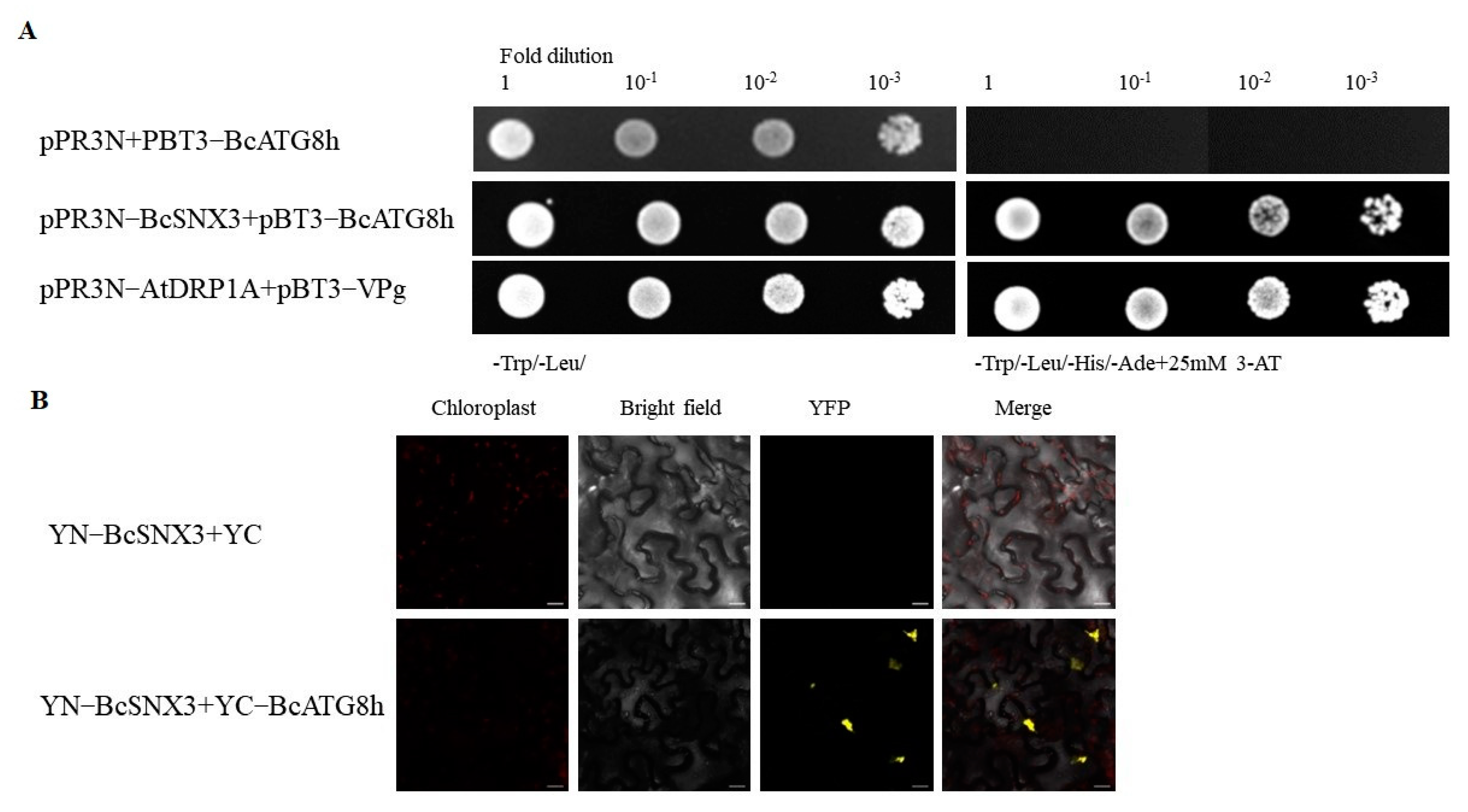
The interactions of BcSNX3 with CP and VPg were verified using YTH and BiFC experiments. The constructed vectors, pPR3N−BcSNX3 and PBT3−BCATG8h, were transformed into yeast competent cells (NMY51 strain). The previous results indicate that the yeast transformed pPR3N-BcSNX3, pBT3 and pPR3N, and PBT3-BcATG8h at the same time could not grow on the defective medium. However, at the same time, yeast transformed pPR3N−BcSNX3, and PBT3−BcATG8h could grow normally on a defective medium (Figure 5A), so there was an interaction between BcSNX3 and BcATG8h.
To further verify the interaction between BcSNX3 and BcATG8h, this study verified the interaction between BcSNX3 and BcATG8h at the protein level through the BiFC experimental system in vivo. The BcSNX3 recombinant vector with YN and BcATG8h recombinant vector with YC was simultaneously transformed into tobacco epidermal cells. After 60-72h infection, the fluorescence signal in the cells was observed using a laser confocal microscope. The results show that the YFP signal could be observed in tobacco epidermal cells when YN−BcSNX3 and YC−BCATG8h were injected simultaneously (Figure 5B). The result further confirmed the interaction between BcSNX3 and BcATG8h.

3.6. Overexpression of BcSNX3 Inhibits the Expression of Autophagy Components and Promotes TuMV Infection in N. benthamiana

We examined the expression of ATG genes in BcSNX3-overexpressed plants using quantitative real-time reverse-transcription PCR (qRT-PCR). The expression levels of all tested six ATG genes, including NbATG2 (Nicotiana benthamiana autophagy-related protein 2), NbATG3 (Nicotiana benthamiana autophagy-related protein 3), NbATG5 (Nicotiana benthamiana autophagy-related protein 5), NbATG7 (Nicotiana benthamiana autophagy-related protein 7), NbATG8a (Nicotiana benthamiana autophagy-related protein 8a) and NbATG9 (Nicotiana benthamiana autophagy-related protein 9), were significantly downregulated in BcSNX3-overexpressed leaves (Figure 6A). We speculated that the overexpression of BcSNX3 inhibits autophagy induced by TuMV infection.
To further study the function of BcSNX3 in response to TuMV infection, agrobacterium infection was used to overexpress BcSNX3 in N. benthamiana leaves and inoculate TuMV at the same time. Quantitative RT-PCR showed that the accumulation of TuMV virus RNA was significantly increased in BcSNX3 overexpressed leaves compared with the leaves-infected buffer and empty vector (Figure 6B).

3.7. BcSNX3 Gene Silencing Inhibits TuMV Infection in Pak-choi

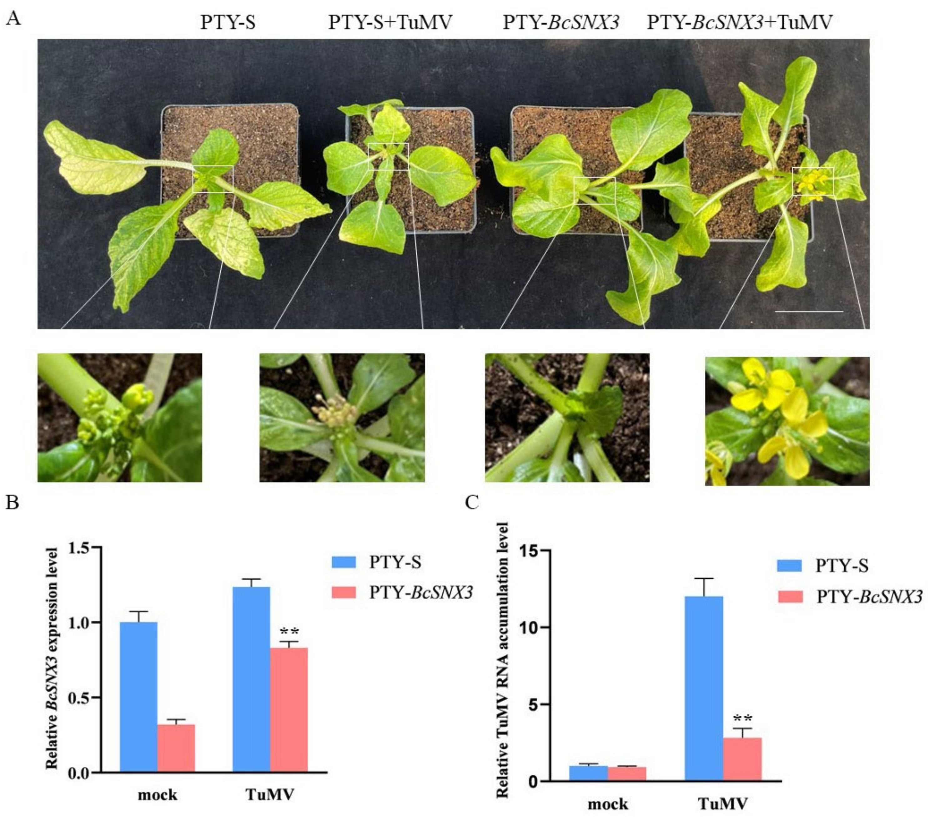
In order to study the effect of BcSNX3 on TuMV infection, the expression of the BcSNX3 gene in pak-choi was silenced by the PTY vector and then inoculated with TuMV to verify the effect of BcSNX3 on TuMV. The expression of the BcSNX3 gene was reduced by the VIGS experiment. Two weeks after 49CX plants were bombarded with a gene gun, the new leaves of silent plants showed obvious symptoms of the PTY virus. The infected plants were sampled, and PTY-S plants were used as a control for quantitative PCR in order to identify the silencing efficiency of BcSNX3. The leaves of silent plants and control plants were selected and ground into slurry by phosphate buffer in turn. Forty-nine plants were pre-inoculated with PTY-BcSNX3 (to silence BcSNX3) and PTY-S (control) juice for 10 days and then inoculated with the ground juice of tobacco leaves containing TuMV-GFP. Compared with the control, BcSNX3-silenced plants showed significantly increased resistance to TuMV. Moreover, the BcSNX3 silenced plants showed normal flowers, while the flower of control plants was withered (Figure 7A). BcSNX3 was silenced in the plants pre-inoculated with PTY-BcSNX3, and we found that the expression level of BcSNX3 decreased in the plants pre-inoculated with PTY-BcSNX3 (Figure 7B). Consistent with the phenotype, lower levels of TuMV genomic RNA were found in BcSNX3-silenced plants compared to controls (Figure 7C). Taken together, these results indicate that BcSNX3 participates in the infection process of TuMV and positively regulates TuMV infection.

3.8. AtSNX3 Gene Knockout Suppressed TuMV Infection in Arabidopsis

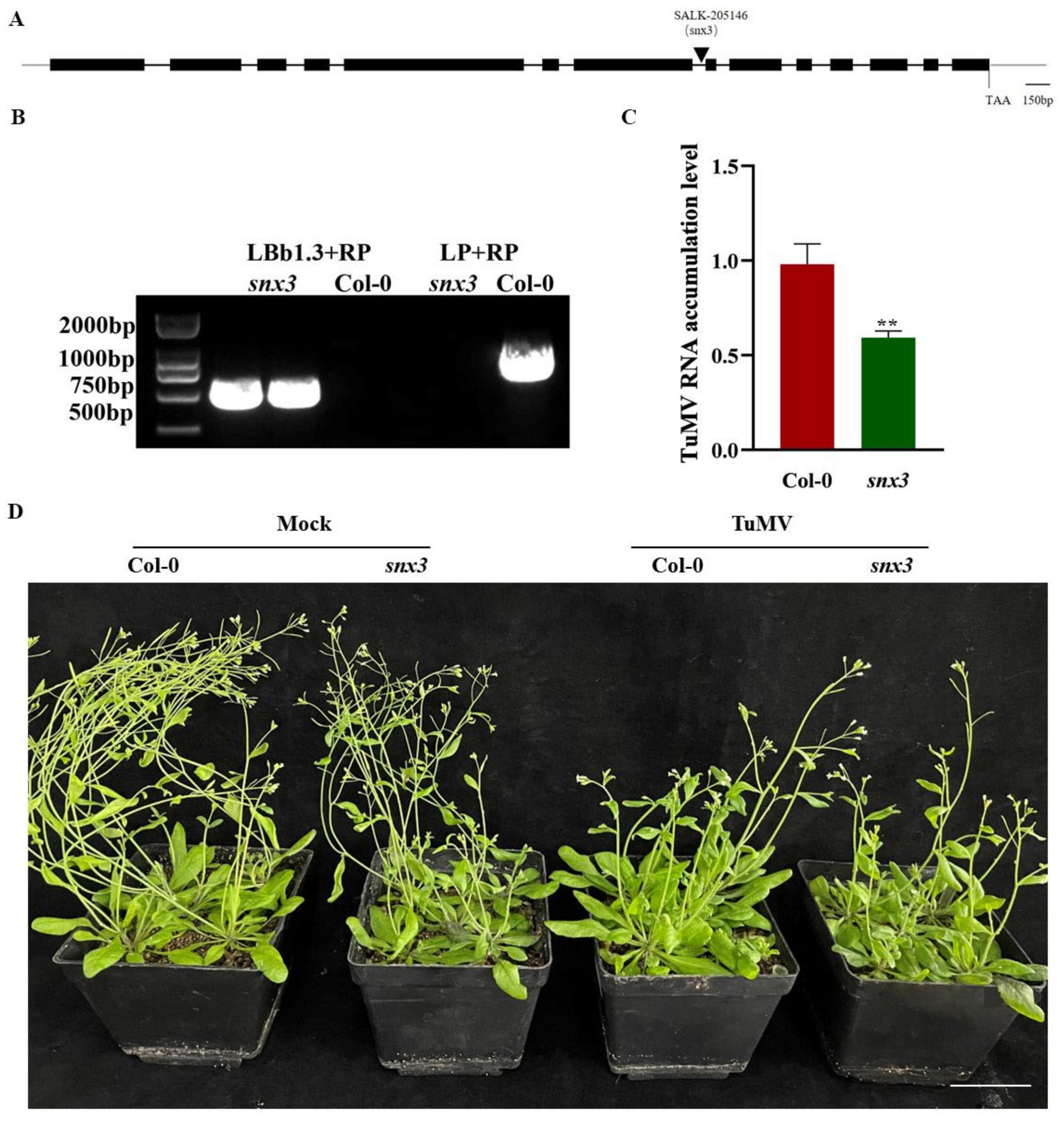
To further test whether SNX3 is essential for TuMV infection, we obtained an Arabidopsis snx3 mutant to conduct the virus inoculation test. Figure 8A is the schematic diagram of the AtSNX3 gene structure. The arrow points to the t-DNA insertion site of the Arabidopsis mutant. In order to detect whether the AtSNX3 mutant (Salk-206146) is homozygous, T-DNA insertion was detected at the DNA level through double-primer PCR. The results are shown in Figure 8B. The length of the 750 bp product could be amplified from the mutant plants, but not from the wild type when LBb1.3+RP primers were used for PCR. However, when using LP+RP primers for PCR, the mutant plants could not be amplified, while the wild type could be amplified with a length of about 1100bp. These results showed that the obtained plants were homozygous mutants with T-DNA insertion of the AtSNX3 gene.
The seeds of identified homozygous mutants (snx3) and wild type (Col-0) were sterilized and seeded on solid MS medium and inoculated with TuMV when the rosette leaves unfolded, with wild type as control. Fifteen days after inoculation, the expression level of CP in the mutant plants was significantly lower than that in the wild-type Arabidopsis (Figure 8C). Meanwhile, the knockout of AtSNX3 suppressed Arabidopsis vegetative growth compared with Col-0 plants (Figure 8D). These results show that AtSNX3 gene knockout inhibited TuMV infection in Arabidopsis.

4. Discussion

Recent studies have shown that SNX genes play an essential role in plant development and involve abiotic stress in plants and defenses against disease in human [27,33,34]. However, the manner in which the SNX gene is activated and regulated in plant–virus interaction remains largely unknown. In this study, we present evidence that TuMV infection induces the expression of an SNX gene, SNX3 in both pak-choi and N. benthamiana (Figure 1). Furthermore, we show that BcSNX3 was localized to the nucleus (Figure 2). The nucleus is where the virus replicates. Therefore, BcSNX3 may play dynamic and contrasting roles in virus infection.
In this study, we found interactions between BcSNX1 and the TuMV CP and VPg through YTH and BiFC experiments (Figure 3). VPg protein is a multifunctional protein that participates in viral replication, translation and movement through an interaction with itself and other viral proteins as host proteins [35,36,37]. NbNdhM may play a protective role in TuMV infection and promote viral infection by inducing the perinuclear aggregation of chloroplast and changing its localization through an interaction with TuMV VPg [38]. CP is the structural protein of TuMV, and it participates in the movement of viruses between cells [39]. CP RNA silencing can improve the disease resistance of transgenic Arabidopsis [40]. In conclusion, we considered that the BcSNX3 may participate in TuMV infection by influencing its replication and diffusion.
The virus is one of the biological stresses suffered by plants, which seriously affects their growth and development. Pak-choi is one of the most important leafy vegetables, and its yield and quality is seriously affected by the virus [41]. Autophagy plays an important role in plant response to virus infection [42,43]. In this study, we found co-localization of BcSNX3 with the autophagosome marker BcATG8b in N. benthamiana (Figure 4). Previous studies have shown that TuMV causes autophagy in plants when it infects them. When TuMV infected plants, the expression of ATG genes was significantly increased in TuMV-inoculated leaves or the new leaves. Therefore, we concluded that BcSNX3 was involved in TuMV-induced autophagy. The interaction between BcATG8h and BcSNX3 was also identified using YTH and BiFC experiments (Figure 5). ATG proteins are associated with autophagy [44], and Atg8 is essential for the formation of autophagic vesicles and their fusion with vacuolar membranes [45,46]. There are nine ATG8 genes in plants, in which silencing ATG8a in tobacco promoted TuMV infection [47] and silencing ATG8f inhibited TuMV infection [48]. This indicates that different ATG8 genes have different effects on virus infection. Therefore, further studies are needed to elucidate how BcSNX3 interacts with BcATG8h to regulate TuMV infection.
In mammals, autophagy is measured by assessing LC3-I to LC3-II conversion and p62 levels through Western blot analyses, and by performing GFP–LC3, PtdIns(3)P and WIPI2 fluorescence microscopy [49]. In plants, Li. et al. observed the structure of an autophagosome through transmission electron microscopy (TEM) and examined the expression of ATG genes using qRT-PCR to measure the activity autophagy [47]. We transiently overexpressed the BcSNX3 gene in N. benthamiana, and the qRT-PCR experiment showed that autophagy-related genes were downregulated and TuMV RNA accumulation was increased in BcSNX3-overexpressed leaves (Figure 6). In mammals, however, SNX induced autophagy and inhibited disease resistance [49]. This requires significant consideration, probably because of the way it works in animals and plants. In addition, in order to further study the function of BcSNX3, the VIGS experiment was used to reduce the expression level of the BcSNX3 gene in pak-choi. After inoculation with TuMV, it was found that the accumulation of viral RNA in silent plants was significantly lower than that in control plants (Figure 7). The accumulation of virus in Arabidopsis SNX3 knockout mutant was also lower than the TuMV inoculation in the wild type (Figure 8). This study shows that TuMV infection accelerates the expression level of BcSNX3, and BcSNX3 protein interacts with BcATG8h to participate in the autophagy process of plant cells. Meanwhile, BcSNX3 promotes viral infection by inhibiting autophagy degradation of the virus and possibly interacting with viral proteins CP and VPg. However, the mechanism of BcSNX3 in response to the TuMV infection of pak-choi still requires further study.

Supplementary Materials

The following supporting information can be downloaded at: https://www.mdpi.com/article/10.3390/agronomy12081757/s1, Table S1: Primers were used in this study.

Author Contributions

Conceptualization, R.Z. and X.H.; formal analysis, R.Z., C.Z. and S.L.; funding acquisition, C.Z., S.L. and X.H.; investigation, R.Z. and Z.F.; methodology, Z.F. and X.H.; project administration, H.Z.; resources, R.Z.; software, S.L.; supervision, Z.F. and X.H.; validation, S.L. and H.Z.; writing—original draft, R.Z.; writing—review and editing, C.Z. All authors have read and agreed to the published version of the manuscript.

Funding

This research was funded by the Fundamental Research Funds for the Central Universities, grant number KYZZ2022004 and the Natural Science Foundation of Jiangsu Province grant number BK20200560.

Institutional Review Board Statement

Not applicable.

Informed Consent Statement

Informed consent was obtained from all subjects involved in the study.

Data Availability Statement

Not applicable.

Acknowledgments

This project was supported by the Fundamental Research Funds for the Central Universities, grant number KYZZ2022004 and the Natural Science Foundation of Jiangsu Province grant number BK20200560. We thank anyone who helped us in this study.

Conflicts of Interest

The authors declare no conflict of interest.

References

  1. Han, S.J.; Yu, B.J.; Wang, Y.; Liu, Y.L. Role of plant autophagy in stress response. Protein Cell 2011, 2, 784–791. [Google Scholar] [CrossRef] [Green Version]
  2. Levine, B.; Klionsky, D.J. Development by Self-Digestion Molecular Mechanisms and Biological Functions of Autophagy. Dev. Cell 2004, 6, 463–477. [Google Scholar] [CrossRef]
  3. Bassham, D.C. Function and regulation of macroautophagy in plants. Biochim. Biophys. Acta (BBA)—Mol. Cell Res. 2009, 1793, 1397–1403. [Google Scholar] [CrossRef] [PubMed] [Green Version]
  4. Zhou, J.; Wang, J.; Yu, J.Q.; Chen, Z.X. Role and regulation of autophagy in heat stress responses of tomato plants. Front. Plant Sci. 2014, 5, 174. [Google Scholar] [CrossRef] [PubMed] [Green Version]
  5. Cheng, F.; Yin, L.-L.; Zhou, J.; Xia, X.-J.; Shi, K.; Yu, J.-Q.; Zhou, Y.-H.; Foyer, C.H. Interactions between 2-Cys peroxiredoxins and ascorbate in autophagosome formation during the heat stress response in Solanum lycopersicum. J. Exp. Bot. 2016, 67, 1919–1933. [Google Scholar] [CrossRef] [PubMed] [Green Version]
  6. Xu, W.; Cai, S.-Y.; Zhang, Y.; Wang, Y.; Ahammed, G.J.; Xia, X.-J.; Shi, K.; Zhou, Y.-H.; Yu, J.-Q.; Reiter, R.J.; et al. Melatonin enhances thermotolerance by promoting cellular protein protection in tomato plants. J. Pineal. Res. 2016, 61, 457–469. [Google Scholar] [CrossRef] [PubMed]
  7. Liu, Y.M.; Xiong, Y.; Bassham, D.C. Autophagy is required for tolerance of drought and salt stress in plants. Autophagy 2009, 5, 954–963. [Google Scholar] [CrossRef] [PubMed] [Green Version]
  8. Ismayil, A.; Yang, M.; Liu, Y.L. Role of autophagy during plant-virus interactions. Semin. Cell Dev. Biol. 2020, 101, 36–40. [Google Scholar] [CrossRef] [PubMed]
  9. Zhang, B.; Shao, L.; Wang, J.; Zhang, Y.; Guo, X.; Peng, Y.; Cao, Y.; Lai, Z. Phosphorylation of ATG18a by BAK1 suppresses autophagy and attenuates plant resistance against necrotrophic pathogens. Autophagy 2021, 17, 2093–2110. [Google Scholar] [CrossRef]
  10. Niu, E.; Liu, H.; Zhou, H.; Luo, L.; Wu, Y.; Andika, I.B.; Sun, L. Autophagy Inhibits Intercellular Transport of Citrus Leaf Blotch Virus by Targeting Viral Movement Protein. Viruses 2021, 13, 2189. [Google Scholar] [CrossRef]
  11. Jiang, L.; Lu, Y.; Zheng, X.; Yang, X.; Chen, Y.; Zhang, T.; Zhao, X.; Wang, S.; Zhao, X.; Song, X.; et al. The plant protein NbP3IP directs degradation of Rice stripe virus p3 silencing suppressor protein to limit virus infection through interaction with the autophagy-related protein NbATG8. New Phytol. 2021, 229, 1036–1051. [Google Scholar] [CrossRef]
  12. Tong, X.; Liu, S.-Y.; Zou, J.-Z.; Zhao, J.-J.; Zhu, F.-F.; Chai, L.-X.; Wang, Y.; Han, C.; Wang, X.-B. A small peptide inhibits siRNA amplification in plants by mediating autophagic degradation of SGS3/RDR6 bodies. EMBO J. 2021, 40, e108050. [Google Scholar] [CrossRef]
  13. Wang, Y.; Wu, Y.; Tang, D. The autophagy gene, ATG18a, plays a negative role in powdery mildew resistance and mildew-induced cell death in Arabidopsis. Plant Signal Behav. 2011, 6, 1408–1410. [Google Scholar] [CrossRef] [Green Version]
  14. Patel, S.; Dinesh-Kumar, S.P. Arabidopsis ATG6 is required to limit the pathogen-associated cell death response. Autophagy 2008, 4, 20–27. [Google Scholar] [CrossRef] [Green Version]
  15. Lu, Y.; He, P.; Zhang, Y.; Ren, Y.; Zhang, L. The emerging roles of retromer and sorting nexins in the life cycle of viruses. Virol. Sin. 2022, 37, 321–330. [Google Scholar] [CrossRef]
  16. Teasdale, R.D.; Loci, D.; Houghton, F.; Karlsson, L.; Gleeson, P.A. A large family of endosome-localized proteins related to sorting nexin 1. Biochem. J. 2001, 358, 7–16. [Google Scholar] [CrossRef]
  17. Worby, C.A.; Dixon, J.E. Sorting out the cellular functions of sorting nexins. Nat. Rev. Mol. Cell Biol. 2002, 3, 919–931. [Google Scholar] [CrossRef]
  18. Heucken, N.; Ivanov, R. The retromer, sorting nexins and the plant endomembrane protein trafficking. J. Cell Sci. 2018, 131, jcs203695. [Google Scholar] [CrossRef] [Green Version]
  19. Barr, V.A.; Phillips, S.A.; Taylor, S.I.; Haft, C.R. Overexpression of a Novel Sorting Nexin, SNX15, Affects Endosome Morphology and Protein Trafficking. Traffic 2000, 1, 904–916. [Google Scholar] [CrossRef]
  20. Rojas, R.; Kametaka, S.; Haft, C.R.; Bonifacino, J.S. Interchangeable but essential functions of SNX1 and SNX2 in the association of retromer with endosomes and the trafficking of mannose 6-phosphate receptors. Mol. Cell Biol. 2007, 27, 1112–1124. [Google Scholar] [CrossRef] [Green Version]
  21. Cullen, P.J.; Korswagen, H.C. Sorting nexins provide diversity for retromer-dependent trafficking events. Nat. Cell Biol. 2011, 14, 29–37. [Google Scholar] [CrossRef]
  22. Harterink, M.; Port, F.; Lorenowicz, M.J.; McGough, I.J.; Silhankova, M.; Betist, M.C.; van Weering, J.R.T.; van Heesbeen, R.G.H.P.; Middelkoop, T.C.; Basler, K.; et al. A SNX3-dependent retromer pathway mediates retrograde transport of the Wnt sorting receptor Wntless and is required for Wnt secretion. Nat. Cell Biol. 2011, 13, 914–923. [Google Scholar] [CrossRef] [Green Version]
  23. Lorenowicz, M.J.; Macurkova, M.; Harterink, M.; Middelkoop, T.C.; De Groot, R.; Betist, M.C.; Korswagen, H.C. Inhibition of late endosomal maturation restores Wnt secretion in Caenorhabditis elegans vps-29 retromer mutants. Cell. Signal. 2014, 26, 19–31. [Google Scholar] [CrossRef]
  24. Lu, D.; Li, Y.; Liu, Q.-R.; Wu, Q.; Zhang, H.; Xie, P.; Wang, Q. Wls promotes the proliferation of breast cancer cells via Wnt signaling. Med. Oncol. 2015, 32, 140. [Google Scholar] [CrossRef]
  25. Zhang, Y.; Zhang, X.; Huang, J.; Dong, Q. Wnt signaling regulation of stem-like properties in human lung adenocarcinoma cell lines. Med. Oncol. 2015, 32, 157. [Google Scholar] [CrossRef]
  26. Yang, M.; Han, Y.-M.; Han, Q.; Rong, X.-Z.; Liu, X.-F.; Ln, X.-Y. KCTD11 inhibits progression of lung cancer by binding to β-catenin to regulate the activity of the Wnt and Hippo pathways. J. Cell Mol. Med. 2021, 25, 9411–9426. [Google Scholar] [CrossRef]
  27. Liang, C.; Li, C.; Wu, J.; Zhao, M.; Chen, D.; Liu, C.; Chu, J.; Zhang, W.; Hwang, I.; Wang, M. SORTING NEXIN2 Proteins Mediate Stomatal Movement and Response to Drought Stress by Modulating Trafficking and Protein Levels of the ABA Exporter ABCG25. Plant J. 2022, 110, 1603–1618. [Google Scholar] [CrossRef]
  28. Nishimura, Y.; Takiguchi, S.; Yoshioka, K.; Nakabeppu, Y.; Itoh, K. Silencing of SNX1 by siRNA stimulates the ligand-induced endocytosis of EGFR and increases EGFR phosphorylation in gefitinib-resistant human lung cancer cell lines. Int. J. Oncol. 2012, 41, 1520–1530. [Google Scholar] [CrossRef] [Green Version]
  29. Wu, G.; Cui, X.; Dai, Z.; He, R.; Li, Y.; Yu, K.; Bernards, M.; Chen, X.; Wang, A. A plant RNA virus hijacks endocytic proteins to establish its infection in plants. Plant J. 2020, 101, 384–400. [Google Scholar] [CrossRef]
  30. Hou, H.; Zhang, C.; Hou, X. Cloning and Functional Analysis of BcMYB101 Gene Involved in Leaf Development in Pak Choi (Brassica rapa ssp. Chinensis). Int. J. Mol. Sci. 2020, 21, 2750. [Google Scholar] [CrossRef] [Green Version]
  31. Yu, J.; Yang, X.D.; Wang, Q.; Gao, L.W.; Yang, Y.; Xiao, D.; Liu, T.K.; Li, Y.; Hou, X.L.; Zhang, C.W. Efficient virus-induced gene silencing in Brassica rapa using a turnip yellow mosaic virus vector. Biol. Plant. 2018, 62, 826–834. [Google Scholar] [CrossRef]
  32. Zhang, R.; Liu, C.; Song, X.; Sun, F.; Xiao, D.; Wei, Y.; Hou, X.; Zhang, C. Genome-wide association study of turnip mosaic virus resistance in non-heading Chinese cabbage. 3 Biotech 2020, 10, 363. [Google Scholar] [CrossRef] [PubMed]
  33. Ivanov, R.; Brumbarova, T.; Blum, A.; Jantke, A.-M.; Fink-Straube, C.; Bauer, P. SORTING NEXIN1 is required for modulating the trafficking and stability of the Arabidopsis IRON-REGULATED TRANSPORTER1. Plant Cell 2014, 26, 1294–1307. [Google Scholar] [CrossRef] [PubMed] [Green Version]
  34. Pourcher, M.; Santambrogio, M.; Thazar, N.; Thierry, A.-M.; Fobis-Loisy, I.; Miège, C.; Jaillais, Y.; Gaude, T. Analyses of sorting nexins reveal distinct retromer-subcomplex functions in development and protein sorting in Arabidopsis thaliana. Plant Cell 2010, 22, 3980–3991. [Google Scholar] [CrossRef] [Green Version]
  35. Cotton, S.; Grangeon, R.; Thivierge, K.; Mathieu, I.; Ide, C.; Wei, T.; Wang, A.; Laliberté, J.-F. Turnip mosaic virus RNA replication complex vesicles are mobile, align with microfilaments, and are each derived from a single viral genome. J. Virol. 2009, 83, 10460–10471. [Google Scholar] [CrossRef] [Green Version]
  36. Huang, T.-S.; Wei, T.; Laliberté, J.-F.; Wang, A. A host RNA helicase-like protein, AtRH8, interacts with the potyviral genome-linked protein, VPg, associates with the virus accumulation complex, and is essential for infection. Plant Physiol. 2010, 152, 255–266. [Google Scholar] [CrossRef] [Green Version]
  37. Léonard, S.; Viel, C.; Beauchemin, C.; Daigneault, N.; Fortin, M.G.; Laliberté, J.-F. Interaction of VPg-Pro of Turnip mosaic virus with the translation initiation factor 4E and the poly(A)-binding protein in planta. J. Gen. Virol. 2004, 85, 1055–1063. [Google Scholar] [CrossRef]
  38. Zhai, Y.; Yuan, Q.; Qiu, S.; Li, S.; Li, M.; Zheng, H.; Wu, G.; Lu, Y.; Peng, J.; Rao, S.; et al. Turnip mosaic virus impairs perinuclear chloroplast clustering to facilitate viral infection. Plant Cell Environ. 2021, 44, 3681–3699. [Google Scholar] [CrossRef]
  39. Dai, Z.; He, R.; Bernards, M.A.; Wang, A. The cis-expression of the coat protein of turnip mosaic virus is essential for viral intercellular movement in plants. Mol. Plant Pathol. 2020, 21, 1194–1211. [Google Scholar] [CrossRef]
  40. Nomura, K.; Ohshima, K.; Anai, T.; Uekusa, H.; Kita, N. RNA Silencing of the Introduced Coat Protein Gene of Turnip mosaic virus Confers Broad-Spectrum Resistance in Transgenic Arabidopsis. Phytopathology 2004, 94, 730–736. [Google Scholar] [CrossRef] [Green Version]
  41. Lyu, S.W.; Gao, L.W.; Zhang, R.J.; Zhang, C.W.; Hou, X.L. Correlation Analysis of Expression Profile and Quantitative iTRAQ-LC-MS/MS Proteomics Reveals Resistance Mechanism Against TuMV in Chinese Cabbage (Brassica rapa ssp. pekinensis). Front. Genet. 2020, 11, 963. [Google Scholar] [CrossRef]
  42. Michaeli, S.; Clavel, M.; Lechner, E.; Viotti, C.; Wu, J.; Dubois, M.; Hacquard, T.; Derrien, B.; Izquierdo, E.; Lecorbeiller, M.; et al. The viral F-box protein P0 induces an ER-derived autophagy degradation pathway for the clearance of membrane-bound AGO1. Proc. Natl. Acad. Sci. USA 2019, 116, 22872–22883. [Google Scholar] [CrossRef] [Green Version]
  43. Liu, G.; Zeng, H.; Li, X.; Wei, Y.; Shi, H. Functional Analysis of MaWRKY24 in Transcriptional Activation of Autophagy-Related Gene 8f/g and Plant Disease Susceptibility to Soil-Borne Fusarium oxysporum f. sp. cubense. Pathogens 2019, 8, 264. [Google Scholar] [CrossRef] [Green Version]
  44. Hayward, A.P.; Tsao, J.; Dinesh-Kumar, S.P. Autophagy and plant innate immunity Defense through degradation. Semin. Cell Dev. Biol. 2009, 20, 1041–1047. [Google Scholar] [CrossRef]
  45. Nakatogawa, H.; Ichimura, Y.; Ohsumi, Y. Atg8, a Ubiquitin-like Protein Required for Autophagosome Formation, Mediates Membrane Tethering and Hemifusion. Cell 2007, 130, 165–178. [Google Scholar] [CrossRef] [Green Version]
  46. Xie, Z.; Nair, U.; Klionsky, D.J. Atg8 controls phagophore expansion during autophagosome formation. Mol. Biol. Cell 2008, 19, 3290–3298. [Google Scholar] [CrossRef] [Green Version]
  47. Li, F.; Zhang, C.; Li, Y.; Wu, G.; Hou, X.; Zhou, X.; Wang, A. Beclin1 restricts RNA virus infection in plants through suppression and degradation of the viral polymerase. Nat. Commun. 2018, 9, 1268. [Google Scholar] [CrossRef]
  48. Li, F.; Zhang, C.; Tang, Z.; Zhang, L.; Dai, Z.; Lyu, S.; Li, Y.; Hou, X.; Bernards, M.; Wang, A. A plant RNA virus activates selective autophagy in a UPR-dependent manner to promote virus infection. New Phytol. 2020, 228, 622–639. [Google Scholar] [CrossRef]
  49. Dong, X.; Yang, Y.; Zou, Z.; Zhao, Y.; Ci, B.; Zhong, L.; Bhave, M.; Wang, L.; Kuo, Y.-C.; Zang, X.; et al. Sorting nexin 5 mediates virus-induced autophagy and immunity. Nature 2021, 589, 456–461. [Google Scholar] [CrossRef]
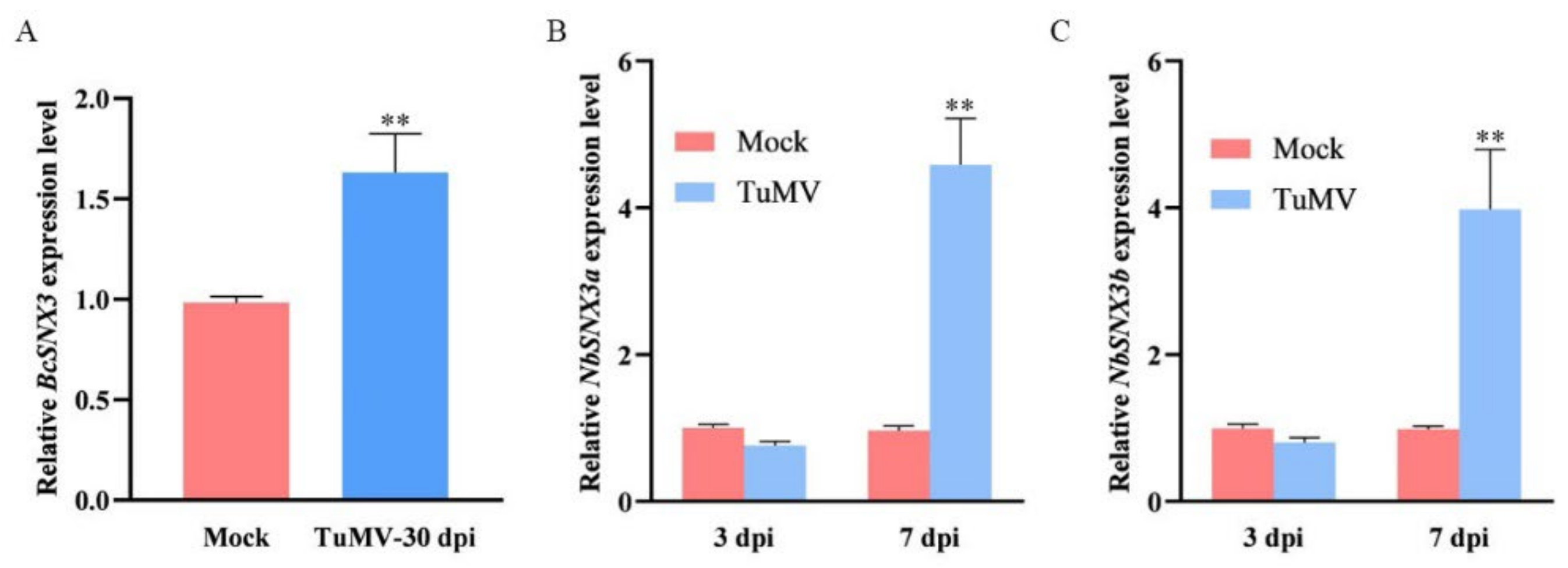
Figure 1. TuMV infection facilitated the expression level of SNX3. (A) RT-qPCR analysis of SNX3 mRNA level in mock (infiltrated with buffer) and TuMV-infected pak-choi. (B,C) RT-qPCR analysis of SNX3a (B) and SNX3b (C) mRNA level in mock (infiltrated with buffer) and TuMV-infected N. benthamiana plants at 3 days post-infiltration (dpi) and 7 dpi. Statistical significance (**, p < 0.05) was determined using Student’s t-test compared with mock infiltration.
Agronomy 12 01757 g001
Figure 2. BcSNX3 is localized to the nucleus. (A) The construct of 35S: YFP and 35S: YFP-BcSNX3. NOS: nopaline synthase gene. (B) Transient expression of 35S: YFP-BcSNX3 fusion protein in tobacco. 35S: YFP was used as a control. Fluorescence images of chloroplast, YFP, and merged were captured with confocal laser scanning microscopy and are displayed in red and yellow, respectively. Scale bars = 20 μm.
Agronomy 12 01757 g002
Figure 3. BcSNX3 interacts with CP and VPg. (A) Yeast-two hybrid (YTH) assays for possible interactions between BcSNX3 and each of the 11 TuMV proteins. BcSNX3 and 11 viral proteins were fused with a GAL4 activation domain (pPR3N−BcSNX3) and a GAL4-binding domain (pBT3−P1, pBT3−HC-Pro, pBT3−P3, pBT3−PIPO, pBT3−6K1, pBT3−CI, pBT3−6K2, pBT3−NIa, pBT3−NIb, pBT3−CP, pBT3−VPg) in turn. NMY51 yeast cells that were co-transformed with the indicated plasmids were subjected to 10-fold serial dilutions and plated on SD/-Trp, -Leu medium to screen for positive interactions at 3 days after transformation. Yeast co-transformed with pPR3N−AtDRP1A+pBT3−VPg served as a positive control; yeast cells co-transformed with pPR3N−BcSNX3 and the empty pBT3 was a negative control. (B) BiFC assays between BcSNX3 on the one hand and CP and VPg on the other in the leaves of N. benthamiana. Confocal imaging was performed at 60-72 hpi. BcSNX3 was fused to the N-terminal fragments of yellow fluorescent protein (YFP)(YN), and CP and VPg were fused to the YC. The BcSNX3−CP and BcSNX3−VPg interaction led to the reconstituted fluorescence-competent structure and the restoration of yellow fluorescence. Bars, 20 μm.
Agronomy 12 01757 g003
Figure 4. Co-localization of BcSNX3 with the autophagosome marker BcATG8b in N. benthamiana. Fluorescence images of YFP, CFP, and merged were captured through confocal laser scanning microscopy and are displayed in green, red and orange, respectively. Bars, 20 μm.
Agronomy 12 01757 g004
Figure 5. BcSNX3 interacts with BcATG8h. (A) Yeast-two hybrid (YTH) assays for possible interactions between BcSNX3 and BcATG8h. (B) BiFC assays between BcSNX3 and BcATG8h in the leaves of N. benthamiana. Confocal imaging was performed at 60–72 hpi. Bars, 20 μm.
Agronomy 12 01757 g005
Figure 6. Overexpression of BcSNX3 inhibits the expression of autophagy components and promotes TuMV infection in N. benthamiana. (A) Effects of BcSNX3 overexpression on the expression of autophagy components at 3 days post-infiltration (dpi). N. benthamiana leaves were agroinfiltrated with 104 (empty vector) and 104-BcSNX3 (overexpress the BcSNX3). Total RNAs were extracted from infiltrated zones at 3 dpi. (B) RT-qPCR analysis of TuMV RNA accumulation in the inoculated leaves. Asterisks indicate significant differences, as determined by ANOVA-LSD (** p < 0.05).
Agronomy 12 01757 g006
Figure 7. The silencing of BcSNX3 suppressed TuMV infection in 49CX, a variety of pak-choi. (A) Viral symptoms in plants pre-inoculated with PTY-S (control), PTY-BcSNX3 (silencing the BcSNX3), for 10 days and then infected by TuMV-GFP. Plants were photographed at 20 dpi. Scale bar = 5 cm. (B) The expression level of BcSNX3 in the above plants. RNA was extracted from systemically infected leaves at 20 dpi. The values are presented as means of fold change ±SD relative to the control plants (pretreated with PTY-S). Error bars represent the SD of three times experiments. (C) Quantification of TuMV genomic RNA in the above plants. Significant differences were calculated using Student’s t-test, ** p < 0.05.
Agronomy 12 01757 g007
Figure 8. TuMV infection was inhibited by AtSNX3 gene knockout in Arabidopsis. (A) Schematic diagram of the AtSNX3 gene structure. The relative position of the T-DNA insertion site is shown. Exons are represented by black boxes. (B) PCR genotyping of the snx3 mutant. (C) qRT-PCR analysis of TuMV RNA level in the wild-type and snx3 mutant under TuMV infection at 15 dpi. The expression level of TuMV genomic RNA in wild-type Col-0 plants was set to 1. Error bars represent the SD of three times experiments. Statistical analysis was performed using Student’s t-test (** p < 0.05). (D) The phenotype of the wild-type (Col-0) and snx3 mutant plants inoculated with TuMV or mock infection at 15 dpi. Bars = 5 cm.
Agronomy 12 01757 g008
Publisher’s Note: MDPI stays neutral with regard to jurisdictional claims in published maps and institutional affiliations.