Article

The Effect of LED and HPS Assimilation Lighting on Leaf Anatomy, Chlorophyll and Carotenoid Autofluorescence Signals, and Some Physiological and Chemical Leaf Traits Related to the Productivity of Cucumber (Cucumis sativus L.) in High-Wire Cultivation

by
Katarzyna Kowalczyk
1,
Leszek Sieczko
2,
Wojciech Borucki
3,
Marzena Sujkowska-Rybkowska
3,
Małgorzata Mirgos
1,
Monika Niedzińska
1,
Magdalena Bederska-Błaszczyk
3,
Waldemar Kowalczyk
4,
Anna Geszprych
1,* and
Janina Gajc-Wolska
1
1
Department of Vegetable and Medicinal Plants, Institute of Horticulture Sciences, Warsaw University of Life Sciences—SGGW, Nowoursynowska 159, 02-776 Warsaw, Poland
2
Department of Biometry, Institute of Agriculture, Warsaw University of Life Sciences—SGGW, Nowoursynowska 159, 02-776 Warsaw, Poland
3
Department of Botany, Institute of Biology, Warsaw University of Life Sciences—SGGW, Nowoursynowska 159, 02-776 Warsaw, Poland
4
Laboratory of Chemical Analysis, The National Institute of Horticultural Research, Konstytucji 3 Maja 1/3, 96-100 Skierniewice, Poland
*
Author to whom correspondence should be addressed.
Agronomy 2022, 12(9), 2004; https://doi.org/10.3390/agronomy12092004
Submission received: 19 July 2022 / Revised: 18 August 2022 / Accepted: 20 August 2022 / Published: 25 August 2022
(This article belongs to the Section Horticultural and Floricultural Crops)

Abstract

:
Supplemental lighting with light-emitting diode (LED) lamps and/or high-pressure sodium (HPS) lamps was applied to increase the activity of the photosynthetic apparatus and thus productivity of greenhouse cucumber (Cucumis sativus L.) in a high-wire growing system. The colocalisation of the chlorophyll of PSII (located mainly in grana) and carotenoid fluorescence signals in chloroplasts of cucumber leaves was studied under confocal microscopy. Leaf anatomy and some chemical quality traits (dry matter, chlorophyll, carotenoids, total soluble solids, total sugars and nitrate reductase activity) as well as selected chlorophyll fluorescence parameters were also investigated and subjected to the multidimensional principal component analysis together with the data on fruit yield. Under LED lighting, a lower correlation between the occurrence of chlorophyll and carotenoid fluorescence signals was observed, especially in older (lower-located) leaves, which may have resulted from changes in the distribution of carotenoids within chloroplasts and/or relative concentrations of chlorophyll and carotenoids. Compared to toplighting with HPS lamps, most commonly used in commercial greenhouse cucumber production, the application of LED interlighting, especially in combination with LED toplighting, led to the increase in chlorophyll and carotenoid content and photosynthetic performance index in older leaves, which was related to the increased cucumber productivity.

1. Introduction

Cucumber (Cucumis sativus L.) is an important crop grown worldwide, both in the open field and under covers. Its total world production in 2020 exceeded 91 million tonnes [1]. In Poland, cucumber production under covers (in greenhouses and plastic tunnels) is carried out in an area of over 1000 hectares and results in harvesting about 300,000 tonnes of fruits per year [2,3]. Cucumber fruits are a low-calorie, alkaline component of the human diet, containing numerous minerals (including potassium, calcium and magnesium), vitamins (e.g., K, C, folate), carotenoids, flavonoids and triterpenes [4,5].
Plant productivity depends on the number, structure and function of cells, leaf size and the entire architecture of the plant, which can be affected by many factors, including environmental conditions and growing method [6]. In cucumber greenhouse production, a high-wire system has been introduced to supply a stable yield of very good quality fruit [7]. In the high-wire system, cucumber plants are grown at high densities per unit area to maximise yield and crop efficiency. In such an intensive production, it is important to provide the plants with optimal conditions for high productivity and fruit quality. During periods of sunlight deficiency (especially winter), assimilation lighting is necessary. Therefore, cultivation studies with different ways of creating supplemental lighting for cucumber plants have been undertaken [8,9,10,11].
In the commercial greenhouse cucumber production, high-pressure sodium (HPS) lamps are predominantly used as a source of assimilation lighting. However, they are not efficient enough in terms of light quality because of low levels of blue light and other photosynthetic sensitive wavelengths [12]. The light-emitting diode (LED) technology has emerged and developed rapidly in the past decades as an alternative to other artificial light sources [13]. The high efficiency of LED lamps allows for lower electric power consumption and production costs [14]. HPS lamps are recommended to be used only as toplighting. LEDs are characterised by their lower production of thermal energy and can be applied both above the canopy and as inter-row lighting to improve vertical light distribution, which is especially important in a large-leaved crop like cucumber [10,15]. Interlighting with LED lamps was found to increase cucumber fruit yield as compared to HPS toplighting [15] and HPS toplighting and inter-row lighting [11].
LEDs emitting near monochromatic light give the possibility to test the influence of light spectral composition on plant growth, yield and accumulation of bioactive compounds [16]. It is widely known that plant growth and physiology (including photosynthesis efficiency) are mainly influenced by red and blue light. Plants grown under monochromatic light, especially a red one, are characterised by reduced growth and CO2 assimilation rate, low maximum quantum yield of chlorophyll fluorescence and unresponsive stomatal conductance, as reported for cucumber [17,18,19]. The application of blue light together with the red one alleviates the symptoms of “red light syndrome” [19]. The positive effects of increasing the blue to red light ratio were found at the stage of cucumber seedling growth [20]. Amoozgar et al. [21] showed that blue LED light alone or a combination of red and blue LED light stimulated the growth of lettuce plants more than the natural light spectrum. In the studies on cucumber, monochromatic blue light was more advantageous for plants than monochromatic red light, whereas the comparison with white light showed diverse results for different physiological parameters [17]. The importance of parts of the spectrum other than red and blue light used in the standard LED panels (far-red light and green light) for the growth and productivity of cucumber was also stressed [22].
Light intensity and quality may affect the structure of the leaves and thus their photosynthetic efficiency. In rape (Brassica napus L.) seedlings, high-intensity LED light (400 µmol·m−2·s−1) caused an increase in leaf thickness, palisade and spongy tissue thickness and resulted in better developed grana compared to the lower light intensities [23]. Pepper (Capsicum annuum L.) leaf thickness, the thickness of palisade and spongy mesophyll tissues and the number of chloroplasts per palisade mesophyll cell were significantly affected by the light source and spectrum, especially the amount of blue light [24].
Both light intensity and quality affect the phytochemical composition of vegetables and thus their nutritional value and role in human health promotion. Manipulation of light spectral quality may increase the yield of vegetables and bioactive compounds content, including flavonoids, carotenoids and vitamins. Photoselective nettings or films are used to modulate light quality in order to improve vegetable quality [25]. In the greenhouse production, the desired light characteristics may be easily achieved by the use of LED lamps. Different illumination spectra of LEDs give an opportunity to compose light conditions for particular plant species and even cultivars and to obtain products with the projected composition and content of phytochemicals [16]. Among the numerous plant active substances, carotenoids belong to the groups of special interest as vitamin A precursors, antioxidants and potential anti-cancer agents [26]. The effect of LED light intensity and quality on the accumulation of carotenoids was mainly studied in leafy green vegetables and microgreens. Brazaitytė et al. [27] reported that the reaction of Brassicaceae microgreens to the irradiance level and spectral quality was species dependent. Samuoliene et al. [28] found that higher blue light intensity was conducive to enriching mustard, beet and parsley microgreens in carotene and xanthophyll pigments and thus to improving their nutritional value. Blue light increased carotenoid concentration also in kale [29] and baby leaf lettuce [30].
In addition to having health-promoting properties, carotenoids are very important components of the photosynthetic antenna, together with chlorophylls a and b, and participate in the photoprotection of photosystems against photodamage [31,32]. Carotenoids, as the parts of photosynthetic antenna, absorb light quanta from the range not covered by chlorophyll, as well as quench triplet chlorophyll and scavenge reactive oxygen species, like singlet oxygen [33,34]. β-carotene and lutein are very important in harvesting blue light and transferring energy to PSII reaction centres [35]. Zeaxanthin participates in the regulation of heat dissipation of excess PSII energy [36]. In excess light, zeaxanthin accumulates and promotes non-photochemical quenching of chlorophyll fluorescence [37]. The objectives of this study were: (1) to check whether light quality (supplemental lighting with LED and/or HPS lamps) affects leaf anatomy, (2) to find whether there is any correlation between localisation of chlorophyll and carotenoid autofluorescence signals under different light conditions, and (3) to evaluate the relationships between some physiological and chemical parameters of leaves and the fruit yield of greenhouse cucumber grown under different supplemental lighting.
In order to achieve the objective (1), light and transmission electron microscopy investigations were carried out. They were focused on the determination of leaf thickness, mesophyll structure and presence of starch in chloroplasts. The setting of the objective (2) was related to the fact that, in spite of a great importance of carotenoids as acceptance compounds in light harvesting photosynthetic complexes, to our knowledge, no attempts had been made to establish the correlation between chlorophyll and carotenoid autofluorescence signals. We hypothesised that, by changes in the distribution of carotenoids towards chlorophyll of PSII, reflected in chlorophyll and carotenoid fluorescence colocalisation, chloroplasts of cucumber acclimate the photosynthetic apparatus to different light quality. A direct analysis of colocalisation is very difficult on the base of merged fluorescence signals emitted by chlorophyll and carotenoids. Therefore, to quantify colocalisation of the signals, the Pearson correlation coefficient (r) was measured, which enabled objective comparisons of different treatments. As for the objective (3), we decided to use principal component analysis to distinguish the leaf traits that most considerably characterise the plants grown under different supplemental lighting and may affect the fruit yield. The studied traits included selected parameters of chlorophyll fluorescence, dry matter, total soluble solids, total sugars, chlorophyll and carotenoid contents and nitrate reductase activity.

2. Materials and Methods

2.1. Plant Material and Growing Conditions

Cucumber plants (Cucumis sativus L. ‘Svyatogor’ F1) were grown in the experimental greenhouse of Warsaw University of Life Sciences (longitude 21° E, latitude 51°15′ N). Commercially available seeds obtained from Rijk Zwaan company were used to prepare seedlings for establishing the experiment. The seedlings were produced in mineral wool cubes in a greenhouse under the following conditions: day/night temperature 22 °C/20 °C, relative humidity of the air 70%, average CO2 concentration 800 ppm, PAR (photosynthetically active radiation) ~170 µmol·m−2·s−1 PPFD (photosynthetic photon flux density) provided by HPS lamps. Cucumber seedlings were planted in the mineral wool slabs on 7 January 2016, 28 days after sowing. The studies were performed in accordance with relevant guidelines and regulations. The research methodology for the cultivation of plants is described in Kowalczyk et al. [38] study. The study was carried out in three greenhouse compartments (each with a usable area of about 40 m2) with different types of supplemental lighting: I-HPS—with HPS toplighting only (HPS lamps Gavita GAN 600 W); II-HPS + LED—with HPS toplighting and LED interlighting (HPS lamps Gavita GAN 600 W and 2 lines of Philips GreenPower LED interlighting modules 2.5 m HO DR/B 100 W); III-LED + LED—with LED toplighting and LED interlighting (Philips GreenPower LED toplighting modules DR/W-LB, 195 W and 2 lines of Philips GreenPower LED interlighting modules 2.5 m HO DR/B 100 W). Supplementary Figure S1 shows the characteristics of light measured at the tops of the plants in particular compartments with handheld Spectral Light Meter MSC15 (Gigahertz-Optik GmbH, Tuerkenfeld, Germany). The light spectrum of the applied LED lamps showed a high peak in the range of deep red light (about 660 nm) and a lower one corresponding to blue light. HPS lamps were characterised by a higher share of the middle part of the PAR spectrum, including green light, and a very low share of blue light. They also emitted far-red light. Light conditions in terms of PAR in every compartment were maintained at the same level, closest to 320 µmol·m−2·s−1 PPFD as possible. PAR was measured with Li-Cor Light meter LI-250A, quantum sensor LI-190 at the tops of the plants and at the level of the 5th and 10th leaf in the middle of the plant canopy. For each compartment, the results of 300 measurements were averaged. The plants were exposed to light for 18 hours a day. In the compartments with both toplighting and inter-row lighting, the plant density was 3.6 plants per m2. In the compartment where only toplighting was used, the plant density was 2.6 plants per m2, as usually applied in the winter production of cucumber with HPS supplemental lighting in order to avoid excessive fruit abortion. In every compartment, computer-controlled microclimate conditions and fertigation were applied. The temperature was maintained at 22–25 °C during the day and 18–22 °C at night. Relative humidity of the air was 70%. The top lamps were turned off when the natural light intensity reached 300 W·m−2 or the external temperature exceeded 30 °C. The experiment was terminated on 5 May 2016, after 17 weeks of cultivation. A high-wire production system was used. Fruits (with a length of about 22 cm and an individual weight of more than 220 g) were harvested every day starting about 3 weeks after planting. Total fruit yield (kg·m−2) was determined on the basis of three repetitions (each of the three benches in the compartment constituted a repetition). The details concerning mineral fertilisation, level of nutrition of cucumber plants and yield in the studied combinations of assimilation lighting were presented in Kowalczyk et al. [38] report.

2.2. The Method of Choosing the Leaves to Be Investigated

The apical buds of cucumber plants contain folded, tightly packed leaves because of short internodes. Unfolded leaves develop below the apical buds. Gradually expanding internodes, petioles and leaf blades, as well as the spiral arrangement of leaves on the stem, ensure their effective light absorption resulting in the highest photosynthetic rate under specific light conditions. Unfolded leaves of cucumber plants were classified into three categories: upper, medium and lower, represented by the 4th, 8th and 12th leaf from the top of the stem, respectively (Supplementary Figure S2). Such leaves corresponded to three levels of the canopy, which differed in light conditions. Plant material for investigations was collected at the full fruiting stage (March) from nine plants from each light treatment (three randomly selected plants from each of the three benches in the greenhouse compartment). Fragments of leaves of each category were taken to be analysed under light microscopy (bright field), transmission electron microscopy and confocal laser scanning microscopy. SPAD index and chlorophyll fluorescence were measured before leaf collecting. Chemical analyses of chosen leaf components were performed in the combined samples of leaves of each category collected from three randomly selected plants per each bench in the compartment.

2.3. Light and Transmission Electron Microscopy Investigations

Hand-cut leaf blade fragments (ca. 2 mm2) were fixed according to Karnovsky [39] at room temperature under vacuum. These fragments were then post-fixed in 2% OsO4 for 2 h at 4 °C, dehydrated in an ethanol series (steps: 10, 20 … 100%, two changes, 30 min each step) and propylene oxide (100%, for 30 min) and embedded in glycid ether 100 epoxy resin (SERVA), a reagent equivalent to Epon 812. Blocks were sectioned using a microtome (Jung RM 2065 and Ultracut UCT, Leica). Semi-thin sections (3 μm) were stained for 1 min with an aqueous mixture of 1% methylene blue and 1% azure A and used for leaf anatomy investigation under light microscopy (Olympus Provis) with the use of Olympus Cell Sens software.
Thin sections (70 nm) were collected on copper grids and stained with uranyl acetate followed by lead citrate for 1 min and examined under transmission electron microscope (Morgagni 268D) to observe chloroplasts under different light conditions. The collected digital microscopic images were saved as jpg files and, if necessary, processed with non-destructive tools (levels and/or contrast) with Photoshop CS 8.0 (Adobe Systems, Inc., San Jose, CA, USA) software.

2.4. Confocal Laser Scanning Microscopy Investigation and Colocalisation Analysis

Fresh, handmade cross sections through the leaves of cucumber plants were prepared using a razor blade. The sections were examined using Leica TCS SP5II laser scanning microscope (Leica Microsystems CMS, Wetzlar, Germany) equipped with a 63 × objective (HCX PLAPO Lambda blue 63.0 × 1.40 OIL UV). The confocal pinhole diameter was automatically set to the so-called ‘1 Airy’ unit to reduce the effect of light diffraction on image formation. As a result, a good compromise between the signal-to-noise ratio and resolution was achieved. Pixel size x/y was 30/30 nm.
Carotenoids were detected at excitation/emission of 488 nm/500–600 nm (false colour green). Excitation at 488 nm favours carotenoid autofluorescence instead of chlorophyll autofluorescence. Autofluorescence of chlorophyll of PSII was detected at excitation/emission of 633 nm/660–705 nm (false colour red). Sequential scanning mode was selected to avoid bleed-through effects. Confocal two-dimensional (2D) optical sections through groups of chloroplasts of palisade mesophyll cells of cucumber plants were collected always under the same experimental settings. Grana, in the top view of the chloroplasts, were represented by bright red fluorescent discs. The measurement of colocalisation was performed by the use of Leica software. The colocalisation of the autofluorescence intensities of chlorophyll and carotenoids was measured.
Image acquisition parameters were as follows:
  • Plan-apochromat objective 63× was used to reduce chromatic shift;
  • Images were acquired by sequential scanning to minimalise bleed-through effect;
  • To avoid saturation, the brightness of red and green channels was adjusted by gain to avoid saturated pixels;
  • To improve the accuracy of the measurements, the background of images was reduced by an appropriate adjustment of the gain and offset of the detector, three-times filtered (line filtered) to minimalise randomness in fluorescence intensity;
  • Chloroplast regions with well visible grana in their upper view were selected as regions of interest (ROIs) to perform Pearson correlation coefficient (r) measurements.
Parameters for fluorescence microscopy were identical for all treatments. All images were processed in the same manner using Leica software. Images were de-convoluted to improve colocalisation analysis [40].

Visualisation of Colocalisation

The representative images of chlorophyll and carotenoid autofluorescence (false colours red and green, respectively) in chloroplasts of upper, medium and lower cucumber leaves were taken. Merged images are presented. Yellow colour indicates strong correlation.

2.5. SPAD Index

The content of chlorophyll (SPAD index) was estimated with the portable equipment chlorophyll meter SPAD-502 (Konica Minolta Sensing, Inc., Osaka, Japan). SPAD index was measured in the upper (4th), middle (8th) and lower (12th) leaf of each of the 9 selected plants from each of the light conditions (see Section 2.2). For each leaf, 5 measurements were taken, and their results were averaged and treated as a single result (n = 9).

2.6. Chlorophyll Fluorescence

Chlorophyll a fluorescence was measured in the upper (4th), middle (8th) and lower (12th) leaf of each of the 9 selected plants from each of the light conditions (n = 9). After 30 min of dark acclimation, leaves were illuminated by red light (3500 μmol·m−2·s−1), and chlorophyll fluorescence was measured using Pocket PEA chlorophyll fluorimeter (Hansatech Instruments Ltd., Pentney, UK). Two indicators were evaluated: Fv/Fm (maximum quantum efficiency of PSII) and PI (Performance Index).

2.7. Analysis of Chosen Components in the Leaves

Dry matter (DM), chlorophyll a (CHa), chlorophyll b (CHb), total carotenoids (CT), total soluble solids (TSS), total sugars (TS) and nitrate reductase activity (NR) were determined in the freshly collected leaves. The analyses were performed in triplicate, in the combined leaf samples of three plants per each of the three benches in the compartment (see Section 2.2) (n = 3).

2.7.1. Dry Matter

The leaf dry matter was determined by drying leaf samples in an oven, at a temperature of 105 °C for 24 h.

2.7.2. Chlorophyll and Carotenoid Content

Chlorophyll a, chlorophyll b and total carotenoid content were determined spectrophotometrically in 80% acetone extract with UV-1700 PharmaSpec spectrophotometer (Shimadzu Corporation, Kyoto, Japan) and calculated to fresh weight of leaves according to the method of Lichtenthaler and Wellburn [41].

2.7.3. Total Soluble Solids and Total Sugars

The content of total soluble solids was determined with the digital refractometer DR-303 (Index Instruments Ltd., Ramsey, UK). Total sugars were analysed according to the Luff-Schoorl method ([42], with modifications) based on the reduction of copper(II) ions from the Luff-Schoorl reagent by reducing sugars present in the investigated sample (free sugars and those released as a result of acidic hydrolysis of sucrose) and iodometric determination of the remaining copper(II) ions by titration with sodium thiosulphate. For this analysis, leaf samples were homogenised with water and the extract was clarified with Carrez I and Carrez II solutions.

2.7.4. Nitrate Reductase Activity

The nitrate reductase activity was determined according to Bar-Akiva et al. [43]. The assay was based on the colorimetric measurement of nitrates(III) produced after 2 h dark incubation of a leaf sample with buffered nitrate(V) solution at 28 °C. Nitrate reductase activity was expressed as the amount of nitrates(III) in nmol per gram of fresh mass of a leaf sample per hour. The colorimetric determination was based on the reaction of nitrates(III) with sulfanilamide and N-(1-naphthyl)ethylenediamine dihydrochloride. The absorption of the obtained colour solution was measured at 540 nm using UV-1700 PharmaSpec spectrophotometer (Shimadzu Corporation, Kyoto, Japan).

2.8. Statistical Analysis

The results of leaf anatomy measurements (n = 3) were statistically analysed using one-way analysis of variance (ANOVA), followed by the Tukey HSD test at the significance level of 0.05.
In order to determine the effect of the experiment factors on the Pearson correlation coefficient between the occurrence of chlorophyll and carotenoids (rCH/C, data from the confocal microscope), a two-factor ANOVA was performed with the following factors: the type of supplemental lighting (HPS, HPS + LED and LED + LED) and the leaf position on the stem (upper, medium and lower leaf representing 4th, 8th and 12th leaf from the apical bud, respectively). The total number of the studied cases (N) was 706, including the following number of measurements for lighting type: HPS − n = 265, HPS + LED − n = 261 and LED + LED − n = 180 and leaf position on the stem: upper − n = 225, medium − n = 229 and lower − n = 252. The null hypothesis was about the lack of influence of the examined factors on the value of the chlorophyll to carotenoid correlation coefficient (rCH/C). In the detailed studies, Tukey HSD test was used to compare mean values of rCH/C at the significance level of 0.05.
In order to check the relations between the chosen examined leaf traits and lighting types, multidimensional statistical analyses were used. Factor analysis using the principal component method (PCA) was performed. Based on ANOVA results and the Tukey HSD test, which did not show significant differences between the mean values of rCH/C for medium and lower leaves, these values were averaged and the leaves were named middle-aged (m). Upper leaves (4th leaf from the apical bud) were called young (y). In order to check how the investigated leaf traits affect cucumber fruit yield, yield values (Yield) were added to the analysis. Based on the data prepared in this way, the principal components were analysed.
All the statistical analyses were calculated in the Statistica v. 13.3 package.

3. Results

3.1. Leaf Structure

Cucumber leaves were fully developed anatomically independently on their position on the stem (upper—the 4th leaf; medium—the 8th leaf; lower—the 12th leaf from the top, Supplementary Figure S2). The microscopic analysis revealed that all leaves showed one palisade parenchyma cell layer and several layers of spongy parenchyma (Figure 1a–i). Lower leaves exhibited more loosely arranged cells of both parenchyma types (Figure 1c,f,i). Supplemental LED lighting caused changes in leaf anatomy (Table 1, Figure 1a–i). The application of LED interlighting led to the significant increase in palisade and spongy parenchyma cell length in the lower leaves of plants grown under HPS toplighting. Both lower and medium leaves of plants grown under LED top- and interlighting were characterised by the significantly longer parenchyma cells in comparison with those subjected to HPS toplighting only (Table 1).
Chloroplasts of the upper leaves of cucumber plants grown in all studied light conditions accumulated distinct amounts of starch (Figure 1a,d,g and Figure 2a,d,g). HPS toplighting resulted in the presence of very small and scarce starch granules in chloroplasts of the medium leaves (Figure 1b and Figure 2b). Larger starch deposits in the medium leaves were found for LED top- and interlighting (Figure 1h and Figure 2h). In case of plants grown under HPS + LED supplemental lighting, medium leaves exhibited the largest starch deposits (Figure 1e and Figure 2e). Chloroplasts of the lower leaves, independently of light conditions, possessed negligible starch amounts (Figure 1c,f,i and Figure 2c,f,i).

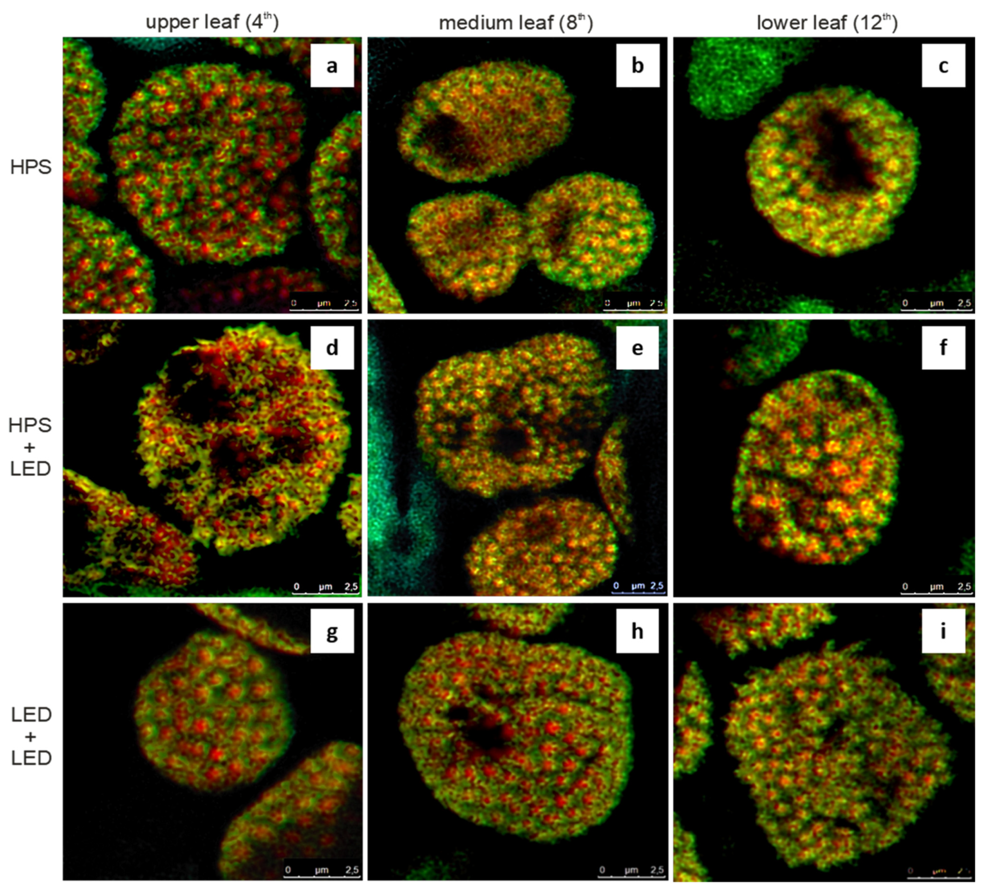
3.2. Colocalisation of Chlorophyll and Carotenoid Fluorescence Signals

Images show merged red and green channels representing chlorophyll and carotenoid fluorescence in median optical sections through chloroplasts of cucumber leaves exposed to different light conditions (Figure 3). Yellow colour represents high correlation between both fluorescence signals. It is worth noting that HPS supplemental lighting alone or in combination with LED interlighting resulted in a larger chloroplast area occupied by yellow colour than in LED + LED lighting treatment. These results are consistent with higher values of rCH/C observed for the plants grown under HPS or HPS + LED compared to those grown under LED + LED supplemental lighting (see Section 3.3).

3.3. Statistics for Data from the Confocal Microscope

On the basis of the results of a two-factor ANOVA (Supplementary Table S1, Supplementary Figures S3–S5), the null hypothesis about the lack of influence of supplemental lighting type on the average rCH/C value (Pearson correlation coefficient between the occurrence of chlorophyll and carotenoids) was rejected at the p-value < 0.001 (Femp = 48.71). The null hypothesis of no effect of leaf position on mean value of rCH/C was rejected at p-value < 0.001 (Femp = 50.80). In the detailed tests, two homogeneous groups for lighting types and two for leaf position on the stem were obtained with the HSD Tukey test. The plants grown under HPS and HPS + LED supplemental lighting were characterised by significantly higher rCH/C than those grown under LED lighting (0.684, 0.672 and 0.613, respectively). Upper leaves were characterised by higher rCH/C (0.704) than lower and medium leaves (0.642 and 0.641, respectively). As rCH/C values for lower and medium leaves did not differ significantly, for further statistical analyses, an average value for these two leaf fractions was used and these leaves were further called “middle-aged” whereas the upper leaves were called “young”.

3.4. The Principal Component Analysis for the Relationships between Experiment Factors, Leaf Quality Parameters and Fruit Yield

The principal component analysis, based on the data presented in Supplementary Table S2, identified three major principal components: PC1 explaining 49.72% of the total variability, PC2 explaining 29.90% and PC3 explaining 11.53% of the total variability (Supplementary Table S3). The first two components taken for interpretation of the results explained 79.62% of the total variability of the examined features. Other components had eigenvalues below 1 and were not included. Principal component analysis showed that PC1 was most correlated with SPAD index, chlorophyll a and total carotenoid content in the leaves and negatively correlated with rCH/C. The yield correlation coefficient with PC1 was 0.47 and, with PC2, 0.74.
The biplot (Figure 4) was used for the graphic presentation. In the space of the first two components, points representing the examined features were plotted by means of the correlation coefficient with the given component, and the same method was used to project the six cases examined. On the biplot, a new axis system for the total fruit yield value was applied (the dashed line). The cases were projected onto this new axis. This transformation was carried out in order to better illustrate the impact of various types of supplemental lighting on the fruit yield in the system of other examined features. Closest to the yield were the cases with LED + LED lighting, both for young and medium leaves. Further from the yield, on the opposite abscissa axis designated for the yield, were HPS and HPS + LED lighting treatments. The exact relations are shown in Figure 4.

4. Discussion

In our experiment, supplementation of the natural light with that emitted by HPS lamps and/or LEDs was used in the high-wire winter cultivation of cucumber in order to improve the intensity of plant irradiance and modify its spectral properties. We hypothesised that light quality could influence cucumber leaf structure, colocalisation of chlorophyll and carotenoid fluorescence signals, chlorophyll fluorescence parameters, and some chemical leaf traits, potentially related to cucumber fruit yield.
LED lighting used in our experiment was a combination of blue and red diodes as it was found that red light alone is insufficient for normal photosynthetic rate and plant growth [18,19]. Hogewoning et al. [44] proved that a combination of red and blue light promotes cucumber plant growth more efficiently that red or blue light alone.
The reports on cucumber leaf anatomical and physiological response to light intensity and quality in the production conditions are scarce. The studies on the effect of supplemental lighting with different light sources are mainly focused on fruit yield and economical aspects of cucumber production [8,9,10,11,15]. Physiological and biochemical reactions of plants to different light treatments are mainly evaluated at the seedling stage [19,20,45,46,47].
The microscopic analysis performed in our study clearly shows that light quality affects leaf anatomical features (Table 1, Figure 1a–i). The application of LED interlighting led to a significant increase of mesophyll cell length compared to plants grown under HPS toplighting. This is in line with the results of other studies that have shown that the thickness of palisade tissue and spongy parenchyma of cucumber seedlings was promoted by LED lighting [48]. Additionally, it has been reported that cucumber leaves become thicker with an increasing blue light ratio and thinner with increasing red light [49]. In our study, we observed that, independently of light quality, chloroplasts of the upper leaves (4th leaf from the top of the stem) show distinct methylene blue-azure affinity (Figure 1a,d,g and Figure 2a,d,g). The affinity probably results from the presence of dense cytoplasm and large starch granules in enlarged chloroplasts, according to the method for staining of epoxy-embedded tissue sections [50]. The starch reserves in the upper leaves indicate high intensity of photosynthesis that exceeds the current demand for photo-assimilates.
In chloroplasts of the medium leaves (8th leaf from the top of the stem), especially those of plants grown under HPS supplemental lighting, starch granules are scarce (Figure 1b and Figure 2b), probably because developing fruits act as strong sinks of photo-assimilates. Induced starch accumulation in the medium leaves was noted for HPS + LED lighting (Figure 1e and Figure 2e). A similar LED-induced increase in starch content in cucumber leaves was observed in another study [51]. Compared to upper and medium leaves, lower leaves (12th leaf from the top) function under the worst light conditions and probably export all current assimilates to the growing fruits.
Confocal laser scanning microscopy was used to check whether the light spectrum affects the distribution of carotenoids and chlorophyll within chloroplasts of cucumber leaves (Figure 3). The analysis of colocalisation of chlorophyll and carotenoid autofluorescence showed that, under LED lighting, substantially lower values of the Pearson correlation coefficient (r) between chlorophyll and carotenoid fluorescence signals in older leaves were obtained. This may result from additional synthesis of carotenoids in chloroplasts of older leaves in response to LED lighting.
It is widely accepted that carotenoids play crucial roles in photosynthesis. They absorb the blue and green portions of the light spectrum and transit the energy to the chlorophylls, which are not able to use green light for photosynthesis [52]. The carotenoids present in lower leaves can absorb the green light reflected or transmitted by the leaves above and thus enhance the photosynthetic rate.
Carotenoids are also structural backbones which stabilise the structures of light harvesting complexes [31,53]. In PSII, β-carotene and lutein play at least two important roles: they are responsible for blue-light harvesting and transferring energy to photosystem reaction centres, and they protect the photosynthetic apparatus from oxidative damage [35]. In the conditions of excess light, carotenoids protect chlorophyll from photodamage by quenching chlorophyll triplet states and singlet oxygen [54,55]. It is widely accepted that carotenoids have antioxidant properties, including radical scavenging and singlet oxygen quenching abilities [56]. The contribution of carotenoids to non-photochemical quenching (transformation of the excess light energy into less harmful heat) is a matter of debate [57].
It was reported that both LED light intensity and spectral composition affected total carotenoid content in Brassicaceae microgreens, but the same light characteristics produced diverse results in different species [27]. In the studies of Brazaitytė et al. [58], the effect of the applied light sources with different spectral characteristics but similar PPFD on the carotenoid content in cucumber seedlings was found to be insignificant, although a slight tendency toward higher carotenoid content was observed when supplemental yellow LEDs were applied. Wang et al. [46] reported that 1–3 h supplemental lighting of cucumber seedlings with LED lamps emitting red and blue light increased the carotenoid and chlorophyll content in leaves.
The results of our experiment showed that LED interlighting applied in combination with HPS or LED toplighting substantially increased carotenoid content in older leaves of cucumber plants as compared to standard HPS toplighting. The application of LED interlighting could have induced the light stress response in older leaves which was expressed in increased carotenoid concentration. The older (lower-located) leaves of plants subjected to LED interlighting were also characterised by higher chlorophyll content, both determined with a spectrophotometric method and estimated using SPAD index (Figure 4, Supplementary Table S2).
Numerous studies, especially on green leafy vegetables, show that light quality affects the content and composition of photosynthetic pigments and photosynthetic efficiency. Red and blue LED supplemental lighting caused the increase in chlorophyll a and b and carotenoid content, improved the efficiency of the photosynthetic apparatus and finally increased the plant weight of lamb’s lettuce [59]. Muneer et al. [60] found that increased blue LED radiation increased biomass and photosynthetic parameters in lettuce as a result of the control of chloroplast protein integrity including PSII-core dimer and PSII-core monomer.
In our study, the results concerning leaf chemical characteristics (including chlorophyll and carotenoid contents) together with chlorophyll fluorescence parameters (Fv/Fm and PI) and yield were subjected to the multidimensional statistical analysis (PCA) to synthetically show the relations between the type of supplemental lighting, leaf traits and productivity of cucumber. The multivariate PCA identified three principal components accounting for 91.15% of the total variation (Supplementary Table S3). The first principal component (PC1) explained 49.72% of the variation, and it was most strongly positively correlated with the chlorophyll a and carotenoid content, SPAD index and PI, which also confirms the relation between the content of photosynthetic pigments and photosynthetic performance. These traits are shown on the biplot (Figure 4) close to each other and close to the cases representing older leaves of plants grown under LED interlighting combined with HPS or LED toplighting. The latter case is located close to the fruit yield, which indicates that top- and interlighting with LEDs is especially favourable for the productivity of cucumber, as we reported earlier [38]. It is worth stressing that identification of leaf traits related to cucumber productivity, especially those that can be determined with non-destructive methods such as SPAD and chlorophyll fluorescence parameters, may be useful in the early diagnosis of plant condition and in cucumber yield modelling and prediction.
Beneficial effects of LED lighting can be sometimes questioned. Ptushenko et al. [61] found that HPS lamps emitting broad light spectrum were more efficient in promoting plant growth of Chinese cabbage than narrow-band red and blue diodes. The authors suggest that light distribution in the plant canopy might be responsible for the effects of LED light. Särkkä et al. [11] reported that the cucumber fruit yield was highest when HPS lamps were used as toplighting and LEDs as interlighting (compared to full HPS and full LED lighting). These authors emphasise that the hybrid illumination (with HPS toplighting and LED inter-row lighting) provided more diverse spectral distribution for the leaves in the middle part of the canopy. Zou et al. [22] propose to use LEDs with the broad light spectrum similar to the solar one in cucumber production. They stress the importance of far-red light for the regulation of plant photomorphogenesis and green light that can penetrate deeper into the plant canopy and increase light utilisation in lower leaves.
Our experiments showed that intra-canopy LED (red and blue) lighting increased both photosynthetic performance index and carotenoid content in lower-located leaves. The yield also increased in response to LED lighting.

5. Conclusions

The application of LED interlighting led to the significant increase of mesophyll cell length in the lower leaves of cucumber plants and to the increase in starch accumulation in medium leaves compared to plants grown under HPS toplighting only. Light spectral conditions influenced the relative distribution of chlorophyll and carotenoid fluorescence signals in chloroplasts of cucumber leaves suggesting an acclimation of light harvesting photosystems. We showed that light spectral properties diversely influenced colocalisation of carotenoid and PSII chlorophyll fluorescence signals. HPS lighting resulted in better correlation (higher values of r—Pearson correlation coefficient) between the distribution of chlorophyll and carotenoid fluorescence than LED lighting. LED lighting may stimulate synthesis of carotenoids and/or change their distribution within chloroplasts leading to a low level of colocalisation of fluorescence signals of the pigments. A low colocalisation value (low Pearson correlation coefficient) of chlorophyll and carotenoid fluorescence signals under certain light conditions may indicate that there is a weak relationship between the concentration of these pigments in particular locations within chloroplasts. It may implicate that cooperation of chlorophyll and carotenoids in photosynthesis depends on the light conditions, particularly light spectral properties. Compared to toplighting with HPS lamps, the application of LED interlighting led to the increase in chlorophyll and carotenoid content and photosynthetic performance index in older (lower-located) leaves. LED inter-row lighting applied together with HPS or LED toplighting resulted in a higher yield of cucumber fruits compared to cultivation with HPS toplighting only, at the same total level of PPFD in all lighting treatments. Top- and interlighting with LED lamps resulted in the highest fruit yield.

Supplementary Materials

The following are available online at https://www.mdpi.com/article/10.3390/agronomy12092004/s1, Supplementary Material file, containing Figure S1: Characteristics of light measured at the tops of the plants in the experimental greenhouse compartments: green and black line—in the compartments with HPS toplighting (HPS and HPS + LED), red line—in the compartment with LED toplighting (LED + LED); Figure S2: Cucumber grown in a high-wire system with application of LED supplemental lighting; Table S1: Parameters of two-factor ANOVA for the effect of supplemental lighting and leaf position on the stem on rCH/C value (α = 0.05); Figure S3: Mean rCH/C values for leaf position on the stem; Figure S4: Mean rCH/C values for supplemental lighting type; Figure S5: Mean rCH/C values for interaction “supplemental lighting × leaf position on the stem”; Table S2: Average values of the cucumber leaf parameters in dependence on leaf age (young—the 4th leaf from the top of the stem; middle—mean values of parameters for the 8th and 12th leaf from the top of the stem) and cucumber yield under different supplemental lighting conditions; Table S3: Correlations between the observed features (variables) and the first three principal components (PCs) in a set of 6 experimental cases (supplemental lighting × leaf age), and variation explained by these PCs.

Author Contributions

Conceptualization, K.K. and J.G.-W.; methodology, K.K., L.S., W.B. and W.K.; formal analysis, L.S.; investigation, K.K., W.B., M.S.-R., M.M., M.N., M.B.-B., W.K. and A.G.; data curation, K.K., L.S., W.B. and A.G.; writing—original draft preparation, K.K., L.S., W.B. and M.M.; writing—review and editing, K.K., A.G. and J.G.-W.; visualization, L.S. and W.B.; supervision, K.K. and J.G.-W. All authors have read and agreed to the published version of the manuscript.

Funding

This research received no external funding.

Institutional Review Board Statement

Not applicable.

Informed Consent Statement

Not applicable.

Data Availability Statement

The data supporting the findings of the study are included in the article and the Supplementary Material file.

Acknowledgments

The study was partially supported by Philips Lighting Holding B.V.

Conflicts of Interest

The authors declare no conflict of interest.

References

  1. FAOSTAT. Crops and Livestock Products. Available online: https://www.fao.org/faostat/en/#data/QCL (accessed on 12 August 2022).
  2. Powierzchnia Uprawy i Produkcja Warzyw Pod Osłonami w 2020 r. Available online: https://www.podoslonami.pl/uprawy-i-odmiany/warzywa/powierzchnia-uprawy-i-produkcja-warzyw-pod-oslonami-2020-r/ (accessed on 12 August 2022).
  3. Statistics Poland. Production of Agricultural and Horticultural Crops in 2021. Available online: https://stat.gov.pl/en/topics/agriculture-forestry/agricultural-and-horticultural-crops/production-of-agricultural-and-horticultural-crops-in-2021,2,6.html (accessed on 12 August 2022).
  4. Murad, H.; Nyc, M.A. Evaluating the potential benefits of cucumbers for improved health and skin care. J. Aging Res. Clin. Pract. 2016, 5, 139–141. [Google Scholar] [CrossRef]
  5. Chakraborty, S.; Rayalu, S. Health beneficial effects of cucumber. In Cucumber Economic Values and Its Cultivation and Breeding, 1st ed.; Wang, H., Ed.; IntechOpen: London, UK, 2021. [Google Scholar] [CrossRef]
  6. De Bossoreille de Ribou, S.; Douam, F.; Hamant, O.; Frohlich, M.W.; Negrutiu, I. Plant science and agricultural productivity: Why are we hitting the yield ceiling? Plant Sci. 2013, 210, 159–176. [Google Scholar] [CrossRef] [PubMed]
  7. Hao, X.; Papadopoulos, A.P.; Khosla, S. Development of a high-wire cucumber production system on raised-troughs with supplemental lighting for year-round production. Acta Hortic. 2007, 761, 337–340. [Google Scholar] [CrossRef]
  8. Hovi, T.; Näkkilä, J.; Tahvonen, R. Interlighting improves production of year-round cucumber. Sci. Hortic. 2004, 102, 283–294. [Google Scholar] [CrossRef]
  9. Hovi-Pekkanen, T.; Tahvonen, R. Effects of interlighting on yield and external fruit quality in year-round cultivated cucumber. Sci. Hortic. 2008, 116, 152–161. [Google Scholar] [CrossRef]
  10. Guo, X.; Hao, X.; Zheng, J.M.; Little, C.; Khosla, S. Response of greenhouse mini-cucumber to different vertical spectra of LED lighting under overhead high pressure sodium and plasma lighting. Acta Hortic. 2016, 1134, 87–94. [Google Scholar] [CrossRef]
  11. Särkkä, L.E.; Jokinen, K.; Ottosen, C.-O.; Kaukoranta, T. Effects of HPS and LED lighting on cucumber leaf photosynthesis, light quality penetration and temperature in the canopy, plant morphology and yield. Agric. Food Sci. 2017, 26, 101–109. [Google Scholar] [CrossRef]
  12. Ouzounis, T.; Rosenqvist, E. Spectral effects of artificial light on plant physiology and secondary metabolism: A Review. HortScience 2015, 50, 1128–1135. [Google Scholar] [CrossRef]
  13. Massa, G.D.; Kim, H.H.; Wheeler, R.M.; Mitchell, C.A. Plant productivity in response to LED lighting. HortScience 2008, 43, 1951–1956. [Google Scholar] [CrossRef]
  14. Gómez, C.; Izzo, L.G. Increasing efficiency of crop production with LEDs. AIMS Agric. Food 2018, 3, 135–153. [Google Scholar] [CrossRef]
  15. Kumar, K.G.S.; Hao, X.; Khosla, S.; Guo, X.; Bennett, N. Comparison of HPS lighting and hybrid lighting with top HPS and intracanopy LED lighting for high-wire mini-cucumber production. Acta Hortic. 2016, 1134, 111–118. [Google Scholar] [CrossRef]
  16. Hasan, M.; Bashir, T.; Ghosh, R.; Lee, S.K.; Bae, H. An overview of LEDs’ effects on the production of bioactive compounds and crop quality. Molecules 2017, 22, 1420. [Google Scholar] [CrossRef]
  17. Wang, H.; Gu, M.; Cui, J.; Shi, K.; Zhou, Y.; Yu, J. Effects of light quality on CO2 assimilation, chlorophyll-fluorescence quenching, expression of Calvin cycle genes and carbohydrate accumulation in Cucumis sativus. J. Photochem. Photobiol. B Biol. 2009, 96, 30–37. [Google Scholar] [CrossRef]
  18. Trouwborst, G.; Hogewoning, S.W.; van Kooten, O.; Harbinson, J.; van Ieperen, W. Plasticity of photosynthesis after the ‘red light syndrome’ in cucumber. Environ. Exp. Bot. 2016, 121, 75–82. [Google Scholar] [CrossRef]
  19. Miao, Y.; Chen, Q.; Qu, M.; Gao, L.; Hou, L. Blue light alleviates ‘red light syndrome’ by regulating chloroplast ultrastructure, photosynthetic traits and nutrient accumulation in cucumber plants. Sci. Hortic. 2019, 257, 108680. [Google Scholar] [CrossRef]
  20. Hernández, R.; Kubota, C. Physiological responses of cucumber seedlings under different blue and red photon flux ratios using LEDs. Environ. Exp. Bot. 2016, 121, 66–74. [Google Scholar] [CrossRef]
  21. Amoozgar, A.; Mohammadi, A.; Sabzalian, M.R. Impact of light-emitting diode irradiation on the photosynthesis, phytochemical composition and mineral element content of lettuce cv. Grizzly. Photosynthetica 2016, 55, 85–95. [Google Scholar] [CrossRef]
  22. Zou, J.; Zhou, C.; Xu, H.; Cheng, R.; Yang, Q.; Li, T. The effect of artificial solar spectrum on growth of cucumber and lettuce under controlled environment. J. Integr. Agric. 2020, 19, 2027–2034. [Google Scholar] [CrossRef]
  23. Yao, X.-Y.; Liu, X.-Y.; Xu, Z.-G.; Jiao, X.-L. Effects of light intensity on leaf microstructure and growth of rape seedlings cultivated under a combination of red and blue LEDs. J. Integr. Agric. 2017, 16, 97–105. [Google Scholar] [CrossRef]
  24. Schuerger, A.C.; Brown, C.S.; Stryjewski, E.C. Anatomical features of pepper plants (Capsicum annuum L.) grown under red light-emitting diodes supplemented with blue or far-red light. Ann. Bot. 1997, 79, 273–282. [Google Scholar] [CrossRef]
  25. Ilić, Z.S.; Fallik, E. Light quality manipulation improves vegetable quality at harvest and postharvest: A review. Environ. Exp. Bot. 2017, 139, 79–90. [Google Scholar] [CrossRef]
  26. Pavlova, T.; Stamatovska, V.; Dimov, I.; Nakov, G.; Kalevska, T. Carotenoids in foods. ARTTE 2018, 6, 321–328. [Google Scholar] [CrossRef]
  27. Brazaitytė, A.; Sakalauskienė, S.; Samuolienė, G.; Jankauskienė, J.; Viršilė, A.; Novičkovas, A.; Sirtautas, R.; Miliauskienė, J.; Vaštakaitė, V.; Dabašinskas, L.; et al. The effects of LED illumination spectra and intensity on carotenoid content in Brassicaceae microgreens. Food Chem. 2015, 173, 600–606. [Google Scholar] [CrossRef]
  28. Samuolienė, G.; Viršilė, A.; Brazaitytė, A.; Jankauskienė, J.; Sakalauskienė, S.; Vaštakaitė, V.; Novičkovas, A.; Viškelienė, A.; Sasnauskas, A.; Duchovskis, P. Blue light dosage affects carotenoids and tocopherols in microgreens. Food Chem. 2017, 228, 50–56. [Google Scholar] [CrossRef]
  29. Lefsrud, M.G.; Kopsell, D.A.; Sams, C.E. Irradiance from distinct wavelength light-emitting diodes affect secondary metabolites in kale. HortScience 2008, 43, 2243–2244. [Google Scholar] [CrossRef]
  30. Li, Q.; Kubota, C. Effects of supplemental light quality on growth and phytochemicals of baby leaf lettuce. Environ. Exp. Bot. 2009, 67, 59–64. [Google Scholar] [CrossRef]
  31. Liu, L.; Shao, Z.; Zhang, M.; Wang, Q. Regulation of carotenoid metabolism in tomato. Mol. Plant 2015, 8, 28–39. [Google Scholar] [CrossRef]
  32. Derks, A.; Schaven, K.; Bruce, D. Diverse mechanisms for photoprotection in photosynthesis. Dynamic regulation of photosystem II excitation in response to rapid environmental change. Biochim. Biophys. Acta Bioenerg. 2015, 1847, 468–485. [Google Scholar] [CrossRef]
  33. Choudhury, N.K.; Behera, R.K. Photoinhibition of photosynthesis: Role of carotenoids in photoprotection of chloroplast constituents. Photosynthetica 2001, 39, 481–488. [Google Scholar] [CrossRef]
  34. Gall, A.; Berera, R.; Alexandre, M.T.A.; Pascal, A.A.; Bordes, L.; Mendes-Pinto, M.M.; Andrianambinintsoa, S.; Stoitchkova, K.V.; Marin, A.; Valkunas, L.; et al. Molecular adaptation of photoprotection: Triplet states in light-harvesting proteins. Biophys. J. 2011, 101, 934–942. [Google Scholar] [CrossRef]
  35. Jahns, P.; Holzwarth, A.R. The role of the xanthophyll cycle and of lutein in photoprotection of photosystem II. Biochim. Biophys. Acta Bioenerg. 2012, 1817, 182–193. [Google Scholar] [CrossRef] [PubMed]
  36. Ramalho, J.C.; Pons, T.L.; Groeneveld, H.W.; Azinheira, H.G.; Nunes, M.A. Photosynthetic acclimation of high light conditions in mature leaves of Coffea arabica L.: Role of xanthophylls, quenching mechanisms and nitrogen nutrition. Austr. J. Plant Physiol. 2000, 27, 43–51. [Google Scholar] [CrossRef]
  37. Bartley, G.E.; Scolnik, P.A. Plant carotenoids: Pigments for photoprotection, visual attraction, and human health. Plant Cell 1995, 7, 1027–1038. [Google Scholar] [CrossRef] [PubMed]
  38. Kowalczyk, K.; Gajc-Wolska, J.; Mirgos, M.; Geszprych, A.; Kowalczyk, W.; Sieczko, L.; Niedzińska, M.; Gajewski, M. Mineral nutrients needs of cucumber and its yield in protected winter cultivation, with HPS and LED supplementary lighting. Sci. Hortic. 2020, 265, 109217. [Google Scholar] [CrossRef]
  39. Karnovsky, M.J. A formaldehyde–glutaraldehyde fixative of high osmolality for use in eletron microscopy. J. Cell Biol. 1965, 27, 137A–138A. [Google Scholar]
  40. Landmann, L. Deconvolution improves colocalization analysis of multiple fluorochromes in 3D confocal data sets more than filtering techniques. J. Microsc. 2002, 208, 134–147. [Google Scholar] [CrossRef]
  41. Lichtenthaler, H.K.; Wellburn, A.R. Determination of total carotenoids and chlorophyll a and b of leaf extracts in different solvents. Biochem. Soc. Trans. 1983, 11, 591–592. [Google Scholar] [CrossRef]
  42. Klepacka, M. (Ed.) Analiza Żywności, 1st ed.; Fundacja Rozwój SGGW: Warszawa, Poland, 1996; pp. 44–47. [Google Scholar]
  43. Bar-Akiva, A.; Sagiv, J.; Leshem, J. Nitrate reductase activity as an indicator for assessing the nitrogen requirement of grass crops. J. Sci. Food Agric. 1970, 21, 405–407. [Google Scholar] [CrossRef]
  44. Hogewoning, S.W.; Trouwborst, G.; Maljaars, H.; Poorter, H.; van Ieperen, W.; Harbinson, J. Blue light dose-responses of leaf photosynthesis, morphology, and chemical composition of Cucumis sativus grown under different combinations of red and blue light. J. Exp. Bot. 2010, 61, 3107–3117. [Google Scholar] [CrossRef]
  45. Song, J.X.; Meng, Q.W.; Du, W.F.; He, D.X. Effects of light quality on growth and development of cucumber seedlings in controlled environment. Int. J. Agric. Biol. Eng. 2017, 10, 312–318. [Google Scholar] [CrossRef]
  46. Wang, S.; Fang, H.; Xie, J.; Wu, Y.; Tang, Z.; Liu, Z.; Lv, J.; Yu, J. Physiological responses of cucumber seedlings to different supplemental light duration of red and blue LED. Front. Plant Sci. 2021, 12, 709313. [Google Scholar] [CrossRef] [PubMed]
  47. Hamedalla, A.M.; Ali, M.M.; Ali, W.M.; Ahmed, M.A.A.; Kaseb, M.O.; Kalaji, H.M.; Gajc-Wolska, J.; Yousef, A.F. Increasing the performance of cucumber (Cucumis sativus L.) seedlings by LED illumination. Sci. Rep. 2022, 12, 852. [Google Scholar] [CrossRef]
  48. Zhang, Y.; Dong, H.; Song, S.; Su, W.; Liu, H. Morphological and physiological responses of cucumber seedlings to supplemental LED light under extremely low irradiance. Agronomy 2020, 10, 1698. [Google Scholar] [CrossRef]
  49. Hernández, R.; Kubota, C. Growth and morphological response of cucumber seedlings to supplemental red and blue photon flux ratios under varied solar daily light integrals. Sci. Hortic. 2014, 173, 92–99. [Google Scholar] [CrossRef]
  50. Humphrey, C.D.; Pittman, F.E. A simple methylene blue-azure II-basic fuchsin stain for epoxy-embedded tissue sections. Stain Technol. 1974, 49, 9–14. [Google Scholar] [CrossRef]
  51. Li, Y.; Xin, G.; Wei, M.; Shi, Q.; Yang, F.; Wang, X. Carbohydrate accumulation and sucrose metabolism responses in tomato seedling leaves when subjected to different light qualities. Sci. Hortic. 2017, 225, 490–497. [Google Scholar] [CrossRef]
  52. Hashimoto, H.; Sugai, Y.; Uragami, C.; Gardiner, A.T.; Cogdell, R.J. Natural and artificial light-harvesting systems utilizing the functions of carotenoids. J. Photochem. Photobiol. C 2015, 25, 46–70. [Google Scholar] [CrossRef]
  53. Pan, X.; Li, M.; Wan, T.; Wang, L.; Jia, C.; Hou, Z.; Zhao, X.; Zhang, J.; Chang, W. Structural insights into energy regulation of light-harvesting complex CP29 from spinach. Nat. Struct. Mol. Biol. 2011, 18, 309–315. [Google Scholar] [CrossRef]
  54. Pospíšil, P. The role of metals in production and scavenging of reactive oxygen species in photosystem II. Plant Cell Physiol. 2014, 55, 1224–1232. [Google Scholar] [CrossRef] [PubMed]
  55. Fischer, B.B.; Hideg, É.; Krieger-Liszkay, A. Production, detection, and signaling of singlet oxygen in photosynthetic organisms. Antioxid. Redox Signal. 2013, 18, 2145–2162. [Google Scholar] [CrossRef]
  56. El-Agamey, A.; Lowe, G.M.; McGarvey, D.J.; Mortensen, A.; Phillip, D.M.; Truscott, T.G.; Young, A.J. Carotenoid radical chemistry and antioxidant/pro-oxidant properties. Arch. Biochem. Biophys. 2004, 430, 37–48. [Google Scholar] [CrossRef] [PubMed]
  57. Holleboom, C.-P.; Gacek, D.A.; Liao, P.-N.; Negretti, M.; Croce, R.; Walla, P.J. Carotenoid-chlorophyll coupling and fluorescence quenching in aggregated minor PSII proteins CP24 and CP29. Photosynth. Res. 2015, 124, 171–180. [Google Scholar] [CrossRef] [PubMed]
  58. Brazaitytė, A.; Duchovskis, P.; Urbonavičiūtė, A.; Samuolienė, G.; Jankauskienė, J.; Kasiulevičiūtė-Bonakėrė, A.; Bliznikas, Z.; Novičkovas, A.; Breivė, K.; Žukauskas, A. The effect of light-emitting diodes lighting on cucumber transplants and after-effect on yield. Zemdirb.-Agric. 2009, 96, 102–118. [Google Scholar]
  59. Wojciechowska, R.; Kołton, A.; Długosz-Grochowska, O.; Żupnik, M.; Grzesiak, W. The effect of LED lighting on photosynthetic parameters and weight of lamb’s lettuce (Valerianella locusta). Folia Hortic. 2013, 25, 41–47. [Google Scholar] [CrossRef]
  60. Muneer, S.; Kim, E.J.; Park, J.S.; Lee, J.H. Influence of green, red and blue light emitting diodes on multiprotein complex proteins and photosynthetic activity under different light intensities in lettuce leaves (Lactuca sativa L.). Int. J. Mol. Sci. 2014, 15, 4657–4670. [Google Scholar] [CrossRef]
  61. Ptushenko, V.V.; Avercheva, O.V.; Bassarskaya, E.M.; Berkovich, Y.A.; Erokhin, A.N.; Smolyanina, S.O.; Zhigalova, T.V. Possible reasons of a decline in growth of Chinese cabbage under a combined narrowband red and blue light in comparison with illumination by high-pressure sodium lamp. Sci. Hortic. 2015, 194, 267–277. [Google Scholar] [CrossRef]
Figure 1. Light microscopy investigations: cucumber leaf anatomy under different light conditions (representative images). (a) HPS, upper leaf; (b) HPS, medium leaf; (c) HPS, lower leaf; (d) HPS + LED, upper leaf; (e) HPS + LED, medium leaf; (f) HPS + LED, lower leaf; (g) LED + LED, upper leaf; (h) LED + LED, medium leaf; (i) LED + LED, lower leaf. Notice that the highest affinity of leaves to methylene blue and azure A staining was found in case of the upper leaves independently of the supplemental light conditions. The affinity was also high for the medium leaves of plants grown under HPS + LED lighting. This affinity probably results from the presence of starch granules in chloroplasts (compare with Figure 2). Abbreviations: arrow—stoma; le/ue—lower/upper epidermis; HPS—high-pressure sodium lamps used as toplighting; HPS + LED—HPS toplighting and LED interlighting; LED + LED—light-emitting diode lamps used as toplighting and interlighting; p/s—palisade/spongy mesophyll tissue. Bars = 100 µm.
Agronomy 12 02004 g001
Figure 2. Electron microscopy investigations: localisation of starch granules within chloroplasts of cucumber leaves exposed to different supplemental light treatments. (a) HPS, upper leaf; (b) HPS, medium leaf; (c) HPS, lower leaf; (d) HPS + LED, upper leaf; (e) HPS + LED, medium leaf; (f) HPS + LED, lower leaf; (g) LED + LED, upper leaf; (h) LED + LED, medium leaf; (i) LED + LED, lower leaf. Notice that large starch granules are present only in chloroplasts of the upper leaves independently of the treatment and in the medium leaves of plants representing HPS + LED treatment. Abbreviations: c—chloroplast; cw—cell wall; is—intercellular space; HPS—high-pressure sodium lamps used as toplighting; HPS + LED—HPS toplighting and LED interlighting; LED + LED—light-emitting diode lamps used as toplighting and interlighting; m—mitochondrium; star—starch granule. Bars = 2 µm.
Agronomy 12 02004 g002
Figure 3. Confocal microscopy investigations: merged red and green channels representing chlorophyll and carotenoid fluorescence, respectively, in the upper view of chloroplasts of cucumber leaves exposed to different light conditions. (a) HPS, upper leaf; (b) HPS, medium leaf; (c) HPS, lower leaf; (d) HPS + LED, upper leaf; (e) HPS + LED, medium leaf; (f) HPS + LED, lower leaf; (g) LED + LED, upper leaf; (h) LED + LED, medium leaf; (i) LED + LED, lower leaf. Yellow colour represents high correlation between both fluorescence signals. Abbreviations: HPS—high-pressure sodium lamps used as toplighting; HPS + LED—HPS toplighting and LED interlighting; LED + LED—light-emitting diode lamps used as toplighting and interlighting. Bars = 2.5 µm.
Agronomy 12 02004 g003
Figure 4. Biplot showing the location of the original features (variables) and the cases examined (supplemental lighting × leaf age) in the system of the first two principal components explaining 79.62% of the total variation. An additional coordinate system (dashed line) illustrates the relationship between the experiment factors and cucumber fruit yield. Crosses—markers for variables (features); circles—markers for cases (supplemental lighting × leaf age). HPS—high-pressure sodium lamps used as toplighting; HPS + LED—HPS toplighting and LED interlighting; LED + LED—light-emitting diode lamps used as toplighting and interlighting; y—young leaves (upper leaves); m—middle-aged leaves (medium and lower leaves). Leaf quality traits: CHa—chlorophyll a; CHb—chlorophyll b; CT—total carotenoids; DM—dry matter; FV/FM—maximum quantum efficiency of PSII; NR—nitrate reductase activity; PI—Performance Index; rCH/C—Pearson correlation coefficient between distribution of chlorophyll and carotenoid fluorescence; SPAD—relative chlorophyll content; TS—total sugars; TSS—total soluble solids.
Agronomy 12 02004 g004
Table 1. Analysed leaf traits depending on the supplemental lighting.
Supplemental
Lighting
LeafPalisade ParenchymaSpongy Parenchyma
Number of Cell LayersCell Length
(µm)
Cell Width
(µm)
Number of Cell LayersCell Length
(µm)
Cell Width
(µm)
HPSupper
(4th)
142.0 ± 1.9
b
9.6 ± 0.8
b
3–412.7 ± 2.1
e
17.9 ± 1.1
bc
medium
(8th)
144.1 ± 4.4
b
12.7 ± 1.2
ab
416.5 ± 0.9
de
17.2 ± 1.8
bc
lower
(12th)
141.3 ± 0.8
b
11.6 ± 0.4
b
415.2 ± 1.9
e
21.1 ± 1.1
ab
HPS
+
LED
upper
(4th)
145.0 ± 1.8
b
11.1± 1.7
b
3–417.6 ± 2.7
cde
13.5 ± 0.3
cd
medium
(8th)
144.3 ± 0.6
b
12.4 ± 1.3
b
423.1 ± 3.6
bcd
22.8 ± 3.4
a
lower
(12th)
155.5 ± 3.3
a
17.1 ± 2.0
a
424.6 ± 3.7
bc
22.8 ± 0.1
a
LED
+
LED
upper
(4th)
139.5 ± 1.6
b
12.4± 2.3
b
3–415.1 ± 2.8
e
12.1 ± 2.1
d
medium
(8th)
155.3 ± 2.0
a
12.4 ± 2.0
b
334.2 ± 2.7
a
21.0 ± 0.8
ab
lower
(12th)
156.2 ± 4.0
a
13.9 ± 0.9
ab
326.3 ± 3.1
b
19.6 ± 1.1
ab
Values are means ± SD of 3 repetitions per treatment. Values marked with the same letter do not differ significantly (Tukey HSD test, α = 0.05). Abbreviations: HPS—high-pressure sodium lamps used as toplighting; HPS + LED—HPS toplighting and LED interlighting; LED + LED—light-emitting diode lamps used as toplighting and interlighting.
Publisher’s Note: MDPI stays neutral with regard to jurisdictional claims in published maps and institutional affiliations.

