Article

Kinetics of Macro and Micronutrients during Germination of Habanero Pepper Seeds in Response to Imbibition

by
Carlos D. Hernández-Pinto
1,
Carlos J. Alvarado-López
2,*,
René Garruña
2,*,
Rubén H. Andueza-Noh
2,
Emanuel Hernández-Núñez
3,
Roberto Zamora-Bustillos
1,
Horacio S. Ballina-Gómez
1,
Esaú Ruiz-Sánchez
1,
Blancka Y. Samaniego-Gámez
4,
Samuel U. Samaniego-Gámez
4 and
Luis Latournerie-Moreno
1
1
National Technological of Mexico/Technological Institute of Conkal, Av. Tecnológico s/n, Conkal 97345, Yucatan, Mexico
2
CONACYT—Technological Institute of Conkal, Av. Tecnológico s/n, Conkal 97345, Yucatan, Mexico
3
Departamento de Recursos del Mar, Centro de Investigación y de Estudios Avanzados del IPN, Unidad Mérida, Antigua Carretera a Progreso Km 6, Cordemex, Loma Bonita Xcumpich, Mérida 97310, Yucatán, México
4
Institute of Agricultural Sciences, Autonomous University of Baja California, Carretera Blvd, Delta s/n Ejido Nuevo León, Mexicali 21705, Baja California, Mexico
*
Authors to whom correspondence should be addressed.
Agronomy 2022, 12(9), 2117; https://doi.org/10.3390/agronomy12092117
Submission received: 1 July 2022 / Revised: 29 August 2022 / Accepted: 30 August 2022 / Published: 6 September 2022

Abstract

:
The aim of this study was to evaluate the concentration and distribution of a number of macro- and micronutrients in response to imbibition in habanero pepper seeds extracted from fruits with different stages of maturity and postharvest storage times, as well as in their seedlings. Fruits were harvested unripe, half-ripe, and ripe, and were subjected to storage for 0 (control), 7, and 14 days postharvest prior to seed extraction. An X-ray microfluorescence analysis was carried out on seeds pre- and post-imbibition, as well as on seedlings at 10 and 14 days after sowing. K, Ca, Fe, P, Mg, and Mn were detected by elemental analysis. The results indicated that the elements had a higher concentration and distribution in seeds extracted from half-ripe fruits and ripe unstored fruits, as well as in seeds extracted from fruits stored for 7 and 14 days postharvest. K and Ca were the elements with the highest distributions and concentrations in seeds and seedlings pre- and post-imbibition compared to the other elements. At all maturity stages, postharvest storage increased the concentration and distribution of mineral nutrients in seeds and seedlings before and after imbibition. Storage translocated mineral elements to the radicle before germination, increased meristem growth in emerged seedlings, decreased electrical conductivity, and increased germination. Fourteen days of postharvest storage increased the distribution of macro- and micronutrients in immature seeds, decreasing electrical conductivity, potentiating germination, and improving mineral element distribution in seedlings.

1. Introduction

The varieties of the species Capsicum chinense, which are highly demanded for their pungency [1], are grown in tropical and subtropical regions [2]. However, one of the main problems is loss of viability and the low germination of seeds [3]. The premature harvesting of fruits carried out by farmers causes physiological alterations during seed formation. This means that they do not complete their formation process and, therefore, there is an insufficient accumulation of reserves (mineral elements, carbohydrates, proteins, among others) in the endosperm, which affects seed viability and vigor [4,5,6].
One of the characteristics of Capsicum fruits is that they are climacteric; that is, they continue their ripening process after being harvested [7]. Therefore, postharvest fruit storage is an alternative for the seeds to finish maturing [8,9]. In this sense, some authors [6,10] have mentioned that postharvest fruit storage modifies biochemical, molecular, and physiological processes at different stages of seed development. These changes affect the maturation of the seed, its size, and moisture content, especially its macro- and micronutrient content [11]. The concentration of mineral elements during the storage of the seed is an essential parameter for internal processes [5]. According to Iwai et al. [4], the accumulation of mineral elements, such as potassium (K), calcium (Ca), phosphorus (P), iron (Fe), magnesium (Mg), and manganese (Mn) contributes to seed viability and seedling growth.
The distribution and translocation of mineral elements in the seed are important processes for seed development due to the fact that these elements are involved in many functions already from the early stages of embryo development, such as maintaining membrane integrity, regulating hormonal concentration (ABA/GA3), regulating carbohydrate and protein synthesis, among others [11,12,13]. During seed imbibition, these elements become highly mobile, translocating to various regions of the embryo, such as the radicle, to provide the necessary conditions to the embryonic axis for the rupture of the endosperm and the testa, enhancing germination and seedling growth [13,14,15].
It is known that some elements, such as Mg, K, and Ca are related to seed viability as they are part of phytin, a salt of phytic acid (myo-inositol) that is involved in the synthesis of proteins and abscisic acid [16]. Moreover, K, Ca, Mg, Mn, P, and Fe act as enzymatic activators and are involved in protein synthesis, among other processes, during seed development [13]. However, the following questions remain to be answered: (1) What happens to mineral elements accumulated during seed development once germination begins? (2) What is the concentration of mineral elements in the different stages of seed maturation? In light of the above, the aim of this research was to evaluate the concentration and distribution of some mineral elements in response to imbibition in habanero pepper (Capsicum chinense) seeds extracted from fruits with different stages of maturity (Figure 1A) and postharvest storage times, as well as in their seedlings.

2. Materials and Methods

2.1. Obtaining Plant Material

One ha (25,000 plants) of habanero pepper (Mayapan variety) was established in Cholul, Yucatan, Mexico. In anthesis, 10,000 flowers were labeled (one flower per plant); only fruits from labeled flowers were used in this research. Harvest, postharvest fruit storage, and seed extraction from the fruits were performed according to Hernández-Pinto et al. [17]. The seeds were extracted from: UR = unripe unstored fruits, UR7 = unripe fruits stored for 7 days, UR14 = unripe fruits stored for 14 days, HR = half-ripe unstored fruits, HR7 = half-ripe fruits stored for 7 days, HR14 = half-ripe fruits stored for 14 days, R = ripe unstored fruits, R7 = ripe fruits stored for 7 days, and R14 = ripe fruits stored for 14 days.

2.2. Mineral Elements Analysis of Seeds and Seedlings

To determine the distribution of mineral elements, the seeds (15 per treatment) were analyzed before and after imbibition. An analysis was also carried out on seedlings at 10 and 14 days after germination. Imbibition was started by immersing the seeds in deionized distilled water for 24 h. An X-ray microfluorescence analyzer (M4 TORNADO, BRUKER, Billerica, MA, USA) was used to perform the elemental analysis mapping [18,19]. The samples were placed on polycarbonate discs and analyzed at 50 kV and 200 µA. The sampled seeds had their testa removed to analyze the embryo and endosperm (Figure 1B,C). To allow better visualization of the location of the elements, the seeds were analyzed by quadrants, taking the position of the micropyle as a reference (quadrants: I = hypocotyl, II = cotyledons, III = hypocotyl-radicle, and IV = radicle) (Figure 1B). Potassium (K), calcium (Ca), phosphorus (P), iron (Fe), magnesium (Mg), and manganese (Mn) were quantified in all seed and seedling samples. The amounts of the elements analyzed were calculated based on the dry weight of the samples (Figure 2, Figure 3, Figure 4, Figure 5, Figure 6 and Figure 7, panels A,B). Other macro- and micronutrients could not be detected by X-ray microfluorescence. Moreover, a concentration scale was generated on the intensity of the signal of X-ray fluorescence spectra (Figure 2, Figure 3, Figure 4, Figure 5, Figure 6 and Figure 7, panels C–F). The seeds used contained about 10% moisture. However, to ensure correct mapping, all samples were dried in an oven at 50 °C until a constant weight was reached (approximately 48 h) and were analyzed at 20 mbar. All elements were quantified with fundamental parameters (FP) quantification algorithms with SPRIT software version 1.5.1.13 (Bruker’s comprehensive analytical software suite) (Bruker Nano GmbH, Berlin, Germany). These methods are based on physical models for the instrumentation and known probabilities of all physical processes involved in the X-ray fluorescence process. They are extremely flexible with regard to different sample matrices. Common X-ray fluorescence quantification is a two-step process that derives net peak intensities and then calculates the sample composition. Based on an assumed sample composition, resulting X-ray fluorescence spectra are simulated with FP. These simulated spectra are compared with the measured spectra and matched by iterating the sample properties. This approach incorporates multiple physical effects, such as self-absorption and secondary excitations. To assign the correct intensities to each element, the software separates the elements by deconvolution, the result is the non-overlapping peak area (intensity) of each element. For each deconvolved fluorescence peak, the absolute standard deviation is calculated based on the statistical error of the entire peak and the background. The standardized squared sum of the differences between the measured and calculated deconvolved intensities is calculated for all. The net peak areas are transferred to the quantification method [18,19].

2.3. Analysis of Germination and Electrical Conductivity

Percentage and accumulated germination (%G) were evaluated in Petri dishes of 90 mm in diameter. Twenty-five seeds were placed in each Petri dish on paper towels moistened with distilled water (4 mL). Four boxes were established per treatment, each box representing a repetition. The growth room where germination was evaluated had a temperature of 22 ± 1 °C, relative humidity of 60%, and was in total darkness. Accumulated germination was counted for 14 days and a seed was considered to have germinated when root protrusion was observed. It was calculated as proposed by Hernández-Pinto et al. [17].
Electrical conductivity (EC) was determined by submerging 50 seeds in a glass container with 50 mL of deionized distilled water for a period of 24 h at a temperature of 25 °C. Readings were taken with a conductivity meter (Consort C931, Debruyne Instruments, Wichelen, Belgium). Four repetitions of 50 seeds were evaluated for each of the treatments evaluated according to Vidigal et al. [6].

2.4. Statistical Analysis and Experimental Design

A completely randomized experimental design with a multi-factorial arrangement was used, with the following factors: factor a, fruit maturity stage (unripe, half-ripe, and ripe); factor b, postharvest storage of fruits (unstored, and stored for 7 and 14 days postharvest); factor c, seed imbibition (seeds pre- and post-imbibition). The distribution of mineral elements in seedlings was determined under a completely random design with the factorial arrangement. As the percentage data were not normally distributed, they were transformed with the arcsine square root, and subsequently the analysis of variance and a comparison of means with Duncan’s test were performed (p ≤ 0.05). The analysis was carried out with Statistica 7 software (StatSoft, Tulsa, OK, USA).

3. Results and Discussion

According to the analysis of variance of the seeds, statistically significant differences were observed in all the evaluated factors, with the exception of Fe, where the fruit maturity stage and postharvest storage factors did not significantly influence the mineral concentration. In the multi-interaction, statistical differences were found in all the elements, as well as in %G and EC (Table 1). Therefore, the distribution of mineral elements changed depending on the evaluated treatments, and the translocation of the elements towards the different embryo structures (cotyledons, radicle, hypocotyl; Figure 1C) was influenced by seed imbibition at the beginning of germination.

3.1. Concentration and Distribution of Mineral Elements (K, Ca, Fe, P, Mg, and Mn) in Seeds and Seedlings

3.1.1. Potassium (K)

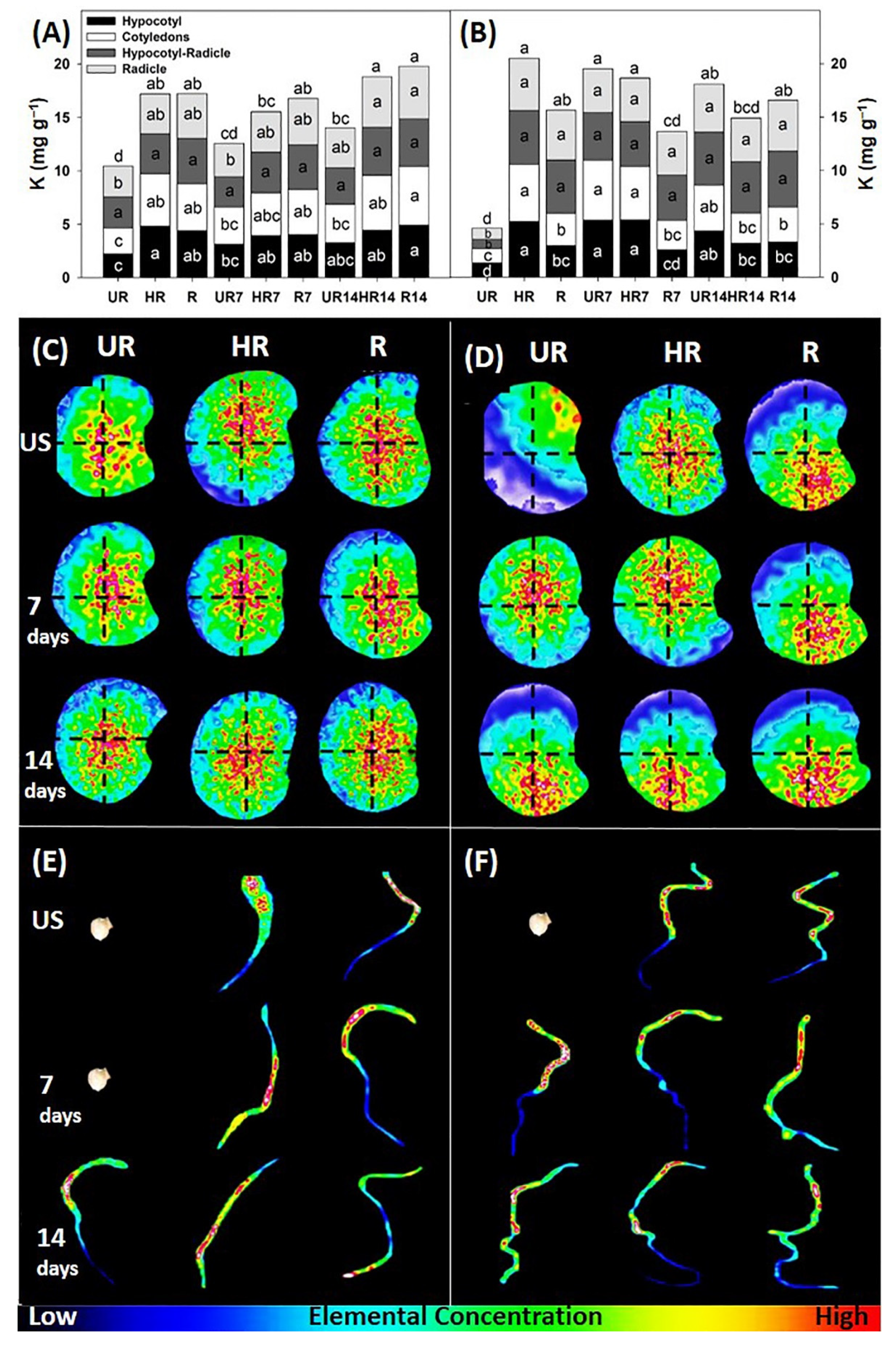
Treatments HR14 and R14 showed the highest total K concentrations before imbibition (18.8 and 19.8 mg g−1, respectively), while after imbibition, treatments HR (20.5 mg g−1), UR7 (19.5 mg g−1), and HR7 (18.6 mg g−1) had the highest concentrations compared to the other treatments (Figure 2A,B). On the other hand, the UR treatment had the lowest concentration in seeds pre- and post-imbibition, being 10.4 and 4.5 mg g−1, respectively. It is evident that K was very mobile in all embryo regions, which suggests that it was translocated towards enzymes, organic acids, sugars, and endogenous hormones, as indicated by Marrush et al. [20], who reported that K is easily translocated throughout the plant because both the developing fruit and seeds are major sinks during plant growth; in addition, the lack of sufficient K may alter the levels of some hormones involved in germination.
The K concentration was higher in the hypocotyl of the seeds pre-imbibition in HR and R14 (4.92 and 4.82 mg g−1, respectively) compared to the other treatments (Figure 2A). The R14 treatment (5.50 mg g−1) registered the highest concentration in the cotyledon. No statistically significant differences were found in the hypocotyl-radicle. In the radicle, HR14 and R14 showed the highest values (4.93 and 4.73 mg g−1, respectively), UR and UR7 treatments (2.89 and 3.11 mg g−1, respectively) had the lowest concentration. The K concentration was higher in the cotyledons and the radicle, both are meristematic regions, which suggests a greater metabolic activity. In this sense, K initiates metabolic reactions during the degradation of nutritional reserves and is involved in the synthesis of carbohydrates and proteins that are essential during seedling growth [21]. On the other hand, in seeds post-imbibition (Figure 2B), the concentration in the hypocotyl was statistically higher in treatments HR, UR7, and HR7 than in the other treatments (5.37, 5.34, and 5.22 mg g−1, respectively), while UR registered the lowest concentration (1.31 mg g−1). In the cotyledons, UR, HR, and HR7 showed the highest concentrations (5.61, 5.33, and 5.02 mg g−1). In the hypocotyl-radicle and the radicle, the treatments were not statistically different from each other, with the exception of the immature seeds, which showed 0.88 and 1.03 mg g−1 (Figure 2B).
With regard to the elemental distribution, the K was accumulated mainly in the endosperm in seeds pre-imbibition (Figure 2C). The K in HR seeds was distributed in the hypocotyl and cotyledons, while in R seeds it was observed in all embryo structures. In the other treatments, it was only found in the hypocotyl and radicle. Postharvest storage for 7 and 14 days increased the distribution of K in all embryo structures (Figure 2C).
In seeds after imbibition, K was translocated to other structures (hypocotyl-radicle and radicle). In the UR treatment, it was leached through the aleurone in the cotyledons (Figure 2D), probably because the seeds were physiologically immature, as indicated by Vidigal et al. [6], who stated that immature seeds leach electrolytes and solutes through the aleurone because they are not fully formed. In seeds post-imbibition, in the HR and R treatments, the K was translocated from the endosperm, cotyledons, and the radicle, respectively, to trigger metabolic reactions and provide the embryo with enzymes, organic acids, sugars, and endogenous hormones to continue its growth (Figure 2D). In this regard, some authors [22,23] indicated that K is more concentrated in the embryo and endosperm to facilitate some metabolic reactions during the germination process. In the treatments with 14 days of postharvest storage, the amount of K increased and it was translocated from the endosperm to the radicle. Thus, K strengthens cell walls and stabilizes cell membranes by binding the phosphate and carboxylate groups of phospholipids and proteins, allowing stabilization of the membranes and aleurone [24].
With regard to the distribution in seedlings, treatment R at 10 days after sowing registered the highest concentration of K. With the exception of treatment R14, in the other treatments K was found in the hypocotyl, while in R14 it was observed in the radicle (Figure 2E). At 14 days, K was visualized in the hypocotyl and cotyledons of seedlings (Figure 2F). In most of the treatments, K was found in the hypocotyl of the seedlings, and after imbibition, the mineral element was translocated to the cotyledons.

3.1.2. Calcium (Ca)

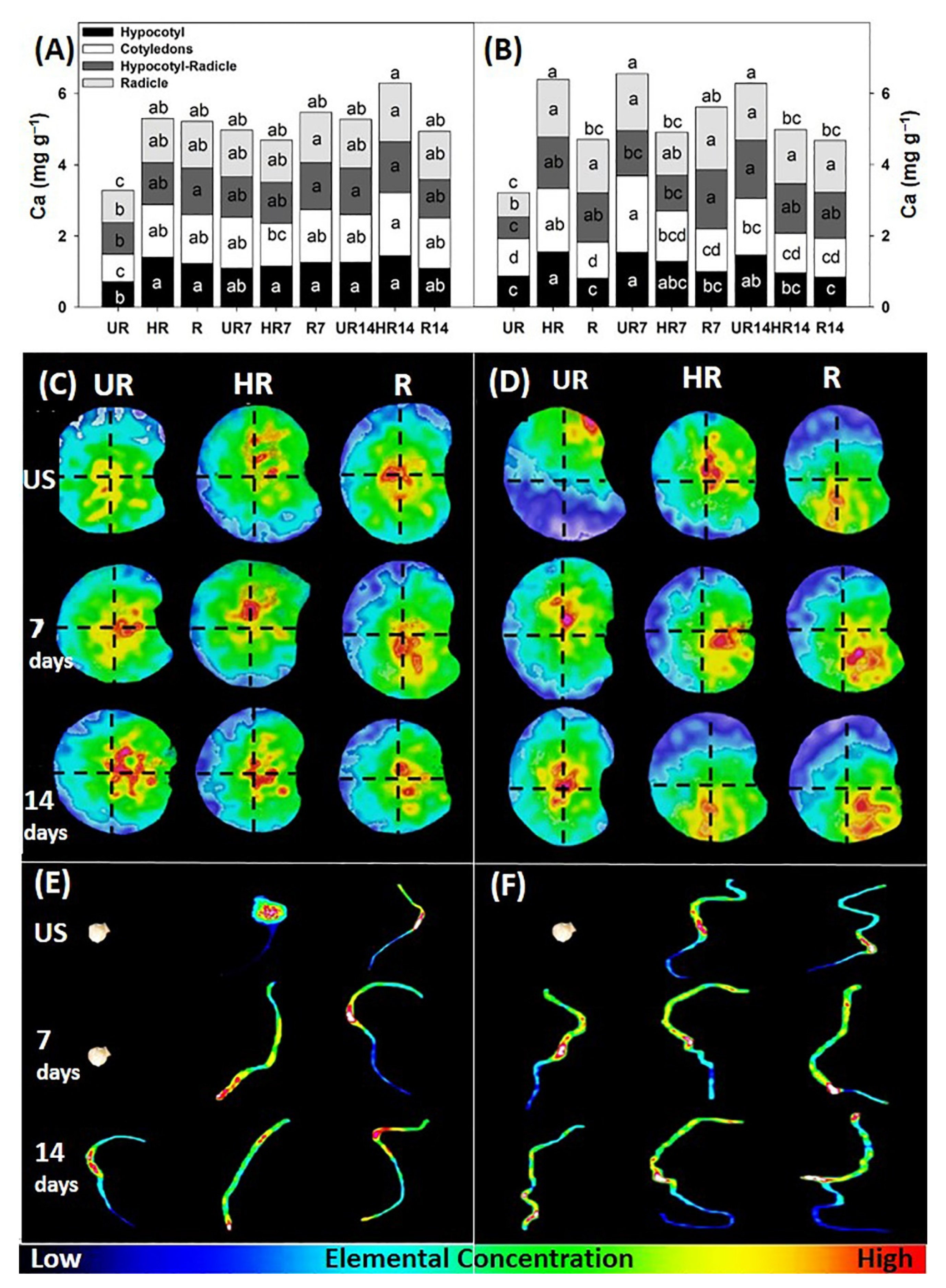
The HR14 treatment had the highest total Ca concentration (6.3 mg g−1) in seeds pre-imbibition. After imbibition, treatments HR, UR7, and UR14 registered the highest values (6.4, 6.5, and 6.2 mg g−1) compared to the other treatments (Figure 3A,B). Ca concentration varied according to embryonic development and seed imbibition. In this regard, the mineral concentration of Ca and its distribution in the seed depend on the stage of development [15].
Regarding the concentration by region, the HR14 treatment presented the highest Ca concentration in the hypocotyl while the UR treatment registered the lowest value (1.43 and 0.71 mg g−1, respectively). In the cotyledons, the concentration in the HR14 treatment was statistically greater than the UR treatment (1.77 and 0.78 mg g−1, respectively). In the hypocotyl-radicle, treatments R, R7, HR14, and UR14 had the highest concentrations of Ca (1.31, 1.31, 1.43, and 1.30 mg g−1, respectively). In the radicle, the HR14 and R7 treatments had the highest concentrations (1.65 and 1.42 mg g−1, respectively) compared to the UR treatment, which registered the lowest value with 0.90 mg g−1 (Figure 3A). In seeds post-imbibition, the HR and UR7 treatments obtained the highest concentrations (1.55 and 1.54 mg g−1, respectively) in the hypocotyl. The cotyledons of UR7 registered the highest concentration (2.14 mg g−1). In the hypocotyl-radicle, treatments R7 and UR14 had the highest concentrations with 1.66 and 1.63 mg g−1, respectively. In the radicle, except for the UR treatment, all other treatments had similar concentrations (Figure 3B).
Ca was distributed in the endosperm, hypocotyl, and cotyledons of seeds pre-imbibition (Figure 3C). Postharvest storage of 14 days in the treatments allowed the translocation of the Ca towards the cotyledons and the radicle (Figure 3C). The translocation of Ca favored the stability of the aleurone layer by decreasing the flow of mineral elements and solutes, and also served as a metabolic activator in the release of fundamental reserves during germination, so its concentration and distribution directly influenced seed viability. In this regard, Hussain et al. [25] reported that a higher concentration of Ca increased the germination and growth of rice seedlings, which suggests an increase in endosperm metabolism, higher respiration rate, and better integrity of the aleurone layer.
In seeds post-imbibition, the UR treatment had greater leaching of Ca in the cotyledons, while in the HR treatment it was concentrated in the endosperm, and in R it was translocated from the endosperm to the hypocotyl-radicle region. Postharvest storage in HR7, R7, HR14, and R14 allowed the translocation of Ca to the hypocotyl-radicle and radicle (Figure 3D). The translocation towards the radicle allowed the embryonic axis to obtain the conditions and capacity to break the endosperm and the testa, which reflected greater germination and vigor. On the contrary, a low distribution and concentration of the mineral elements reflect low viability. In this regard, Welch [14] mentioned that seeds with a high mineral distribution had high germination and emergence, while Ca deficient seeds obtained low germination and abnormal seedlings. At 10 days after sowing (das) in seedlings, the Ca was located in the hypocotyl (Figure 3E), while at 14 das the distribution was in the hypocotyl and cotyledons. The UR14 treatment had the highest concentration of Ca in the radicle (Figure 3F), and this distribution suggests that Ca, together with K, translocates to meristems where they are involved in protein synthesis and growth regulators. In this regard, some authors [24,26] indicated that K and Ca are involved in multiple enzyme systems and the activity of growth regulators, as well as in the formation and balance of the osmotic cell wall and vacuole.

3.1.3. Phosphorus (P)

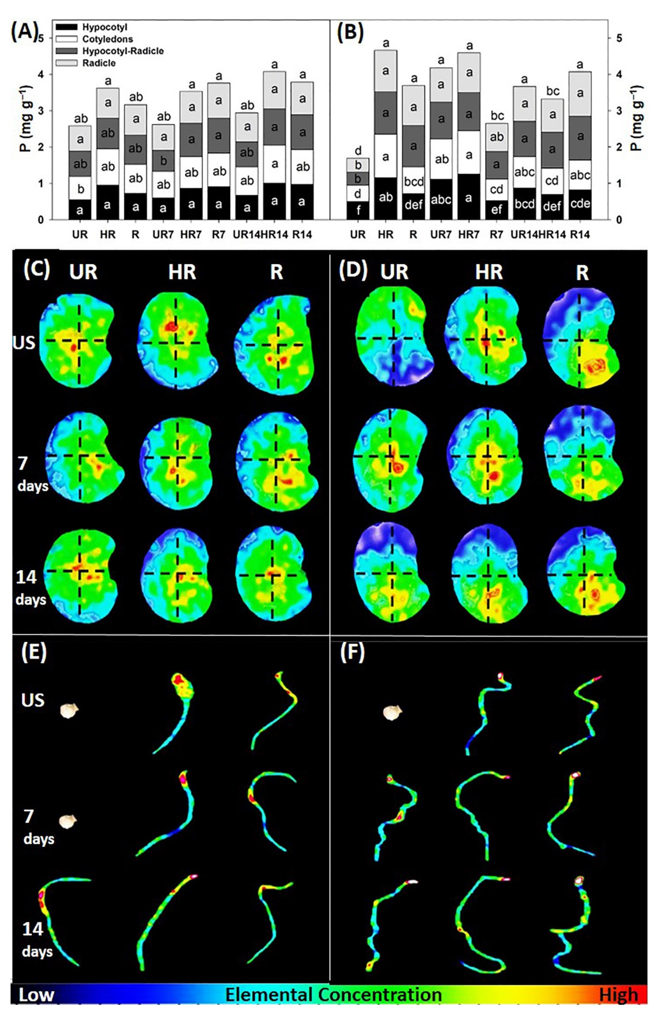
Treatments HR, HR7, R7, HR14, and R14 had the highest total P concentrations (3.6, 3.5, 3.8, 4.1, and 3.8 mg g−1, respectively). After imbibition, treatments HR, R, UR7, HR7, UR14, and R14 registered the highest values (4.7, 3.7, 4.2, 4.6, 3.7, and 4.1 mg g−1, respectively) compared to the other treatments (Figure 4A,B). The UR treatment had the lowest concentration in seeds pre- and post-imbibition (2.6 and 1.6 mg g−1, respectively).
No significant differences were found among treatments in the hypocotyl of seeds pre-imbibition. The HR14 treatment showed the highest concentration (1.05 mg g−1) in the cotyledons. Treatments HR7, R7, HR14, and HR14 had the highest concentrations (0.92, 0.97, 1.00, and 0.96 mg g−1, respectively) in the hypocotyl-radicle. No significant differences were found in the concentration of P in the radicle (Figure 4A). In seeds post-imbibition, the HR7 treatment showed the highest concentration (1.25 mg g−1) in the hypocotyl, while HR and HR7 had the highest concentrations (both with 1.20 mg g−1) in the cotyledons. In the hypocotyl-radicle, UR had the lowest value (0.38 mg g−1); the other treatments were similar to each other (Figure 4B). The concentration of P indicates that it was distributed throughout the seed in order to maintain the aleurone structure. The P constantly accumulates in the aleurone during the maturation of the embryo, which reflects that P accumulation in the aleurone layer allows the formation of globoid phytin [27].
In the HR treatment, the P was translocated to the cotyledons, and in R to the cotyledons and the radicle (Figure 4C). Fourteen days of postharvest storage increased the distribution of P in treatments UR7 and UR14 and it was located in the endosperm, cotyledons, and radicle. In this regard, Mori et al. [28] mentioned that seeds treated with a higher concentration of P increased the activity of the enzyme α-amylase, which resulted in a greater release of reserves and energy availability, thereby improving viability and root growth. In the seeds post-imbibition, the P was leached in the UR treatment, while in treatments HR and R it remained in the endosperm and radicle. On the other hand, in UR7 and HR7 the P was distributed in the endosperm, and in R7 in the hypocotyl-radicle and radicle (Figure 4D).
In the seedlings of the UR14, R7, and R14 treatments at 10 das, the P was observed in the hypocotyl, in HR in the endosperm, while in HR7, HR14, and R it was found in the cotyledons (Figure 4E). At 14 das, P was visualized in the cotyledons in all treatments (Figure 4F). The distribution of P in cotyledons suggests its involvement in chlorophyll synthesis and primordial growth, which allows greater seedling growth. In this regard, Mondal et al. [29] mentioned that a higher concentration and distribution of P allowed greater growth because the element participates in the synthesis of chlorophyll, involved in photosynthesis, respiration, and nitrogen fixation. Likewise, in other studies, P influenced the emergence, height, and dry weight of the seedlings [28].

3.1.4. Iron (Fe)

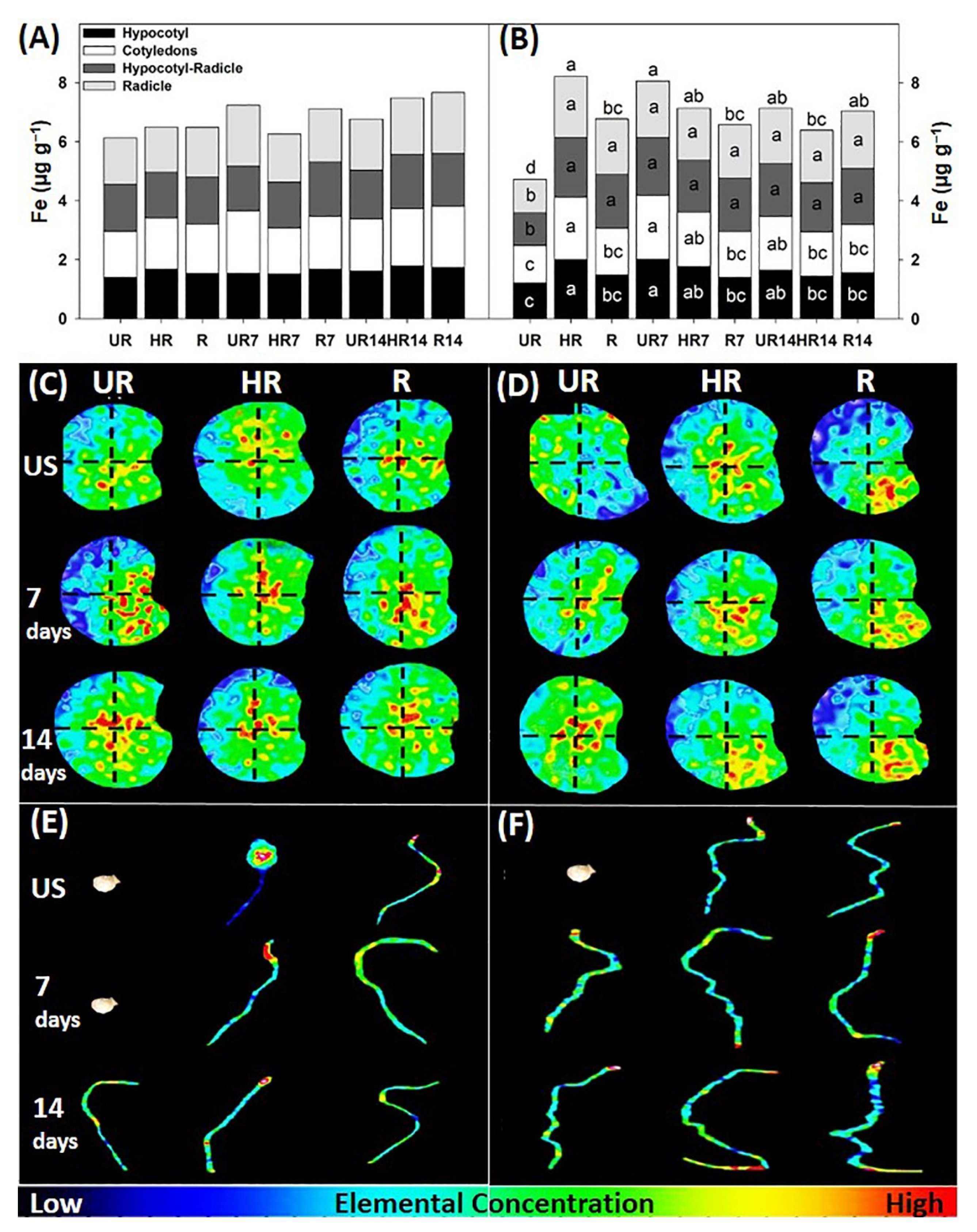
There were no statistically significant differences between treatments in the total concentration of Fe in seeds pre-imbibition. After imbibition, the highest concentrations were found in the HR and UR7 treatments (8.2 and 8.1 µg g−1, respectively) (Figure 5A,B). However, in seeds post-imbibition, significant differences were found. In the hypocotyl and cotyledons, the treatments HR (2.01 and 2.17 µg g−1, respectively) and UR7 (2.01 and 2.12 µg g−1, respectively) registered the highest concentrations. In the hypocotyl-radicle and the radicle, with the exception of UR, all the other treatments had similar concentrations between them (Figure 5B). The Fe concentration was similar in all structures of the embryo, which suggests that its distribution is mainly in the endosperm and aleurone to maintain the integrity of the seeds. In this sense, Lombi et al. [5] mentioned that Fe is concentrated mainly in the ventral part of the endosperm to accelerate enzymatic reactions during the degradation of nutrients. Likewise, Borg et al. [30] mentioned that approximately one-fifth of the Fe was located in the central endosperm and was stored in proteins of vacuoles; Fe is involved in the synthesis of proteins and carbohydrates, in addition to its joint involvement with P during photosynthesis in plants.
In the UR treatment, Fe was distributed in the endosperm, while in HR it was found in the cotyledons, and in R in the cotyledons and endosperm (Figure 5C). Postharvest storage of the fruit (7 and 14 days) increased the concentration of Fe. It was distributed in the endosperm, which allowed increased the degradation of carbohydrates present during germination. In this regard, Lu et al. [31] determined that the Fe caused an increase in oxygen reactions during the degradation of reserves, which facilitated the availability of energy during the germination process. After imbibition, the treatments with postharvest storage of 7 and 14 days had a higher Fe concentration, also allowing the translocation of the Fe to the endosperm and radicle (Figure 5D). In this regard, Lee et al. [1] mentioned that the translocation of Fe towards the endosperm is due to transporters such as nicotianamine, which transports the Fe to growth areas, such as the radicle. In the HR7, R7, HR14, and R14 treatments, Fe was translocated from the endosperm to the radicle (Figure 5D), which suggests that it was moved to this area to facilitate chemical reactions and promote the rupture of the endosperm.
In seedlings at 10 das, the Fe had a low distribution in the UR14 treatment. In HR, it lodged in the endosperm; in HR7, HR14, and R in the cotyledons, and in R7 and R14 in the hypocotyl (Figure 5E). At 14 days, the HR3 and R14 seedlings registered the highest distribution in the cotyledons and in the radicle (Figure 5F).

3.1.5. Magnesium (Mg)

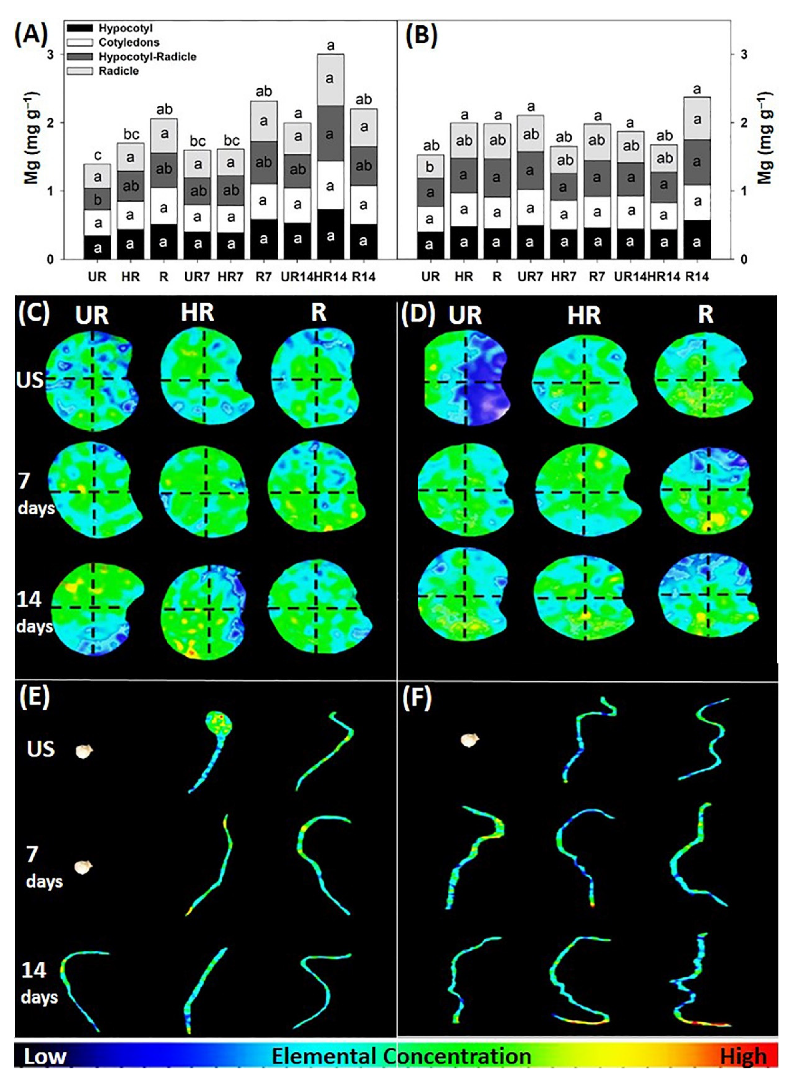
The HR14 treatment had the highest total concentration of Mg (3.0 mg g−1) in seeds pre-imbibition; and in seeds, post-imbibition treatments HR, R, UR7, R7, UR14, and R14 had the highest values (1.9, 1.9, 2.1, 1.9, 1.9, and 2.4 mg g−1, respectively) compared to the other treatments (Figure 6A,B). In seeds pre-imbibition, significant differences were only found in the hypocotyl-radicle, the UR14 treatment had the highest concentration (0.81 mg g−1) (Figure 6A). In seeds post-imbibition, in the radicle, the R14 treatment had the highest value (0.62 mg g−1) compared to the UR treatment (0.34 mg g−1) (Figure 6B). Mg is a mineral element with a low concentration and distribution compared to other elements. In barley grains, a lower concentration (18%) of Mg was obtained with respect to Zn and Cu (25% and 43%, respectively) but participated in essential functions during seed development and germination [5].
In the UR treatment, the Mg was lower than in the other treatments. In the HR treatment it was located in the hypocotyl, and in R treatment in the endosperm. With postharvest storage, the Mg was translocated to various structures of the embryo. In the HR7 treatment it was found in the cotyledons, in R7 in the hypocotyl-radicle, while in UR14 and HR14, the Mg was translocated to the hypocotyl and hypocotyl-radicle (Figure 6C). After imbibition, in the UR treatment, the Mg was translocated from the endosperm to the hypocotyl, while in HR and R it was maintained in the endosperm. In UR7, HR7, and UR14 the Mg was translocated to the cotyledons, and in R7, HR14, and R14 to the hypocotyl-radicle and radicle (Figure 6D). The translocation of Mg towards different structures of the embryo during its development allows the cellular structures of the different compounds to be maintained, as well as for it to participate in the synthesis of proteins and carbohydrates [32,33]. Both Shaul [32] and White et al. [33] mentioned that the mobilization of Mg is due to the synthesis of proteins and the regulation of the balance of cellular ions that occurs in the various stages of formation and development of the embryo. In seedlings at 10 das, Mg was found in the hypocotyl in the treatments UR14, R, R7, and R14, in the endosperm in HR, and in the cotyledons in HR7 and HR14 (Figure 6E), while at 14 das the Mg was found in the hypocotyl in UR7, R1, and R7; but in UR14, HR, HR7, HR14, and R14 it was translocated towards the radicle (Figure 6F).

3.1.6. Manganese (Mn)

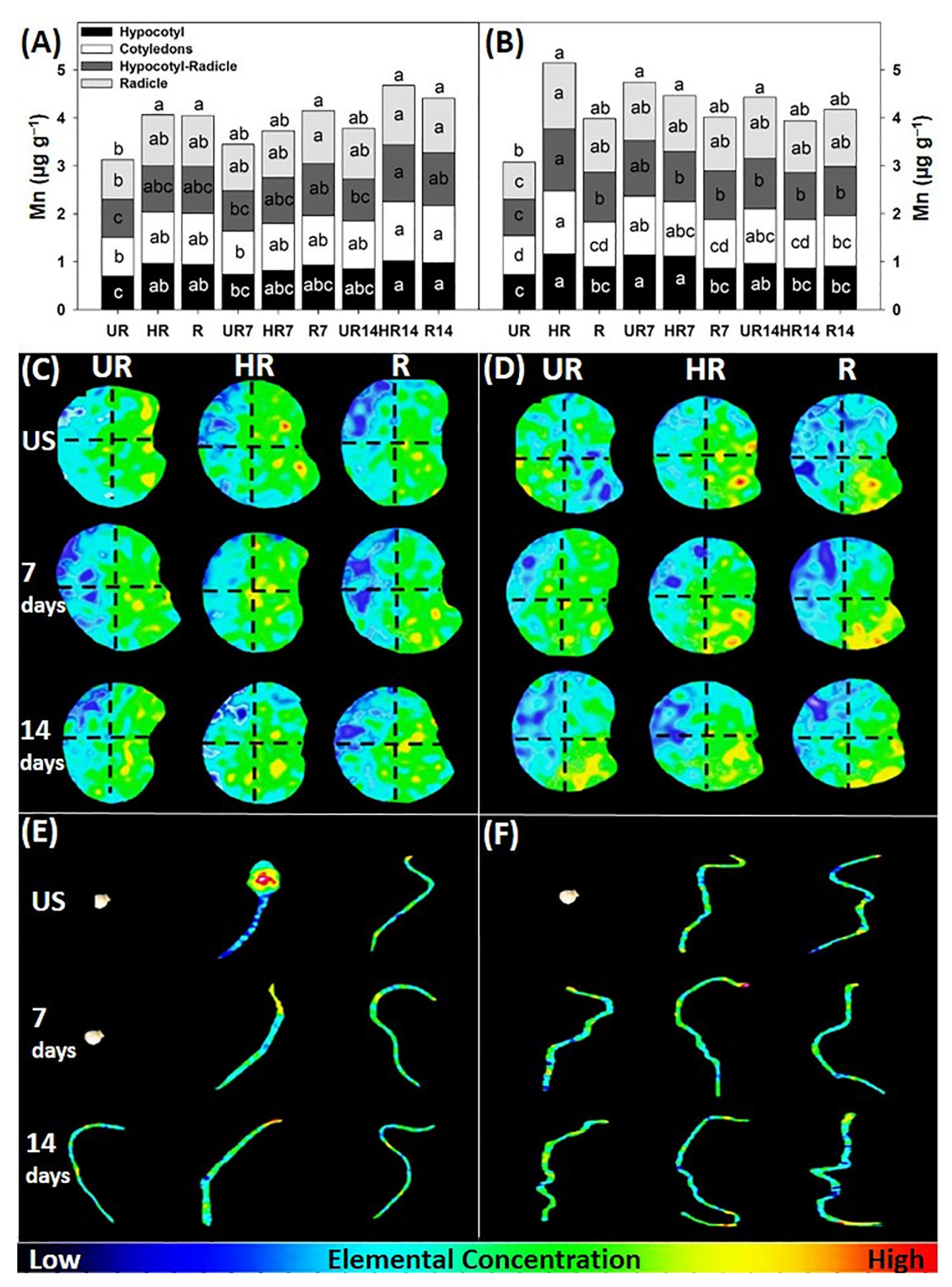
Treatments HR, R, R7, HR14, and R14 had the highest total concentrations of Mn in seeds pre-imbibition (4.1, 4.0, 4.1, 4.7, and 4.4 µg g−1, respectively) (Figure 7A). After imbibition, the treatments HR, UR7, HR7, and UR14 had the highest values (5.1, 4.7, 4.5, and 4.4 µg g−1, respectively) with respect to the other treatments (Figure 7B). The UR treatment had the lowest concentration in seeds pre- and post-imbibition (3.1 and 3.0 µg g−1, respectively). Treatments HR14 and R14 had the highest concentrations in regions of the embryo of seeds pre-imbibition (Figure 7A). In seeds post-imbibition, the HR treatment had the highest concentration in the hypocotyl, cotyledons, hypocotyl-radicle, and radicle (1.16, 1.31, 1.29, and 1.38 µg g−1, respectively) (Figure 7B). The concentration of Mn allows the activity of protection mechanisms against pathogens, as well as photosynthetic and respiratory capacity [34,35]. However, its absence or a deficiency in its concentration negatively influences seed viability [36]. In this regard, Longenecker et al. [36] reported slow germination and low development of barley and lupine seedlings in treatments with a lower Mn content.
In the UR, HR, and R treatments, the Mn was found to be distributed in the cotyledons and the radicle (Figure 7C). Postharvest storage for 7 and 14 days did not modify the distribution of the Mn in the embryo structures (Figure 7C). After imbibition, in the UR treatment, the Mn was leached through the aleurone, while in HR and R the Mn was found in the radicle. Postharvest storage for 7 and 14 days translocated Mn to the root region (Figure 7D). The highest distribution of Mn was recorded in the radicle because it participates in growth processes in the root apex in the meristematic zone, as well as participating in photosynthesis and respiration in plants [5]. In this regard, Lombi et al. [5] obtained higher root growth in treatments where the concentration of Mn was higher with respect to the other treatments. Moreover, Chen et al. [21] registered greater development in treatments where Mn was distributed and concentrated in the embryonic root and the cotyledon.
In the UR14, R7, and R14 treatments the Mn was located in the hypocotyl of the seedlings at 10 das (Figure 7E). At 14 das, in the UR7, UR14, HR, HR7, R, and R7 treatments, the Mn was distributed in the cotyledons, while in HR14 and R14, the highest distribution was in the radicle (Figure 7F). The Mn was distributed throughout the seedling mainly in growing areas. In this sense, Mondal et al. [29] mentioned that Mn performs functions in the physiology of plants, influencing germination, leaf size, and the lengths of the apex and roots. Likewise, Babaeva et al. [37] reported that the concentration of Mg and Mn increased germination (36%) and emergence (27%) in Echinacea purpurea L.

3.2. Germination and Electrical Conductivity

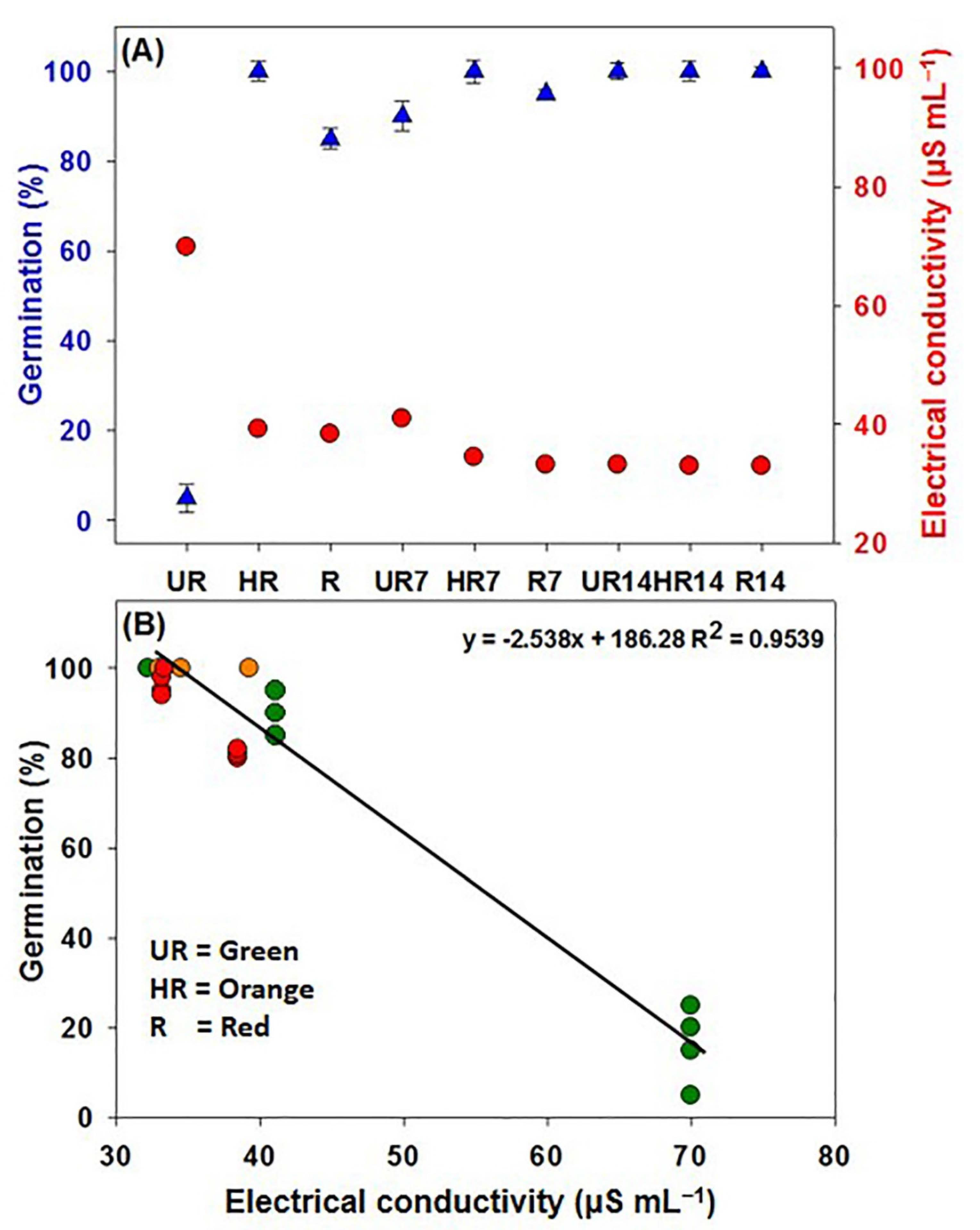
Maximum germination was registered in treatments HR, UR14, HR14, and R14 (All with 100%). These treatments had greater mineral concentrations, as well as lower electrical conductivities (34.5, 32.2, 33, and 33.4 µS mL−1, respectively). This suggests that the seeds extracted from fruits with postharvest storage continued their embryo and seed maturation process (Figure 8A). The UR treatment showed the highest mineral elements flux (70 µS mL−1) through aleurone and the lowest germination percentage, which indicates that these seeds did not complete their maturation process. Indeed, immature seeds often exude more mineral elements during the first hours of imbibition compared to mature seeds [6,8]. Fourteen days of postharvest storage in UR14 reduced the flow of elements and solutes through the aleurone layer by up to 53.72%, postharvest storage allowed the seeds to reach physiological maturity (Figure 8A). In this regard, EC is an indication of the physiological maturity of the seed. Therefore, the flow of mineral elements in the external environment of the seeds represents the degree of aleurone integrity; that is, low values of electrical conductivity are associated with mature seeds [38,39,40]. Both seed vigor and viability are related to membrane integrity [39], size [41], seed characteristics [42,43], and the concentration and distribution of mineral elements in the seeds [12,15].
In the correlation graph (Figure 8B), it can be observed that the electrical conductivity is inversely related to seed viability (germination). The seeds from fruits with postharvest storage of 7 and 14 days prior to extraction completed their physiological maturation and had the highest germination; in contrast, the seeds with a high electrical conductivity had the lowest viability.

4. Conclusions

X-ray fluorescence is an alternative means to visualize and determine the concentrations of some macro- and micronutrients in seeds and seedlings. The concentration and distribution of mineral elements inside seeds depend on the development stage of the embryo and are modified by the postharvest storage of fruits and imbibition during the germination process. There were greater concentrations of potassium and calcium in seeds pre- and post-imbibition. Fourteen days of postharvest storage increased the concentration and modified the translocation of macro- and micronutrients in the different regions of the embryo in seeds pre- and post-imbibition. The imbibition of the seeds allowed the translocation of the elements to the root region of the embryo, which enhanced seed viability and vigor. The mineral element distribution in the seedlings increased as a function of embryonic development and postharvest storage. Furthermore, the postharvest storage of fruits translocated the mineral elements to the different structures of the seedling to enhance its growth. The 14-day postharvest storage decreased the leaching of mineral elements and solutes from the aleurone layer, increasing the maximum germination rate. This could be considered by seedling growers because they could harvest unripe fruits to avoid competition among fruits in plants and finalize the ripeness of embryos during the storage of fruits before extracting the seeds. Thus, they could decrease losses in seedling production.

Author Contributions

Conceptualization, C.D.H.-P., R.G., R.Z.-B. and H.S.B.-G.; formal analysis, C.D.H.-P., C.J.A.-L., R.G.; E.H.-N., B.Y.S.-G. and S.U.S.-G.; funding acquisition, R.G. and L.L.-M.; investigation, R.Z.-B., H.S.B.-G., and S.U.S.-G.; methodology, C.D.H.-P., C.J.A.-L., E.R.-S. and S.U.S.-G.; project administration, E.R.-S.; resources, R.G. and L.L.-M.; validation, C.J.A.-L.; visualization, R.H.A.-N.; writing—original draft preparation, C.D.H.-P. and B.Y.S.-G.; writing—review and editing, C.J.A.-L., R.G., R.H.A.-N., E.H.-N. and L.L.-M. All authors have read and agreed to the published version of the manuscript.

Funding

This research received no external funding.

Institutional Review Board Statement

Not applicable.

Informed Consent Statement

Not applicable.

Data Availability Statement

The data presented in this study are available upon request from the primary or correspondence author.

Acknowledgments

We acknowledge the National Council of Science and Technology (CONACyT) for the postgraduate scholarship (number 703739) granted to the first author. Likewise, we thank the Instituto Tecnológico de Conkal for the facilities to carry out this research.

Conflicts of Interest

The authors declare no conflict of interest.

References

  1. Lee, I.O.; Lee, K.H.; Pyo, J.H.; Choi, Y.J.; Lee, Y.C. Anti- inflammatory effect of capsaicin in Helicobacter pylori infected gastric epithelial cells. Helicobacter 2007, 12, 510–517. [Google Scholar] [CrossRef] [PubMed]
  2. Rithesh, M.; Dangi, R.S.; Dass, S.C.; Malhothra, R.C. The hottest chilli variety in India. Curr. Sci. 2000, 79, 287–288. [Google Scholar]
  3. Garruña-Hernández, R.; Latournerie-Moreno, L.; Ayala-Garay, O.; Santamaría, J.M.; Pinzón-López, L. Acondicionamiento pre-siembra: Una opción para incrementar la germinación de semillas de chile habanero (Capsicum chinense Jacq.). Agrociencia 2014, 48, 413–423. [Google Scholar]
  4. Iwai, T.; Takahashi, M.; Oda, K.; Terada, Y.; Yoshida, K. Dynamic changes in the distribution of minerals in relation to phytic acid accumulation during rice seed development. Plant Physiol. 2012, 160, 2007–2014. [Google Scholar] [CrossRef] [PubMed]
  5. Lombi, E.; Smith, E.; Hansen, T.H.; Paterson, D.; De Jonge, M.; Howard, D.L.; Persson, D.P.; Husted, S.; Ryan, C.; Schjoerring, J.K. Megapixel imaging of (micro) nutrients in mature barley grains. J. Exp. Bot. 2010, 62, 273–282. [Google Scholar] [CrossRef]
  6. Vidigal, D.S.; Dias, D.C.F.S.; Von-Pinho, E.R.V.; Dias, L.A.S. Sweet pepper seed quality and lea-protein activity in relation to fruit maturation and post-harvest storage. Seed Sci. Technol. 2009, 37, 192–201. [Google Scholar] [CrossRef]
  7. Ayala-Villegas, M.J.; Ayala-Garay, O.J.; Aguilar-Rincón, V.H.; Corona-Torres, T. Evolución de la calidad de semilla de Capsicum annuum L. durante su desarrollo en el fruto. Rev. Fitotec. Mex. 2014, 37, 79–87. [Google Scholar] [CrossRef]
  8. Dias, D.C.F.S.; Ribeiro, F.P.; Dias, L.A.S.; Silva, D.J.H.; Vidigal, D.S. Tomato seed quality harvested from different trusses. Seed Sci. Technol. 2006, 34, 681–689. [Google Scholar] [CrossRef]
  9. Vidigal, D.S.; Dias, D.C.F.S.; Dias, L.A.S.; Finger, F.L. Changes in seed quality during fruit maturation of sweet pepper. Sci. Agric. 2011, 68, 535–539. [Google Scholar] [CrossRef]
  10. Caixeta, F.; Von Pinho, E.V.R.; Guimarães, R.M.; Pereira, P.H.A.R. Determinação de ponto de colheita na produção de sementes de pimenta malagueta e alterações bioquímicas durante o armazenamento e a germinação. Científica 2014, 42, 187. [Google Scholar] [CrossRef]
  11. Bewley, J.D.; Black, M. Seeds: Physiology of Development and Germination, 2nd ed.; Plenum Press: New York, NY, USA, 1994; Volume 445. [Google Scholar]
  12. Smiciklas, K.D.; Mullen, R.E.; Carlson, R.E.; Knapp, A.D. Drought-induced stress effect on soybean seed calcium and quality. Crop Sci. 1989, 29, 1519–1523. [Google Scholar] [CrossRef]
  13. Xu, G.; Kafkafi, U.; Wolf, S.; Sugimoto, Y. Mother plant nutrition status and growing condition affect assimilates and emergence quality of hybrid sweet pepper seeds. J. Plant Nutr. 2002, 25, 1645–1665. [Google Scholar] [CrossRef]
  14. Welch, R.M. Effects of nutrient deficiencies on seed production and quality. In Advances in Plant Nutrition; Tinker, P.B., Lauchli, A., Eds.; Praeger: New York, NY, USA, 1986; Volume 2, pp. 205–247. [Google Scholar]
  15. Keiser, J.R.; Mullen, R.E. Calcium and relative humidity effects on soybean seed nutrition and seed quality. Crop Sci. 1993, 33, 1345–1349. [Google Scholar] [CrossRef]
  16. Lott, J.N.A.; Greenwood, J.S.; Batten, G.D. Mechanisms and regulation of mineral nutrient storage during seed development. In Seed Development and Germination; Kigel, J., Galili, G., Eds.; Marcel Dekker: New York, NY, USA, 1995; pp. 215–235. [Google Scholar]
  17. Hernández-Pinto, C.; Garruña, R.; Andueza-Noh, R.; Hernández-Núñez, E.; Zavala-León, M.J.; Pérez-Gutiérrez, A. Post-harvest storage of fruits: An alternative to improve physiological quality in habanero pepper seeds. Rev. Bio Cienc. 2020, 7, 1–15. [Google Scholar]
  18. Hill, T.; Sirbescu, M. Micro-XRF mapping of chemically zoned beryl: Fast, non-destructive, and precise. Microsc. Microanal. 2022, 28, 624–628. [Google Scholar] [CrossRef]
  19. Pacheco, N.; Méndez-Campos, G.K.; Herrera-Pol, I.E.; Alvarado-López, C.J.; Ramos-Díaz, A.; Ayora-Talavera, T.; Talcott, S.U.; Cuevas-Bernardino, J.C. Physicochemical composition, phytochemical analysis and biological activity of ciricote (Cordia dodecandra A. D.C.) fruit from Yucatán. Nat. Prod. Res. 2022, 36, 440–444. [Google Scholar] [CrossRef]
  20. Marrush, M.; Yamaguchi, M.; Saltveit, M.E. Effect of potassium nutrition during bell pepper seed development on vivipary and endogenous levels of abscisic acid (ABA). J. Am. Soc. Hortic. Sci. 1998, 123, 925–930. [Google Scholar] [CrossRef]
  21. Chen, P.; Lott, J.N.A. Studies of Capsicum annuum seeds: Structure, storage reserves, and mineral nutrients. Can. J. Bot. 1992, 70, 518–529. [Google Scholar] [CrossRef]
  22. Mazzolini, A.P.; Pallaghy, C.K.; Legge, G.J.F. Quantitative microanalysis of Mn, Zn and other elements in mature wheat seed. New Phytol. 1985, 100, 483–509. [Google Scholar] [CrossRef]
  23. Xu, G.; Kafkafi, U. Seasonal differences in mineral content, distribution and leakage of sweet pepper sedes. Ann. Appl. Biol. 2003, 143, 45–52. [Google Scholar] [CrossRef]
  24. Marschner, H. Mineral Nutrition of Higher Plants, 2nd ed.; Academic Press: San Diego, CA, USA, 1995; pp. 285–312. [Google Scholar]
  25. Hussain, S.; Khan, F.; Hussain, H.A.; Nie, L. Physiological and biochemical mechanisms of seed priming induced chilling tolerance in rice cultivars. Front. Plant Sci. 2016, 7, 116. [Google Scholar] [CrossRef]
  26. McCarty, D.R. Genetic control and integration of maturation and germination pathways in seed development. Annu. Rev. Plant Physiol. Plant Mol. Biol. 1995, 46, 71–93. [Google Scholar] [CrossRef]
  27. Ogawa, M.; Tanaka, K.; Kasai, Z. Isolation of high phytin containing particles from rice grains using an aqueous polymer two phase system. Agric. Biol. Chem. 1975, 39, 695–700. [Google Scholar] [CrossRef]
  28. Mori, S.; Fujimoto, H.; Watanabe, S.; Ishioka, G.; Okabe, A.; Kamei, M.; Yamauchi, M. Physiological performance of iron-coated primed rice seeds under submerged conditions and the stimulation of coleoptile elongation in primed rice seeds under anoxia. Soil Sci. Plant Nutr. 2012, 56, 469–478. [Google Scholar] [CrossRef]
  29. Mondal, S.; Bandana, B. Impact of micronutrient seed priming on germination, growth, development, nutritional status and yield aspects of plants. J. Plant Nutr. 2019, 42, 2577–2599. [Google Scholar] [CrossRef]
  30. Borg, S.; Brinch-Pedersen, H.; Tauris, B.; Holm, P.B. Iron transport, deposition and bioavailability in the wheat and barley grain. Plant Soil 2009, 325, 15–24. [Google Scholar] [CrossRef]
  31. Lu, L.; Tian, S.; Liao, H.; Zhang, J.; Yang, X.; Labavitch, J.; Chen, W. Analysis of metal element distributions in rice (Oryza sativa L.) seeds and relocation during germination based on X-Ray fluorescence imaging of Zn, Fe, K, Ca, and Mn. PLoS ONE 2013, 8, e57360. [Google Scholar] [CrossRef]
  32. Shaul, O. Magnesium transport and function in plants: The tip of the iceberg. Biometals 2002, 15, 309–323. [Google Scholar] [CrossRef]
  33. White, P.J.; Broadley, M.R. Biofortification of crops with seven mineral elements often lacking in human diets-iron, zinc, copper, calcium, magnesium, selenium and iodine. New Phytol. 2009, 182, 49–84. [Google Scholar] [CrossRef]
  34. Yao, Y.; Wang, J.; Ma, X.; Lutts, S.; Sun, C.; Ma, J.; Yang, Y.; Achal, V.; Xu, G. Proteomic analysis of Mn-induced resistance to powdery mildew in grapevine. J. Exp. Bot. 2012, 63, 5155–5170. [Google Scholar] [CrossRef]
  35. Borisjuk, L.; Neuberger, T.; Schwender, J. Seed architecture shapes embryo metabolism in oilseed rape. Plant Cell 2013, 25, 1625–1640. [Google Scholar] [CrossRef]
  36. Longenecker, N.E.; Marcar, N.E.; Graham, R.D. Increased manganese content of barley seeds can increase grain yield in manganese-deficient conditions. Aust. J. Agric. Res. 1991, 42, 1065–1074. [Google Scholar] [CrossRef]
  37. Babaeva, E.Y.; Volobueva, V.F.; Yagodin, B.A.; Klimakhin, G.I. Sowing quality and productivity of Echinacea purpurea in relation to soaking the seed in manganese and zinc solutions. Izv. Timiryazevskoi Sel’skokhozyaistvennoi Akad. 1999, 4, 73–80. [Google Scholar]
  38. Wann, E.V. Leaching of metabolites during imbibition of sweet corn seed of different endosperm genotypes. Crop Sci. 1986, 26, 731–733. [Google Scholar] [CrossRef]
  39. McDonald, M.B. Seed quality assessment. Seed Sci. Res. 1998, 8, 265–275. [Google Scholar] [CrossRef]
  40. Bewley, J.D.; Bradford, K.J.; Hilhorst, W.M.H.; Nonogaky, H. Seed: Physiology of Development, Germination and Dormancy, 3rd ed.; Springer: New York, NY, USA, 2013; p. 392. [Google Scholar]
  41. Sanchez, V.M.; Sundstrom, F.J.; Lang, N.S. Plant size influences bell pepper seed quality and yield. HortScience 1993, 28, 809–811. [Google Scholar] [CrossRef]
  42. Shephard, H.L.; Naylor, R.E.L. Effect of the seed coat on water uptake and electrolyte leakage of sorghum (Sorghum bicolor L. Moench) seeds. Ann. Appl. Biol. 1996, 129, 125–136. [Google Scholar] [CrossRef]
  43. Rehman, S.; Harris, P.J.C.; Bourne, W.F. The effect of sodium chloride on the Ca2+, K+ and Na+ concentrations of the seed coat and embryo of Acacia tortilis and A. coriacea. Annu. Appl. Biol. 1998, 133, 269–279. [Google Scholar] [CrossRef]
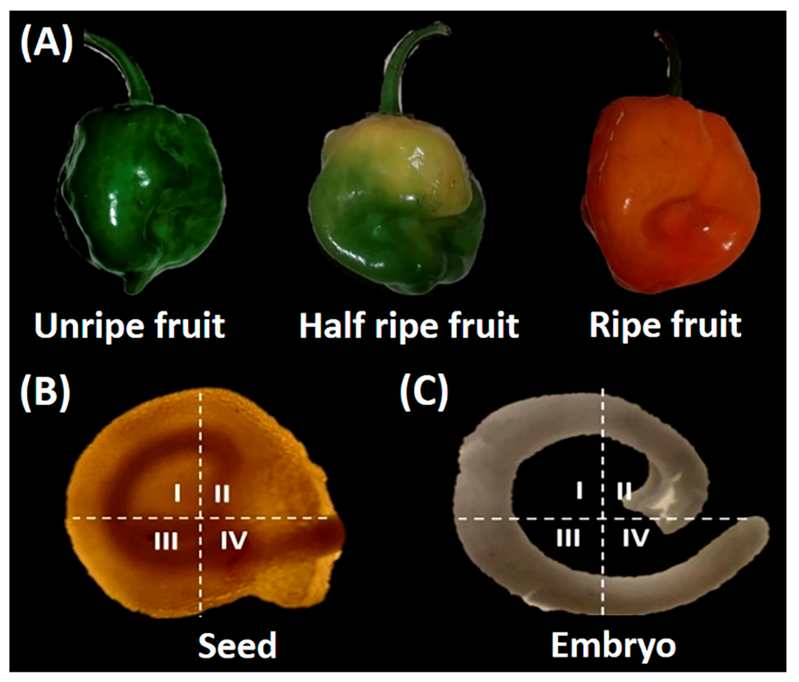
Figure 1. (A) Maturity stages of the habanero pepper fruit. (B) Complete seed divided into the central region of the micropyle and (C) habanero pepper embryo, quadrants I (hypocotyl), II (cotyledons), III (hypocotyl-radicle), and IV (radicle).
Agronomy 12 02117 g001
Figure 2. K concentration in seeds pre-imbibition (A) and post-imbibition (B), K distribution in habanero pepper seeds pre-imbibition (C) and post-imbibition (D), habanero seedlings at 10 (E) and 14 days (F) after sowing, of seeds of unripe (UR), half-ripe (HR), and ripe (R) fruits; unstored (US) and stored for 7 and 14 days postharvest. Colors in panels (CF) are based on a relative intensity scale (reported at the bottom of the figure) generated on the intensity of the X-ray fluorescence Kα peak of K in the samples. Data are means. Different letters indicate statistically significant differences among treatments (Duncan, p ≤ 0.05, n = 50).
Agronomy 12 02117 g002
Figure 3. Ca concentration in seeds pre-imbibition (A) and post-imbibition (B), Ca distribution in habanero pepper seeds pre-imbibition (C) and post-imbibition (D), habanero seedlings at 10 (E) and 14 days (F) after sowing, of seeds of unripe (UR), half-ripe (HR), and ripe (R) fruits; unstored (US) and stored for 7 and 14 days postharvest. Colors in panels (CF) are based on a relative intensity scale (reported at the bottom of the figure) generated on the intensity of the X-ray fluorescence Kα peak of Ca in the samples. Data are means. Different letters indicate statistically significant differences among treatments (Duncan, p ≤ 0.05, n = 50).
Agronomy 12 02117 g003
Figure 4. P concentration in seeds pre-imbibition (A) and post-imbibition (B), P distribution in habanero pepper seeds pre-imbibition (C) and post-imbibition (D), habanero seedlings at 10 (E) and 14 days (F) after sowing, of seeds of unripe (UR), half-ripe (HR), and ripe (R) fruits; unstored (US) and stored for 7 and 14 days postharvest. Colors in panels (CF) are based on a relative intensity scale (reported at the bottom of the figure) generated on the intensity of the X-ray fluorescence Kα peak of P in the samples. Data are means. Different letters indicate statistically significant differences among treatments (Duncan, p ≤ 0.05, n = 50).
Agronomy 12 02117 g004
Figure 5. Fe concentration in seeds pre-imbibition (A) and post-imbibition (B), Fe distribution in habanero pepper seeds pre-imbibition (C) and post-imbibition (D), habanero seedlings at 10 (E) and 14 days (F) after sowing, of seeds of unripe (UR), half-ripe (HR), and ripe (R) fruits; unstored (US) and stored for 7 and 14 days postharvest. Colors in panels (CF) are based on a relative intensity scale (reported at the bottom of the figure) generated on the intensity of the X-ray fluorescence Kα peak of Fe in the samples. Data are means. Different letters indicate statistically significant differences among treatments (Duncan, p ≤ 0.05, n = 50).
Agronomy 12 02117 g005
Figure 6. Mg concentration in seeds pre-imbibition (A) and post-imbibition (B), Mg distribution in habanero pepper seeds pre-imbibition (C) and post-imbibition (D), habanero seedlings at 10 (E) and 14 days (F) after sowing, of seeds of unripe (UR), half-ripe (HR), and ripe (R) fruits; unstored (US) and stored for 7 and 14 days postharvest. Colors in panels (CF) are based on a relative intensity scale (reported at the bottom of the figure) generated on the intensity of the X-ray fluorescence Kα peak of Mg in the samples. Data are means. Different letters indicate statistically significant differences among treatments (Duncan, p ≤ 0.05, n = 50).
Agronomy 12 02117 g006
Figure 7. Mn concentration in seeds pre-imbibition (A) and post-imbibition (B), Mn distribution in habanero pepper seeds pre-imbibition (C) and post-imbibition (D), habanero seedlings at 10 (E) and 14 days (F) after sowing, of seeds of unripe (UR), half-ripe (HR), and ripe (R) fruits; unstored (US) and stored for 7 and 14 days postharvest. Colors in panels (CF) are based on a relative intensity scale (reported at the bottom of the figure) generated on the intensity of the X-ray fluorescence Kα peak of Mn in the samples. Data are means. Different letters indicate statistically significant differences among treatments (Duncan, p ≤ 0.05, n = 50).
Agronomy 12 02117 g007
Figure 8. Percentage of germination and electrical conductivity (A) of habanero pepper seeds and correlation between the germination percentage and electrical conductivity (B) of habanero pepper seeds from unripe (UR = Green circles), half-ripe (HR = Orange circles), and ripe (R = Red circles) fruits; unstored (US) and stored for 7 and 14 days postharvest.
Agronomy 12 02117 g008
Table 1. Analysis of variance of mineral elements concentration (K, Ca, P, Fe, Mg, and Mn), germination (%G), and electrical conductivity (EC) of habanero pepper seeds extracted from fruits with different stages of maturity (unripe, half-ripe, and ripe) and postharvest storage times (unstored and stored for 7 and 14 days postharvest).
FactorsKCaMgFePMn%GEC
FMS20.95 **12.35 *8.79 *3.58 ns8.13 *20.41 *86.09 *20.60 **
PSF9.15 **10.98 *10.16 *1.50 ns13.92 *10.85 *162.53 **53.60 **
SI127.88 **11.49 *4.89 *0.80 *5.76 *8.70 *20.77 **14.20 **
FMS × PSF × SI7.21 **11.31 *3.65 *1.18 *2.85 *1.77 *55.04 **23.15 **
CV5.669.395.498.236.718.179.2510.74
ns = no significance; * = statistically significant differences (ANOVA p ≤ 0.05, n = 50); ** = highly significant differences (ANOVA p ≤ 0.01, n = 50); FMS = fruit maturity stages; PSF = postharvest storage of fruits; SI = seed imbibition; CV = coefficient of variation.
Publisher’s Note: MDPI stays neutral with regard to jurisdictional claims in published maps and institutional affiliations.