Article

Characteristics of Oil Body Development and the Cloning and Expression Analysis of PDAT Genes in Eucommia ulmoides

1
Research Institute of Non-Timber Forestry Chinese Academy of Forestry (CAF), Zhengzhou 450003, China
2
Key Laboratory of Non-Timber Forest Germplasm Enhancement & Utilization of State Forestry and Grassland Administration, Zhengzhou 450003, China
3
Central South University of Forestry and Technology, Changsha 410004, China
*
Authors to whom correspondence should be addressed.
These authors contributed equally to this work.
Agronomy 2022, 12(9), 2197; https://doi.org/10.3390/agronomy12092197
Submission received: 10 August 2022 / Revised: 9 September 2022 / Accepted: 12 September 2022 / Published: 15 September 2022
(This article belongs to the Section Crop Breeding and Genetics)

Abstract

:
Studies in multiple species have shown that phospholipid:diacylglycerol acyltransferase (PDAT) and oil bodies are important factors affecting plant oil accumulation. However, little information is available about the PDAT genes and oil bodies in Eucommia ulmoides Oliv. a high-quality oil tree. In this study, the oil bodies in Eucommia ulmoides seeds at different developmental stages were observed by frozen section technology combined with Nile red staining. It was found that there was a significant positive correlation between oil content and oil body number. In addition, seven Eucommia ulmoides PDAT (EuPDAT) genes were cloned from Eucommia ulmoides seeds based on genome data. They were assembled into three subgroups according to their classifications in Arabidopsis. EuPDAT genes in the same subgroup had similar gene structures and conserved motifs. Putative cis-element analysis of EuPDAT gene promoter regions uncovered numerous elements related to stress and plant hormones response. The expression patterns showed that EuPDAT2 and EuPDAT7 expression levels were positively related to oil content in different periods, and the correlation coefficients were 0.78 and 0.91, respectively. Our results suggest that oil body number and EuPDAT genes are important factors affecting oil accumulation in E. ulmoides seeds, and this work provides a theoretical reference for the directional cultivation of new high-oil-content E. ulmoides varieties.

1. Introduction

Plant oils play important roles in plant growth, human diet, and industrial development, and they are mainly stored in oil bodies in the form of triacylglycerols (TAGs) [1]. The oil body is mainly composed of TAGs, phospholipids, and oil body proteins, and it exists in plant seeds and pollen [2,3]. In recent years, there have been two main views on the relationship between oil bodies and plant oil content. One view is that during the process of plant growth and development, based on the mutual recognition of oil body proteins, small oil bodies fuse to form large oil bodies. According to this view, it is believed that the size of oil bodies is positively correlated with oil content [4,5]. Another view is that the oil content is mainly determined by the total cross-sectional area of the intracellular oil bodies but has no significant correlation with the size of intracellular oil bodies [6]. Eucommia ulmoides Oliv, as an important economic tree species, has excellent oil quality. Therefore, it is of great significance to study the correlation between oil content and the oil bodies of E. ulmoides for the development of the E. ulmoides oil industry.
At present, the Kennedy pathway is known as the main pathway of oil accumulation; it relies on acyl-CoA and synthesizes TAG mainly under the catalysis of glycerol-3-phos-phate acyltransferase (GPAT) [7,8]. In addition, a subsidiary pathway for oil synthesis that is independent of acyl-CoA has been found. PDAT directly catalyzes the binding of diacylglycerol (DAG) with phospholipids to produce TAG and lysophospholipids [9,10,11,12,13]. PDAT was first discovered by Voelker in yeast and was later studied in species including Arabidopsis thaliana (L.) Heynh, Helianthus annuus L., Brassica napus L., and Camellia oleifera Abel. It was found that there were differences in PDAT functions in different species [14,15]. When RNAi technology was used to knock down the PDAT gene in Arabidopsis thaliana, the oil content of A. thaliana seeds decreased by about 75%, and the embryos did not develop normally [9]. When the diacylglycerol acyl-transferase (DGAT) gene, the other key gene involved in TAG synthesis, was knocked down, the oil content of A. thaliana seeds decreased by about 30%, and the embryo development was also hindered [16,17]. Therefore, it is speculated that the PDAT gene and DGAT gene overlap in function in A. thaliana [18,19]. In flax, it has been demonstrated that the PDAT gene has a specific preference for linolenic acid substrate, which can not only improve the oil content of flax seeds, but can also improve the content of linolenic acid in oil [20]. The PDAT gene in Linum usitatissimum L was transferred to A. thaliana, and it was found that the linolenic acid content in A. thaliana oil was significantly increased [21,22]. The PDAT gene was cloned from Ricinus communis Mill, and it was found that its substrate preference contained phospholipids with castor acyl chains, which could significantly increase the content of hydroxyl fatty acids in oil. This further supports the conclusion that the PDAT gene in different species has different substrate preferences [16].
E. ulmoides is a unique economic tree species that has been utilized in China for more than 2000 years [23]. E. ulmoides seed oil is rich in unsaturated fatty acids, which are beneficial to the human body [24,25]. Among these acids, the alpha-linolenic acid content is as high as 65%, which is one of the highest linolenic acid contents found in plant oils at present. Alpha-linolenic acid is essential to the human body and can only be obtained through dietary intake. It can be converted into docosahexaenoic acid (DHA) and eicosapentaenoic acid (EPA) in the human body, which can effectively enhance intelligence and reduce the incidence of cerebral thrombosis, hypertension, myocardial infarction, and other diseases [26,27,28,29,30]. Therefore, E. ulmoides oil has great development and utilization value. However, the relationships between oil bodies, PDAT genes, and oil content have not been reported in E. ulmoides. Therefore, in this study, the oil bodies of E. ulmoides seeds at different growth stages were investigated. PDAT genes were identified and cloned from E. ulmoides seeds, and their gene structure, subcellular localization, and expression pattern were analyzed. These findings provide a theoretical reference for the directional cultivation of new varieties of E. ulmoides with high oil contents.

2. Materials and Methods

2.1. Plant Materials and Stress Treatment Methods

The leaves and bark of E. ulmoides were obtained from the Institute of Economic Forestry, Chinese Academy of Forestry, the forest planting base in Yuanyang County, China (34°55′–34°56′ N, 113°46′–113°47′ E) [31]. ‘Huazhong 8’ E. ulmoides were selected as typical representative plants, and samples were collected once per week. Fruit samples were collected from 60 days after fertilization (DAF) (when the seeds had fully formed) to 180 DAF (when the seeds had fully matured). Samples were collected a total of 18 times. After each sampling, the seeds were peeled quickly and divided into three sets. One set of seeds was frozen using liquid nitrogen and brought back to the laboratory for storage in a freezer at −80 °C. The other set of seeds was oven-dried at 65 °C to constant weight, then stored in a refrigerator at 4 °C for further testing. The last set of seeds was placed into a sampling tube, fixed with a formalin–glacial acetic acid 50% ethanol mixture (FAA fixative), and placed in a refrigerator at 4 °C for further testing [32].

2.2. Determination of E. ulmoides Oil

The Soxhlet extraction method was used to extract oil from E. ulmoides seeds. The dried seeds were crushed, and the extraction bottle (m1) and 10 g sample (m2) were weighed to an accuracy of 0.01 g. The samples were placed into a filter paper bag and set into a Soxhlet extractor. Petroleum ether (boiling range 60–90 °C) was used as the extraction solvent for extraction at 80 °C for 8 h. After the extraction, the filter paper bag was taken out, the petroleum ether was removed by evaporation, and the sample was cooled to room temperature (26 °C). The total mass of the extraction bottle and the oil (m3) was weighed three times, and the average value was taken as the weight. The oil content was calculated as follows:
Oil content = (m3 − m1)/m2 × 100%.

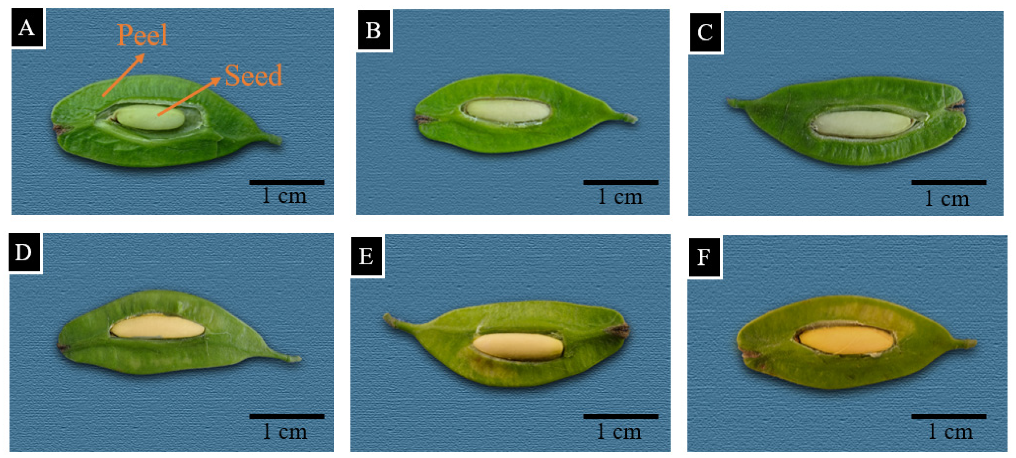
2.3. Observation of E. ulmoides Seed Oil Bodies

Frozen sections were taken, and fluorescent Nile red dye staining and laser scanning confocal microscopy were used to observe the oil bodies of E. ulmoides seeds at different growth stages. The treated seeds were randomly selected and sliced using a frozen cutting machine (KD-III). The temperature of the blade and the cutting table was set to −20 °C, and the slice thickness was 6 μm. Slices were dyed in 5 ug/mL Nile red for 1 h. Then, 0.1 mol/L phosphate-buffered saline buffer was used to clean the slices three times, and finally, the 20-fold and 40-fold objective lenses of the laser confocal laser microscope (FV-3000) were used for observation, with an absorption wavelength of 552 nm [33].

2.4. Identification of the PDAT Gene Family in E. ulmoides

E. ulmoides genome data were downloaded from the Genome Warehouse (https://ngdc.cncb.ac.cn/gwh/Assembly/13/show) (PRJCA000677) (accessed on 4 May 2022). The Arabidopsis PDAT gene sequences were downloaded from TATR (https://www.arabidopsis.org/) [34] (accessed on 4 May 2022). Blast software (v. 2.2.20; NCBI, Altschul, Bethesda, MD, USA) was used to construct the PDAT protein sequence library in Arabidopsis and blast search the E. ulmoides protein sequences. The PDAT conserved domain (PF02450) HMM file was downloaded from the Pfam database (http://pfam.sanger.ac.uk/) (accessed on 4 May 2022), and the E. ulmoides protein sequences were searched by HMMER3.0. Nine candidate genes were preliminarily obtained, and two incomplete protein sequences were manually deleted. After online comparison with NCBI CDD (http://www.ncbi.nlm.nih.gov/cdd/) (accessed on 4 May 2022) and Pfam (https://pfam.xfam.org/) (accessed on 4 May 2022), it was determined that all seven sequences contained PDAT conserved domains. The PDAT gene cloning experiment was performed using E. ulmoides seeds as experimental materials to further detect the PDAT prediction genes. Primer software(v. 5.0; Premier Company, San Francisco, CA, USA) was used to design the primers. The primers are shown in Table 1. Total RNA was extracted using the RNAprep Pure Plant Kit (TIANGEN, Beijing, China) and reverse-transcribed into cDNA [35]. The polymerase chain reaction (PCR) cycling conditions were as follows: 94 °C for 3 min; 34 cycles of 94 °C for 10 s, 58 °C for 15 s, and 72 °C for 10 s; and final extension at 72 °C for 5 min. The target fragment was purified and ligated into the pEASY-T5-ZO vector. The recombinant plasmid was transformed into Escherichia coli DH5α cells, and the positive clones were sequenced.

2.5. Bioinformatics Analysis of PDAT Protein

A total of 42 PDAT protein sequences from E. ulmoides, A. thaliana, Carya cathayensis, R. communis, and L. usitatissimum were compared using clustalW, and the phylogenetic tree of the PDAT gene family was constructed using MEGA 7.0 maximum likelihood (ML) and 500 bootstrap tests (bootstraps = 500). Based on the genome annotation information of E. ulmoides, TBtools (v.1.098; CJ-Chen, China) was used to analyze the position and chromosome length of each EuPDAT gene on chromosomes. Based on the EuPDAT full-length sequences and CDS sequences of E. ulmoides, the GSDS (http://gsds.cbi.pku.edu.cn/) (accessed on 5 May 2022) online tool was used to draw a gene structure intron and exon map [35]. The MEME tool (http://meme.nbcr.net/meme/tools/meme) (accessed on 5 May 2022) was used to analyze the conserved domains of the EuPDAT protein. The parameters were set as follows: the number of repeats was any, the maximum number of motifs was 10, and the optimal length of each motif was 6–30. ExPASy (https://web.expasy.org/protparam/) (accessed on 6 May 2022) was used to predict the protein isoelectric point and molecular weight. The online tool CELLO v. 2.5 (http://cello.life.nctu.edu.tw/) (accessed on 6 May 2022) was used to perform a subcellular localization prediction analysis of EuPDAT. The sequence 2000 bp upstream of the 5′ end of the PDAT gene was obtained from the E. ulmoides genome using TBtools and was submitted to the plantCare (http://bioinformatics.psb.ugent.be/webtools/plantcare/html/) (accessed on 6 May 2022) website for cis-acting element prediction. GSDS online tool was used to analyze the EuPDAT cis-acting elements. The STRING protein interaction database (http://string-db.org/) (accessed on 7 May 2022) was used to analyze the interaction network of seven EuPDAT proteins. The plant model was set to A. thaliana [36].

2.6. RNA Extraction and Quantitative Reverse Transcription PCR Assay

The EZgene Plant Easy Spin RNA Miniprep Kit (TIANGEN, Beijing, China) was used to extract total RNA from E. ulmoides seeds at different growth stages. Reverse transcription of RNA into cDNA was performed according to the novoprotein E047-01B reverse transcription kit instructions. The primers used were designed using Primer 5.0, and actin was used as the reference gene [28]. The primers are shown in Table 2. Expression analysis was performed using ChamQ Universal SYBR qPCR Master Mix fluorescent dye in a DLAB Precision 96 fluorescence quantitative PCR instrument. The reaction program was as follows: 95 °C for 5 min, 95 °C for 10 s, 60 °C for 10 s, 72 °C for 10 s, and 75 °C for 5 s, for a total of 40 cycles. The program was repeated three times. SPSS (v.23.0; Norman H. Nie, Chicago, IL, USA) was used to analyze the significant differences.

3. Results

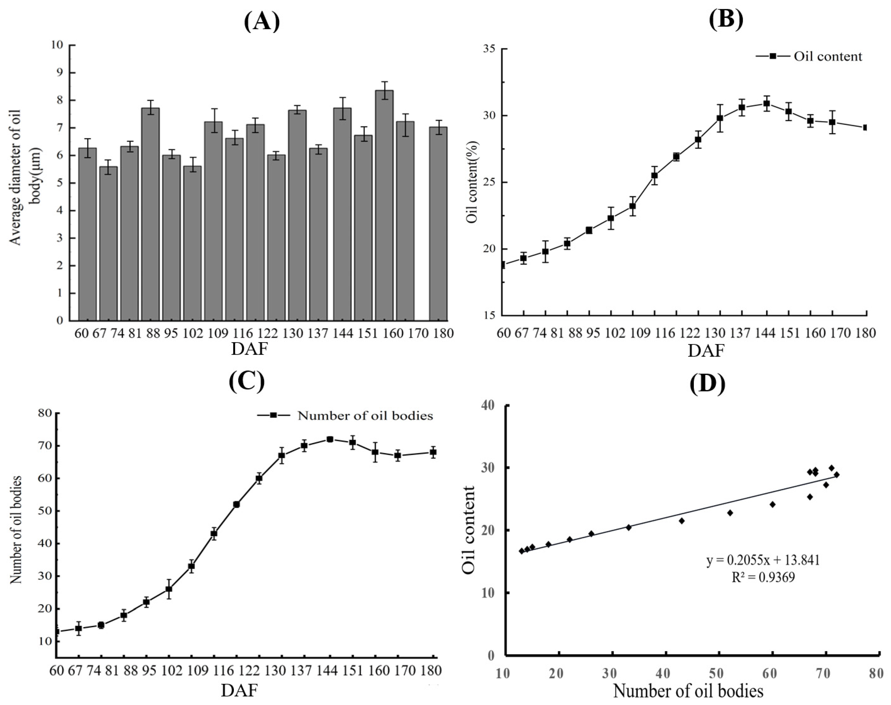
3.1. Correlation Analysis between Oil Bodies and Oil Content

Seeds have the highest oil content of all E. ulmoides tissues. The observation of seeds (Figure 1) revealed that there were differences in the distribution, size, and quantity of oil bodies at different developmental stages. The oil bodies in the seeds were dyed red using Nile red dye and observed using a laser confocal microscope. As shown in Figure 2, there were clear oil bodies in the seeds at 60 DAF; these were small in volume and irregular in shape and were mainly distributed around the cell wall, with large gaps between the oil bodies (Figure 2A). With the development of seeds, the number of oil bodies gradually increased, and the shape gradually changed from irregular to spherical and ellipsoidal. The distribution of oil bodies began to spread from around the cell wall to the center of the cell, and the gaps between oil bodies gradually narrowed (Figure 2B,C). At 120 DAF, the number of oil bodies in seeds further increased, and the shapes were mostly spherical and ellipsoidal. The gap between oil bodies further narrowed, and the number of large oil bodies increased (Figure 2D). At 140 DAF, the number of oil bodies in seeds increased significantly and filled whole cells (Figure 2E). The number, size, and distribution of oil bodies in seeds at 180 DAF exhibited no significant change compared with those at 140 DAF (Figure 2F). This indicates that oil bodies are formed at the early stage of seed development. From 60 DAF to 140 DAF, the number, size, and distribution of oil bodies changed significantly, and these became stable after 140 DAF.
In order to understand the correlation between the oil content and oil bodies during the development of seeds, this study used laser confocal microscopy at different stages to take pictures of seeds after staining, and three to five cells were randomly selected to obtain the diameter and number of oil bodies. There were some differences in the numbers and shapes of oil bodies in kernels at different developmental stages. The oil body diameters at different developmental stages ranged from 5 to 8 μm (Figure 3A). The number of oil bodies and oil content of Eucommia ulmoides seeds showed the same “S”-shaped change trend (Figure 3B,C). The correlation analysis showed that the oil content was significantly positively correlated with the number of oil bodies, with a correlation coefficient of 0.94 (Figure 3D), but was not significantly correlated with the size of oil bodies. Therefore, it can be speculated that the oil content of Eucommia ulmoides seed is mainly affected by the oil body number during the development of E. ulmoides seeds but has no significant correlation with the size of oil bodies.

3.2. Identification and Analysis of the Physicochemical Properties of EuPDAT Proteins in E. ulmoides

In this study, based on the whole genome data of E. ulmoides, seven target gene bands were obtained through PCR amplification. After sequencing, it was confirmed that the target gene was a EuPDAT gene. The longest CDS sequence was found in the EuPDAT2 gene at 1178 bp, encoding 392 amino acids. The shortest CDS sequence was found in the EuPDAT1 gene at 225 bp, encoding 75 amino acids (Table 3). The physicochemical properties of the EuPDAT proteins were analyzed using ExPasy (v.3.0; SIB, CHE). The results showed that the molecular weight of EuPDAT proteins ranged from 37.8 to 95.4 kDa; the theoretical isoelectric point was between 5.12 and 8.29, among which four theoretical isoelectric points were less than 7, indicating that most were alkaline proteins. The instability coefficient of EuPDAT proteins ranged from 31.28 to 49.23, among which five EuPDAT protein instability coefficients were greater than 40, indicating that most EuPDAT proteins were unstable proteins. The average hydrophobic coefficient was greater than 0, indicating that the EuPDAT proteins were hydrophobic proteins. The results of subcellular localization prediction showed that the PDAT proteins were mainly distributed in the plasma membrane, endoplasmic reticulum, and nucleus, which suggests that the different distributions of the EuPDAT proteins might be related to the diversity of their functions.

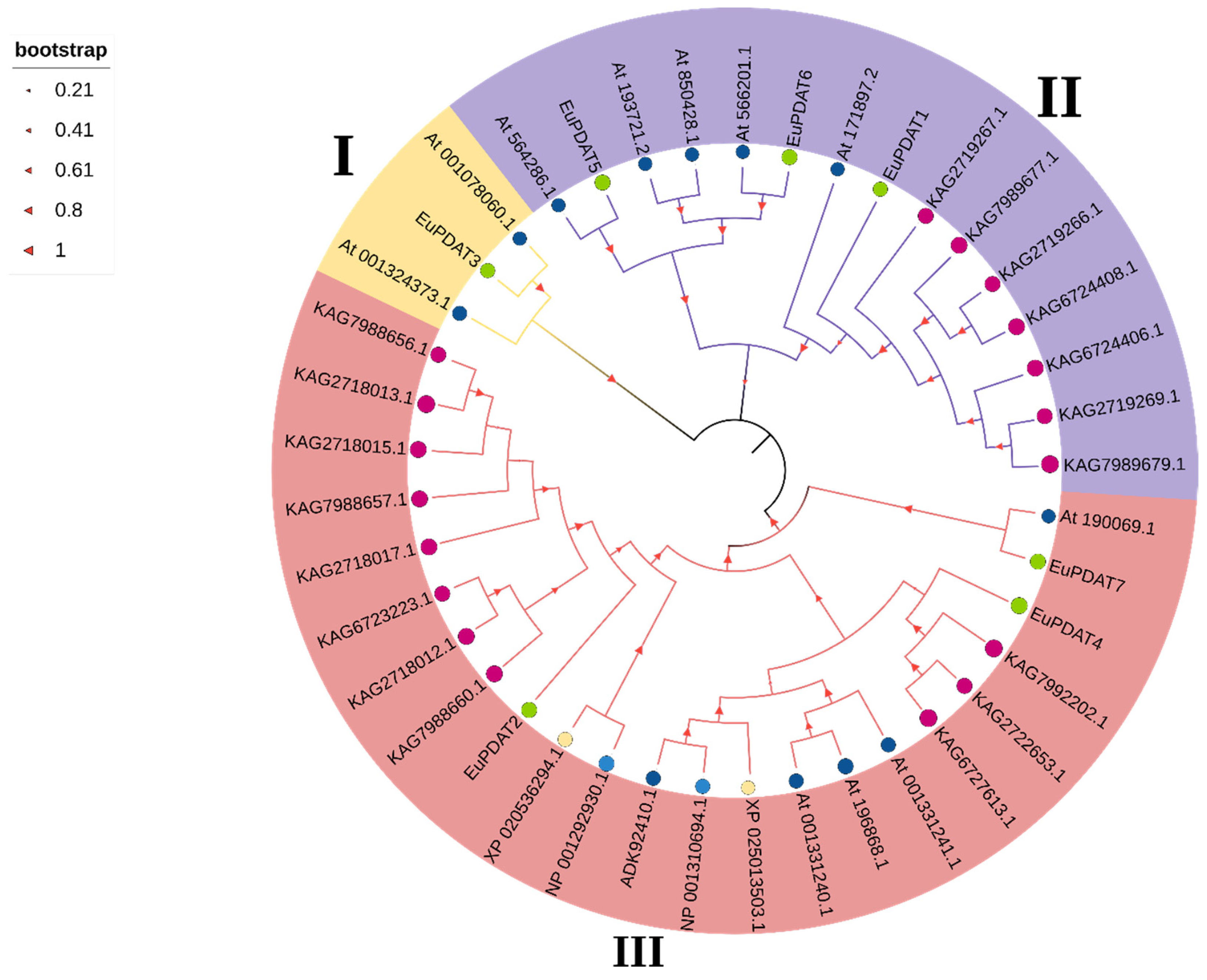
3.3. Phylogenetic Tree, Gene Structure, and Conserved Motif Analysis of the EuPDAT Genes in E. ulmoides

To explore the evolutionary relationships among the PDAT proteins in E. ulmoides, C. cathayensis, R. communis, L. usitatissimum, and Arabidopsis, a phylogenetic tree was constructed using MEGA 7.0 based on the PDAT protein sequences (Figure 4). According to the classification of PDAT proteins in Arabidopsis, the seven PDAT proteins in E. ulmoides were divided into three subfamilies, namely, PDAT group I, group II, and group III. Among them, the group III subfamily had the smallest number of members, consisting of only EuPDAT1, while the other two groups (I and II) both contained three members. The phylogenetic tree analysis demonstrated that PDAT proteins in Arabidopsis and E. ulmoides share a high similarity.
The exon and intron structures of EuPDAT genes in E. ulmoides were constructed using GSDS (v.2.0; GUO An-Yuan, China). As shown in Figure 5A, the seven EuPDAT genes were divided into three groups. The EuPDAT genes within the same group had similar intron–exon gene structures, and different subfamilies of the EuPDAT structures showed various differences. The exon number of the EuPDAT in group I was about 8–11, group III contained the maximum number of exons (16), and group II contained about 10–14 exons (Figure 5B). The results indicate that the gene structures of EuPDAT in group II and III were more complex and diverse than those in group I. The online MEME program was used to analyze the conserved structures of EuPDAT proteins in E. ulmoides. A total of 10 distinct conserved motifs were found; motif 2 and motif 10 were found to encode the PDAT domain. As illustrated in Figure 5, most PDAT members within the same clade, especially the most closely related members, usually shared common motif compositions (e.g., EuPDAT4 and EuPDAT7), indicating potential functional similarities among PDAT proteins. Motif 3, Motif 4, Motif 5, Motif 6, Motif 7, and Motif 9 were unique to the members in Clade II, which may be important to the functions of unique EuPDAT III proteins. Taken together, these results suggest that there is functional similarity of the EuPDAT proteins in the same group, and the EuPDAT proteins in group II contain a greater variety of complex functions compared with proteins in groups I and III.

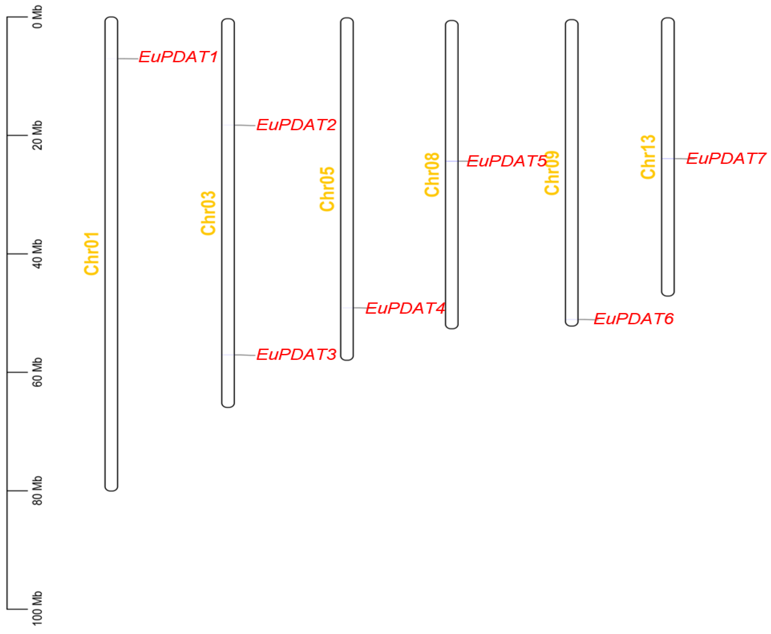
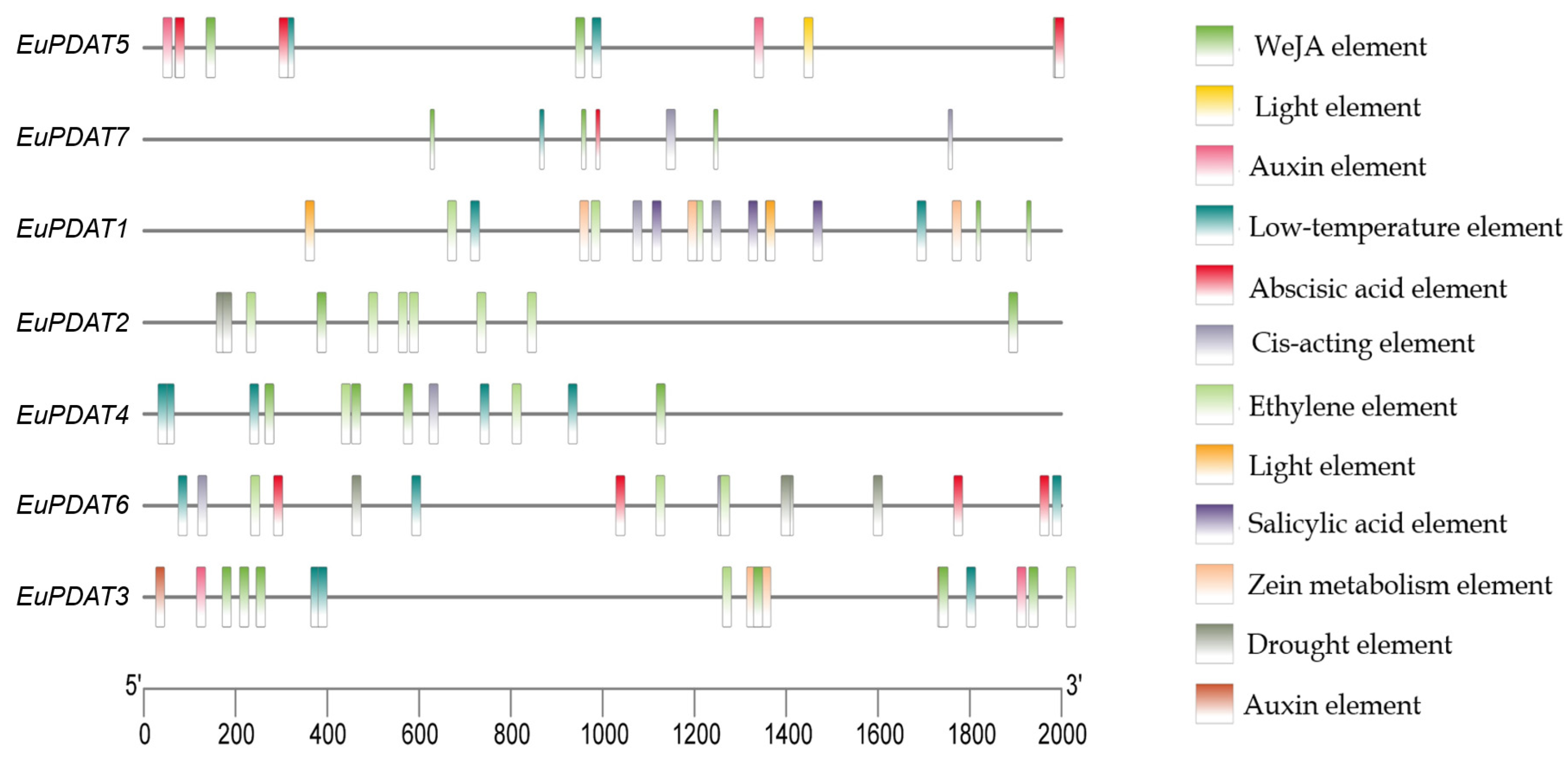
3.4. Chromosomal Locations and Putative Cis-Element Analysis of the EuPDAT Genes in E. ulmoides

The results of chromosome mapping showed that the seven EuPDAT genes were mapped onto six chromosomes (Figure 6). Among them, chromosome 3 contained the most genes (EuPDAT2 and EuPDAT3), while the other five chromosomes contained only one gene each.
To identify the functions of the EuPDAT genes in response to stresses, the putative cis-elements related to stress responsiveness in the upstream 2000 bp promoter sequence for each EuPDAT gene were identified using PlantCARE (Figure 7). Several stress responsive elements, such as drought response, low-temperature response, defense, and stress response, and hormone responsive elements, including abscisic acid response, methyl jasmonate response, and salicylic acid response, were identified. As shown in Figure 7, multiple cis-elements were found in the promoters of all EuPDAT genes. The cis-element analysis suggests that EuPDAT genes play an important role in the response to various stresses.

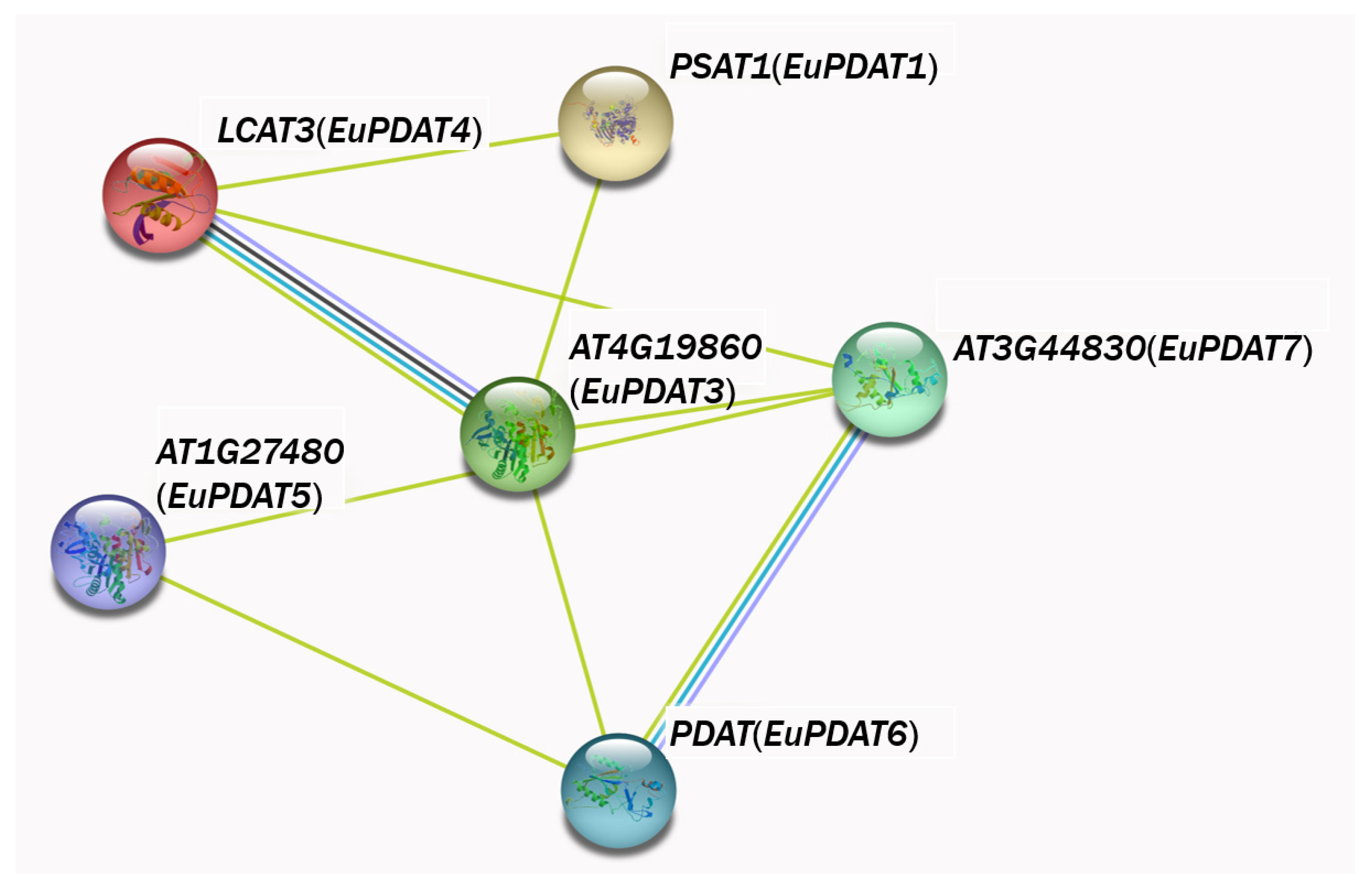
3.5. PDAT Protein Interaction Network Analysis of E. ulmoides

The interactions between proteins are the foundation for complex biological functions. Therefore, in this study, A. thaliana was used as the plant model, and the STRING protein interaction database was used to analyze the protein interaction network of the seven PDAT genes in E. ulmoides (Figure 8). The results showed that, with the exception of EuPDAT2, the other six proteins all interact. Among them, EuPDAT3, EuPDAT5, and EuPDAT7 correspond to AT4G19860, AT1G27480, and AT3G44830, respectively, which are involved in the regulation of low temperature and drought in A. thaliana, suggesting that PDAT genes in E. ulmoides play an important role in oil synthesis and could also interact with and resist various abiotic stresses. Based on the fact that the EuPDAT2 protein did not correspond to any protein in A. thaliana in the interaction network, it is speculated that EuPDAT2 may be a unique protein in E. ulmoides.

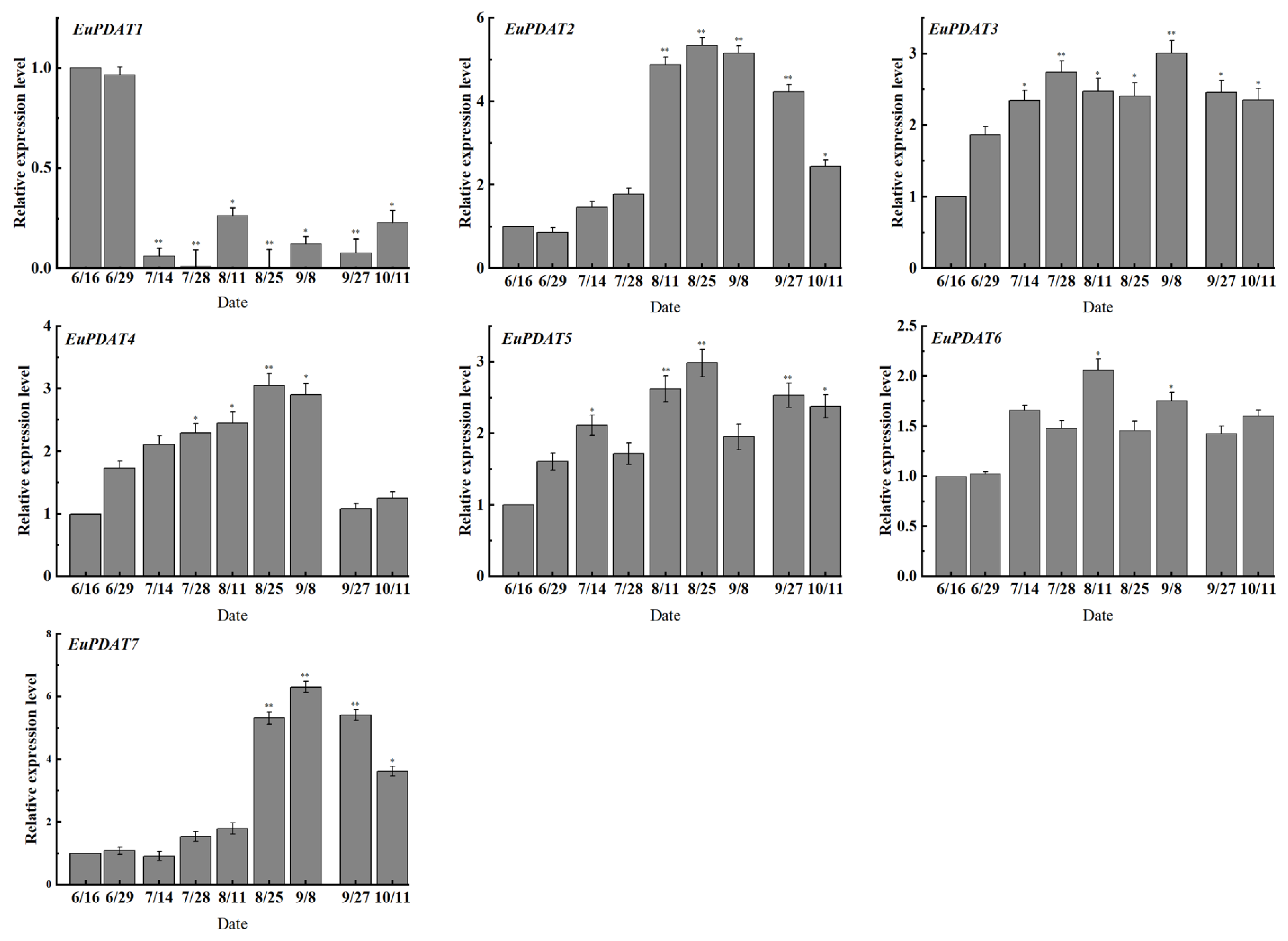
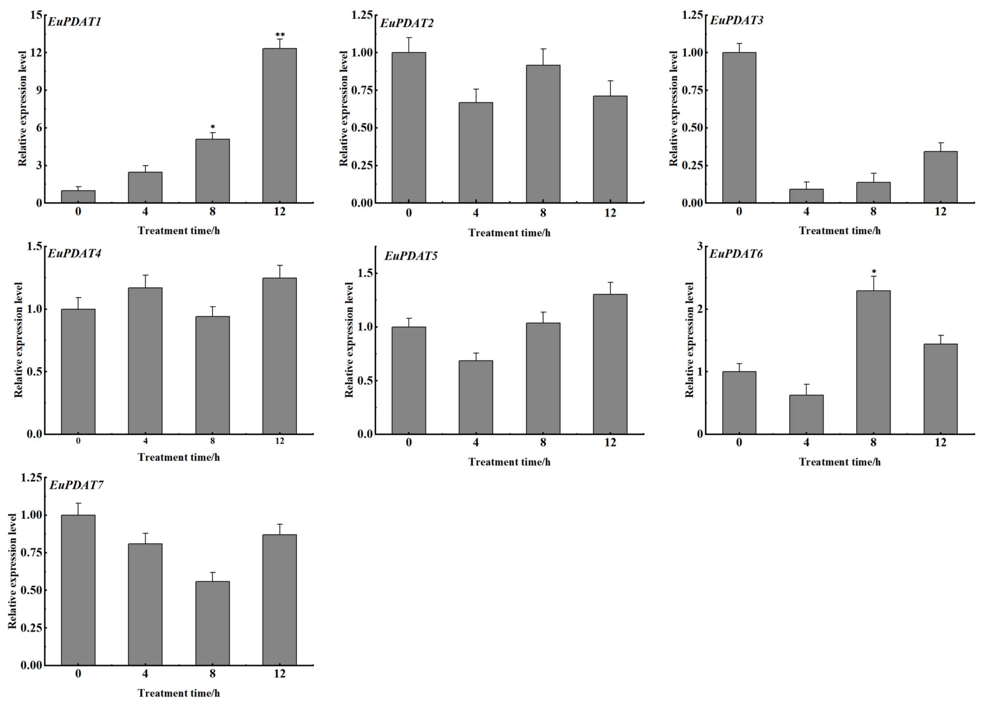
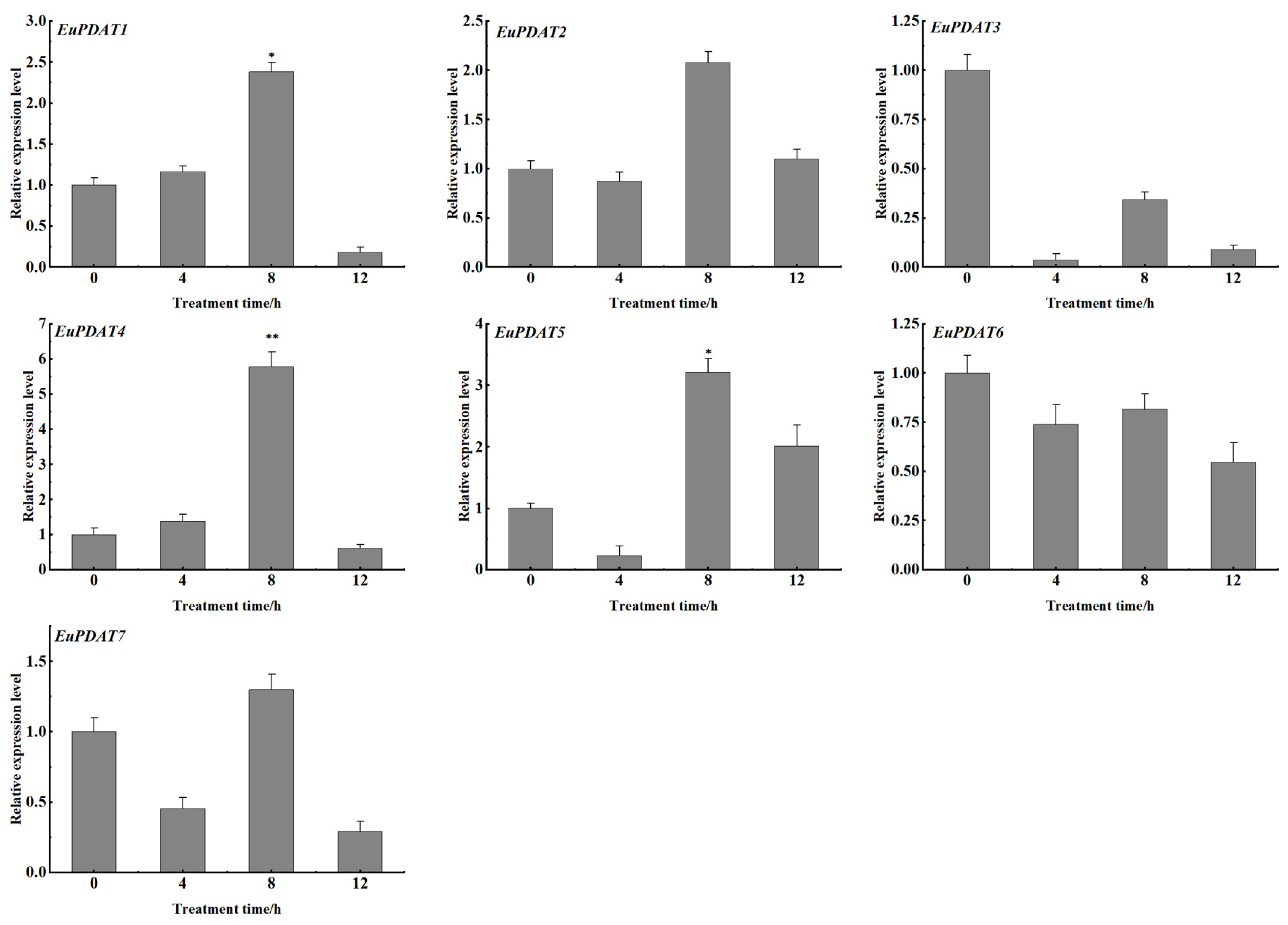
3.6. Expression Profiles of EuPDAT Genes at Different Stages and Treatments

To explore the regulatory mechanism of EuPDAT genes in the oil synthesis process of E. ulmoides seeds, we selected nine periods in which the oil content changed significantly during the oil synthesis process and used real-time fluorescence quantitative PCR technology to analyze the expression levels of EuPDAT genes, with the first period used as the control (Figure 9). The results showed that the seven EuPDAT genes were differentially expressed in the nine periods. The expression of EuPDAT1 decreased rapidly and then increased slowly with the passage of time, and the expression of EuPDAT6 did not change significantly in each period. The expression of EuPDAT2, EuPDAT4, EuPDAT5, and EuPDAT7 increased first and then decreased with the passage of time, and the expression levels were the highest at 140 DAF, which was consistent with the highest oil content period of the E. ulmoides seeds. The correlation analysis between the EuPDAT gene expression levels and the oil content at different stages (Figure 10) showed that EuPDAT2, EuPDAT4, and EuPDAT7 were positively correlated with the oil content, and EuPDAT2 and EuPDAT7 were significantly positively correlated with the oil content. In summary, the oil synthesis of E. ulmoides is regulated by multiple EuPDAT genes, of which EuPDAT2, EuPDAT4, and EuPDAT7 play a major regulatory role in seed oil synthesis.
To explore the responses of EuPDAT genes under different stress conditions in E. ulmoides, we analyzed the expression profiles of the EuPDAT genes treated with methyl jasmonate (WeJA), ethylene, and low temperature. The qRT-PCR analysis showed that most of the EuPDATs responded to these stresses in varying degrees. For WeJA stress (Figure 11), the expression trends of EuPDAT1, EuPDAT4, and EuPDAT5 were increased continuously and reached a peak after 12 h of treatment. For ethylene stress (Figure 12), the expression trend of EuPDAT1 was increased significantly, up to 13-fold after 12 h of treatment. For low-temperature stress (Figure 13), the expression trends of EuPDAT1 and EuPDAT4 were initially increased and then decreased, and they reached a peak after 8 h of treatment. In addition, the expression patterns of EuPDAT2, EuPDAT5, and EuPDAT7 were consistent with each other. These various expression results imply that EuPDAT genes may be complex and diverse in their response to ethylene, WeJA, and low-temperature stresses.

4. Discussion

Lipid metabolism is not only one of the basic metabolic pathways that sustains plant life but also an important source of energy for human health and development [37,38]. The oil body is the smallest organelle in plants, and it is mainly composed of triacylglycerol (TAG), phospholipid (PL) and oil body proteins. Therefore, studying the dynamic changes to oil bodies in E. ulmoides seeds is helpful to understanding the regulation of oil accumulation and degradation during seed development [32,39]. The diameter of oil bodies in the seeds of E. ulmoides ranged from 5 to 8 μm. With the development of seeds, the number of oil bodies gradually increased and then slowly decreased, and the oil body distribution gradually gathered from the cell wall to the center of the cell. Compared with common oil-bearing crops, such as Arachis hypogaea (1–3 μm), Juglans regia (1–4 μm), B. napus (0.5–2 μm), and C. oleifera (2–4 μm), E. ulmoides had the same change trend in oil body number and distribution, but there were great differences in oil body diameter, indicating that the morphological diversity of oil bodies in different species may be an important reason for their differing oil contents [4,40,41]. Heneen found that large oil bodies were formed by the fusion of small oil bodies, which indicated that oil content was positively correlated with oil body size [42]. Dong found that the number and cross-sectional area of oil bodies were positively correlated with oil content in B. napus [6]. In this study, frozen sections, Nile red staining, and laser confocal microscopy were combined for the first time to analyze the correlation between the oil content and oil bodies in E. ulmoides. The results showed that the number of oil bodies was positively correlated with the oil content, but the size of oil bodies was not correlated with the oil content. These findings further indicate that the correlation between oil bodies and oil content is related to species.
The PDAT gene, as one of the key genes regulating oil synthesis in plants, was first discovered by Dahlqvist in H. annuus and R. communis. At present, it has been studied in species including Arabidopsis, C. oleifera, A. hypogaea, and L. usitatissimum [18,20,43,44]. In this study, seven PDAT genes were cloned from the seeds of E. ulmoides based on the whole genome data. Subcellular localization prediction showed that EuPDAT genes were mainly located in the plasma membrane and endoplasmic reticulum. This is consistent with the conclusion that TAG is mainly synthesized in the plasma membrane and endoplasmic reticulum [19,45]. Multiple sequence alignment showed that EuPDAT genes had high homology with the PDAT genes of C. oleifera, C. cathayensis, and L. usitatissimum, and they all belonged to woody oil tree species. It is speculated that EuPDAT genes may have functional mechanisms in regulating TAG synthesis that are similar to those of PDAT genes in these species. The EuPDAT family in E. ulmoides was divided into three subfamilies according to the relatedness of these genes with homologs in Arabidopsis, in line with earlier studies in other species, such as H. pluvialis, poplar, A. hypogaea, and C. oleifera [46,47]. The gene structure and conserved motif analysis showed that EuPDAT genes had structural similarity in the same group, and subfamilies exhibited various differences. This suggests that EuPDAT genes have a variety of functions. Recent studies have found that the PDAT gene not only plays an important role in regulating TAG synthesis, but also participates in various abiotic and biotic stress responses in plants [9,22]. In Arabidopsis, it was found that the PDAT gene enhanced heat tolerance by regulating TAG synthesis. Overexpression of the PDAT gene increased seedling survival by about 50% under high temperature [16,48]. Studies in Chlorophyta showed that MiPDAT accelerated the conversion of membrane lipids to TAG under nitrogen deficiency stress, thereby reducing the damage to plants [21]. This study found that EuPDAT genes had different degrees of response to low temperature, ethylene, and methyl jasmonate treatment. Among these EuPDAT genes, the expression levels of EuPDAT1, EuPDAT4, and EuPDAT5 were obviously induced by almost all stress; in particular, the expression of EuPDAT1 was upregulated more than 13-fold after the ethylene treatment, showing significantly higher expression than the other genes. These results indicate that EuPDAT genes not only play an important role in the synthesis of plant oil, but also play an important role in the process of biotic and abiotic stress.
To explore the regulating role of EuPDAT genes in oil synthesis by E. ulmoides, the oil of E. ulmoides in different periods was obtained by Soxhlet extraction, and the expression levels of EuPDAT genes in different periods were determined by qRT-PCR. The results showed that the variation trends of the expression levels of EuPDAT2, EuPDAT3, EuPDAT5, and EuPDAT7 were correlated with oil content, and the variation trends of the expression levels of EuPDAT2 and EuPDAT7 were significantly correlated with oil content. It is speculated that oil synthesis in E. ulmoides is regulated by multiple genes, with EuPDAT2 and EuPDAT7 playing major regulatory roles and other EuPDAT members possibly mainly involved in other physiological processes. The substrate-specific preference of the PDAT gene was observed in Arabidopsis, C. oleifera, flax, and other species, resulting in significant differences in the fatty acid composition of various species. The content of linolenic acid in E. ulmoides oil is about 65%, which is relatively high. However, whether the PDAT gene also exhibits substrate preference needs to be further explored. The quality of E. ulmoides oil is good, but the low oil content seriously restricts the development of E. ulmoides oil industrialization [49,50,51]. Therefore, the correlations between oil body characteristics, oil content, and the PDAT gene, which plays an important regulatory role in the process of oil synthesis, were analyzed in this study. The aim of this study was to clarify the correlation between morphological changes to oil bodies and oil content, and to investigate the regulatory effect of EuPDAT genes on oil synthesis. Taken together, these findings can not only provide a reference for further exploring the law of oil accumulation in Eucommia ulmoides, but also provide a theoretical basis for directional cultivation of new varieties of Eucommia ulmoides with high oil content.

5. Conclusions

There was a positive correlation between the oil content and oil body number in E. ulmoides seeds at different developmental stages. The PDAT gene family of E. ulmoides contains seven members and can be divided into three subgroups according to the classification in Arabidopsis. EuPDATs in the same subgroup had similar gene structures and conserved protein motifs. The EuPDAT genes, which are mainly distributed in the endoplasmic reticulum and plasma membrane, play an important role in the lipid synthesis pathway and participate in a variety of abiotic stress response processes. The expression levels of EuPDAT2, EuPDAT4, and EuPDAT7 at different stages were significantly positively correlated with oil content. It is speculated that EuPDAT2, EuPDAT4, and EuPDAT7 are the main regulatory genes in oil synthesis in E. ulmoides. This study can provide a theoretical reference for further exploration of the correlation between oil body characteristics and oil content and the regulatory role of the PDAT gene in the synthesis of plant oils.

Author Contributions

J.Z. and L.W. conceived and designed the experiments; J.Z. wrote the article, analyzed the data, and produced the figures; J.Q., C.L., Q.W., H.D., P.L., Q.D., L.W. and L.D. provided support and experimental guidance for this study. All authors have read and agreed to the published version of the manuscript.

Funding

This work was supported by the National Key Research and Development Program of China (Grant No. 2017YFD0600702).

Data Availability Statement

Not applicable.

Conflicts of Interest

The authors declare that they do not have any competing financial or commercial interest that represents a conflict of interest in connection with this paper.

References

  1. Wang, X.Q.; Liang, X.Q.; Zhao, J.; Huang, B.B. Cultivar Characterization of Tea Seed Oils by Their Active Components and Antioxidant Capacity. J. Am. Oil Chem. Soc. 2014, 91, 629–639. [Google Scholar] [CrossRef]
  2. Frandsen, G.I.; Mundy, J.; Tzen, J. Oil bodies and their associated proteins, oleosin and caleosin. Physiol. Plant. 2010, 112, 301–307. [Google Scholar] [CrossRef]
  3. Song, Y.; Wang, X.D.; Rose, R.J. Oil body biogenesis and biotechnology in legume seeds. Plant Cell Rep. 2017, 36, 1519–1532. [Google Scholar] [CrossRef]
  4. Siloto, R.M.P.; Findlay, K.; Lopez-Villalobos, A.; Yeung, E.C.; Nykiforuk, C.L.; Moloney, M.M. The Accumulation of Oleosins Determines the Size of Seed Oilbodies in Arabidopsis. Plant Cell 2006, 18, 1961–1974. [Google Scholar] [CrossRef]
  5. Tzen, J.T.; Huang, A.H. Surface structure and properties of plant seed oil bodies. J. Cell Biol. 1992, 117, 327–335. [Google Scholar] [CrossRef]
  6. Dong, J.S.; Shi, D.Q.; Gao, J.Q.; Li, C.L.; Liu, J.; Yang, W.C. Correlation Between the Quantity and the Sum of Areas of Oil Bodies and Oil Content in Rapeseed (Brassica napus). Chin. Bull. Bot. 2009, 44, 79–85. [Google Scholar]
  7. Kennedy, E.P. Biosynthesis of complex lipids. Fed. Proc. 1961, 20, 934–940. [Google Scholar]
  8. Liu, X.; Ouyang, L.; Zhou, Z. Phospholipid: Diacylglycerol acyltransferase contributes to the conversion of membrane lipids into triacylglycerol in Myrmecia incisa during the nitrogen starvation stress. Sci. Rep. 2016, 6, 26610. [Google Scholar] [CrossRef]
  9. Fan, J.; Yan, C.; Xu, C. Phospholipid:diacylglycerol acyltransferase-mediated triacylglycerol biosynthesis is crucial for protection against fatty acid-induced cell death in growing tissues of Arabidopsis. Plant J. 2013, 76, 930–942. [Google Scholar] [CrossRef] [PubMed]
  10. Ståhl, U.; Carlsson, A.S.; Lenman, M.; Dahlqvist, A.; Huang, B.; Banaś, W.; Banaś, A.; Stymne, S. Cloning and Functional Characterization of a Phospholipid:Diacylglycerol Acyltransferase from Arabidopsis. Plant Physiol. 2004, 135, 1324–1335. [Google Scholar] [CrossRef]
  11. Zheng, L.; Shockey, J.; Guo, F.; Shi, L.; Li, X.; Shan, L.; Wan, S.; Peng, Z. Discovery of a new mechanism for regulation of plant triacylglycerol metabolism: The peanut diacylglycerol acyltransferase-1 gene family transcriptome is highly enriched in alternative splicing variants. J. Plant Physiol. 2017, 219, 62–70. [Google Scholar] [CrossRef] [PubMed]
  12. Pan, X.; Peng, F.Y.; Weselake, R.J. Genome-Wide Analysis of PHOSPHOLIPID:DIACYLGLYCEROL ACYLTRANSFERASE (PDAT) Genes in Plants Reveals the Eudicot-Wide PDAT Gene Expansion and Altered Selective Pressures Acting on the Core Eudicot PDAT Paralogs. Plant Physiol. 2015, 167, 887–904. [Google Scholar] [CrossRef] [PubMed] [Green Version]
  13. Fenyk, S.; Woodfield, H.K.; Romsdahl, T.B.; Wallington, E.J.; Bates, R.E.; Fell, D.A.; Chapman, K.D.; Fawcett, T.; Harwood, J.L. Overexpression of phospholipid: Diacylglycerol acyltransferase inBrassica napus results in changes in lipid metabolism and oil accumulation. Biochem. J. 2022, 479, 805–823. [Google Scholar] [CrossRef] [PubMed]
  14. Voelker, T.A.; Worrell, A.C.; Anderson, L.; Bleibaum, J.; Fan, C.; Hawkins, D.J.; Radke, S.E.; Davies, H.M. Fatty acid biosynthesis redirected to medium chains in transgenic oilseed plants. Science 1992, 257, 72–74. [Google Scholar] [CrossRef]
  15. Simpson, J.P.; Ohlrogge, J.B. A Novel Pathway for Triacylglycerol Biosynthesis Is Responsible for the Accumulation of Massive Quantities of Glycerolipids in the Surface Wax of Bayberry (Myrica pensylvanica) Fruit. Plant Cell 2016, 28, 248–264. [Google Scholar] [CrossRef]
  16. Banaś, W.; Sanchez Garcia, A.; Banaś, A.; Stymne, S. Activities of acyl-CoA:diacylglycerol acyltransferase (DGAT) and phospholipid:diacylglycerol acyltransferase (PDAT) in microsomal preparations of developing sunflower and safflower seeds. Planta 2013, 237, 1627–1636. [Google Scholar] [CrossRef]
  17. Kroon, J.T.M.; Wei, W.; Simon, W.J.; Slabas, A.R. Identification and functional expression of a type 2 acyl-CoA:diacylglycerol acyltransferase (DGAT2) in developing castor bean seeds which has high homology to the major triglyceride biosynthetic enzyme of fungi and animals. Phytochemistry 2006, 67, 2541–2549. [Google Scholar] [CrossRef]
  18. Kim, H.U.; Lee, K.; Go, Y.S.; Jung, J.H.; Suh, M.; Kim, J.B. Endoplasmic Reticulum-Located PDAT1-2 from Castor Bean Enhances Hydroxy Fatty Acid Accumulation in Transgenic Plants. Plant Cell Physiol. 2011, 52, 983–993. [Google Scholar] [CrossRef]
  19. Chapman, K.D.; Ohlrogge, J.B. Compartmentation of Triacylglycerol Accumulation in Plants. J. Biol. Chem. 2012, 287, 2288–2294. [Google Scholar] [CrossRef] [PubMed]
  20. Pan, X.; Siloto, R.M.; Wickramarathna, A.D.; Mietkiewska, E.; Weselake, R.J. Identification of a pair of phospholipid:diacylglycerol acyltransferases from developing flax (Linum usitatissimum L.) seed catalyzing the selective production of trilinolenin. J. Biol. Chem. 2013, 288, 24173–24188. [Google Scholar] [CrossRef]
  21. Furmanek, T.; Demski, K.; Banaś, W.; Haslam, R.; Napier, J. The Utilization of the Acyl-CoA and the Involvement PDAT and DGAT in the Biosynthesis of Erucic Acid-Rich Triacylglycerols in Crambe Seed Oil. Lipids 2014, 49, 327–333. [Google Scholar] [CrossRef] [PubMed]
  22. Yang, X.; Caldo, K.; Nath, D.P.; Ozga, J.; Lemieux, M.J.; Weselake, R.J.; Chen, G. Properties and Biotechnological Applications of Acyl-CoA:diacylglycerol Acyltransferase and Phospholipid:diacylglycerol Acyltransferase from Terrestrial Plants and Microalgae. Lipids 2018, 53, 663–688. [Google Scholar]
  23. Du, H.Y.; Du, Q.X. Foundation, problems and countermeasures of Eucommia ulmoides industry high-quality development in China. Nonwood For. Res. 2020, 38, 1–10. [Google Scholar]
  24. Tu, T.H.; Kim, H.; Yang, S.; Kim, J.K.; Kim, J.G. Linoleic acid rescues microglia inflammation triggered by saturated fatty acid. Biochem. Biophys. Res. Commun. 2019, 513, 201–206. [Google Scholar] [CrossRef]
  25. Inaba, Y.; Nakahigashi, K.; Ito, T.; Tomita, M. Alteration of fatty acid chain length of Chlamydomonas reinhardtii by simultaneous expression of medium-chain-specific thioesterase and acyl carrier protein. Phycol. Res. 2017, 65, 94–99. [Google Scholar] [CrossRef]
  26. Asefy, Z.; Tanomand, A.; Hoseinnejhad, S.; Ceferov, Z.; Oshaghi, E.A.; Rashidi, M. Unsaturated fatty acids as a co-therapeutic agents in cancer treatment. Mol. Biol. Rep. 2021, 48, 2909–2916. [Google Scholar] [CrossRef]
  27. Feng, Y.; Wang, L.; Fu, J.; Wuyun, T.; Du, H.; Tan, X.; Zou, F.; Li, F. Transcriptome sequencing discovers genes related to fatty acid biosynthesis in the seeds of Eucommia ulmoides. Genes Genom. 2016, 38, 275–283. [Google Scholar] [CrossRef]
  28. Kaseke, T.; Opara, U.L.; Fawole, O.A. Fatty acid composition, bioactive phytochemicals, antioxidant properties and oxidative stability of edible fruit seed oil: Effect of preharvest and processing factors. Heliyon 2020, 6, e4962. [Google Scholar] [CrossRef]
  29. Were, B.A.; Onkware, A.O.; Gudu, S.; Welander, M.; Carlsson, A.S. Seed oil content and fatty acid composition in East African sesame (Sesamum indicum L.) accessions evaluated over 3 years. Field Crops Res. 2006, 97, 254–260. [Google Scholar] [CrossRef]
  30. Heil, C.S.; Wehrheim, S.S.; Paithankar, K.S.; Grininger, M. Fatty Acid Biosynthesis: Chain-Length Regulation and Control. Chembiochem 2019, 20, 2298–2321. [Google Scholar] [CrossRef]
  31. Qing, J.; Du, Q.; Meng, Y.; Liu, P.; Du, H.; Wang, L. Genome-wide identification and expression pattern analysis of the ribonuclease T2 family in Eucommia ulmoides. Sci. Rep. 2021, 11, 6900. [Google Scholar] [CrossRef] [PubMed]
  32. Luo, L.P.; Xiao, P.; Du, S.G.; Jiang, X.M.; Fu, Y.X.; Guo, X.L. Oil Bodies Observation and the Correlation Between the Oil Bodies and Oil Content in Camellia oleifera Seeds. J. Chin. Cereals Oils Assoc. 2014, 29, 82–85. [Google Scholar]
  33. Chen, H.; Pan, C.D.; Wang, B.; Xiao, Z.Z.; Hu, Y.; Hu, G.J. Oil Body Observation in Seed Development and Its Analysis in Seed of Juglans regia ‘Wen185’ andJ. regia ‘Xinxin2’ in Period of Seed Maturity. Sci. Agric. Sin. 2015, 48, 3899–3909. [Google Scholar]
  34. Zhang, Q.; Chen, T.; Wang, X.; Wang, J.; Gu, K.; Yu, J.; Hu, D.; Hao, Y. Genome-wide identification and expression analyses of homeodomain-leucine zipper family genes reveal their involvement in stress response in apple (Malus × domestica). Hortic. Plant J. 2022, 8, 261–278. [Google Scholar] [CrossRef]
  35. Zhang, T.; Li, X.; Zhao, Q.; Shi, Y.; Hao, Y.; You, C. Genome-wide identification and functional characterization of the MdCLE peptide family in apple (Malus × domestica). Hortic. Plant J. 2022, 8, 279–288. [Google Scholar] [CrossRef]
  36. Xie, T.; Chen, C.; Li, C.; Liu, J.; Liu, C.; He, Y. Genome-wide investigation of WRKY gene family in pineapple: Evolution and expression profiles during development and stress. BMC Genom. 2018, 19, 490. [Google Scholar] [CrossRef] [Green Version]
  37. Lin, T.K.; Zhong, L.; Santiago, J.L. Anti-Inflammatory and Skin Barrier Repair Effects of Topical Application of Some Plant Oils. Int. J. Mol. Sci. 2018, 19, 70. [Google Scholar] [CrossRef]
  38. Marmon, S.; Sturtevant, D.; Herrfurth, C.; Chapman, K.; Stymne, S.; Feussner, I. Two Acyltransferases Contribute Differently to Linolenic Acid Levels in Seed Oil. Plant Physiol. 2017, 173, 2081–2095. [Google Scholar] [CrossRef]
  39. Tzen, J.T.C. Integral Proteins in Plant Oil Bodies. ISRN Bot. 2012, 2012, 173954. [Google Scholar] [CrossRef]
  40. Kodad, O.; Rafel Socias I Company. Variability of Oil Content and of Major Fatty Acid Composition in Almond (Prunus amygdalus Batsch) and Its Relationship with Kernel Quality. J. Agric. Food Chem. 2008, 56, 4096–4101. [Google Scholar] [CrossRef]
  41. Hu, Z.; Wang, X.; Zhan, G.; Liu, G.; Hua, W.; Wang, H. Unusually large oilbodies are highly correlated with lower oil content in Brassica napus. Plant Cell Rep. 2009, 28, 541–549. [Google Scholar] [CrossRef] [PubMed]
  42. Heneen, W.K.; Karlsson, G.; Brismar, K.; Gummeson, P.; Marttila, S.; Leonova, S.; Carlsson, A.S.; Bafor, M.; Banas, A.; Mattsson, B.; et al. Fusion of oil bodies in endosperm of oat grains. Planta 2008, 228, 589–599. [Google Scholar] [CrossRef] [PubMed]
  43. Ghosal, A.; Banas, A.; St Hl, U.; Dahlqvist, A.; Lindqvist, Y.; Stymne, S. Saccharomyces cerevisiae phospholipid:diacylglycerol acyl transferase (PDAT) devoid of its membrane anchor region is a soluble and active enzyme retaining its substrate specificities. BBA Mol. Cell Biol. Lipids 2007, 1771, 1457–1463. [Google Scholar] [CrossRef]
  44. Dahlqvist, A.; Stahl, U.; Lenman, M.; Banas, A.; Lee, M.; Sandager, L.; Ronne, H.; Stymne, S. Phospholipid:diacylglycerol acyltransferase: An enzyme that catalyzes the acyl-CoA-independent formation of triacylglycerol in yeast and plants. Proc. Natl. Acad. Sci. USA 2000, 97, 6487–6492. [Google Scholar] [CrossRef]
  45. Yoon, K.; Han, D.; Li, Y.; Sommerfeld, M.; Hu, Q. Phospholipid:Diacylglycerol Acyltransferase Is a Multifunctional Enzyme Involved in Membrane Lipid Turnover and Degradation While Synthesizing Triacylglycerol in the Unicellular Green MicroalgaChlamydomonas reinhardtii. Plant Cell 2012, 24, 3708–3724. [Google Scholar] [CrossRef] [PubMed]
  46. Xu, J.; Carlsson, A.S.; Francis, T.; Zhang, M.; Hoffman, T.; Giblin, M.E.; Taylor, D.C. Triacylglycerol synthesis by PDAT1 in the absence of DGAT1 activity is dependent on re-acylation of LPC by LPCAT2. BMC Plant Biol. 2012, 12, 4. [Google Scholar] [CrossRef] [Green Version]
  47. Wang, H.; Zhang, J.; Gai, J.; Chen, S. Cloning and comparative analysis of the gene encoding diacylglycerol acyltransferase from wild type and cultivated soybean. Theor. Appl. Genet. 2006, 112, 1086–1097. [Google Scholar] [CrossRef]
  48. Mueller, S.P.; Unger, M.; Guender, L.; Fekete, A.; Mueller, M.J. Phospholipid:Diacylglycerol Acyltransferase-Mediated Triacylglyerol Synthesis Augments Basal Thermotolerance. Plant Physiol. 2017, 175, 486–497. [Google Scholar] [CrossRef]
  49. Liu, P.F.; Wang, L.; Du, Q.X.; Du, L.Y. Estimation of potential suitable distribution area and the ecological characteristics of Eucommia ulmoides Oliv. in China. Acta Ecol. Sin. 2020, 40, 5674–5684. [Google Scholar]
  50. Li, R. Urgent Need to Improve Professional Cooperation in Eucommia Seed Oil Industry. China Forestry Industry. 2016, 8, 63–64. [Google Scholar]
  51. Alrashidi, M.; Derawi, D.; Salimon, J.; Firdaus Yusoff, M. An investigation of physicochemical properties of Nigella sativa L. Seed oil from Al-Qassim by different extraction methods. J. King Saud Univ. Sci. 2020, 32, 3337–3342. [Google Scholar] [CrossRef]
Figure 1. Seeds at different growth stages. (AF): Seeds collected on 60, 74, 95, 122, 145, and 180 DAF, respectively.
Agronomy 12 02197 g001
Figure 2. Laser confocal microscopy images of the seeds’ different development periods. (AF): Seeds collected on 60, 74, 95, 122, 145, and 180 DAF, respectively. Ob: Oil body; Cw: Cell wall.
Agronomy 12 02197 g002
Figure 3. Variation characteristics of oil bodies and their correlation with oil content: (A) Changes in the oil body diameter of E. ulmoides seeds at different developmental stages; (B) Changes in the oil content of E. ulmoides seeds at different development stages; (C) Changes in the oil body number of E. ulmoides seeds at different development stages; (D) Correlation between the number of oil bodies and oil content of E. ulmoides seeds in different developmental stages.
Agronomy 12 02197 g003
Figure 4. Unrooted phylogenetic tree representing relationships among the PDAT domains of Eucommia ulmoides, Arabidopsis, Carya cathayensis, Ricinus communis, and Linum usitatissimum. I, II and III represent three groups that are close according to genetic relationship, respectively. The different colored arcs indicate different groups (or subgroups) of PDAT domains. The green, yellow, burgundy, and blue circles represent PDAT domains from E. ulmoides, Ricinus communis, Carya cathayensis, and Arabidopsis, respectively. PDAT proteins from E. ulmoides with the prefix “Eu” indicate “EuPDAT”. PDAT: phospholipid:diacylglycerol acyltransferase.
Agronomy 12 02197 g004
Figure 5. Phylogenetic relationships, gene structure, and architecture of conserved protein motifs in PDAT genes from Eucommia ulmoides. (A) The phylogenetic tree was constructed based on the full-length sequences of E. ulmoides PDAT proteins using MEGA (v.7.0; Mega Limited, Auckland, New Zealand). Cluster details are shown in different colors. (B) The motif composition of E. ulmoides PDAT proteins. Motifs 1–10 are shown in different colored boxes. The length of each protein can be estimated using the scale at the bottom. (C) Exon–intron structure of E. ulmoides PDAT genes. Green boxes indicate exons; black lines indicate introns. The number indicates the phases of the corresponding introns. PDAT: phospholipid:diacylglycerol acyltransferase.
Agronomy 12 02197 g005
Figure 6. Genome locations of the seven Eucommia ulmoides PDAT genes on six chromosomes. The length of each chromosome can be estimated using the scale at the left. PDAT: phospholipid:diacylglycerol acyltransferase.
Agronomy 12 02197 g006
Figure 7. Putative cis-elements related to stress responsiveness in the promoter sequence for EuPDATs. The scale represents the length of the upstream segment of the EuPDAT.
Agronomy 12 02197 g007
Figure 8. The predicted protein interaction network of the EuPDAT proteins based on orthologs in Arabidopsis using the STRING database.
Agronomy 12 02197 g008
Figure 9. EuPDAT gene expression profiles in Eucommia ulmoides at different developmental stages. * and ** indicate significant differences at the p < 0.05 and p < 0.01 levels, respectively.
Agronomy 12 02197 g009
Figure 10. Correlations between EuPDAT gene expression and oil content. The deeper the blue color, the more significant the correlation.
Agronomy 12 02197 g010
Figure 11. Expression profiles of EuPDAT genes under WeJA stress conditions. * and ** indicate significant differences at the p < 0.05 and p < 0.01 levels, respectively.
Agronomy 12 02197 g011
Figure 12. Expression profiles of EuPDAT genes under ethylene stress conditions. * and ** indicate significant differences at the p < 0.05 and p < 0.01 levels, respectively.
Agronomy 12 02197 g012
Figure 13. Expression profiles of EuPDAT genes under low-temperature stress conditions. * and ** indicate significant differences at the p < 0.05 and p < 0.01 levels, respectively.
Agronomy 12 02197 g013
Table 1. Primers used in PCR analysis.
NamePrimer Sequence
EuPDAT1-FATGGGAATCAAAGTGGCAGTC
EuPDAT1-RTCACCGTGAAACGTTAATCTGAG
EuPDAT2-FATGGCTTCTTCCTCTAAAAATTCTG
EuPDAT2-RTTACACATAATGCAAGAGGAGATCA
EuPDAT3-FATGGCTTCTTCAGTTCTTCGGTT
EuPDAT3-RTCACAACTGAATATTCAATCTCTCCG
EuPDAT4-FATGTCGATGTTGAGGCGGAG
EuPDAT4-RTCAAGCTTTCCCCATACTATTTACA
EuPDAT5-FATGCTCGGGGGATGCTGTT
EuPDAT5-RTTAGAGAACTCGGGAGCTTTCCTC
EuPDAT6-FATGGCGCTGATTCGAAGAAG
EuPDAT6-RCTAAAAGTCTCTTCCTTTGTACTGCA
EuPDAT7-FATGGACCTTGATCTATGGATGTTG
EuPDAT7-RTCATATGATAGATGGTCCCTTACCC
Table 2. Primers used in qRT-PCR analysis.
NamePrimer Sequence
EuPDAT1-FAGACCTCCGACGGTTTTTCC
EuPDAT1-RGGTTCCTTTGGAGGACCGAG
EuPDAT2-FGTGAGTTGCCTTGCGGATTC
EuPDAT2-RCAACCTGCAGCCGTAGTACA
EuPDAT3-FGAGAACACCAAAGCGGAGGA
EuPDAT3-RGCGCCACGAATTTCATCTCC
EuPDAT4-FGGACTCCGCAAAGAAAGGGA
EuPDAT4-RGTCTCGGTCAGCTCGGTAAG
EuPDAT5-FTTCCCGCATATCTGGAACCG
EuPDAT5-RTAACCAGGCCTCCACCTTCT
EuPDAT6-FCGCTATGGATCCCACACCTC
EuPDAT6-RCCGAGAGCGTCGATGAGAAA
EuPDAT7-FAAAACGACTCCGATGCGACT
EuPDAT7-RTCAGTGGCAGTGGACCAATC
Actin-FTTGTTAGCAACTGGGATGATATGG
Actin-RCAGGGTGTTCTTCAGGAGCAA
Table 3. Genome-wide characterization of EuPDAT proteins in Eucommia ulmoides.
Gene NameNumber of Amino AcidsMolecular Weight (kDa)Theoretical pIInstability IndexAliphatic IndexGrand Average of Hydropathicity Subcellular Location
EuPDAT 17537.85.1241.0791.640.634Plasma membrane
EuPDAT 239295.48.2945.4080.550.721Endoplasmic reticulum
EuPDAT 331376.55.9543.0581.010.737Plasma membrane
EuPDAT 49686.87.5637.0277.940.846Endoplasmic reticulum
EuPDAT 518265.75.9144.7785.980.959Plasma membrane
EuPDAT 613881.45.5949.2370.470.781Nucleus
EuPDAT 717878.67.0931.2897.070.813Endoplasmic reticulum
Publisher’s Note: MDPI stays neutral with regard to jurisdictional claims in published maps and institutional affiliations.

