Article

Detection of ‘Candidatus Phythoplasma prunorum’ in Apricot Trees and its Associated Psyllid Samples

1
Institute of Plant Protection, Hungarian University of Agriculture and Life Sciences, Ménesi Street 44, 1118 Budapest, Hungary
2
Institute of Mathematics and Basic Science, Hungarian University of Agriculture and Life Sciences, Villányi Street 29, 1118 Budapest, Hungary
3
Department of Plant Sciences, Albert Kázmér Faculty of Mosonmagyaróvár, Széchenyi István University, Vár Square 2, 9200 Mosonmagyaróvár, Hungary
4
ELKH-SZE PhatoPlant-Lab, Széchenyi Isván University, Vár Square 2, 9200 Mosonmagyaróvár, Hungary
5
Directorate of Agricultural Genetic Resources, Variety Testing Department for Field Crops, National Food Chain Safety Office, Keleti Károly Street 24, 1024 Budapest, Hungary
6
Directorate of Plant Protection, Soil Conservation and Agri-Environment, National Food Chain Safety Office, Budaörsi Street 141–145, 1118 Budapest, Hungary
*
Author to whom correspondence should be addressed.
Agronomy 2023, 13(1), 199; https://doi.org/10.3390/agronomy13010199
Submission received: 11 December 2022 / Revised: 22 December 2022 / Accepted: 27 December 2022 / Published: 9 January 2023
(This article belongs to the Section Pest and Disease Management)

Abstract

:
Candidatus Phytoplasma prunorum’ is causing ever increasing economic losses through the decline of apricot trees in European countries, e.g., Hungary. In this study, the pathogen was identified from plant tissues and insects by nested-PCR. The insect species were identified via morphology and molecular methods. The incidence of the pathogen was 29.6% in randomly selected apricot trees. Most of the infected trees with symptoms died within a year. These results show that phytoplasma is significantly present and causes damage in the investigated plantations. The only known insect vector of this phytoplasma is the plum psyllid, Cacopsylla pruni, which was regularly encountered in the sampled apricot orchards and in their surroundings. In a two-year study, several adults among the sampled specimens were observed to be infected by the pathogen. This observation further confirms the role of the plum psyllid in vectoring the phytoplasma. All the sampled plum psyllid adults belonged to the ‘B’ biotype. Besides C. pruni, Cacopsylla crataegi was abundant in the samples. Several adults of the latter species were also infected by the pathogen ‘Ca. Phytoplasma prunorum’. The rates of occurrence of this phytoplasma in male and female adults of the two psyllid species appeared to be similar. The examined C. crataegi individuals showed genetic differences from each other and from specimens included in a previous investigation.

1. Introduction

Candidatus Phytoplasma prunorum’ is one causative agent of disease for several host plants of the Prunus genus [1]. These diseases are collectively called European Stone Fruit Yellows (ESFY) [2]. This group of diseases severely affects Prunus armeniaca (Family: Rosaceae) in southern and central Europe [3]. Studies on phytoplasma infestation of apricot trees have shown a wide range of results, with an infection rate of 11% in Poland [4], 24% in Croatia [5], 44.9% in Austria, 47% in Hungary [6], 18.5 to 56% in the Czech Republic [7,8], 82% in France [9], and 67% to 83% in Germany [10,11]. In one of the reports, researchers found 26.6% disease incidence of P. cerasifera rootstocks [6]. Apricot trees usually show yellowing, rolling and wilting of the leaves, and death of the woody parts or the whole tree as a result of the disease [8,12,13]. ’Myrobalan’ does not show or shows only few foliar symptoms; however, some phytoplasma strains can cause significant mortality [12,14]. In addition to the destruction of trees, the economic loss is increased by the deterioration of fruit quantity and quality in the case of several varieties [15,16]. There are no registered plant protection products that are effective against this disease or that have proven to be useful in combatting the infection, so its spread must be prevented; thus, our current knowledge should be expanded.
The currently known vector of the causative agent of ESFY is the plum psyllid, Cacopsylla pruni (Scopoli, 1763), which is member of the order Hemiptera and belongs to the family Psylloidea [17,18]. Psyllids have been found in several countries of Europe, for example, the Czech Republic, Germany, Hungary, Italy, Serbia, Slovakia, and Slovenia [19,20]. In Hungary, the presence of C. pruni has been confirmed in the south and western part of the country, specifically, in the Pest, Somogy, Vas, and Veszprém counties [21,22,23]. Two biotypes have been established in C. pruni: ‘A’ and ‘B’ [24]. These groups have not yet been morphologically distinguished [25]. Both biotypes are capable of transmitting the causative agent of ESFY [26,27]. The Internal Transcribed Spacer 2 (ITS2) region is used for the molecular identification of these biotypes. Three primer sets are available for the test, as different primer sets may be required for the identification of certain samples. It is worth mentioning that currently no other species of Cacopsylla can be identified by this procedure [25]. Interestingly, samples originating from Turkey and from many parts of Europe (for example, Bulgaria, the Czech Republic, and Hungary) contained only specimens of biotype ‘B’ [20,23,25,28,29]. Another method can also identify two variants. This test uses the cytochrome c oxidase subunit I (COI) region, with which variants 1 and 2 have been identified [30].
C. pruni is a univoltine, oligophagus species. It feeds on Prunus species, upon which they lay eggs [31,32,33,34]. In one study, the host preference of C. pruni in the genus Prunus has been inferred based on the presence of individuals caught by the beating tray method, namely, Prunus spinosa, P. cerasifera, P. salicina, P. domestica, and P. armeniaca (given in decreasing order of host preference) [35]. Rootstock suckers of apricots in orchards and wild host plants from the genus Prunus in the surroundings of apricot orchards play a role in supporting vector psyllids (in feeding and breeding) [35,36,37,38,39]. It has also been observed that greater quantities of psyllids were captured in orchards where P. domestica or P. cerasifera rootstocks were used because they produce more suckers that are preferred by the psyllids [35]. In another survey, a large number of C. pruni specimens were collected on wild host plants of Prunus spinosa and P. cerasifera, but few specimens were collected from cultivated host plants in Germany [10]. From apricot trees, few or no specimens have been collected in Germany, France, and Hungary [23,35,37]. In some cases, other researchers have found important populations on these plant species in Germany and Hungary [10,29].
The wild plant species that could serve as reservoirs for this phytoplasma may be present either in the immediate surroundings of or far from orchards [11,36]. Based on the results of field tests, C. pruni generally acquires the pathogen from wild Prunus species [27]. Plum psyllids transmit the pathogen through phloem-sap feeding in a persistent-propagative manner [34]. The specimens retain their infectivity through their entire lifespan [40] In some surveys, plum psyllid specimens were collected from cultivated and/or wild hosts and examined for the presence of ‘Ca. Phytoplasma prunorum’. In Germany, from 0.8 to 3% [11]; in Bulgaria, 2.3% [41]; in Poland, from 1.5 to 4.2% [42]; in France, 4.8% [27]; in Turkey, 23% [43]; and in Hungary from 15% up to 64.2% [23] of the specimens were carriers of the phytoplasma. Researchers also investigated the gender distribution and carrier capabilities of plum psyllids on various Prunus hosts. Most of the experiments revealed that females were more abundant after returning from their winter hosts [11,23,29,44,45]. Regarding the carrier capabilities of the sexes, more males (from 2.8% up to 47%) were carriers of the phytoplasma than females (from 1.1% up to 31%) [11,29]. In contrast, another study found that females (52.1%) were more infected with phytoplasma than males (12.1%) [44]. Others have found no significant difference between the infection rates of females and males [23,45].
Cacopsylla crataegi (Schrank, 1801) is also a univoltine species living on host plants such as hawthorns (Crataegus sp.; Family: Rosaceae) and apples (Malus sp.; Family: Rosaceae) [31,32,33,46]. The hawthorn psyllids have been found in many countries in Europe, including Hungary [19], where they were collected from Crataegus sp., Malus sp., and Ambrosia artemisiifolia (Family: Asteraceae) [47,48,49,50,51]. In Croatia and Italy, the abundance of C. crataegi was low compared to other species on apple trees (from 1.4 up to 3.7%) and on hawthorn (0.38%) [52,53]. In Poland, Spain, and Italy, researchers were able to collect few specimens from plum, cherry, and apple trees [42,54,55]. Two studies have also investigated the infection of psyllids with phytoplasma. The estimated infection rates of individuals from hawthorn were 2.63% and 4.36% in two years. ‘Ca. Phytoplasma pyri’ was identified from the infected individuals [53]. In another survey, 16.7% of psyllids (from apple trees) carried the ‘Ca. Phytoplasma mali’ pathogen [54].
The aim of this study was to reveal the diversity, abundance, and ‘Ca. Phytoplasma prunorum’ infection rate of psyllid species, which occurred in high numbers in apricot orchards and were present to a lesser extent in their immediate surroundings in the study areas.

2. Materials and Methods

2.1. Plant Samples Collection

The sample collection was carried out in two commercial apricot plantations (Pomáz, Sóskút) and one cultivar collection (Soroksár) in Pest County, Hungary, in 2014. The cultivars of Pomáz orchard were mixed and plant samples were collected from ‘Ceglédi bíbor’, ‘Ceglédi Piroska’, ‘Harcot’, ‘Hargrand’, ‘Gönci magyar kajszi’, and ‘Pannónia’. At Sóskút, we sampled the ‘Bergeron’, ‘Gönci magyar kajszi’, ‘Magyar kajszi C.235’, and ‘Mandula kajszi’ varieties. At Soroksár, samples were collected from the following varieties: ‘Ceglédi Piroska’, ‘Ceglédi óriás’, ‘Goldrich’, ‘Gönci magyar kajszi’, ‘Harcot’, ‘Magyar kajszi C.235’, and ‘Mandula kajszi’. The examined scions were grafted onto ‘Myrobalan’ at all orchards. At each site, we randomly selected 27–27 trees, of which 50% were symptomatic and the other 50% were asymptomatic. In 2015, the symptoms of apricot trees were evaluated again. The trees were at least 10 years of age; from these trees, we collected 1-year-old woody parts as samples. Rootstock suckers had been removed from all sites as part of the planned plantation maintenance; thus, we could only collect woody samples from the ‘Myrobalan’ rootstocks in 14 cases. We also sampled 5 blackthorn (P. spinosa) and 5 hawthorn (C. monogyna) bushes in the near vicinity of the orchards at Sóskút and Soroksár, but only 1 hawthorn has been sampled at Pomáz, as there were no other specimens nearby. At the same time as the samples were collected, we recorded the symptoms of rootstock suckers and wild plants.

2.2. Insect Sample Collection

Psyllids (Cacopsylla spp.) were collected using beating trays at the sampling sites mentioned above. In 2015 and 2016, collection of samples was started in the first days of March until no psyllids were found in several consecutive sampling attempts (May and June). The samples were collected weekly at Pomáz, and every two weeks at Soroksár and Sóskút. At each sampling date, the plants were randomly selected. Each time, we beat 20 apricot trees (on ‘Myrobalan’) and 6–7 ‘Myrobalan’ rootstock suckers. Additionally, we sampled plum trees near Pomáz four and five times in 2015 and 2016, respectively. Samples from blackthorn were taken in the near vicinity of the orchards; each time, 5 bushes were beaten. In addition, we sampled five hawthorn bushes near each orchard, and one near Pomáz, as there was only one plant growing in the near vicinity. In the case of the trees, one branch was selected for beating, while in the case of the bushes, one bushy part of the polycormon was selected for this purpose. The plant parts were stuck three consecutive times with a beater and the fallen psyllid adults were collected within one minute. They were preserved immediately and individually in microcentrifuge tubes containing 70% ethanol.
There were three insecticide applications at Pomáz, which were carried out immediately after the flowering of the apricot (active ingredient: esfenvalerate), one week later (active ingredient: indoxakarb), and finally two weeks later (active ingredient: acetamiprid). At Sóskút, after flowering, trees were thoroughly sprayed with the following insecticide active ingredients three to four times: fenoxycarb, indoxacarb, lambda-cyhalothrin, or pirimicarb + lambda-cyhalothrin. At Soroksár, the orchard was sprayed tree times with insecticide during the investigation period (active ingredients: acetamiprid, indoxacarb, beta-cyfluthrin, deltamethrin, and pirimicarb + lambda-cyhalothrin, or thiacloprid). Prunus species (e.g., plum, peach, etc.) in the surrounding area were sprayed with insecticide several times after flowering when treatments were necessary.

2.3. Nucleic Acid Extraction

Deoxyribonucleic acid (DNA) extraction of plant samples was carried out following the modified protocol of Daire et al. [56]. We used 4 mL of extraction buffer per sample. The homogenized plant samples were incubated for a longer time at 65 °C for 50 min. During this time, the homogenized plant tissues were vortexed every 10 min to enhance extraction. Total DNA extraction of psyllid individuals was performed using the modified method of Doyle and Doyle [57], which was described by Marzachì et al. [58]. The DNA pellets of plant and psyllid samples were resuspended in 50 µL or 30 µL of 1X TE (10 mM TRIS (2-Amino-2-(hydroxymethyl)propane-1,3-diol) at pH 7.6 and 1 mM of EDTA (2,2′,2″,2′″-(Ethane-1,2-diyldinitrilo)tetraacetic acid disodium salt) pH 8) buffer, respectively. The DNA quality and quantity of the plant and psyllid samples were determined with NanoDrop2000 spectrophotometer (Thermo Fisher Scientific Baltics UAB, Vilnius, Lithuania). Nucleic acids were stored at −20 °C until further processing.

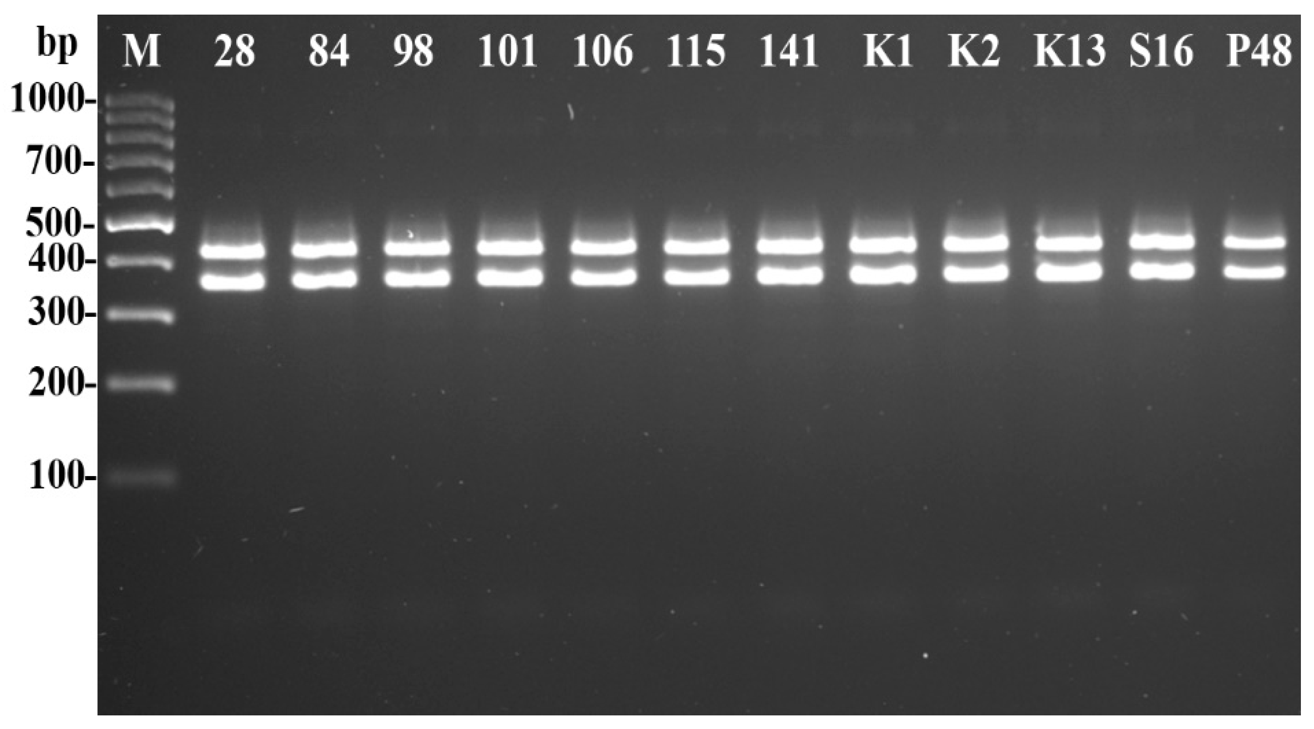
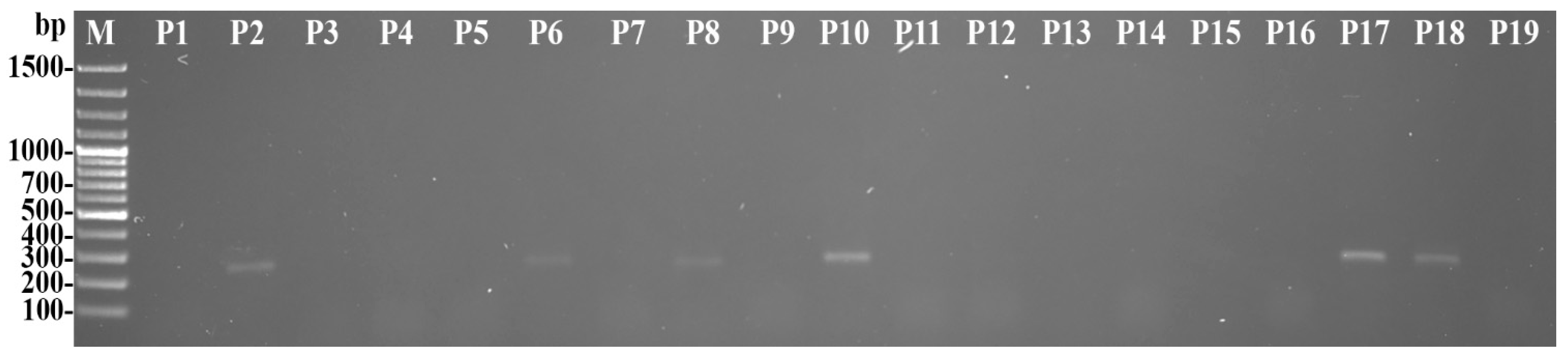
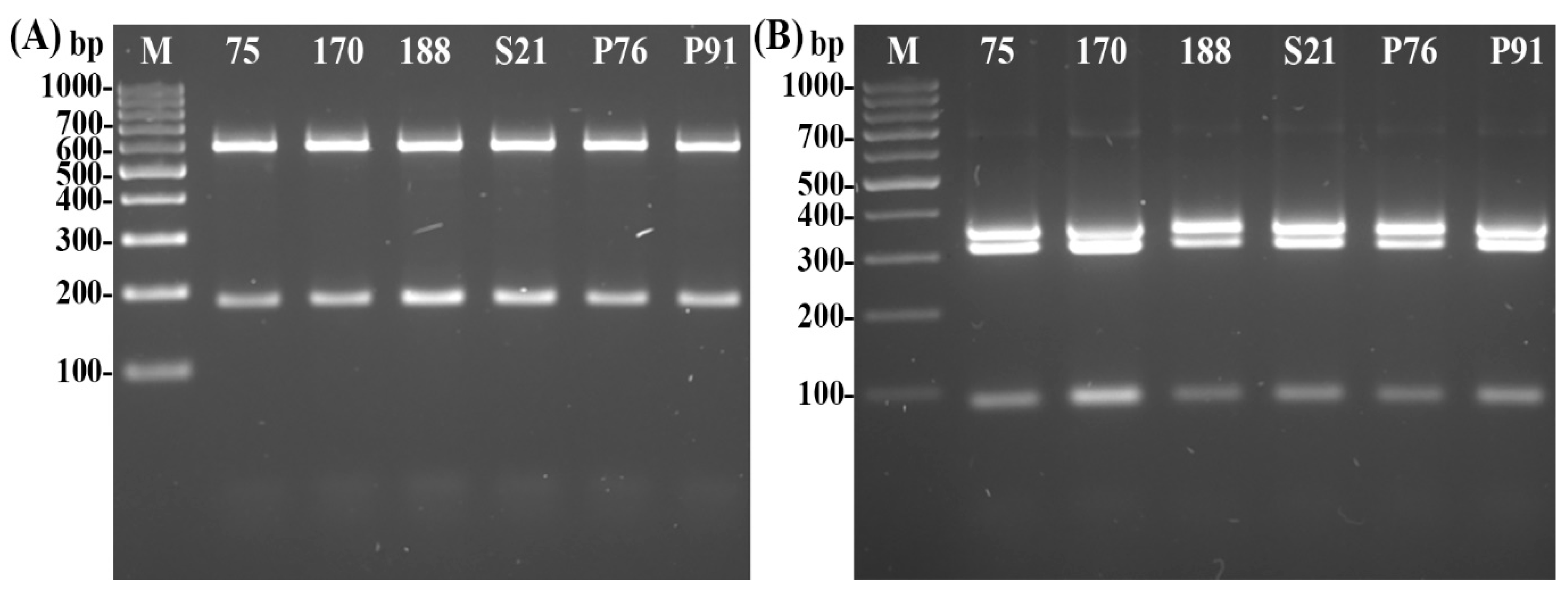
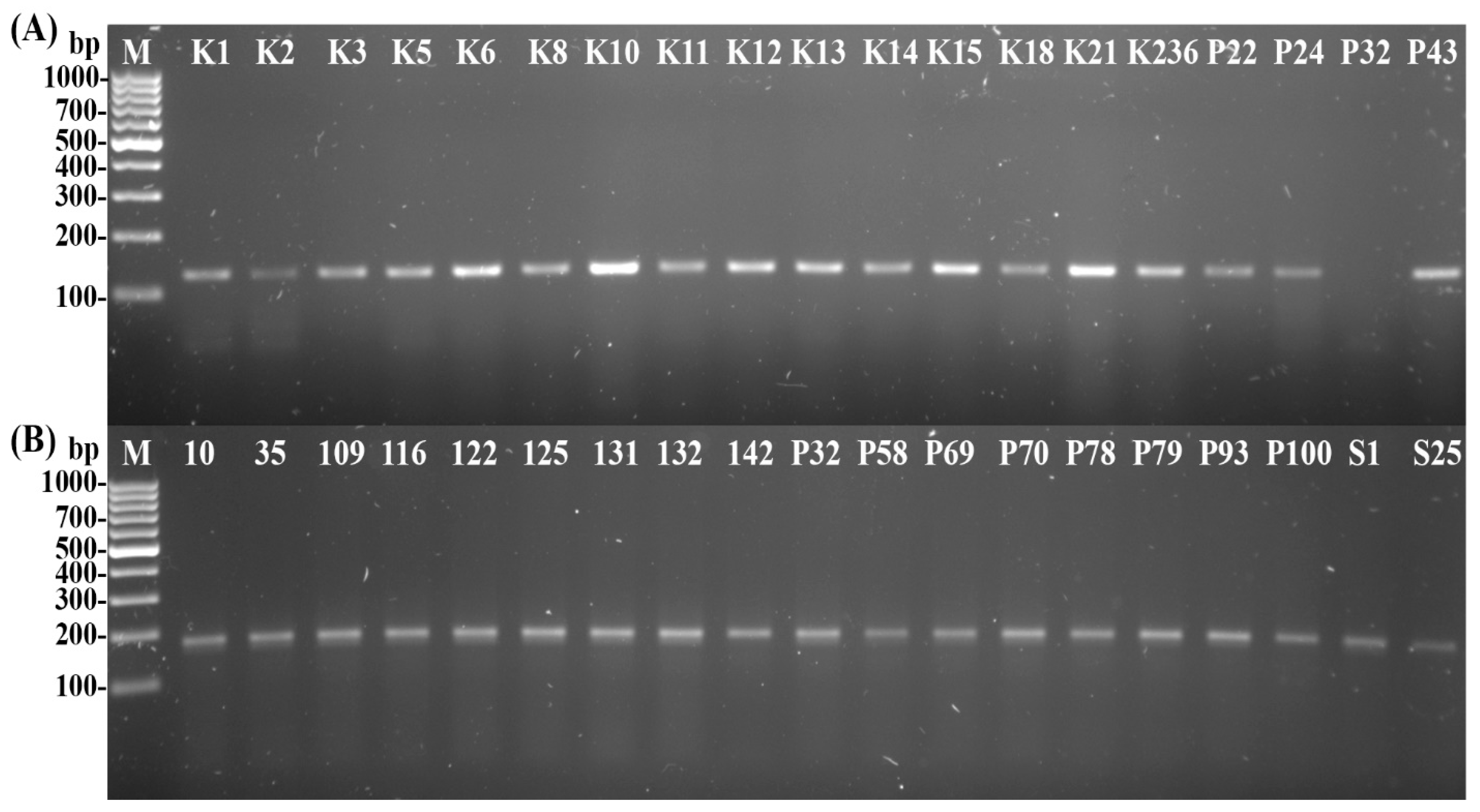
2.4. Pathogen Identification

Ca. Phytoplasma prunorum’ identification was carried out using phloem tissues from plants and from the vector psyllids. All samples were tested using a nested-Polymerase Chain Reaction (nested-PCR) method. Amplification was performed in a 12.5 µL PCR mix, containing 0.75 µL of DNA template, 0.25 µL of both forward and reverse primers (10 µM), 6.25 µL of Dream Taq Green PCR Master Mix 2X buffer (Thermo Fisher Scientific Baltics UAB, Vilnius, Lithuania), and 5 µL of sterilized, distilled water. In the first round of amplification, the universal primer pair of Eof and Eor were used (Table S1) [59]. The second round of amplification was performed with 0.75 µL of the PCR mixture from the first round as template, using ACE1 and ACE2 specific primer pair (Table S1) [9]. Both of the PCRs were conducted with an initial denaturation at 95 °C for 5 min and a final elongation at 72 °C for 10 min. In the first PCR, 35 cycles were run as follows: denaturation at 95 °C for 60 s, annealing at 55 °C for 60 s, and elongation at 72 °C for 60 s. The second PCR program involved 30 cycles: denaturation at 95 °C for 30 s, annealing at 55 °C for 30 s, and elongation at 72 °C for 45 s. The amplified DNA fragments were visualized in 1% TBE agarose gels using electrophoresis and then documented (Uvidoc HD6, UVItec Ltd., Cambridge, UK).

2.5. Psyllid Identification

First, the preserved psyllid adults were identified by classical methods, using stereomicroscope (Olympus CX21, Olympus Corporation, Tokyo, Japan) and the dichotomous identification keys [32,60,61].
The classical identification of ‘Ca. Phytoplasma prunorum’ carrier psyllids was additionally confirmed using the PCR-RFLP method previously described by Oettl and Schlink [30]. In the case of C. pruni, the PCR mix contained the following ingredients at a total of 25 µL: 1.5 µL of psyllid DNA, 0.5 µL of VPm_COI_F2 forward primer and 0.5 µL of VPm_COI_R4 reverse primers (Table S1) [30] (20 µM), 12.5 µL of Dream Taq Green PCR Master Mix 2X buffer (Thermo Fisher Scientific Baltics UAB, Vilnius, Lithuania), and 10 µL of sterilized distilled water. The same components were used in the case of C. crataegi, but the final volume of the PCR mix was 75 µL. The PCR protocol comprised initial denaturation at 95 °C for 2 min, followed by 45 cycles of denaturation at 95 °C for 30 s, annealing at 46 °C for 30 s, elongation at 72 °C for 1 min, and a final elongation at 72 °C for 5 min. From each sample, 10 µL of the PCR product was directly digested with TaqI FastDigest restriction endonuclease (Thermo Fisher Scientific Baltics UAB, Vilnius, Lithuania). The TaqI enzyme produces well-distinguishable fragments (including flanking primers) between the C. pruni-1 and -2 variants, but fragment sizes were similar for C. pruni-1 (593 bp/208 bp) and C. crataegi (593 bp/190 bp/19 bp). Due to the similarities in the digested fragment sizes, an additional AluI digestion step was carried out. This enzyme produced different number of fragments (including flanking primers) that enabled the precise identification of the two psyllid species (C. pruni-1: 362 bp/247 bp/134 bp/37 bp/21 bp; C. crataegi: 684 bp/91 bp/21 bp). Furthermore, in the case of C. crataegi, 50 µL of PCR product was purified with High Pure PCR Product Purification Kit (Roche, Basel, Switzerland) according to the manufacturer’s instructions. Nucleotide sequences of the specific PCR products were determined by direct Sanger sequencing [62] with the VPm_COI primer pairs. Sequencing was carried out by service providers. BLAST analysis (http://www.ncbi.nlm.nih.gov/blast, accessed on 25 November 2022) was used to examine the sequences and determine the most identical sequences in GenBank. After receiving the chromatogram files, they were loaded into Unipro Ugene v 38.0 [63], where the reads were mapped to a reference sequence with Trimming quality threshold set to 0 and Mapping min. similarity set to 50%. Mapped reads were then manually corrected. Corrected sequences were deposited in GenBank.
The biotypes of all collected plum psyllids were determined using the primer sets 2 (CpA50F, CpB350F, and Cp480R) and 3 (Cp135F, CpA425R, and CpB315R) (Table S1) [25]. The PCR products expected for biotype ‘A’ are 421 bp and 293 bp for primer set 2 and primer set 3, while the PCR products of biotype ‘B’ using primer set 2 and primer set 3 are 151 bp and 177 bp, respectively. Amplification was performed in a 12.5 µL PCR mix, containing 0.75 µL of DNA template, 0.25–0.25 µL of both forward and reverse primers (10 µM), 6.25 µL of Dream Taq Green PCR Master Mix 2X buffer (Thermo Fisher Scientific Baltics UAB, Vilnius, Lithuania), and 5 µL of sterilized distilled water. The PCR cycles were as follows: initial denaturation at 94 °C for 5 min, followed by 30 cycles at 94 °C for 30 s, 65 °C for 20 s, and 72 °C for 30 s, with a final 5 min extension period at 72 °C.
All PCRs and nested-PCRs were carried out in a GeneAmp 9700 PCR (Applied Biosystems, Foster City, CA, USA) cycler. The amplified DNA fragments were visualized in 1.5% TBE agarose gels, while the digested fragments were visualized in 2% TBE agarose gels using electrophoresis and then documented.

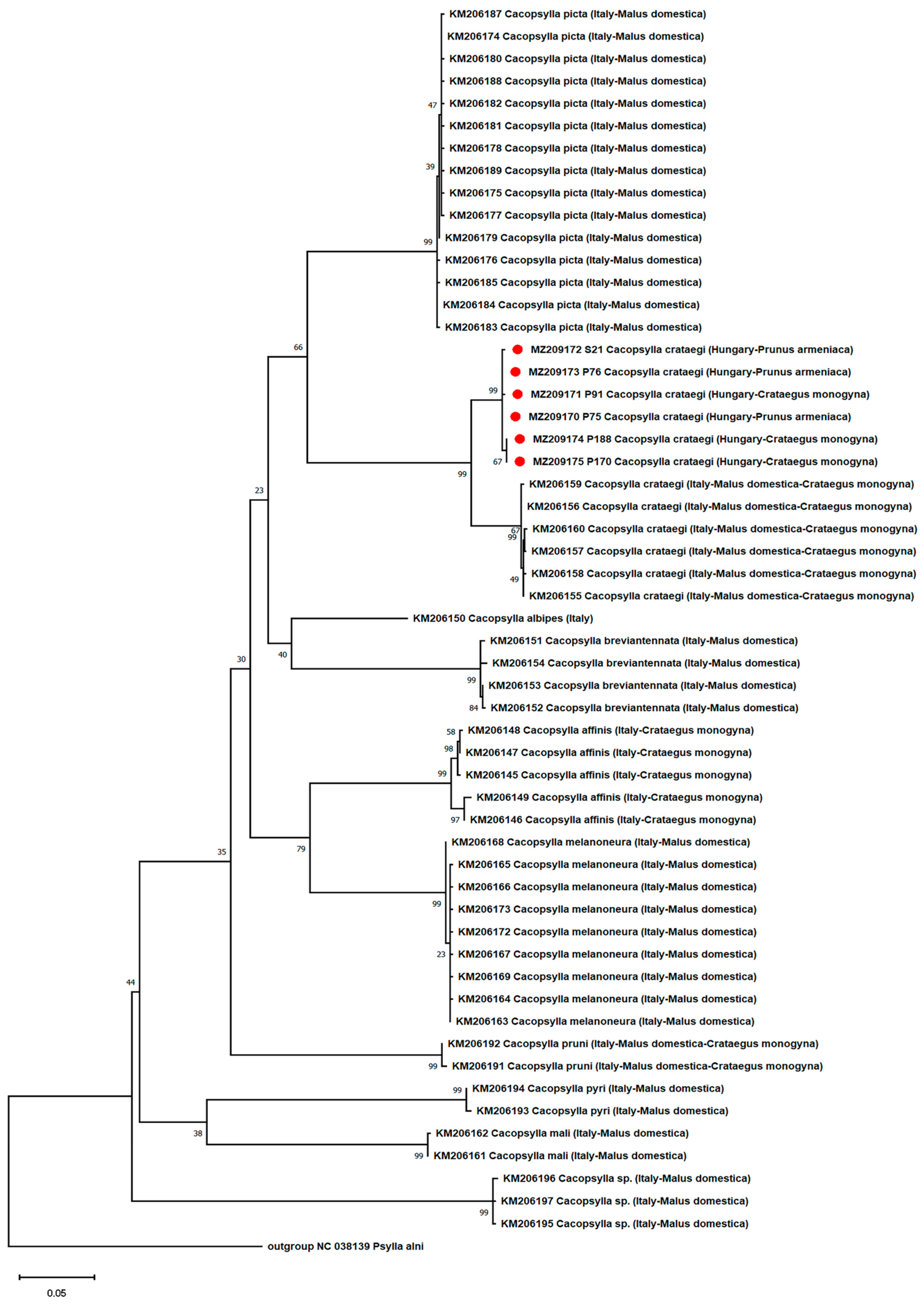
2.6. Phylogenetic and Recombination Analysis of Psyllids

All phylogenetic analyses were carried out using the MEGA X program [64]. We compared our sequences to other GenBank sequences for phylogenetic and recombination analyses, the latter of which are listed in Table S2.
First, ClustalW [65] alignment was generated using ClustalW (1.6) matrix, with gap-opening penalty of 15.00 and gap extension penalty of 6.66. Then, we sought out the best-fitting DNA substitution model using all sites. The model with the lowest Bayesian Information Criterion score (BIC) was considered to most accurately describe the substitution pattern [66]. With this model, we built a Maximum Likelihood phylogenetic tree [67,68]. The reliability of the tree was tested with the Bootstrap method [69], employing 1000 pseudo-replicates.
Recombination detection was carried out with RDP v4.98beta [70,71] with the following parameters: sequences were linear, highest acceptable p-value was 0.05, and with Bonferroni correction applied. Recombination events detected with fewer than three methods were rejected.

2.7. Statistical Analysis of Abundance and Phytoplasma Presence of Psyllids

The abundance rates of C. pruni and C. crataegi on different plants were compared with one-sample Z test. The proportions of phytoplasma-positive male and female adults were compared by Fisher’s exact test for both psyllid species and for all examined plants. The incidences of ‘Ca. Phytoplasma prunorum’ in C. pruni and C. crataegi specimens were also compared with Fisher’s exact test. The statistical analysis was performed in statistical software environment R version 4.1.2 (R Core Team, 2021) [72].

3. Results

3.1. Disease Occurrence in Plants

A considerable number of the tested trees were positive for ‘Ca. Phytoplasma prunorum’ (Figure 1).
More than half of the infected trees were also symptomatic (Table 1). All of these symptomatic trees died within a year after the detection of the pathogen, except two. Besides the infection of the apricot trees, the wild plants also tested positive for the pathogen (Table 1).

3.2. Insect Samples

  • Classical Identification of Psyllids
All 414 collected specimens were successfully identified; the vast majority of the collected individuals were either C. pruni (plum psyllid) or C. crataegi (hawthorn psyllid). Five specimens belonged to C. affinis and two to C. picta, but they were not included in further analysis due to the low number of individuals.
  • Psyllids Collected on Apricot
Due to their numbers, apricot trees were the most frequently examined. From these trees, high quantities of psyllids were collected. The abundance rate of C. pruni on P. armeniaca was significantly higher (Z test, p < 0.01; Table 2).
Besides species identification, we also counted the number of males and females present in each of the orchards. The ratio of females was significantly greater than that of males in the case of the C. pruni specimens collected from P. armeniaca (Z test, p < 0.001; Table 3), while the female ratio was just slightly significantly higher in the case of the C. crataegi specimens collected from P. armeniaca (Z test, p = 0.06; Table 3).
  • Psyllids Collected on Non-Apricot Hosts
It is important to point out that fewer European plums, blackthorns, common hawthorn plants, and cherry plum suckers of apricot were sampled compared to the apricot trees.
During the study, we collected few psyllids from the European plums. The proportion of collected C. pruni was significantly higher than that of the hawthorn psyllids (Z test, p < 0.001; Table 2). Most of the collected adults were females for both psyllid species. In the case of C. pruni, the number of females collected from P. domestica was significantly higher (Z test, p < 0.001; Table 3). In the case of C. crataegi, no significant differences between male and female collection rates were detected, which was probably due to the low sample size (Z test, p = 0.62; Table 3). The rate of plum psyllids and hawthorn psyllids collected from blackthorn (Z test, p = 0.453; Table 2) and the gender ratio of C. pruni (Z test, p = 0.453; Table 3) and C. crataegi (Z test, p = 1.00; Table 3) showed no significant difference. There were only four C. pruni specimens collected from cherry plum suckers (Table 2). The abundance rates of C. crataegi on C. monogyna were significantly greater (Z test, p < 0.001; Table 2). Among the C. crataegi individuals, there were slightly significantly more females than males (Z test, p = 0.063; Table 3). In the case of plum psyllids, the gender ratio was not significantly different (Z test, p > 0.999; Table 3).
  • Candidatus Phytoplasma prunorum’ Incidence in Psyllids
Nested-PCR tests were carried out on 183 C. pruni psyllids, of which 12 (6.6%) specimens tested positive for the pathogen. After an examination of the infection rate of C. pruni, it was determined that 2.6% (1 from 39) of males and 7.6% (11 from 144) of females were carriers (Table 4). However, there were no significant differences in the infection rates of males and females (Fisher’s exact test, p = 0.47; Table 4).
The determination of the infection rates of the psyllids on different host plants resulted in the following prevalence rates: 7.5% on apricot (10 adults out of 133) and 7.7% on plum (2 adults out of 26). None of the plum psyllid specimens collected from the other studied plant species tested positive for ‘Ca. Phytoplasma prunorum’.
From the 231 collected C. crataegi specimens, 2.6% of the adults (6 specimens) tested positive for ‘Ca. Phytoplasma prunorum’. Slightly significantly lower levels of infection were identified in the collected C. crataegi individuals compared to C. pruni (Fisher’s exact test, p = 0.06; Table 5).
Among the specimens, the number of infected females and males were equal to three (Table 4), with no significant difference between them (Fisher’s exact test, p = 0.69). An examination of the infection rates of the psyllids on different plant species showed incidence rates of 3.2% on apricot (3 adults out of 89) and 2.2% on hawthorn (3 adults out of 128), while the rest of the tested individuals (14 specimens) did not carry phytoplasma.
  • Molecular Identification of Psyllids
Molecular identification of ‘Ca. Phytoplasma prunorum’-positive adult psyllids (Figure 2) confirmed the morphological identification.
Based on the fragment lengths of the PCR-RFLP, we could only identify C. pruni-2 variants among the 12 infected plum psyllids. In the case of six infected C. crataegi specimens, the restriction analysis of the PCR products with the TaqI enzyme resulted in the predicted fragment lengths shown in Figure 3A. However, the digestion of infected hawthorn psyllids with AluI revealed different fragment lengths (Figure 3B), which was different from the results of an earlier study [30].
Our samples had an additional fragment of the COI region that the AluI enzyme could digest. Thus, we identified four fragments instead of the expected three. To better understand this phenomenon, we sequenced the PCR products. A BLAST analysis of all of our isolates (accession #MZ209170-MZ209175) revealed 99.60% to 100% identity with respect to each other, for which the highest identity was confirmed with the formerly published Cacopsylla crataegi (accession #KM206155) from Italy (100% query cover and 95.39% to 95.79% identity). To map the reads, we used this Italian accession to manually correct the sequencing ambiguities of our isolates. In addition, sequences were examined using the CLC Sequence Viewer program (v 8.0) restriction site search engine. The following fragments were predicted when tested with the TaqI and AluI enzymes: 593 bp/190 bp/19 bp and 363 bp/327 bp/91 bp/21 bp, respectively. These findings were confirmed by the results of PCR + RFLP assays mentioned above (Figure 3).
The biotyping of all the collected C. pruni specimens was also carried out. For some samples, the PCR did not amplify the target sequence when using primer set 2 (Figure 4A). These samples yielded a PCR product with the primer set 3 (Figure 4B). Among the collected samples, only the ‘B’ biotype was present.
  • Phylogenetic and Recombination Analysis of the Psyllids
The model with the lowest BIC score (9667.00725) was the Hasegawa–Kishino–Yano model [73] with a gamma distribution and invariant sites. A phylogenetic tree was built with this model, and the accuracy of this tree was tested with the Bootstrap method with 1000 pseudo-replicates (Figure 5).
On the phylogenic tree, C. crataegi forms a distinct clade from the other species, which is supported by a high (99%) Bootstrap value. In addition, the Hungarian isolates form a distinct clade within C. crataegi, which is supported by a 99% Bootstrap value. Interestingly, two isolates (P170 and P188) were separated from the other Hungarian isolates by an 89% Bootsrap value, which is considered to be very high (Figure 5). Recombination detection of the accessions revealed only weak recombination signals, but they were rejected, as none of them were identified by three or more statistical methods.

4. Discussion

In this study, we examined the ‘Ca. Phytoplasma prunorum’ infection in apricot trees older than 10 years. From the collected rootstock suckers and scions 28.6% and 25.9% tested positive for phytoplasma, respectively, which coincides with earlier reports [5,6,7]. In contrast to the infected scions, no symptoms appeared on the diseased rootstock suckers. Summarizing the symptom appearance and ESFY phytoplasma presence of trees, 61.9% of the infected trees had foliar symptoms, and all except two died within a year after the pathogen’s identification. Our results show that the phytoplasma infection of trees is a major limiting factor of successful apricot cultivation on the sites examined, which further underlines the importance of surveying the infection of the vector and other Cacopsylla species in orchards and their near vicinity. The latter is necessary, because ESFY phytoplasma-infected wild host plants were identified near the plantations. We sampled psyllids using the beating tray method. First, all of the adult psyllids were identified with classical methods. Based on external morphological characteristics, the vast majority of the samples were C. pruni and C. crataegi. C. affinis and C. picta were only present in very low numbers; thus, they were not part of the further investigation. In the case of C. pruni, our plum psyllids biotype study [25] confirmed the morphological results. Furthermore, the test resulted in the determination of type ‘B’ only. The result confirmed the hypothesis that C. pruni is unique or dominant in central and eastern Europe [28]. Since then, type ‘A’ specimens have been found in Northern Italy and Western Germany [11,20]. Thus, the results of previous studies and our current investigation induced the conclusion that biotype ‘B’ of C. pruni remains a unique variant in east-central and south-east Europe [20,23,25,28,29].
Former studies found great numbers of plum psyllids on P. armeniaca [10,29], which is in accordance with our results, while other researchers found no or only a few specimens from apricot [23,35,37]. These contrary phenomena could be a result of environmental and climatic conditions or the adaptation of insects. The sex ratio of C. pruni on the apricot trees in our study showed a higher proportion of females (79.7%) than males, which is in accordance with former reports [29,45]. Infected samples were identified among the rootstock suckers of apricot trees. However, few individuals could be collected from them. Based on these findings, this part of the plant is the reservoir of the pathogen, but contrary to other reports [35,37], it is a less important host plant for C. pruni in our test plantations. The latter was attributable to the low number of rootstock suckers caused by their regular removal. Crataegus monogyna seemed to be a less preferred plant species for C. pruni compared to Prunus species; we could collect only a few individuals from these bushes.
From C. pruni, 6.6% of the 183 collected specimens tested positive for the ESFY phytoplasma. The infection rate measured in this study is higher than the results of former reports: 0.8–4.8% [10,11,27,41,42]. Although, in some studies, the infection rate was much higher than ours: 15–64.2% [23,29,43,44]. Similar to other investigations, we could not evince either sex having a significantly higher phytoplasma infection rate, even though we observed that the infection rates of females were somewhat higher than those of males [23,45]. Infected individuals were collected from two plant species. The infection rate of C. pruni was 7.5% in the case of P. armeniaca and 6.7% in P. domestica. P. spinosa is an important host of C. pruni and a reservoir of the pathogen of ESFY [36]. Psyllids mainly acquire the phytoplasma from wild Prunus plants [27]. Similar to other reports, we were able to collect plum psyllids from P. spinosa [10,11,23,42]. However, among the tested adults, there were no positive results for ‘Ca. Phytoplasma prunorum’, which was probably due to the low number of sampled plants. Some of the examined blackthorns were latently infected with the pathogen, which is in concordance with other reports [6,36]. Based on the identified phytoplasma-positive plants and the psyllids present on them, we believe that P. spinosa plays a role in the spread of ESFY phytoplasma in Hungary.
In the collected samples, there were hawthorn psyllids (C. crataegi) in vast numbers. On the studied plants, the occurrence of C. crataegi was slightly higher than that of C. pruni. On hawthorn bushes we found significantly more C. crataegi than C. pruni, which is not surprising, because hawthorn is only a host for C. crataegi [32]. It is worth mentioning that about one third of the hawthorn bushes examined for the presence of phytoplasma were identified as infected. Furthermore, we were also able to collect hawthorn psyllids from apricot trees (89 specimens), but in lower numbers than in C. pruni (133 specimens). To the best of our knowledge, the presence of hawthorn psyllids on apricot has not been reported before. We were able to identify the pathogen of ESFY from some C. crataegi individuals collected on P. armeniaca (with a 4.4% infection rate) and C. monogyna (with a 2.8% infection rate). Considering all the examined plants, the rate of female abundance of hawthorn psyllids was significantly higher than that of males. While the phytoplasma-carrying rates of the C. crataegi males were a bit higher than those of the females, the difference was not significant. Among all the collected hawthorn psyllids, 2.6% tested positive for ‘Ca. Phytoplasma prunorum’. To our knowledge, this is the first report of C. crataegi being infected by the ESFY phytoplasma. The pathogen-carrying rate of hawthorn psyllids was lower than that of plum psyllids, though the difference was not significant. On this basis, if the infectivity of C. crataegi is confirmed in ‘Ca. Phytoplasma prunorum’ transmission tests, this psyllid species will have been proved to be an important vector much like the C. pruni species.
PCR-RFLP analyses were performed on individuals carrying the pathogen of ESFY [30]. These analyses confirmed the classical identification of C. pruni; we found that these specimens belonged to the C. pruni-2 cluster. Individuals of the hawthorn psyllid species (phytoplasma-positive) identified by classical methods were also subjected to the digestion of the specific PCR products with TaqI and AluI restriction endonucleases. The digestion of the fragments with AluI yielded different fragment lengths than anticipated based on the work of Oettl and Schlink [30]. Therefore, the nucleotide sequences of the PCR products of six C. crataegi samples were determined. BLAST analyses of the sequences resulted in the identification of C. crataegi having the highest sequence identity with an Italian specimen (accession number KM206155) [30]. On the phylogenetic tree, the Hungarian psyllids formed a distinct clade within the C. crategi species, but shared a common ancestor with the Italian isolates, which suggests that the two are genetically distinct populations. Based on the results of Oettl and Schlink [30] and our investigation, C. crataegi could be highly reliably identified by PCR + RFLP without sequencing the PCR products. Moreover, the difference in the number of the digested fragments could be a valuable marker with which to differentiate between the Italian and Hungarian populations.

5. Conclusions

In this work, a considerable number of the tested apricot trees tested positive for phytoplasma. Among the infected trees, the vast majority of the symptomatic ones died within a year, which underlines the destructiveness of ‘Ca. Phytoplasma prunorum’ at the test sites. The sampled apricot orchards and their immediate surroundings were colonized by the ’B’ biotype of Cacopsylla pruni. As in other studies, we also collected several plum psyllid adults, and some of them tested positive for the pathogen ‘Ca. Phytoplasma prunorum’. We provided further evidence that the plum psyllid plays an unquestionable role in the spread of pathogen of ESFY in the studied apricot production areas in Hungary. In our opinion, it is necessary to control plum psyllids with pesticide applications— if the presence of the insect is justified by monitoring —in order to reduce the disease’s occurrence. Based on the contrast between reports similar to our results in terms of abundance and other works, we consider it to be worthwhile to examine the role of apricots in the life cycle of C. pruni and to determine which factors may influence it. Examinations of insect–plant relationships (such as between pest and hosts or food plants) could be the basis of new research projects.
Besides C. pruni, C. crataegi individuals were also collected in high numbers. Based on our results, it would also be worthwhile to examine the role of apricots in the life cycle of C. crataegi. Furthermore, several of the hawthorn psyllids tested positive for ‘Ca. Phytoplasma prunorum’. If further transmission studies confirm C. crataegi as a vector of ESFY phytoplasma, professional growers should monitor and control the abundance of hawthorn psyllids by employing good agricultural practices, as in the case of the plum psyllid. We consider it to be important to expand the existing data on C. crataegi (e.g., its distribution, population dynamics, etc.) to acquire more comprehensive knowledge with respect to this species, which will be necessary if it is to be controlled.

Supplementary Materials

The following supporting information can be downloaded at: https://www.mdpi.com/article/10.3390/agronomy13010199/s1, Table S1: Primer pairs used in the tests; Table S2: GenBank sequences used in the phylogenetic and recombination analysis; Supplementary Material: Data of apricot trees, wild plants and Cacopsylla sp.

Author Contributions

Conceptualization, L.S.K. and B.P.; methodology, L.S.K., B.P. and L.P.; data curation, M.L. and L.S.K.; formal analysis, M.L., J.Á. and L.S.K.; investigation, L.S.K., J.Á., B.P., P.G., M.P. and L.P.; resources, G.N., B.P., J.F. and L.P.; writing—original draft preparation, L.S.K. and J.Á.; writing—review and editing, L.S.K., B.P., P.G., J.F., M.L., J.Á., G.N., M.P. and L.P.; visualization, L.S.K., J.Á., M.P. and L.P.; supervision J.F. and L.P.; project administration, L.S.K. and L.P.; funding acquisition, M.P., J.F. and L.P. All authors have read and agreed to the published version of the manuscript.

Funding

The research of the ELKH-SZE PhatoPlant-Lab was supported by the ELKH TKI, under the number 3200107. The APC was funded by Hungarian University of Agriculture and Life Sciences.

Data Availability Statement

The data presented in this study are available in Supplementary material. Table S1 contains information of primers used in the tests. Table S2 shows information of the Gen Bank sequences used in phylogenetic and recombination analysis.

Acknowledgments

We are grateful for professional support from Ibolya Ember, former employee of Hungarian University of Agriculture and Life Sciences.

Conflicts of Interest

The authors declare no conflict of interest. The funders had no role in the choice of research project; design of the study; in the collection, analyses or interpretation of data; in the writing of the manuscript; or in the decision to publish the results.

References

  1. Marcone, C.; Jarausch, B.; Jarausch, W. Candidatus Phytoplasma prunorum, the causal agent of European stone fruit yellows: An overview. J. Plant Pathol. 2010, 92, 19–34. [Google Scholar]
  2. Lorenz, K.; Dosba, F.; Poggi-Pollini, C.; Llacer, G.; Seemüller, E. Phytoplasma diseases of Prunus species in Europe are caused by genetically similar organisms. J. Plant Dis. Prot. 1994, 101, 567–575. [Google Scholar]
  3. Steffek, R.; Follak, S.; Sauvion, N.; Labonne, G.; MacLeod, A. Distribution of ‘Candidatus Phytoplasma prunorum’ and its vector Cacopsylla pruni in European fruit-growing areas: A review. EPPO Bull. 2012, 42, 191–202. [Google Scholar] [CrossRef]
  4. Cieślińska, M.; Morgaś, H. Detection and identification of ‘Candidatus Phytoplasma prunorum’,‘Candidatus Phytoplasma mali’ and ‘Candidatus Phytoplasma pyri’in stone fruit trees in Poland. J. Phytopathol. 2011, 159, 217–222. [Google Scholar] [CrossRef]
  5. Ivić, D.; Plavec, J.; Ivančan, G. The occurrence of ‘Candidatus Phytoplasma prunorum’in apricot orchards in Baranja. Pomol. Croat. Glas. Hrvat. Agron. Društva 2017, 21, 101–106. [Google Scholar]
  6. Riedle-Bauer, M.; Paleskić, C.; Schwanzer, J.; Kölber, M.; Bachinger, K.; Antonielli, L.; Schönhuber, C.; Elek, R.; Stradinger, J.; Emberger, M.; et al. Epidemiological and molecular study on ‘Candidatus Phytoplasma prunorum’in Austria and Hungary. Ann. Appl. Biol. 2019, 175, 400–414. [Google Scholar] [CrossRef]
  7. Ludvikova, H.; Franova, J.; Sucha, J. Phytoplasmas in apricot, peach and sour cherry orchards in East Bohemia, Czech Republic. Bull. Insectol. 2011, 64, S67–S68. [Google Scholar]
  8. Nečas, T.; Ondrášek, I.; Krška, B. ‘Candidatus Phytoplasma prunorum’-a pathogen spreading uncontrollably in apricot orchards in the Czech Republic. Acta Hortic. 2015, 1105, 131–136. [Google Scholar] [CrossRef]
  9. Jarausch, W.; Lansac, M.; Saillard, C.; Broquaire, J.; Dosba, F. PCR assay for specific detection of European stone fruit yellows phytoplasmas and its use for epidemiological studies in France. Eur. J. Plant Pathol. 1998, 104, 17–27. [Google Scholar] [CrossRef]
  10. Jarausch, B.; Mühlenz, I.; Beck, A.; Lampe, I.; Harzer, U.; Jarausch, W. Epidemiology of European Stone Fruit Yellows in Germany. Acta Hortic. 2008, 781, 417–422. [Google Scholar] [CrossRef]
  11. Jarausch, W.; Jarausch, B.; Fritz, M.; Runne, M.; Etropolska, A.; Pfeilstetter, E. Epidemiology of European stone fruit yellows in Germany: The role of wild Prunus spinosa. Eur. J. Plant Pathol. 2019, 154, 463–476. [Google Scholar] [CrossRef]
  12. Morvan, G. Apricot chlorotic leaf roll. EPPO Bull. 1977, 7, 37–55. [Google Scholar] [CrossRef]
  13. Žežlina, I.; Rot, M.; Kač, M.; Trdan, S. Causal agents of stone fruit diseases in Slovenia and the potential for diminishing their economic impact–a review. Plant Prot. Sci. 2016, 52, 149–157. [Google Scholar]
  14. Kison, H.; Seemüller, E. Differences in strain virulence of the European stone fruit yellows phytoplasma and susceptibility of stone fruit trees on various rootstocks to this pathogen. J. Phytopathol. 2001, 149, 533–541. [Google Scholar] [CrossRef]
  15. Gazel, M.; Caglayan, K.; Serce, C.U.; Son, L. Evaluations of apricot trees infected by ‘Candidatus Phytoplasma prunorum’for horticultural characteristics. Rom. Biotechnol. Lett. 2009, 14, 4123–4129. [Google Scholar]
  16. Nečas, T.; Kiss, T.; Eichmeier, A.; Nečasova, J.; Ondrašek, I. The effect of phytoplasma disease caused by ‘Candidatus Phytoplasma prunorum’on the phenological and pomological traits in apricot trees. Not. Bot. Horti Agrobot. Cluj-Napoca 2018, 46, 107–114. [Google Scholar] [CrossRef]
  17. Carraro, L.; Osler, R.; Loi, N.; Ermacora, P.; Refatti, E. Transmission of European stone fruit yellows phytoplasma by Cacopsylla pruni. J. Plant Pathol. 1998, 80, 233–239. [Google Scholar]
  18. Weintraub, P.G.; Beanland, L. Insect vectors of phytoplasmas. Annu. Rev. Entomol. 2006, 51, 91–111. [Google Scholar] [CrossRef]
  19. Ouvrard, D. Psyl’list—The World Psylloidea Database. Available online: http://www.hemiptera-databases.com/psyllist (accessed on 2 February 2022).
  20. Sauvion, N.; Peccoud, J.; Meynard, C.N.; Ouvrard, D. Occurrence data for the two cryptic species of Cacopsylla pruni (Hemiptera: Psylloidea). Biodivers. Data J. 2021, 9, e68860. [Google Scholar] [CrossRef]
  21. Kiss, E.; Mergenthaler, E.; Kiss, B.; Viczián, O. A csonthéjasok európai sárgulása (ESFY) magyarországi terjedésének hátterében álló okok. In Proceedings of the 61. Növényvédelmi Tudományos Napok, Budapest, Hungary, 21 February 2015; p. 60. [Google Scholar]
  22. Mergenthaler, E.; Kiss, B.; Kiss, E.; Viczián, O. Survey on the occurrence and infection status of Cacopsylla pruni, vector of European stone fruit yellows in Hungary. Bull. Insectol. 2017, 70, 171–176. [Google Scholar]
  23. Viczián, O.; Kiss, B.; Kiss, E.; Orosz, S.; Juhász, A.L.; Mergenthaler, E. Mit tudunk a ‘Ca. Phytoplasma prunorum’ fitoplasma terjedéséről ma és mit gondolunk ugyanerről? [The significance of phytoplasma (‘Ca. Phytoplasma prunorum’) disease of stone fruits-what do we know and what is need to be done?]. Növényvédelem 2017, 78, 525–531. [Google Scholar]
  24. Sauvion, N.; Lachenaud, O.; Genson, G.; Rasplus, J.Y.; Labonne, G. Are there several biotypes of Cacopsylla pruni ? Bull. Insectol. 2007, 60, 185–186. [Google Scholar]
  25. Peccoud, J.; Labonne, G.; Sauvion, N. Molecular test to assign individuals within the Cacopsylla pruni complex. PLoS ONE 2013, 8, e72454. [Google Scholar] [CrossRef] [PubMed] [Green Version]
  26. Jarausch, B.; Weintraub, P.; Sauvion, N.; Maixner, M.; Foissac, X. Diseases and insect vectors. In Phytoplasma and Phytoplasma Disease Management: How to Reduce their Economic Impact; Bertaccini, A., Ed.; International Phytoplasmologist Working Group (IPWG): Bologna, Italy, 2014; pp. 111–121. [Google Scholar]
  27. Marie-Jeanne, V.; Bonnot, F.; Thébaud, G.; Peccoud, J.; Labonne, G.; Sauvion, N. Multi-scale spatial genetic structure of the vector-borne pathogen ‘Candidatus Phytoplasma prunorum’ in orchards and in wild habitats. Sci. Rep. 2020, 10, 5002. [Google Scholar] [CrossRef] [Green Version]
  28. Etropolska, A.; Jarausch, W.; Jarausch, B.; Trenchev, G. Molecular typing of Bulgarian specimen of the phytoplasma vectors Cacopsylla pruni Scopoli and Cacopsylla melanoneura (Foerster). Bulg. J. Agric. Sci. 2016, 22, 98–102. [Google Scholar]
  29. Lepres, L.A.; Mergenthaler, E.; Viczián, O.; Tóth, F. A szilva levélbolha (Cacopsylla pruni Scopoli, 1763) jelenlétének felmérése és „Candidatus Phytoplasma prunorum′′ kórokozóval való fertőzöttségének vizsgálata egy heves megyei kajszibarack ültetvényben. Növényvédelem 2018, 54, 197–203. [Google Scholar]
  30. Oettl, S.; Schlink, K. Molecular Identification of Two Vector Species, Cacopsylla melanoneura and Cacopsylla picta (Hemiptera: Psyllidae), of Apple Proliferation Disease and Further Common Psyllids of Northern Italy. J. Econ. Entomol. 2015, 108, 2174–2183. [Google Scholar] [CrossRef]
  31. Hodkinson, I.D. Life cycle variation and adaptation in jumping plant lice (Insecta: Hemiptera: Psylloidea): A global synthesis. J. Nat. Hist. 2009, 43, 65–179. [Google Scholar] [CrossRef]
  32. Ossiannilsson, F. The Psylloidea (Homoptera) of Fennoscandia and Denmark. In Fauna Entomologica Scandinavica; E. J. Brill: Leiden, The Netherlands, 1992; Volume 26, p. 346. [Google Scholar]
  33. Ripka, G. Levélbolhák 1; Agroinform Kiadó: Budapest, Hungary, 2010. [Google Scholar]
  34. Thébaud, G.; Yvon, M.; Alary, R.; Sauvion, N.; Labonne, G. Efficient transmission of ‘Candidatus Phytoplasma prunorum’ is delayed by eight months due to a long latency in its host-alternating vector. Phytopathology 2009, 99, 265–273. [Google Scholar] [CrossRef] [Green Version]
  35. Labonne, G.; Lichou, J. Data on the life cycle of Cacopsylla pruni, Psyllidae vector of European stone fruit yellows (ESFY) phytoplasma, in France. Acta Hortic. 2004, 657, 465–470. [Google Scholar] [CrossRef]
  36. Carraro, L.; Ferrini, F.; Ermacora, P.; Loi, N. Role of wild Prunus species in the epidemiology of European stone fruit yellows. Plant Pathol. 2002, 51, 513–517. [Google Scholar] [CrossRef]
  37. Jarausch, W.; Danet, J.L.; Labonne, G.; Dosba, F.; Broquaire, J.; Saillard, C.; Garnier, M. Mapping the spread of apricot chlorotic leaf roll (ACLR) in southern France and implication of Cacopsylla pruni as a vector of European stone fruit yellows (ESFY) phytoplasmas. Plant Pathol. 2001, 50, 782–790. [Google Scholar] [CrossRef]
  38. Yvon, M.; Labonne, G.; Thébaud, G. Survival of European stone fruit yellows phytoplasma outside fruit crop production areas: A case study in southeastern France. Acta Hortic. 2004, 657, 477–481. [Google Scholar] [CrossRef]
  39. Gallinger, J.; Jarausch, B.; Jarausch, W.; Gross, J. Host plant preferences and detection of host plant volatiles of the migrating psyllid species Cacopsylla pruni, the vector of European Stone Fruit Yellows. J. Pest Sci. 2020, 93, 461–475. [Google Scholar] [CrossRef]
  40. Carraro, L.; Loi, N.; Ermacora, P. Transmission characteristics of the European stone fruit yellows phytoplasma and its vector Cacopsylla pruni. Eur. J. Plant Pathol. 2001, 107, 695–700. [Google Scholar] [CrossRef]
  41. Etropolska, A.; Jarausch, W.; Jarausch, B.; Trenchev, G. Detection of European fruit tree phytoplasmas and their insect vectors in important fruit-growing regions in Bulgaria. Bulg. J. Agric. Sci. 2015, 21, 1248–1253. [Google Scholar]
  42. Warabieda, W.; Soika, G.; Cieślińska, M. Cacopsylla pruni in Poland and its significance as a vector of ‘Candidatus Phytoplasma prunorum’. Žemdirb. (Agric.) 2018, 105, 177–182. [Google Scholar] [CrossRef] [Green Version]
  43. Serçe, Ç.U.; Yvon, M.; Kaya, K.; Gazel, M.; Cengız, F.C.; Çağlayan, K.; Sauvion, N. Survey on the presence of Cacopsylla pruni in Turkey: Preliminary results. Bull. Insectol. 2011, 64, S145–S146. [Google Scholar]
  44. Bodnár, D.; Szalai, B.; Tarcali, G.; Viczián, O.; Mergenthaler, E. Phytoplasma infection status survey in plum psyllid (Cacopsylla pruni) population. Acta Agrar. Debr. 2019, 2, 45–48. [Google Scholar] [CrossRef]
  45. Ermacora, P.; Ferrini, F.; Loi, N.; Martini, M.; Osler, R. Population dynamics of Cacopsylla pruni and ‘Candidatus Phytoplasma prunorum’infection in North-Eastern Italy. Bull. Insectol. 2011, 64, S143–S144. [Google Scholar]
  46. Burckhardt, D.; Sharma, A.; Raman, A. Checklist and comments on the jumping plant-lice (Hemiptera: Psylloidea) from the Indian subcontinent. Zootaxa 2018, 4457, 1–38. [Google Scholar] [CrossRef]
  47. Horváth, G. Ordo. Hemiptera. In A Magyar Birodalom Állatvilága (Fauna Regni Hungariae). III. Arthropoda. (Insecta. Hemiptera.); Királyi Magy; Természettudományi Társulat: Budapest, Hungary, 1897; p. 72. [Google Scholar]
  48. Kontschán, J.; Kiss, E.; Ripka, G. Új adatok a hazai levélbolhák (Insecta: Psylloidea) előfordulásaihoz. [New data to occurrences of the Hungarian jumping plant lice (Insecta: Psylloidea).]. Növényvédelem 2020, 81, 197–202. [Google Scholar]
  49. Kontschán, J.; Ripka, G. Új adatok a hazai levélbolhák (Insecta: Psylloidea) előfordulásaihoz II. [New data to occurrences of the Hungarian jumping plant lice (Insecta: Psylloidea) II.]. Növényvédelem 2021, 82, 336–341. [Google Scholar]
  50. Ripka, G. Újabb adatok a díszfa- és díszcserjefajok levélbolha-faunájának ismeretéhez (Homoptera, Psylloidea) [Recent data on the psyllid fauna of ornamental trees and shrubs of Hungary (Homoptera: Psylloidea)]. Növényvédelem 1997, 33, 269–273. [Google Scholar]
  51. Ripka, G.; Kiss, B. További adatok a hazai parlagfűállományokban előforduló levélbolha-fajok (Hemiptera: Psylloidea) ismeretéhez [Recent data to the knowledge on psyllid species (Hemiptera: Psylloidea) occurring on common ragweed in Hungary]. Növényvédelem 2008, 44, 257–261. [Google Scholar]
  52. Križanac, I.; Mikec, I.; Budinščak, Ž.; Musić, M.Š.; Škorić, D. Diversity of phytoplasmas infecting fruit trees and their vectors in Croatia. J. Plant Dis. Prot. 2010, 117, 206–213. [Google Scholar] [CrossRef]
  53. Tedeschi, R.; Lauterer, P.; Brusetti, L.; Tota, F.; Alma, A. Composition, abundance and phytoplasma infection in the hawthorn psyllid fauna of northwestern Italy. Eur. J. Plant Pathol. 2009, 123, 301–310. [Google Scholar] [CrossRef]
  54. Miñarro, M.; Somoano, A.; Moreno, A.; García, R.R. Candidate insect vectors of apple proliferation in Northwest Spain. SpringerPlus 2016, 5, 1240. [Google Scholar] [CrossRef] [Green Version]
  55. Fischnaller, S.; Parth, M.; Messner, M.; Stocker, R.; Kerschbamer, C.; Reyes-Dominguez, Y.; Janik, K. Occurrence of different Cacopsylla species in apple orchards in South Tyrol (Italy) and detection of apple proliferation phytoplasma in Cacopsylla melanoneura and Cacopsylla picta. Cicadina 2017, 17, 37–51. [Google Scholar]
  56. Daire, X.; Clair, D.; Reinert, W.; Boudon-Padieu, E. Detection and differentiation of grapevine yellows phytoplasmas belonging to the elm yellows group and to the stolbur subgroup by PCR amplification of non-ribosomal DNA. Eur. J. Plant Pathol. 1997, 103, 507–514. [Google Scholar] [CrossRef]
  57. Doyle, J.J.; Doyle, J.L. Isolation of plant DNA from fresh tissues. Focirs 1990, 12, 13–15. [Google Scholar]
  58. Marzachì, C.; Veratti, F.; Bosco, D. Direct PCR detection of phytoplasmas in experimentally infected insects. Annals of Applied Biology 1998, 133, 45–54. [Google Scholar] [CrossRef]
  59. Mergenthaler, E. Fitoplazmás Betegségek Magyarországon: Korszerű Diagnosztikai Módszerek Fejlesztése [Phytoplasma Diseases in Hungary: Development of Improved Diagnostical Methods]. Ph.D. Thesis, Budapesti Közgazdaságtudományi és Államigazgatási Egyetem, Budapest, Hungary, 2004. [Google Scholar]
  60. Burckhardt, D. Identification Key for the Central European Cacopsylla Species. Available online: https://www.dlr.rlp.de/Internet/global/Themen.nsf/b81d6f06b181d7e7c1256e920051ac19/5f722b729e24cfccc12584430022b32e/$FILE/cacopsylla_pruni.pdf (accessed on 15 January 2015).
  61. Burckhardt, D. Identification Key for the Central European Cacopsylla Species. Available online: https://www.dlr.rlp.de/Internet/global/Themen.nsf/7683c11d82324367c1256ea600533a09/7e0b2d87f5d7df3bc1258443001fed2f/$FILE/cacopsylla_crataegi.pdf (accessed on 15 January 2015).
  62. Sanger, F.; Coulson, A.R. A rapid method for determining sequences in DNA by primed synthesis with DNA polymerase. J. Mol. Biol. 1975, 94, 441–448. [Google Scholar] [CrossRef]
  63. Okonechnikov, K.; Golosova, O.; Fursov, M. Unipro UGENE: A unified bioinformatics toolkit. Bioinformatics 2012, 28, 1166–1167. [Google Scholar] [CrossRef] [Green Version]
  64. Kumar, S.; Stecher, G.; Li, M.; Knyaz, C.; Tamura, K. MEGA X: Molecular Evolutionary Genetics Analysis across Computing Platforms. Mol. Biol. Evol. 2018, 35, 1547–1549. [Google Scholar] [CrossRef] [PubMed]
  65. Larkin, M.A.; Blackshields, G.; Brown, N.P.; Chenna, R.; McGettigan, P.A.; McWilliam, H.; Valentin, F.; Wallace, I.M.; Wilm, A.; Lopez, R.; et al. Clustal W and Clustal X version 2.0. Bioinformatics 2007, 23, 2947–2948. [Google Scholar] [CrossRef] [Green Version]
  66. Nei, M.; Kumar, S. Molecular Evolution and Phylogenetics; Oxford University Press: New York, NY, USA, 2000; p. 352. [Google Scholar]
  67. Felsenstein, J. Evolutionary trees from DNA sequences: A maximum likelihood approach. J. Mol. Evol. 1981, 17, 368–376. [Google Scholar] [CrossRef] [PubMed]
  68. Guindon, S.; Gascuel, O. A Simple, Fast, and Accurate Algorithm to Estimate Large Phylogenies by Maximum Likelihood. Syst. Biol. 2003, 52, 696–704. [Google Scholar] [CrossRef] [Green Version]
  69. Felsenstein, J. Confidence limits on phylogenies: An approach using the bootstrap. Evolution 1985, 39, 783–791. [Google Scholar] [CrossRef]
  70. Martin, D.P.; Murrell, B.; Golden, M.; Khoosal, A.; Muhire, B. RDP4: Detection and analysis of recombination patterns in virus genomes. Virus Evol. 2015, 1, vev003. [Google Scholar] [CrossRef] [Green Version]
  71. Martin, D.P.; Murrell, B.; Khoosal, A.; Muhire, B. Detecting and analyzing genetic recombination using RDP4. In Bioinform; Keith, J.M., Ed.; Springer: New York, NY, USA, 2017; Volume 1525, pp. 433–460. [Google Scholar]
  72. Team, R.C. R: A Language and Environment for Statistical Computing [Software Package], 4.1.2; R Foundation for Statistical Computing: Vienna, Austria, 2021. [Google Scholar]
  73. Hasegawa, M.; Kishino, H.; Yano, T.-A. Dating of the human-ape splitting by a molecular clock of mitochondrial DNA. J. Mol. Evol. 1985, 22, 160–174. [Google Scholar] [CrossRef] [PubMed]
Figure 1. The results of ‘Ca. Phytoplasma prunorum’ infection tests on apricot scions and rootstock suckers (from the examined samples: P1–P19) displayed on agarose gel (1.0%). Lane M represents a 100 bp DNA ladder (Thermo Fisher Scientific Baltics UAB, Vilnius, Lithuania).
Agronomy 13 00199 g001
Figure 2. Result of the PCR-RFLP analysis with TaqI endonuclease. Restriction fragment patterns of all infected Cacopsylla pruni-2 (samples: 28–P48) specimens displayed on agarose gel (2.0%). Lane M represents a 100 bp DNA ladder (Thermo Fisher Scientific Baltics UAB, Vilnius, Lithuania).
Agronomy 13 00199 g002
Figure 3. Results of the PCR-RFLP analysis (A) digested with TaqI endonuclease and (B) digested with AluI endonuclease. Restriction fragment patterns of all infected Cacopsylla crataegi (samples: 75–P91) specimens displayed on agarose gel (2.0%). Lane M represents a 100 bp DNA ladder (Thermo Fisher Scientific Baltics UAB, Vilnius, Lithuania).
Agronomy 13 00199 g003
Figure 4. The results of the Cacopsylla pruni biotype assay displayed on agar gels. (A) with primer set 2 (from the examined samples: K1–P43-‘B’ biotype, 151 bp-). (B) with primer set 3 (from the examined samples: 10–S25-‘B’ biotype, 177 bp-). Lane M represents a 100 bp DNA ladder (Thermo Fisher Scientific Baltics UAB, Vilnius, Lithuania).
Agronomy 13 00199 g004
Figure 5. Maximum Likelihood tree of Cacopsylla sequences, which was built with Hasegawa–Kishino–Yano nucleotide substitution model using gamma distribution, invariant sites, and tested with the Bootstrap method with 1000 replicates. Outgroup is Psylla alni (RefSeq: NC_038139). Bootstrap values are indicated on the branches as percentages. Hungarian isolates are marked with red dots.
Agronomy 13 00199 g005
Table 1. Number of specimens collected, ‘Candidatus Phytoplasma prunorum’ infection state, and symptom appearance of the infected tested plant samples at all sites.
Plant MaterialN 1I 2 (%)S 3 (%)
ApricotScion8125.961.9
Rootstock1428.60
Graft8129.662.5
Wild plantsBlackthorn1526.70
Hawthorn1127.30
1 N: number of plant materials; 2 I: Ratio of phytoplasma infection; 3 S: Appearance of symptoms of infected plant materials.
Table 2. Abundance of Cacopsylla pruni and C. crataegi on different plants and the comparative ratios, with 95% confidence intervals, Z test for equality, and their significance levels.
Examined PlantsSpeciesN 1Comparative Ratios (%)95% Confidence IntervalZ 5p 4
LCI 2UCI 3
All examined plantsC. pruni18344.20.390.492.310.021
C. crataegi23155.80.510.61
P. armeniacaC. pruni13359.90.530.662.890.004
C. crataegi8940.10.340.47
P. domesticaC. pruni2686.70.680.963.83<0.001
C. crataegi413.30.040.32
P. spinosaC. pruni1661.50.410.790.980.327
C. crataegi1038.50.210.59
P. cerasiferaC. pruni41000.401.001.50.13
C. crataegi000.000.60
C. monogynaC. pruni43.00.010.0810.71<0.001
C. crataegi12897.00.920.99
1 N: Abundance of Cacopsylla sp. individuals on investigated plants; 2 LCI: Lower limit of confidence intervals; 3 UCI: Upper limit of confidence intervals; 5 Z: Z test for equality; 4 p: Significance level.
Table 3. The number and proportions of males and females within the Cacopsylla pruni and C. crataegi species on different plants, with 95% confidence intervals, Z test for equality, and their significance levels.
C. pruniC. crataegi
Examined PlantsSex 1N 2Proportion (%)95% Confidence IntervalZ 5p6N 2Proportion (%)95% Confidence IntervalZ 5p 6
LCI 3UCI 4LCI 3UCI 4
All examined plants4021.90.160.297.54<0.0019340.30.340.472.890.004
14378.10.710.8413859.70.530.66
P. armeniaca2720.30.160.296.76<0.0013539.30.290.501.910.056
10679.70.720.865460.70.500.71
P. domestica311.50.030.313.73<0.001125.00.010.780.500.62
2388.50.690.97375.00.220.99
P. spinosa637.50.160.640.750.453550.00.240.760.001.00
1062.50.360.84550.00.240.76
P. cerasifera125.00.010.780.500.620
375.00.220.990
C. monogyna250.00.150.850.001.005341.40.330.501.860.063
250.00.150.857558.60.500.67
1 ♂: males; ♀: females; 2 N: Abundance of males and females of Cacopsylla sp. on investigated plants; 3 LCI: Lower limit of confidence intervals; 4 UCI: Upper limit of confidence intervals; 5 Z: Z test for equality; 6 p: Significance level.
Table 4. Incidence, odds ratio (with 95% confidence interval), and relative risk of the presence of ‘Candidatus Phytoplasma prunorum’ in all Cacopsylla pruni and C. crataegi specimens. Infected and non-infected individuals were sorted by males and females. Fisher’s exact test results are given for the infection rates (in bold), comparing the males’ and female’ incidence rates.
Pathogen IncidenceSex 1Statistical Analysis
C. pruniN 2Infected111
Non-infected38133
Ratio of individuals within males/females (%)Infected2.67.6Odds ratio = 0.32 (CI 3: 0.007;2.330)
Relative risk = 0.34
Fisher test p 4 = 0.47
Non-infected97.492.4
C. crataegiN2Infected33
Non-infected91134
Ratio of individuals within males/females (%)Infected3.22.2Odds ratio = 1.47 (CI 3: 0.193;11.218)
Relative risk = 1.46
Fisher test p 4 = 0.69
Non-infected96.897.8
1 ♂: males; ♀: females; 2 N: Number of males and females of infected and non-infected Cacopsylla sp; 3 CI: confidence intervals; 4 p: Significance level.
Table 5. Incidence, odds ratio (with 95% confidence interval), and relative risk of ‘Candidatus Phytoplasma prunorum’ infection in all Cacopsylla pruni and C. crataegi specimens. Fisher’s exact test results are given for the infection rates (in bold) comparing the specimen incidences.
Pathogen IncidenceSpeciesStatistical Analysis
C. pruniC. crataegi
N 1Infected126
Non-infected171225
Ratio of individuals within species (%)Infected6.62.6Odds ratio = 2.63 (CI 2: 0.890;8.705)
Relative risk = 2.52
Fisher test p 3 = 0.06
Non-infected93.497.4
1 N: Number of infected and non-infected Cacopsylla inviduals; 2 CI: confidence intervals; 3 p: Significance level.
Disclaimer/Publisher’s Note: The statements, opinions and data contained in all publications are solely those of the individual author(s) and contributor(s) and not of MDPI and/or the editor(s). MDPI and/or the editor(s) disclaim responsibility for any injury to people or property resulting from any ideas, methods, instructions or products referred to in the content.

