Article

Influence of Fruit Wounding on Subsequent Monilinia laxa Infection of Nectarines

by
Maximiliano Dini
1,2,†,
Maria do Carmo Bassols Raseira
3,
Marie-Noëlle Corre
4,
Véronique Signoret
4 and
Bénédicte Quilot-Turion
4,*
1
Programa de Pós-Graduação em Agronomia (PPGA), Faculdade de Agronomia Eliseu Maciel, Universidade Federal de Pelotas (UFPel), Caixa Postal 354, Pelotas CEP 96010-900, Rio Grande do Sul, Brazil
2
Instituto Nacional de Investigación Agropecuaria (INIA), Estación Experimental INIA Las Brujas, Ruta 48 km 10, Rincón del Colorado CP 90100, Canelones, Uruguay
3
Empresa Brasileira de Pesquisa Agropecuária, Embrapa Clima Temperado, BR 392, km 78, Caixa Postal 403, Pelotas CEP 96010-971, Rio Grande do Sul, Brazil
4
INRAE, GAFL, F-84143 Montfavet, France
*
Author to whom correspondence should be addressed.
Part of the PhD thesis of first author.
Agronomy 2023, 13(5), 1235; https://doi.org/10.3390/agronomy13051235
Submission received: 31 March 2023 / Revised: 20 April 2023 / Accepted: 24 April 2023 / Published: 27 April 2023
(This article belongs to the Special Issue Monilinia on Stone Fruit Species)

Abstract

:
Despite the fact that brown rot is the most economically important disease in stone fruits, the relationship between mechanical stress and infection has never been explored. Thus, to explore this connection, we carried out four experiments linking nectarine wounding and M. laxa infection. First, we evaluated a possible systemic reaction of the fruit to wounding that could impact the M. laxa infection. Afterward, we study the impact of the fruit in the environment on M. laxa colonies grown in vitro. Subsequently, we tested the disease susceptibility of fruits inoculated with M. laxa placed in the same environment as wounded fruits. Finally, in the fourth experiment, the effect of wounding on the subsequent fruit infections was evaluated at three fruit stages. As a result, we observed that there was no evidence of a fruit systemic reaction to wounding and M. laxa infection. In the study related to the impact of the fruit in the environment of M. laxa, the findings suggest that M. laxa “perceived” the fruit’s presence, resulting in accelerated in vitro growth. Moreover, the presence of wounded fruits in the box increased the susceptibility to brown rot. Inoculated fruits showed a delayed and reduced infection 7 h after being wounded, during the second stage of fruit development (15–19 weeks after full bloom). Moreover, a red reaction associated with inoculation was detected immediately after the fruits were wounded. Nine phenolic compounds exclusively related to the red areas were extracted, while six other compounds were present in higher proportions. These compounds may be actively involved in plant–pathogen interactions and the activation of metabolic pathways involved in nectarine susceptibility/resistance to M. laxa.

1. Introduction

A variety of diseases attack stone fruits. Brown rot (BR), which is primarily caused by three species of the genus Monilinia, is one of the most economically significant diseases affecting these fruit trees. M. laxa (Aderh. and Ruhl.) Honey is still predominant in Europe, China, South Africa, Chile, North America, and the Middle East. M. fructicola (Wint.) Honey is important in South and North America and sometimes becomes important in Europe and Asia, whereas M. fructigena (Aderh. and Ruhl.) Honey, present in Europe and China, has a narrow distribution and less importance [1,2,3]. The disease occurs from the bloom to postharvest stages, with the main symptoms being blossom blight, branch canker, and lesions on immature and mature fruits, the latter being the most sensitive host phenological phase in stone fruits [1,2,4,5]. The fruit development stage determines its susceptibility to BR infection, and three phases of susceptibility were established: the first phase involves small immature fruits; the second phase, with less susceptibility, occurs during pit hardening; and the third phase is fruit ripening, when the susceptibility to disease increases [6,7,8].
Frequent application of fungicides required for BR control remains a major impediment to sustainable production. Consequently, genetic BR resistance is highly desirable, and it is a priority for several breeding programs worldwide; however, commercial peach and nectarine cultivars resistant or immune to BR are not available [2,9,10]. Thus, it is targeted by institutional research groups searching for molecular markers of resistance; the FruitBreedomics (Europe) and the RosBREED (North American) projects were the most important collective initiatives thus far [11,12]. The main challenge in genetic improvement is that BR resistance is quantitatively inherited [13,14,15], so its heritability is medium to low with a high environmental influence, resulting in small and slow progress [10,16,17,18].
Plants respond to stress by activating a variety of mechanisms that vary according to the development stage, stress intensity and duration, and tissue type [19]. Mechanical damages, such as physical injuries that can occur in the orchard and after harvesting, are examples of abiotic stresses. Physiological, biochemical, and molecular responses to plant wounds result in metabolic alterations that target different purposes, for instance, sealing and recovering wounded tissue, creating mechanical barriers, and activating defense mechanisms against pathogenic or opportunistic, invading organisms [20,21,22,23,24]. These changes involve the selective modulation of gene expression. Several wound-related genes have been identified, and their expression studied [25,26,27,28,29,30] in relation to various hormones such as ethylene, abscisic acid, and jasmonic acid [27,31,32,33,34]. Some of the identified genes encode signaling molecules. Cheong et al. [22], for example, proposed that after an injury, a cascade of gene regulation occurs in Arabidopsis, with early genes encoding regulatory proteins (transcription factors) that modulate the expression of other response genes. Wound-induced transcription factors were identified in several species, such as kaki (Diospyros kaki Thunb.) [35], tobacco (Nicotiana tabacum L.) [36], poplar (Populus spp.) [25,37], and rubber tree (Hevea brasiliensis Muell. Arg.) [38]. Response genes, modulated by transcription factors, primarily encode effector proteins, including those that enhance the resistance or recovery of stressed or damaged cells, such as heat-shock proteins, cell-wall-modifying enzymes, secondary metabolites, and proteins related to the pathogenesis [22].
Limited information is available on the molecular mechanisms of wounded fruits in comparison to other plant organs or tissues [19]. Wounds in ripe fruits increase the respiration rate and ethylene production, resulting in flesh softening, membrane rupture, browning, accelerated senescence, weight and water loss, and the development of microbial disease [39]. The expression of the phenylalanine ammonia lyase gene was found to be positively regulated in mature banana flesh after the accumulation of RNAs from different heat-shock proteins [40]. Mechanical stress responses can vary depending on the development stage. Su et al. [41] demonstrated that wounding Gala apples at different stages of development could activate the initial accumulation of H2O2 and the wound healing capacity to defend against Botrytis cinerea penetration.
In the case of the peach fruit, Tosetti et al. [19] studied the molecular and biochemical responses to mesocarp wounds by comparing mesocarp samples from wedges (as in minimal processing), with the whole fruit as the control. Up to 2218 genes were reported to be differentially expressed between the minimally processed and control fruit in this study. However, no research has been conducted on wounds as potential activators of reactions that result in induced resistance to pathogens. In that context, the objective of this study was to evaluate the influence of wounds in nectarine fruits on the subsequent infection by M. laxa under different experimental conditions [42], by studying the growth kinetics of the M. laxa colony and characteristics of the BR infection.

2. Materials and Methods

2.1. Fruit Material

The nectarine cultivar Zephyr and four selections (C207, C216, F115, and H165) from the Génétique et Amélioration des Fruits et Légumes (GAFL) research unit of the Institut national de recherche pour l’agriculture, l’alimentation, et l’environnement (INRAE), Avignon, were used in the experiments. The plants were located at the experimental orchard at Saint Paul station (INRAE, Avignon). Full bloom was recorded for Zephyr and C207 on 10 February 2018; C216 on 23 February 2018; F115 on 27 February 2018; and H165 on 8 March 2018. Fruits were collected and transported to the laboratory at Saint Maurice station (INRAE, Avignon), where they were selected for the absence of apparent lesions or infections. Individual fruit weight, height, and equatorial diameter on and perpendicular to the suture were all measured for each fruit. Fruit was disinfected for 30 s with 60 °C water [42,43].

2.2. Pathogen Culture and Incubation Conditions

On the day of inoculation, the inoculum was prepared under aseptic conditions using the same strain of M. laxa (INRAE accession ID: Ml3) preserved in Petri dishes with potato dextrose agar (PDA) media (4 g L−1 potato extract, 20 g L−1 dextrose, and 15 g L−1 agar). This strain was already used in former experimentations [44,45]. For seven to ten days, these Petri dishes were incubated in a growth room at 25 ± 1 °C, 80% relative humidity, and 12 h of photoperiod. Using a Mallassez chamber and an optical microscope, the suspension of M. laxa was adjusted to 1.0 × 10⁵ conidia mL−1. To break the surface tension and improve the homogeneity of the suspension, distilled water with a drop of Tween-80® (100 ppm) was used. The fruits were placed on metal rings inside clear plastic boxes (40 × 28 × 18 cm) that had been disinfected with 0.2% sodium hypochlorite and then 75% alcohol. The boxes were maintained in a growth chamber at 25 ± 1 °C, 80% relative humidity, and a photoperiod of 12 h [42].

2.3. Experiments

Four experiments were carried out to evaluate the effect of wounds on fruit and the relationship with BR susceptibility (Table 1).
The first three experiments were performed with immature fruit of the Zephyr cultivar on 14 June, 124 days after full bloom (DAFB) and 20 days before the harvest date (DBHD).

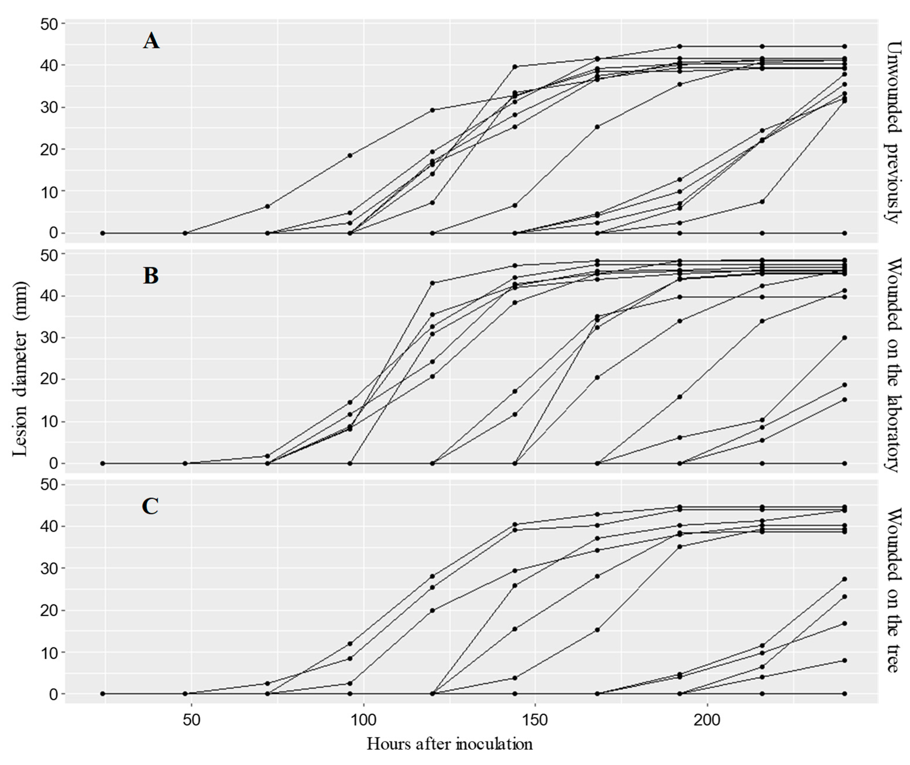
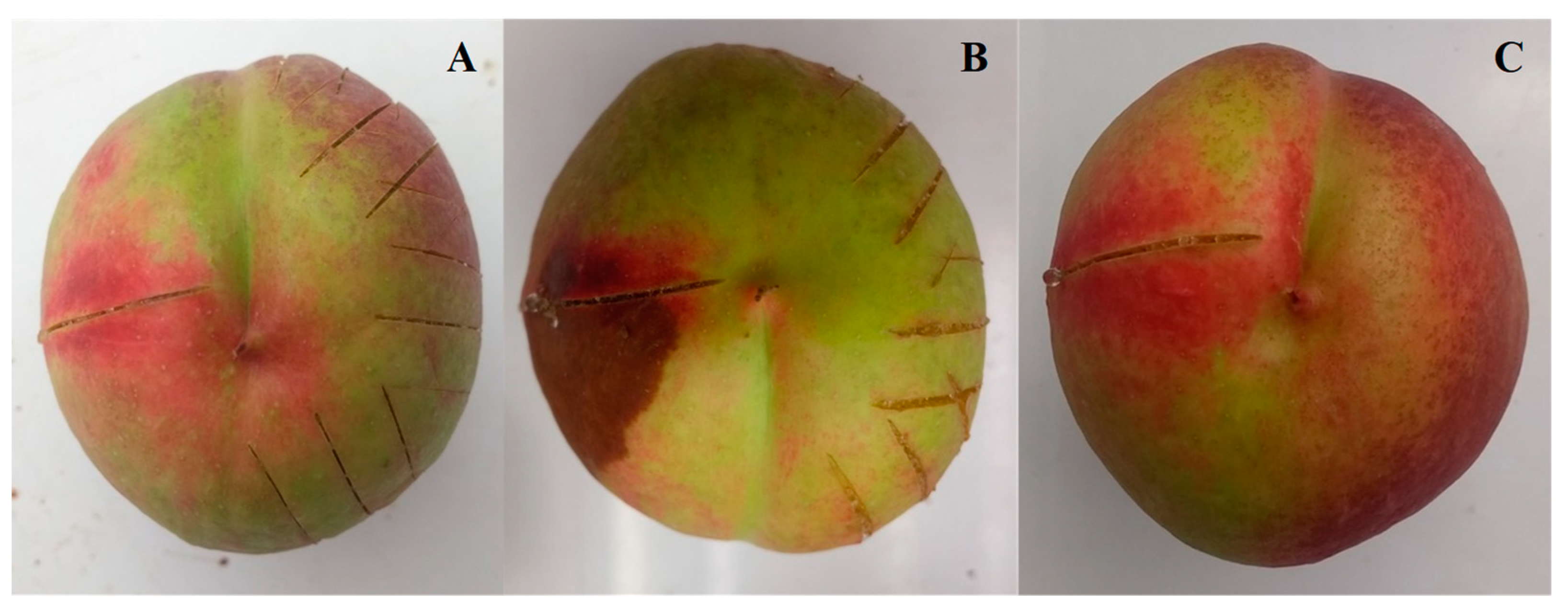
2.3.1. First Experiment: Evaluating a Possible Systemic Reaction to Wounding That May Impact Subsequent M. laxa Infection

To test the existence of a systemic reaction, 60 immature fruits were preconditioned with three different treatments, each with 20 fruits.
Fruits were collected, transported to the laboratory, and placed inside the boxes without wounds for the first treatment (control). For the second treatment, multiple longitudinal wounds with a razor blade were made on one side of the fruit (apex to peduncle) at a depth of no more than 3 mm. For the third treatment, the fruits were marked and wounded in the orchard using the same razor blade method as before, but they were left on the tree for 7 h. They were then harvested, transported to the laboratory, and placed in boxes as with the fruits from the other two treatments.
After 7 h, all fruits (Treatments 1, 2, and 3) were wounded with a razor blade making longitudinal cuts from the apex to the peduncle at a depth not exceeding 3 mm. In the second and third treatments, the fruits were wounded on the side that had not previously been wounded. The inoculation was performed immediately after wounding using a micropipette, depositing 10 µL of inoculum along the wound of each fruit. After that, the boxes were then sealed and placed in the growth chamber.
The incidence and growth kinetics of M. laxa lesions and sporulation were studied. Each fruit was evaluated every 24 h until 240 h after infection (hai). The presence or absence of infection was determined by the appearance of the necrotic lesion that is characteristic of BR. A digital caliper was used to measure the lesion perpendicular to the wound in the largest lesion zone. The presence and absence of M. laxa sporulation were also recorded; when present, the sporulation diameter was measured the same way as the lesions.

2.3.2. Second Experiment: Evaluating the Influence of Fruit on the In Vitro Development of M. laxa

Thirty Petri dishes (5 cm in diameter) containing PDA media were inoculated with 10 µL fungus suspension prepared under the same conditions as for the first experiment. Petri dishes without a lid were separated into three groups of ten and placed in three boxes.
One of the three boxes was sealed hermetically and placed in the growth chamber. Ten immature fruits were placed in the second box, which was then closed and placed in the growth chamber. Ten fruits with multiple wounds (made with a razor blade) were placed in the third box; then, the box was closed and placed under the same conditions as the other two.
The follow-up was conducted daily by visual observations, but the boxes were opened only 168 h after inoculation for the final evaluation, which consisted of two perpendicular measurements of the M. laxa colony for each Petri dish. For the statistical analysis, the mean value of the two measurements of each Petri dish was used, considering each Petri dish as a replication.

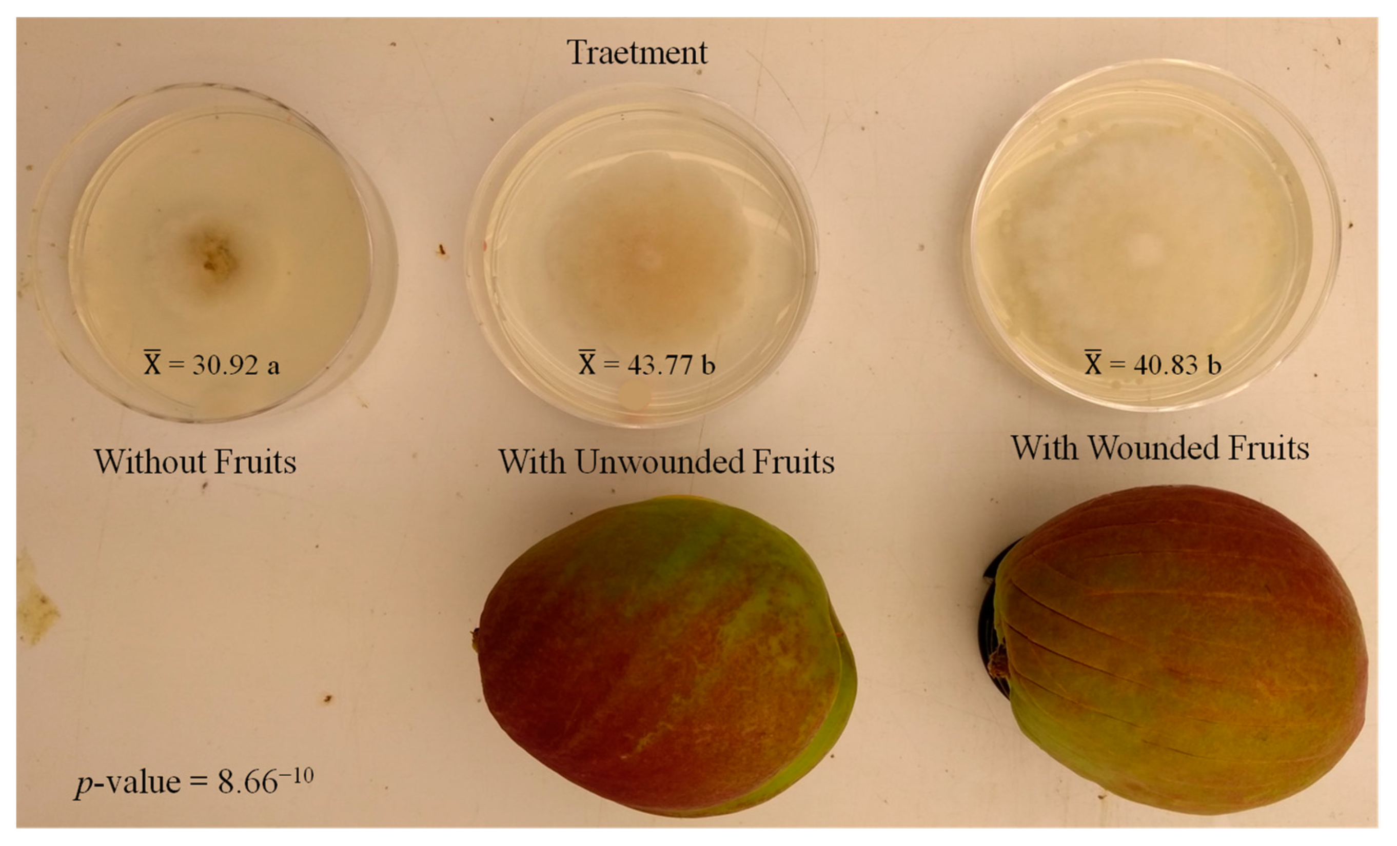
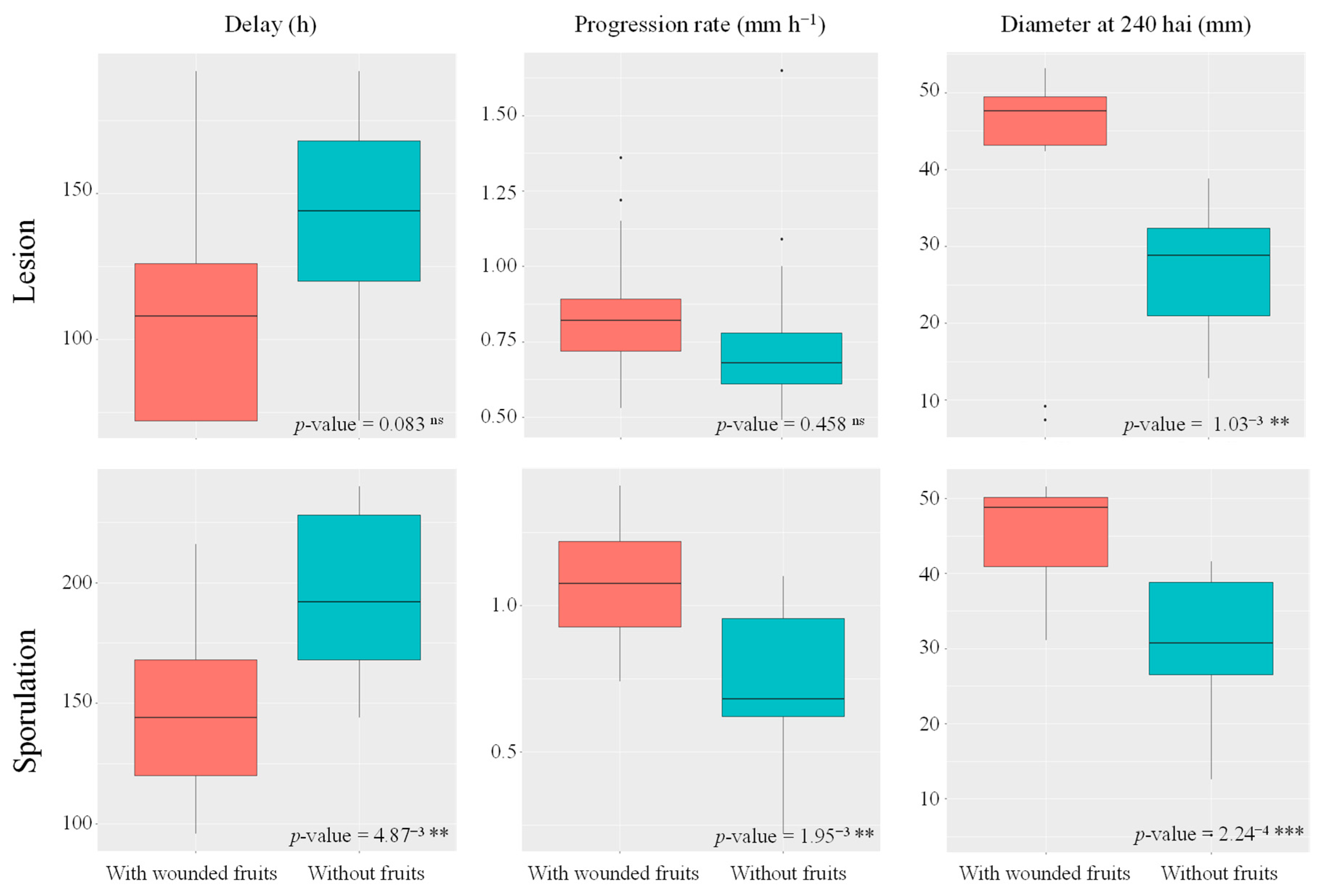
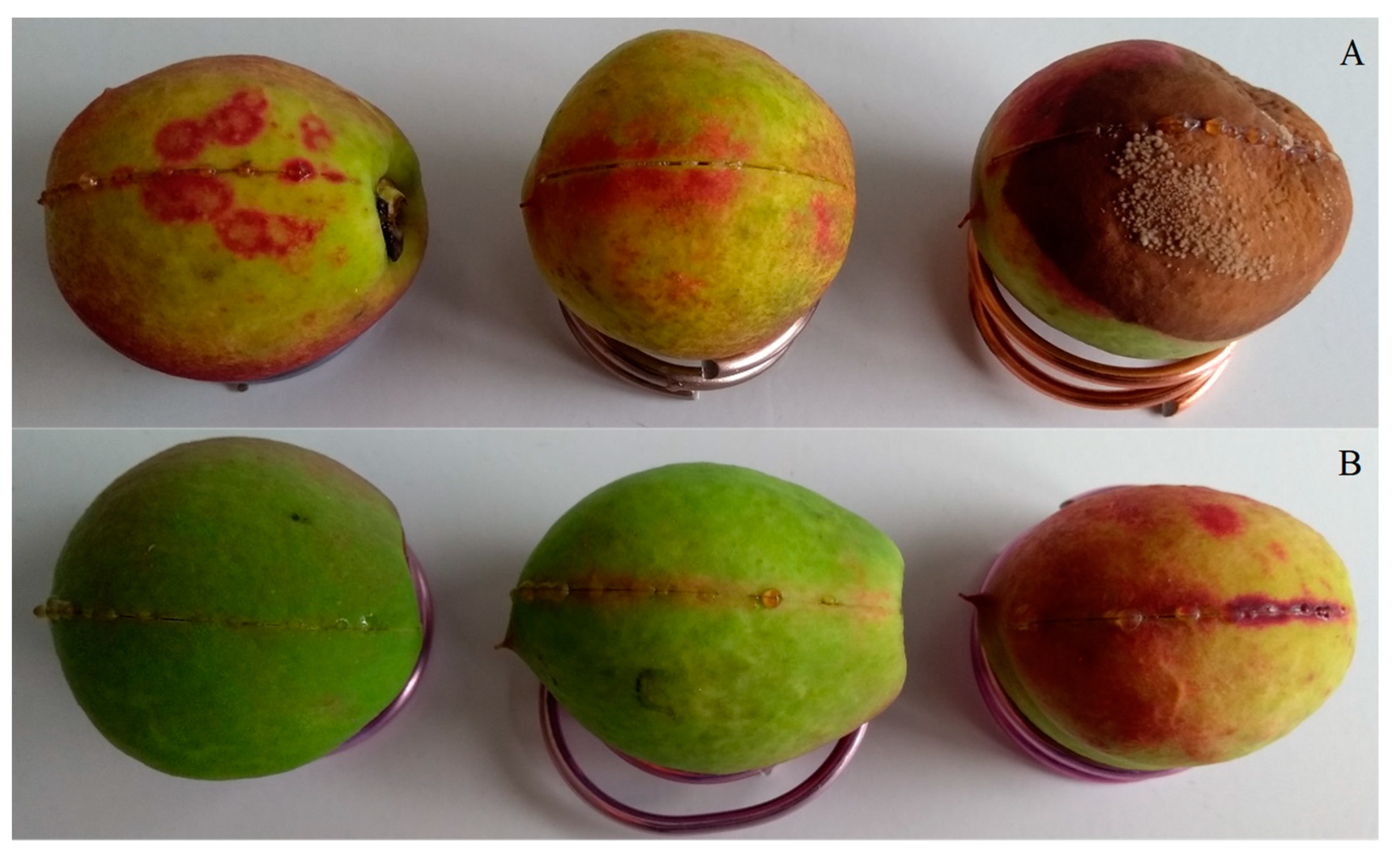
2.3.3. Third Experiment: Evaluating the Influence of the Presence of Wounded Fruits on the Infection of Other Fruits Inoculated with M. laxa

The purpose of this experiment was to determine the susceptibility of fruits inoculated with M. laxa and placed in the same environment as wounded fruits. For that, two treatments were compared. Both treatments consisted in placing five unwounded fruits inside a box. However, for the second treatment, another ten fruits with multiple wounds were added to each box of five unwounded fruits. Fruits were kept in the boxes for 7 h. Four repetitions (boxes) of each treatment were used.
The previously wounded fruits were eliminated after 7 h. The remaining fruits from the two treatments were wounded with a razor blade, making longitudinal cuts from the apex to the peduncle with a depth of no more than 3 mm. Immediately after that, inoculation was performed with a micropipette, depositing 10 µL of inoculum on one wound of each fruit. The hermetically sealed boxes were placed in the growth chamber.
The infection evaluations were carried out in the same way as in the first experiment.

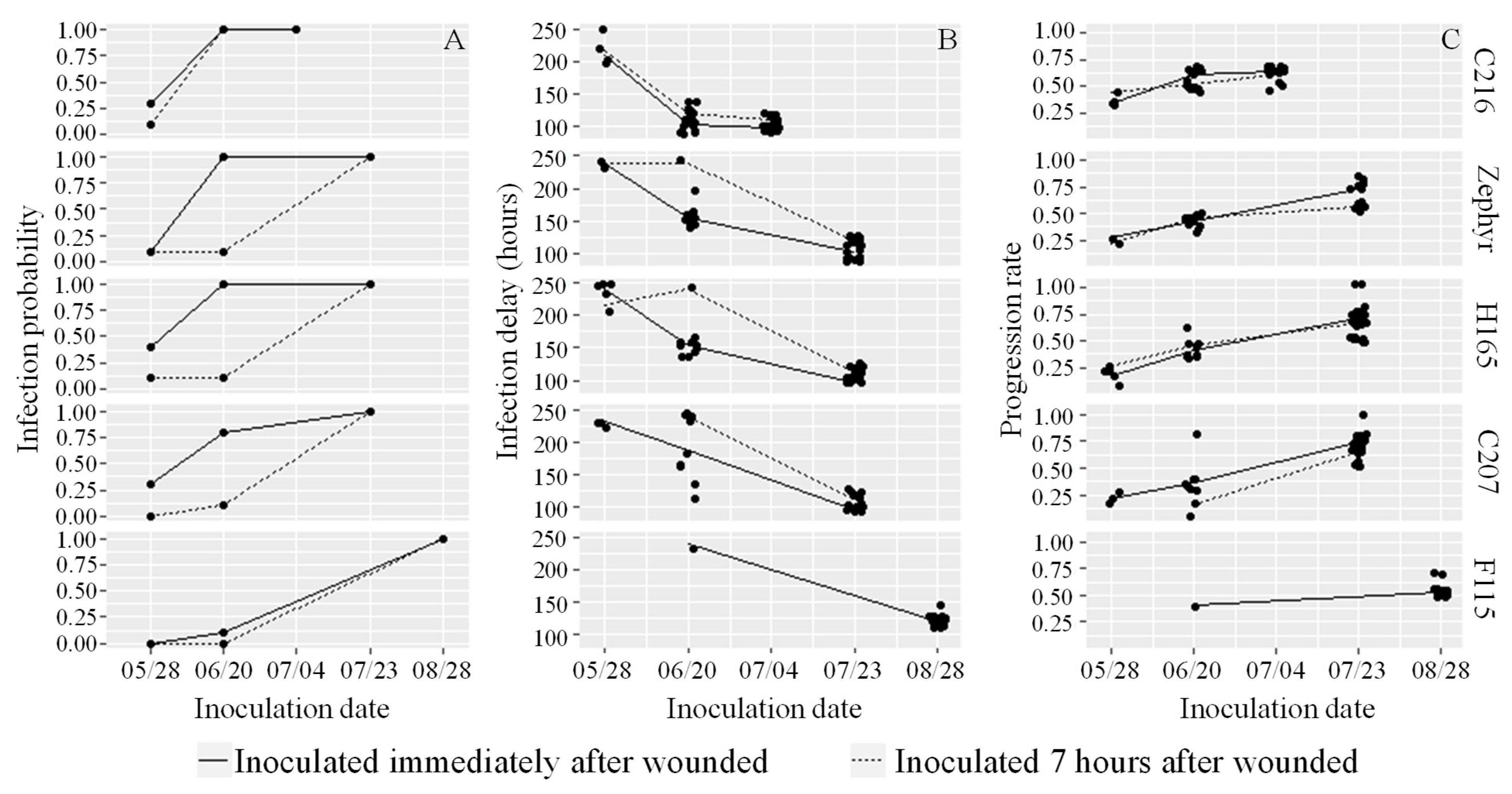
2.3.4. Forth Experiment: Evaluating the Fruit Reaction to Brown Rot at Different Development Stages and Inoculation Times after Wounding

The experimental design was completely randomized, with a triadic arrangement consisting of five nectarine genotypes, two inoculation times—immediately after wounding (IAW) and 7 h after wounding (I7hAW)—and three stages of fruit development (three inoculation dates referred to as date1, date2, and date3). Ten fruits were used for each genotype, inoculation time, and harvest date, with each fruit being considered as a repetition. The first inoculation began on 28 May 2018, corresponding to 11–15 weeks after full bloom (WAFB) when the fruits were at the pit-hardening stage. The second inoculation began on 20 June 2018, at 15–19 WAFB. The third evaluation corresponded to the commercial maturity date of each used genotype. For the early maturing nectarine C216, it was 4 July 2018 (21 WAFB); for the nectarines Zephyr, H165, and C207, 23 July 2018 (23, 21, and 20 WAFB, respectively), and for the late nectarine F115, it was 28 August 2018 (26 WAFB).
The infection evaluations were carried out exactly as described in the first experiment.

2.4. Exploration of Red Reaction

Skin samples from all genotypes were analyzed using high-performance liquid chromatography (HPLC) at the end of the first evaluation (pit hardening) of the fourth experiment (7 June 2018). For each genotype and both inoculation times, skin samples from all fruit without infection symptoms were collected. The samples were taken and pooled from the areas with red reaction. When the red reaction did not occur, samples were taken and pooled in the same quantity and position as the previous ones. The tissues were immediately frozen in liquid nitrogen after sampling and stored at −80 °C. They were then ground to a fine powder and lyophilized for three days. The HPLC analyses were performed at INRAE’s laboratories (GAFL INRAE, Avignon).
The specialized metabolites were extracted from 50 mg of the lyophilized dry powder. The material was homogenized for 1 min in an ULTRA-TURRAX homogenizer (IKA labortechnik, JK Janke & Kunkel) with 8 mL of extraction solution (ethanol 95%), then placed in a rotary shaker (Rotator SB3, Stuart) under controlled environment at 4 °C for 4 h, then centrifuged (Sigma 4K15, 5000 rpm, 5 min, 4 °C). The volume of the supernatant was carefully recovered to prevent contamination with the pellet. The supernatant was collected and placed in a Pyrex tube (12 mL), which was then placed in a Speed Vac Concentrator (SC210A, Thermo Electron Corporation) for solvent evaporation. The resultant residue was dissolved in 1000 μL of 100% methanol (Prolabo Hypersolv Chromanorm), filtered (membrane PTFE 0.45 μm), and collected into an autosampler vial of 1.5 mL for HPLC analysis and stored at −20 °C until analysis.
Extracts were analyzed using an HPLC system (Shimadzu Prominence) equipped with a reverse-phase C18 column (Merck Superspher RP18 end-capped) coupled with a photodiode array detector operated by Shimadzu software (CBM-20A/20Alite, LC Solutions). The extracted content was determined using a method developed for measuring specialized metabolites in peach and nectarine fruits. The mobile phases were dissolved in acidified pH 2.6 water (with H3PO4) and 100% methanol (Prolabo Hypersolv Chromanorm). The HPLC solvents were programmed based on the polarity of the extracted molecules, starting with 97% water and 3% methanol to extract the most polar molecules and ending with 0% water and 100% methanol to extract the most apolar molecules (triterpenoids). A 10 μL aliquot of the filtered extract was injected, and the column was kept at 30 °C.
The methanol extracts were analyzed simultaneously for free forms of triterpenoids (210 nm), flavan-3-ols and flavanones (280 nm), hydroxycinnamic acids and derivate forms of triterpenoids (315 nm), flavonols (350 nm), and anthocyanidins (520 nm). The compounds were characterized according to their UV lambda maximum, retention time, co-chromatography with known standards, and data previously obtained by mass spectrometry.

2.5. Statistical Analysis and Graphic Representations

The first three experiments were analyzed using a completely randomized design, with each fruit or Petri dish as replication. The experimental design of the fourth experiment was previously described in Section 2.3.4. The probability of infection was estimated by the percentage of fruit with lesions on each inoculation date. Delay was calculated by the time at which the infection was first observed (in hours), and the progression rate was calculated by the maximum progression of infection between two observation dates. For the sporulation data, the same parameters were calculated (percentage of fruit with sporulation, delay, and progression rate).
Graphical representations and statistical analyses were performed using R (version 4.0.5) [46]. Results of infection parameters (delay, progression rate, diameter for the lesion, and sporulation) underwent a one-way analysis of variance with an F-test to test the effect of treatment. In the fourth experiment, the effects on the infection parameters of genotype, inoculation time, development stage, and their interactions were tested with a multi-factor analysis of variance with an F-test. Regarding concentration of compounds analyzed by HPLC, the statistical analyses were performed using the peak areas from HPLC analyses. Data from the different genotypes were analyzed together to compare red reaction and reactionless zones: the five samples that presented red spots (C216 IAW, Zephyr IAW, H165 IAW, C207 IAW, and C207 I7hAW) and the five that did not show reaction (C216 I7hAW, Ze I7hAW, H165 I7hAW, F115 IAW, and F115 I7hAW) were taken as repetitions. A one-way analysis of variance was used with an F-test to compare the mean differences between red reaction and areas without reaction for concentration of each individual compound and the family contents. Treatments were compared using a p-value of 0.01 to indicate significance. The package ggplot2 was used to generate graphs.

3. Results

3.1. First Experiment: Evaluating a Possible Systemic Reaction to Wounding That May Impact Subsequent M. laxa Infection

The progression of the lesion diameter over time displayed slight differences between the treatments (Figure 1 and Figure 2). However, the infection probability at 240 hai reached different levels, depending on the treatments: 65%, 70%, and 50%, respectively, for fruits that were not previously wounded (Figure 1A), fruits previously wounded on one side in the laboratory (Figure 1B), and fruits wounded in the tree (Figure 1C).
M. laxa sporulation was observed in 55% of the fruits that had not previously been wounded, 60% of the fruits that had previously been wounded on one of their sides in the laboratory, and 30% of the fruits that had previously been wounded in the tree (Supplementary Figure S1). Despite these tendencies, it is not possible to form a conclusion on a treatment effect of the infection probability. Indeed, the integrative nature of these traits (infection and sporulation probabilities) prevents comparative statistics. However, the data on individual fruit traits (delay, progression rate, and final diameter (240 hai)) obtained for both lesion and sporulation progressions were not significantly different among treatments (Figure 2).

3.2. Second Experiment: Evaluating the Influence of Fruit on M. laxa In Vitro Development

When M. laxa growth was recorded under in vitro conditions, there were significant differences (p-value 8.66−10) between the tested treatments. The colony diameter was larger in treatments where the Petri plates were incubated together with fruits (43.77 and 40.83 mm vs. 30.92 mm, mean values), without significant differences between the wounded and unwounded fruits (Figure 3).

3.3. Third Experiment: Influence of the Presence of Wounded Fruits on the Infection of Other Fruits Inoculated with M. laxa

The presence of wounded fruits in the vicinity of inoculated fruits increased the BR infection and M. laxa sporulation. When fruits were inoculated after 7 h in a closed box with other wounded fruits, 80% showed BR infection (Figure 4A). In the case of fruits which were inoculated without being in the presence of other wounded fruits, 65% of the fruits presented infection (Figure 4B). Figure 4C,D show that M. laxa sporulation occurred in 70% and 55% of the fruits, respectively.
The delay and progression rate of the lesion were not significantly different between the two treatments (p > 0.01). However, the final diameter of the lesion (240 hai) was considerably higher (p-value 1.03−3) for fruits that had previously been maintained with wounded fruits for 7 h (Figure 5).
In the case of sporulation, the three calculated parameters revealed significant differences (p ≤ 0.01). The delay proved to be longer without exposure to wounded fruits prior to the inoculation, suggesting that the M. laxa took longer to sporulate on these fruits. In turn, the progression rate and the final diameter (240 hai) were greater when the fruits were first maintained for 7 h alongside wounded fruits, indicating that M. laxa sporulation occurred faster in this condition and reached a larger final diameter (Figure 5).

3.4. Fourth Experiment: Fruit Reaction to Brown Rot at Different Development Stages and Inoculation Times after Wounding

On the first inoculation date (May 28), 14% of the small immature fruit (26.7 g of average mass, 11–15 weeks after full bloom) were infected, and 9% displayed sporulation of M. laxa (Supplementary Figure S2) at the end of the evaluation period (240 hai). On the second inoculation date (20 June), with intermediate-size immature fruit (46.7 g of average mass, 15–19 weeks after full bloom), 100% of the C216 fruit showed M. laxa infection and sporulation, regardless of the inoculation time. In contrast, the Zephyr, H165, and C207 genotypes had a higher number of fruits with lesions and sporulation when they were inoculated immediately after wounding (IAW). Only one fruit with BR lesions and fungus sporulation was found in the F115 genotype. On the third inoculation date, which corresponded to the harvest date of each genotype, M. laxa infection and sporulation were found in 100% of the fruits.
The infection probability on the first inoculation date ranged between 0 to 0.40 and 0 to 0.10 (Figure 6A) for the IAW and I7hAW inoculation conditions, respectively. The highest variability was observed on the second inoculation date, with the probability of infection ranging between 0.10 and 1.00 and between 0 and 1.00, for IAW and I7hAW, respectively. When mature fruits were inoculated, the infection probability was 1.00 for all genotypes and both inoculation conditions.
Except for the F115 genotype, the infection delay for inoculation IAW was between 200 and 240 h for the first inoculation date, between 24 and 200 h for the second, and between 24 and 120 h for the third (Figure 6B). The delay for fruit inoculated I7hAW ranged from 216 to 240 h for the first inoculation, between 24 and 240 h for the second, and between 24 and 144 h for the third inoculation of the C216, Zephyr, and H165 genotypes. In the case of the F115 genotype, no fruit was infected on the first inoculation date, and on the second, only one fruit of the group inoculated IAW showed symptoms of the disease.
The progression rate was between 0.10 to 0.40 on the first inoculation date, between 0.10 to 0.70 on the second inoculation date, and between 0.40 to 1.10 on the inoculation at the mature stage (Figure 6C).
The sporulation probability, delay, and infection rate had a similar tendency as the lesion data and are provided in Supplementary Figure S3.
The effect of different inoculation times on the lesions (LD) and sporulation (SpoD) proved to be the same regardless of observation time (ANOVA analysis); thus, only the analysis of the latest evaluation (240 hai) is presented. The triple interaction (genotype–inoculation time–development stage) was significant only for the sporulation delay (p-value = 0.0008). Among the double interactions, genotype–inoculation time was not significant for any of the variables tested (p-value > 0.01); the genotype–development stage was significant for all variables (p-value < 0.01), except sporulation rate (p-value = 0.1491); and the inoculation time–development stage interaction was significant for infection delay, LD, and SpoD. Analyzing the main factors, it was observed that the inoculation time was not significant (p-value > 0.01) for any of the variables tested since its effect depended largely on the development stage. The genotype was significant for all variables except for the lesion rate, and the development stage was highly significant (p-value < 0.0001) for all variables (Table 2).
On the first and last inoculation dates, no significant difference (p-value > 0.01) was observed between the two different inoculation times. In contrast, on the second date, the LD and SpoD in fruit inoculated IAW were larger (p-value < 0.01) than in fruit inoculated 7hAW. These differences can be attributed to the Zephyr, C207, and H165 genotypes. Similarly, the infection delay decreased as the fruit grew. On the second date, significant differences were found between inoculation times (p-value < 0.01), with a greater delay in inoculations performed 7 h after wounding (Figure 6B).

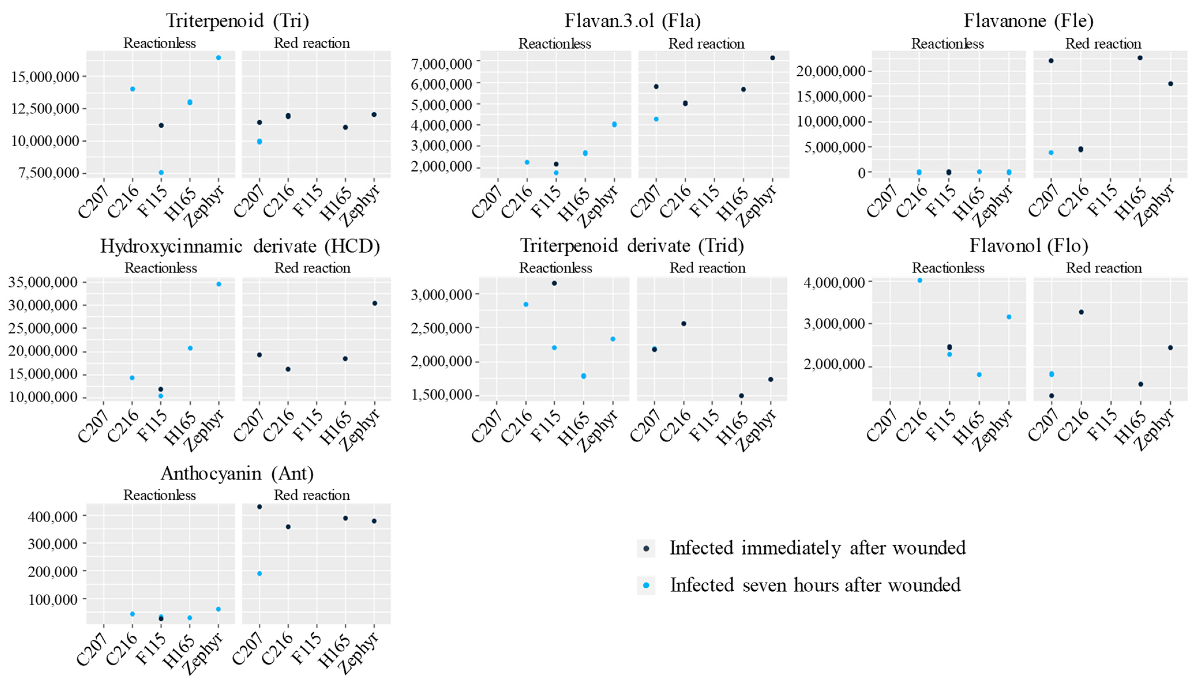
3.5. Exploration of Compounds Present in Red Reaction Zones

Red reactions were detected in the first and fourth experiments. In the first experiment, red reactions were associated with double stress (wounding plus M. laxa inoculation) (Figure 7). This reaction was not detected in the absence of inoculation in both cases when the multiple wounding was performed in the laboratory and on the tree (Figure 3A,B; see the right side of the fruit).
In the fourth experiment, two inoculation times were performed at three developmental stages using the Zephyr cultivar as an example (Supplementary Figure S4). It is noteworthy that red reactions were observed on the first date only. In the particular case of Zephyr, the red spots were associated only with the inoculation IAW and were located around the wound and at some scattered points near it. The same was observed in the selections H165 and C216, whereas the genotype C207 presented red spots in fruit inoculated at both inoculation times (IAW and 7hAW). However, genotype F115 did not show any red reaction in either of the two inoculation times. It should be emphasized that this genotype did not present any BR-infected fruit on the first inoculation date and only a few on the second date (Figure 8).
HPLC analyses of tissue from red reaction zones revealed the presence of 44 phenolic and terpenoid compounds, four of which belonged to the triterpenoid family (Tri), while five were flavan-3-ols (Fla), six flavanones (Fle), ten hydroxycinnamic derivatives (HCD), eleven triterpenoid derivatives (Trid), six flavonols (Flo), and two anthocyanins (Ant) (Table 3).
It should be noted that some compounds were only found in the red reaction samples, including six compounds of the flavanone family and three hydroxycinnamic derivatives (HCD1, HCD3, and HCD4). Furthermore, six other compounds showed significant differences (p-value < 0.01), with a higher proportion in the samples that had the red reaction. These compounds were four flavan-3-ols (Fla1, Fla2, Fla3, and catechin), a hydroxycinnamic derivative (5-p-coumaroylquinic acid), and an anthocyanin (cyanidin-3-glucoside). On the other hand, a hydroxycinnamic derivative (cis-neochlorogenic acid) was significantly (p-value = 0.00919) lower in the red reactions samples (Table 3). Supplementary Figure S5 shows an HPLC chromatogram of the red reaction and nonreaction samples using Zephyr as an example.
When the peak areas of the compounds detected according to their family were added together, the flavan-3-ol and anthocyanin stand out as being in higher proportion in the red reactions, regardless of genotype, besides the flavanone only present in this condition (Table 3 and Figure 9). Supplementary Figure S6 shows boxplot graphs comparing the contents of each compound between the samples for the red reaction and reactionless zones. Two of the six flavanones featured large peak heights. They were identified as eriodictyol-7-glucoside and naringenin-7-glucoside (syn.: prunin) using pure standards.

4. Discussion

4.1. Wounding Has No Systemic Effect on M. laxa Infection

Several studies reported that wounds may increase pathogen resistance [20,21,22,32,41]. However, according to our results, the wounds do not appear to have a systemic effect on the whole fruit. Indeed, although the lowest values were associated with the treatment of the wounds on the tree, no significant differences were obtained between the treatments. Therefore, if wounding increases resistance to pathogens, it may only be at the site of the wound, preventing pathogen entry and activating local defense mechanisms [22].

4.2. Fruit Presence Enhances M. laxa Growth Both In Vitro and in Fruit

The presence of fruits near the culture in Petri dishes and fruits stored for 7 h near other fruits, wounded or not, resulted in a higher growth of M. laxa in vitro. M. laxa appears to be able to detect the presence of fruit. It most likely recognizes some of the volatile compounds released by the fruit. This theory is supported by reports on how pathogens perceive host volatile compounds [47,48,49,50].
Wilson et al. [51] investigated 16 natural volatile fruit compounds for the control of M. fructicola and B. cinerea in peach fruit during the postharvest period, some of which had an inhibitory effect on the germination and growth of the pathogens such as benzaldehyde, methyl salicylate, and ethyl benzoate. On the other hand, other compounds, such as d-limonene, favored the fungus, increasing the percentage of spore germination. In this case, the volatile compounds were applied as isolated compounds and at known concentrations; however, it is important to point out that the fruit synthesizes a great complexity of volatile compounds [52,53] that may evolve with fruit development. The association of different volatile compounds in different concentrations certainly affects the influence of each independent compound on the pathogen, probably canceling out opposite effects. There are reports of this perception in other species modulating plant–pathogen interactions either with host or nonhost plants [34,48,49,54]. However, the type of wounding may have an effect on the volatile compounds released, but no literature on this was found.

4.3. A Delay of 7 h between Wounding and Subsequent Inoculation Decreases Infection in Immature Fruit

To evaluate if a delay between wounding and inoculation influences M. laxa fruit susceptibility, we chose to experiment at three different dates. Susceptibility to Monilinia spp. infection is high during the early stages of fruit development, decreases during the green fruit stages (pit hardening), and then increases again as the fruit matures, being the highest during the preharvest ripening stage [2,6,8,55,56,57]. In agreement with these observations from the literature, in our experiment, the fruits were resistant at date1 (corresponding to Stage 2, pit hardening), and susceptible at date3 (harvest time). Consequently, the date2 proved to be the most efficient for our analysis, with a significant difference in infection parameters between the two inoculation times (IAW and 7hAW) for three genotypes (Zephyr, H165, and C207). The reduction in infection symptoms may be associated with a rapid and efficient defense response of the fruit 7 h after wounding. For the two other genotypes, C216 and F115, the fruit stage was probably shifted: C216 (early harvest) probably had already entered the fruit ripening period, and F115 (late harvest) was still in the green fruit stage.
Microcracks and wounds can heal in minutes to several hours, depending on the degree of injury, the fruit’s physiological stage, and the environmental conditions [58,59]. It has already been shown, in other species, that healing can induce defense pathways [23,24,37,60,61]. In the inoculation performed 7 h after wounding, the injury may have begun to heal, preventing the conidia penetration into the fruit and/or triggering the resistance mechanisms.

4.4. Specialized Metabolites Detected in Red Reaction Zones

The red reaction appears to be associated with double stress (wounding and immediate infection) and could be related to the fruit’s active resistance mechanisms against the pathogen. An HPLC analysis of the tissues showing red reactions revealed changes in the phenolic compounds, including six compounds from the flavanone family (Fle1, Fle2, Fle3, Fle4, E7glu, and N7glu) and three hydroxycinnamic derivatives (HCD1, HCD3, and HCD4). They were either not present in the samples without a red reaction or were present in such low concentrations that they were impossible to measure. In addition to these compounds, four other flavan-3-ols (Fla1, Fla2, Fla3, and catechin), a hydroxycinnamic derivative (5-p-coumaroylquinic acid), and an anthocyanin (cyanidin-3-glucoside) were present in a significantly larger proportion in samples isolated from red spots.
Guidarelli et al. [8] performed a microarray-based transcriptome analysis to compare the expression of genes between susceptible (ripening phase) and resistant fruit (green fruit, at the pit-hardening stage). They observed that genes related to flavonoids and phenylpropanoids were differentially expressed between the two stages, supporting the fact that some of these metabolites may be present constitutionally in the fruit at some stages. A transcriptomic analysis after infection and after wounding plus infection would provide more information on the role of these metabolites in the fruit response to M. laxa.
It is worth noting that there was no change in the triterpenoids, triterpenoid derivatives, or chlorogenic acid, the major phenolic compounds in nectarine skin. Indeed, Lino et al. [45] showed that the terpenoids and derivatives present on the nectarine surface accumulate during the second stage of fruit development when the fruit is resistant. In the same way, Lee et al. [62] demonstrated the role of chlorogenic acid in inhibiting appressorium formation of germinated M. fructicola conidia and inhibiting cell-wall degradation by changes in the redox environment. However, in our study, these compounds were not modified in red spots. They may not be involved in the response to the double stress provoking red reactions.
The role of E7glu and N7glu, which were only found in significant amounts in the red reaction zones, had already been reported in a number of studies in several fruit species, such as apple and pear leaves [63,64,65], Prunus davidiana stems [66], tomato fruit cuticles [67], several Citrus species [68,69,70,71,72], and even in peach buds [73]. However, this is the first study reporting these two compounds in nectarine fruit. Studying the effect of dioxygenase inhibitors on the resistance-related flavonoid metabolism of apples and pears, Roemmelt et al. [63] reported changes in the pattern of flavonoids and phenylpropanoids in apple leaves treated with prohexadione-Ca. More specifically, they observed an increase in the concentration of luteoliflavan, E7glu, N7glu, and HCD. Such changes induced in the flavonoid composition of the leaves correlate with reduced susceptibility to the pathogenic bacterium Erwinia amylovora. The authors postulated that these changes in the flavonoid composition are responsible for the observed pathogen resistance induced by prohexadione-Ca treatment. Among the compounds mentioned by the authors, E7glu, N7glu (=Prunin), and HCD correspond with the compounds we detected only or in a greater proportion in the red reactions. This suggests a resistance-induced response to M. laxa when inoculated simultaneously with the injury. On the other hand, working with the silencing of flavanone-3-hydroxylase in apple trees, Flachowsky et al. [64] were able to increase the accumulation of flavanones but failed to reduce susceptibility to fire blight caused by E. amylovora.
Petkovsek et al. [74] showed that there was a higher content of total hydroxycinnamic acids after the infection of apple leaves with Venturia inaequalis. In our study, the analyzed HCD exhibited various responses. Three of them were specific to the red reaction zones, but they were in low amounts. One of them, the pCQ, was increased, whereas the caffeoylquinic acids, the major subfamily of HCD in our samples, did not present any changes. Only certain specific compounds of a family can be involved in a defense response.
Regarding the flavan-3-ol family, four of the five compounds analyzed showed a significant increase in the red samples. Petkovsek et al. [74] reported an increase in catechin and epicatechin (monomeric flavan-3-ols) in apple leaves after infection with Venturia inaequalis, while Procyanidin B2 (oligomeric flavan-3-ol) decreased. They explained that it was a consequence of the increase in monomeric flavan-3-ols—precursors of oligomeric flavan-3-ols. In our results, Procyanidin B1 did not show any changes, while the other flavan-3-ols exhibited significant variations.
Finally, the anthocyanin family also showed an increase in red zones. Anthocyanins are most commonly associated with abiotic stress, but some authors have linked their involvement to biotic stress [75,76]. Shaefer [76] observed that the anthocyanin content increased when V. vinifera was infected with Botrytis cinerea and demonstrated that cyanidin-3-glucoside had a strong antifungal effect in vitro.
The case of these red reactions is particularly interesting to investigate in the light of the preliminary results obtained. The compounds detected are in fact already known to be involved in defense reactions. Analyses of volatile compounds coupled with gene expression should make it possible to increase our understanding of the mechanisms involved in the response to this double stress (wounding plus infection) and identify genes, metabolic pathways, and compounds that may curb infection. Similarly, such multi-level analyses, linking metabolites, volatile compounds, and gene expression, should be performed to compare fruit samples immediately after wounding and seven hours after wounding.

5. Conclusions

The present study provides some elements related to nectarine fruit–M. laxa interactions, particularly in the case of wound-induced fruit defense. We found no evidence of a systemic effect of injury on the whole fruit. The wounds caused in another part of the fruit (aside from the inoculation spot) did not affect the susceptibility of the nectarine fruits to brown rot. We observed that M. laxa could perceive the fruit presence resulting in accelerated in vitro growth. In addition, the presence of wounded fruits in the proximity of inoculated fruit increased the brown rot susceptibility. These results suggest the intervention of biochemical interplay between the fruit and the fungi, triggered by prior injury. Finally, based on the findings, it is hypothesized that flavonoid and phenylpropanoid compounds may influence the fruit response to M. laxa infection.
Further exploration of these host–pathogen interactions related to mechanical stress, such as wounding, is essential for understanding resistance mechanisms.

Supplementary Materials

The following supporting information can be downloaded at: https://www.mdpi.com/article/10.3390/agronomy13051235/s1. Figure S1: Brown rot lesion growth in Zephyr immature fruits wounded and immediately inoculated with Monilinia laxa in three conditions: unwounded previously (A); previously wounded on the laboratory in one side of the fruit (B); and previously wounded on the tree in one side of the fruit (C). Figure S2: (A) Lesion and (B) sporulation diameter in five nectarine genotypes inoculated with M. laxa immediately after wounding (gray line) and inoculated 7 h after wounding (black line), in three stages of fruit development (fourth experiment): date1 = May 28, 11–15 weeks after full bloom (WAFB); date2 = June 20, 15–19 WAFB; and date3 = corresponds to the ripening date of each genotype (20–26 WAFB). Figure S3: Monilinia laxa sporulation probability (A), sporulation delay (B), and sporulation rate (C) in five nectarine genotypes inoculated immediately after wounding (IAW) and inoculated 7 h after wounding (I7hAW), in three stages of fruit development (fourth experiment): first date (May 28), 11 to 15 weeks after full bloom (WAFB); second date (June 20), 15–19 WAFB; and third, the ripening date of each genotype (20 to 26 WAFB). Figure S4: Fruit from Zephyr infected by M. laxa, inoculated immediately after wounding (IAW) or 7 h after wounding (I7hAW), at three fruit development stages (fourth experiment): date1 = May 28, 15 weeks after full bloom (WAFB); date2 = June 20 (19 WAFB); and date3 = July 23, ripening stage (21 WAFB). Photos were taken 240 h after inoculation. Figure S5: HPLC chromatogram (280 nm) of immature Zephyr fruit (15 weeks after full bloom) with red reaction (wounded + Monilinia laxa immediate inoculation) and no red reaction (wounded + M. laxa inoculation seven hours after wounding). Marked peaks correspond to compounds that differed significantly between the two samples. Abbreviations: Fla, Flavan-3-ol; Cat, Catechin; Fle, Flavanone; HCD, Hydroxycinnamic derivative; pCQ, 5-p-Coumaroylquinic acid; and c3CQ, cis-Neochlorogenic acid. Figure S6: Boxplot of the HPLC peak area for compounds identified with no reaction (Not_red) and red reaction (Red) in immature fruits (11–15 weeks after full bloom), 2018 season. Abbreviations of compounds in Table 2.

Author Contributions

Conceptualization, B.Q.-T.; methodology, M.-N.C. and V.S.; investigation, M.D., M.-N.C. and V.S.; software, M.D. and B.Q.-T.; data curation M.D.; writing—original draft preparation, M.D.; writing—review and editing, B.Q.-T., M.d.C.B.R., M.-N.C. and M.D.; supervision, B.Q.-T.; project administration, B.Q.-T.; funding acquisition, B.Q.-T., M.D. and M.d.C.B.R. All authors have read and agreed to the published version of the manuscript.

Funding

This research was funded by the Coordination for the Improvement of Higher Education Personnel (CAPES-Brazil), awarding the first author’s doctoral scholarship, and by INRAE (GAFL, Avignon, France).

Data Availability Statement

Not applicable.

Conflicts of Interest

The authors declare no conflict of interest.

References

  1. Ogawa, J.M.; Zehr, E.I.; Bird, G.W.; Ritchie, D.F.; Uriu, K.; Uyemoto, J.K. Compendium of Stone Fruit Diseases, 1st ed.; The American Phytopathological Society: Saint Paul, MN, USA, 1995; 98p, ISBN 9780890541746. [Google Scholar]
  2. Adaskaveg, J.E.; Schnabel, G.; Forster, H. Diseases of peach caused by fungi and fungal-like organisms: Biology, epidemiology and management. In The Peach: Botany, Production and Uses; Layne, D.R., Bassi, D., Eds.; CAB International: Wallingford, UK, 2008; pp. 352–406. ISBN 9781845933869. [Google Scholar] [CrossRef]
  3. Hu, M.J.; Cox, K.D.; Schnabel, G.; Luo, C.X. Monilinia species causing brown rot of peach in China. PLoS ONE 2011, 6, e24990. [Google Scholar] [CrossRef] [PubMed]
  4. May-De-Mio, L.L.; Moreira, L.M.; Monteiro, L.B.; Justiniano Júnior, P.R. Infection of Monilinia fructicola in budding stages and incidence of brown rot on fruits in two peach production systems. Trop. Plant Pathol. 2008, 33, 227–234. [Google Scholar] [CrossRef]
  5. May-De-Mio, L.L.; Garrido, L.R.; Ueno, B.; Fajardo, T.V.M. Doenças da cultura do pessegueiro e métodos de controle. In Pessegueiro; Raseira, M.C.B., Pereira, J.F.M., Carvalho, F.L.C., Eds.; Embrapa: Brasília, Brazil, 2014; pp. 355–432. ISBN 978-85-7035-371-9. [Google Scholar]
  6. Mari, M.; Casalini, L.; Baraldi, E.; Bertolini, P.; Pratella, G.C. Susceptibility of apricot and peach fruit to Monilinia laxa during phenological stages. Postharvest Biol. Technol. 2003, 30, 105–109. [Google Scholar] [CrossRef]
  7. Gell, I.; De Cal, A.; Torres, R.; Usall, J.; Melgarejo, P. Relationship between the incidence of latent infections caused by Monilinia spp. and the incidence of brown rot of peach fruit: Factors affecting latent infection. Eur. J. Plant Pathol. 2008, 121, 487–498. [Google Scholar] [CrossRef]
  8. Guidarelli, M.; Zubini, P.; Nanni, V.; Bonghi, C.; Rasori, A.; Bertolini, P.; Baraldi, E. Gene expression analysis of peach fruit at different growth stages and with different susceptibility to Monilinia laxa. Eur. J. Plant Pathol. 2014, 140, 503–513. [Google Scholar] [CrossRef]
  9. Raseira, M.C.B.; Franzon, R. Melhoramento genético. In Pessegueiro; Raseira, M.C.B., Pereira, J.F.M., Carvalho, F.L.C., Eds.; Embrapa: Brasília, Brazil, 2014; pp. 57–72. ISBN 978-85-7035-371-9. [Google Scholar]
  10. Fresnedo-Ramírez, J.; Famula, T.R.; Gradziel, T.M. Application of a Bayesian ordinal animal model for the estimation of breeding values for the resistance to Monilinia fruticola (G.Winter) Honey in progenies of peach [Prunus persica (L.) Batsch]. Breeding Sci. 2017, 67, 110–122. [Google Scholar] [CrossRef]
  11. Oliveira Lino, L.; Pacheco, I.; Mercier, V.; Faoro, F.; Bassi, D.; Bornard, I.; Quilot-Turion, B. Brown rot strikes Prunus fruit: An ancient fight almost always lost. J. Agric. Food Chem. 2016, 64, 4029–4047. [Google Scholar] [CrossRef]
  12. Fu, W.; Burrell, R.; Linge, C.S.; Schnabel, G.; Gasic, K. Breeding for brown rot (Monilinia spp.) tolerance in Clemson University Peach Breeding Program. J. Am. Pom. Soc. 2018, 72, 94–100. [Google Scholar] [CrossRef]
  13. Martínez-García, P.J.; Parfitt, D.E.; Bostock, R.M.; Fresnedo-Ramírez, J.; Vazquez-Lobo, A.; Ogundiwin, E.A.; Gradziel, T.M.; Crisosto, C.H. Application of genomic and quantitative genetic tools to identify candidate resistance genes for brown rot resistance in peach. PLoS ONE 2013, 8, e78634. [Google Scholar] [CrossRef]
  14. Pacheco, I.; Bassi, D.; Eduardo, I.; Ciacciulli, A.; Pirona, R.; Rossini, L.; Vecchietti, A. QTL mapping for brown rot (Monilinia fructigena) resistance in an intraspecific peach (Prunus persica L. Batsch) F1 progeny. Tree Genet. Genomes 2014, 10, 1223–1242. [Google Scholar] [CrossRef]
  15. Baró-Montel, N.; Eduardo, I.; Usall, J.; Casals, C.; Arús, P.; Teixidó, N.; Torres, R. Exploring sources of resistance to brown rot in an interspecific almond × peach population. J. Sci. Food Agric. 2019, 99, 4105–4113. [Google Scholar] [CrossRef] [PubMed]
  16. Scariotto, S.; Dini, M.; Raseira, M.C.B.; Santos, J. Estimates of genetic parameters for brown rot resistance in Prunus persica. Acta Hort. 2021, 1304, 289–297. [Google Scholar] [CrossRef]
  17. Dini, M.; Scariotto, S.; Raseira, M.C.B.; Ueno, B. Heritability and segregation of resistance to brown rot in peach fruits. Acta Hort. 2021, 1304, 339–346. [Google Scholar] [CrossRef]
  18. Dini, M.; Raseira, M.C.B.; Scariotto, S.; Ueno, B. Breeding peaches for brown rot resistance in Embrapa. Agronomy 2022, 12, 2306. [Google Scholar] [CrossRef]
  19. Tosetti, R.; Tardelli, F.; Tadiello, A.; Zaffalon, V.; Giorgi, F.M.; Guidi, L.; Trainotti, L.; Bonghi, C.; Tonutti, P. Molecular and biochemical responses to wounding in mesocarp of ripe peach (Prunus persica L. Batsch) fruit. Postharvest Biol. Technol. 2013, 90, 40–51. [Google Scholar] [CrossRef]
  20. Zhou, L.; Thornburg, R. Wound-inducible genes in plants. In Inducible Gene Expression; Reynolds, P.H.S., Ed.; CAB International: Wallingford, UK, 1999; pp. 127–158. [Google Scholar]
  21. Bruxelles, G.L.; Roberts, M.R. Signals regulating multiple responses to wounding and herbivores. Crit. Rev. Plant Sci. 2001, 20, 487–521. [Google Scholar] [CrossRef]
  22. Cheong, Y.H.; Chang, H.S.; Gupta, R.; Wang, X.; Zhou, T.; Luan, S. Transcriptional profiling reveals novel interactions between wounding, pathogen, abiotic stress, and hormonal responses in Arabidopsis. Plant Physiol. 2002, 129, 661–677. [Google Scholar] [CrossRef] [PubMed]
  23. Shanker, A.K.; Venkateswarlu, B. Abiotic Stress Response in Plants—Physiological, Biochemical and Genetic Perspectives, 1st ed.; Intech: Rijeka, Croatia, 2011; 360p. [Google Scholar] [CrossRef]
  24. Mostafa, S.; Wang, Y.; Zeng, W.; Jin, B. Plant responses to herbivory, wounding, and infection. Int. J. Mol. Sci. 2022, 23, 7031. [Google Scholar] [CrossRef]
  25. Christopher, M.E.; Miranda, M.; Major, I.T.; Constabel, C.P. Gene expression profiling of systemically wound-induced defenses in hybrid poplar. Planta 2004, 219, 936–947. [Google Scholar] [CrossRef]
  26. Mitsuda, N.; Iwase, A.; Yamamoto, H.; Yoshida, M.; Seki, M.; Shinozaki, K.; Ohme-Takagia, M. NAC transcription factors, NST1 and NST3, are key regulators of the formation of secondary walls in woody tissues of Arabidopsis. Plant Cell 2007, 19, 270–280. [Google Scholar] [CrossRef]
  27. Koo, A.J.K.; Howea, G.A. The wound hormone jasmonate. Phytochemistry 2009, 70, 1571–1580. [Google Scholar] [CrossRef] [PubMed]
  28. Trinidade, I.; Santos, D.; Dalmay, T.; Fevereiro, P. Facing the environment: Small RNAs and the regulation of gene expression under abiotic stress in plants. In Abiotic Stress Response in Plants—Physiological, Biochemical and Genetic Perspectives; Shanker, A.K., Venkateswarlu, B., Eds.; Intech: Rijeka, Croatia, 2011; pp. 113–136. ISBN 978-953-307-672-0. [Google Scholar]
  29. Leide, J.; Hildebrandt, U.; Hartung, W.; Riederer, M.; Vogg, G. Abscisic acid mediates the formation of a suberized stem scar tissue in tomato fruits. New Phytol. 2012, 194, 402–415. [Google Scholar] [CrossRef]
  30. Tsaballa, A.; Nikolaidis, A.; Trikka, F.; Ignea, C.; Kampranis, S.C.; Makris, A.M.; Argiriou, A. Use of the de novo transcriptome analysis of silver-leaf nightshade (Solanum elaeagnifolium) to identify gene expression changes associated with wounding and terpene biosynthesis. BMC Genom. 2015, 16, 504. [Google Scholar] [CrossRef]
  31. Birkenmeier, G.F.; Ryan, C.A. Wound signalling in tomato plants—Evidence that ABA is not a primary signal for defense gene activation. Plant Physiol. 1998, 117, 687–693. [Google Scholar] [CrossRef]
  32. León, J.; Rojo, E.; Sánchez-Serrano, J.J. Wound signalling in plants. J. Exp. Bot. 2001, 52, 1–9. [Google Scholar] [CrossRef] [PubMed]
  33. Delessert, C.; Wilson, I.W.; Van Der Straeten, D.; Dennis, E.S.; Dolferus, R. Spatial and temporal analysis of the local response to wounding. Plant Mol. Biol. 2004, 55, 165–181. [Google Scholar] [CrossRef]
  34. Broekaert, W.F.; Zelauré, S.L.; De Bolle, M.F.C.; Cammue, B.P.A. The role of ethylene in host-pathogen interactions. Annu. Rev. Phytopathol. 2006, 44, 393–416. [Google Scholar] [CrossRef] [PubMed]
  35. Akagi, T.; Ikegami, A.; Yonemori, K. DkMyb2 wound-induced transcription factor of persimmon (Diospyros kaki Thunb.), contributes to proanthocyanidin regulation. Planta 2010, 232, 1045–1059. [Google Scholar] [CrossRef]
  36. Hara, K.; Yagi, M.; Kusano, T.; Sano, S. Rapid systemic accumulation of transcripts encoding a tobacco WRKY transcription factor upon wounding. Mol. Gen. Genet. 2000, 263, 30–37. [Google Scholar] [CrossRef]
  37. Smith, C.M.; Rodriguez-Buey, M.; Karlsson, J.; Campbell, M.M. The response of the poplar transcriptome to wounding and subsequent infection by a viral pathogen. New Phytol. 2004, 164, 123–136. [Google Scholar] [CrossRef]
  38. Chen, Y.Y.; Wang, L.F.; Dai, L.J.; Yang, S.G.; Tian, W.M. Characterization of HbEREBP1, a wound-responsive transcription factor gene in laticifers of Hevea brasiliensis Muell. Arg. Mol. Biol. Rep. 2012, 39, 3713–3719. [Google Scholar] [CrossRef] [PubMed]
  39. Toivonen, P.M.A.; Brummell, D.A. Biochemical bases of appearance and texture changes in fresh-cut fruit and vegetables. Postharvest Biol. Technol. 2008, 48, 1–14. [Google Scholar] [CrossRef]
  40. Chen, J.; He, L.; Jiang, Y.; Kuang, J.; Lu, C.; Joyce, D.; Macnish, A.; He, Y.; Lu, W. Expression of PAL and HSPs in fresh-cut banana fruit. Environ. Exp. Bot. 2009, 66, 31–37. [Google Scholar] [CrossRef]
  41. Su, J.; Tu, K.; Cheng, L.; Tu, S.; Wang, M.; Xu, H.; Zhan, G. Wound-induced H2O2 and resistance to Botrytis cinerea decline with the ripening of apple fruit. Postharvest Biol. Technol. 2011, 62, 64–70. [Google Scholar] [CrossRef]
  42. Dini, M. Resistência à Podridão-Parda no Pessegueiro. Ph.D. Thesis, Programa de Pós-Graduação em Agronomia, Faculdade de Agronomia Eliseu Maciel, Universidade Federal de Pelotas, Pelotas, Brazil, 2019; 237p. [Google Scholar]
  43. Lurol, S.; Tabaries, P.; Buffet, B.; Sobas, M.-A.; Portal, A. Lutte post récolte contre Monilia. Application d´eau chaude sur pêche. InfosCTIFL 2009, 250, 32–36. [Google Scholar]
  44. Lino, L.O.; Confolent, C.; Signoret, V.; Génard, M.; Quilot-Turion, B. Genetic variability in the susceptibility of immature peach fruit to Monilinia laxa is associated with surface conductance but not stomatal density. J. Plant Sci. Phytopathol. 2022, 6, 91–100. [Google Scholar] [CrossRef]
  45. Lino, L.O.; Quilot-Turion, B.; Dufour, C.; Corre, M.-N.; Lessire, R.; Génard, M.; Poëssel, J.-L. Cuticular waxes of nectarines during fruit development in relation to surface conductance and susceptibility to Monilinia laxa. J. Exp. Bot. 2020, 71, 5521–5537. [Google Scholar] [CrossRef]
  46. R Core Team. R: A Language and Environment for Statistical Computing. R Foundation for Statistical Computing, Vienna. 2018. Available online: https://www.R-project.org (accessed on 15 June 2018).
  47. Knogge, W. Molecular basis of specificity in host/fungus interactions. Eur. J. Plant Pathol. 1996, 102, 807–816. [Google Scholar] [CrossRef]
  48. Cowan, M.M. Plant products as antimicrobial agents. Clin. Microbiol. Rev. 1999, 12, 564–582. [Google Scholar] [CrossRef]
  49. Kishimoto, K.; Matsui, K.; Ozawa, R.; Takabayashi, J. Volatile C6-aldehydes and allo-ocimene activate defense genes and induce resistance against Botrytis cinerea in Arabidopsis thaliana. Plant Cell Physiol. 2005, 46, 1093–1102. [Google Scholar] [CrossRef]
  50. Chisholm, S.T.; Coaker, G.; Day, B.; Staskawicz, B.J. Host-microbe interactions: Shaping the evolution of the plant immune response. Cell 2006, 124, 803–814. [Google Scholar] [CrossRef]
  51. Wilson, C.L.; Franklin, J.D.; Otto, B.E. Fruit volatiles inhibitory to Monilinia fructicola and Botrytis cinerea. Plant Dis. 1987, 71, 316–319. [Google Scholar] [CrossRef]
  52. Aubert, C.; Milhet, C. Distribution of the volatile compounds in the different parts of a white-fleshed peach (Prunus persica L. Batsch). Food Chem. 2007, 102, 375–384. [Google Scholar] [CrossRef]
  53. Eduardo, I.; Chietera, G.; Bassi, D.; Rossini, L.; Vecchitti, A. Identification of key odor volatile compounds in the essential oil of nine peach accessions. J. Sci. Food Agric. 2009, 90, 1146–1154. [Google Scholar] [CrossRef]
  54. Lee, H.A.; Lee, H.Y.; Seo, E.; Lee, J.; Kim, S.B.; Oh, S.; Choi, E.; Choi, E.; Lee, S.E.; Choi, D. Current understandings of plant nonhost resistance. Mol. Plant Microbe Interact. 2017, 30, 5–15. [Google Scholar] [CrossRef] [PubMed]
  55. Biggs, A.R.; Northover, J. Early and late-season susceptibility of peach fruits to Monilinia fructicola. Plant Dis. 1988, 72, 1070–1074. [Google Scholar] [CrossRef]
  56. Gradziel, T.M. Changes in susceptibility to brown rot with ripening in three clingstone peach genotypes. J. Am. Soc. Hort. Sci. 1994, 119, 101–105. [Google Scholar] [CrossRef]
  57. Emery, K.M.; Michailides, T.J.; Scherm, H. Incidence of latent infection of immature peach fruit by Monilinia fructicola and relationship to brown rot in Georgia. Plant Dis. 2000, 84, 853–857. [Google Scholar] [CrossRef]
  58. Dean, B.B.; Kolattukudy, P.E. Synthesis of suberin during wound-healing in jade leaves, tomato fruit, and bean pods. Plant Physiol. 1976, 58, 411–416. [Google Scholar] [CrossRef]
  59. Spotts, R.A.; Sanderson, P.G.; Lennox, C.L.; Sugar, D.; Cervantes, L.A. Wounding, wound healing and staining of mature pear fruit. Postharvest Biol. Technol. 1998, 13, 27–36. [Google Scholar] [CrossRef]
  60. Janisiewicz, W.J.; Nichols, B.; Bauchan, G.; Chao, T.C.; Jurick II, W.M. Wound responses of wild apples suggest multiple resistance mechanism against blue mold decay. Postharvest Biol. Technol. 2016, 117, 132–140. [Google Scholar] [CrossRef]
  61. Han, C.; Jin, P.; Li, M.; Wang, L.; Zheng, Y. Physiological and transcriptomic analysis validates previous findings of changes in primary metabolism for the production of phenolic antioxidants in wounded carrots. J. Agric. Food Chem. 2017, 65, 7159–7167. [Google Scholar] [CrossRef] [PubMed]
  62. Lee, M.-H.; Bostock, R.M. Fruit exocarp phenols in relation to quiescence and development of Monilinia fructicola infections in Prunus spp.: A role for cellular redox? Phytopathology 2007, 97, 269–277. [Google Scholar] [CrossRef]
  63. Roemmelt, S.; Fischer, T.C.; Halbwirth, H.; Peterek, S.; Schlangen, K.; Speakman, J.B.; Treutter, D.; Forkmann, G.; Stich, K. Effect of dioxygenase inhibitors on the resistance-related flavonoid metabolism of apple and pears: Chemical, biochemical and molecular biological aspects. Eur. J. Hort. Sci. 2003, 68, 129–136. [Google Scholar]
  64. Flachowsky, H.; Halbwirth, H.; Treutter, D.; Richter, K.; Hanke, M.V.; Szankowski, I.; Gosch, C.; Stich, K.; Fischer, T.C. Silencing of flavanone-3-hydroxylase in apple (Malus × domestica Borkh.) leads to accumulation of flavanones, but not to reduced fire blight susceptibility. Plant Physiol. Biochem. 2012, 51, 18–25. [Google Scholar] [CrossRef] [PubMed]
  65. Halbwirth, H.; Fischer, T.C.; Roemmelt, S.; Spinellid, F.; Schlangena, K.; Peterekc, S.; Sabatini, E.; Messinae, C.; Speakman, J.B.; Andreotti, C.; et al. Induction of antimicrobial 3-deoxyflavonoids in pome fruit trees controls fire blight. Z. Naturforsch. 2003, 58, 765–770. [Google Scholar] [CrossRef]
  66. Choi, J.S.; Yokozawa, T.; Oura, H. Improvement of hyperglycemia and hyperlipemia in streptozotocin-diabetic rats by a methanolic extract of Prunus davidiana stems and its main component, Prunin. Planta Med. 1990, 57, 208–211. [Google Scholar] [CrossRef] [PubMed]
  67. Domínguez, E.; Luque, P.; Heredia, A. Sorption and interaction of the flavonoid naringenin on tomato fruit cuticles. J. Agric. Food Chem. 2009, 57, 7560–7564. [Google Scholar] [CrossRef]
  68. Castillo, J.; Benavente, O.; Del Rio, J.A. Hesperetin 7-O-glucoside and prunin in citrus species (C. aurantium and C. paradisi). A study of their quantitative distribution in immature fruits and as immediate precursors of Neohesperidin and Naringin in C. aurantium. J. Agric. Food Chem. 1993, 41, 1920–1924. [Google Scholar] [CrossRef]
  69. Del Río, J.A.; Fuster, M.D.; Sabater, F.; Porras, I.; García-Lidón, A.; Ortuño, A. Effect of benzylaminopurine on the flavanones hesperidin, hesperetin 7-O-glucoside, and prunin in tangelo Nova fruits. J. Agric. Food Chem. 1995, 43, 2030–2034. [Google Scholar] [CrossRef]
  70. Frydoonfar, H.R.; McGrath, D.R.; Spigelman, A.D. The variable effect on proliferation of a colon cancer cell line by the citrus fruit flavonoid Naringenin. Color. Dis. 2003, 5, 149–152. [Google Scholar] [CrossRef]
  71. Mir, I.A.; Tiku, A.B. Chemopreventive and therapeutic potential of “naringenin,” a flavanone present in citrus fruits. Nutr. Cancer. 2014, 67, 27–42. [Google Scholar] [CrossRef]
  72. Orallo, F.; Camiña, M.; Álvarez, E.; Basaran, H.; Lugnier, C. Implication of cyclic nucleotide phosphodiesterase inhibition in the vasorelaxant activity of the citrus-fruits flavonoid (+/−)-naringenin. Planta Med. 2005, 71, 99–107. [Google Scholar] [CrossRef] [PubMed]
  73. Erez, A.; Lavee, S. Prunin identification, biological activity and quantitative change in comparison to Naringenin in dormant peach buds. Plant Physiol. 1969, 44, 342–346. [Google Scholar] [CrossRef] [PubMed]
  74. Petkovsek, M.; Slatnar, A.; Stampar, F.; Veberic, R. Phenolic compounds in apple leaves after infection with apple scab. Biol. Plantarum. 2011, 55, 725–730. [Google Scholar] [CrossRef]
  75. Kaur, S. Protective and defensive role of anthocyanins under plant abiotic and biotic stresses: An emerging application in sustainable agriculture. J. Biotechnol. 2023, 361, 12–29. [Google Scholar] [CrossRef] [PubMed]
  76. Shaefer, H.M.; Rentzsch, M.; Breuer, M. Anthocyanins reduce fungal growth in fruits. Nat. Prod. Commun. 2008, 3, 1267–1272. [Google Scholar] [CrossRef]
Figure 1. Brown rot lesion growth in Zephyr immature fruits wounded and immediately inoculated with Monilinia laxa under three conditions: previously unwounded (A), previously wounded in the laboratory on one side of the fruit (B), and previously wounded on the tree on one side of the fruit (C).
Agronomy 13 01235 g001
Figure 2. Boxplots of the delay, progression rate, and diameter of M. laxa lesions and sporulation in differently conditioned Zephyr immature fruits 240 h after inoculation (hai), inoculated immediately after wounding: previously unwounded (red); one side of the fruit previously wounded in the laboratory (green); and one side of the fruit previously wounded on the tree (blue). ns—not significant by F-test at p ≤ 0.01.
Agronomy 13 01235 g002
Figure 3. In vitro growth of M. laxa inoculated in open Petri dishes with PDA, placed inside sealed plastic boxes with three treatments: without fruits, with unwounded fruits, and with wounded fruits. The diameter means of the M. laxa colony at 168 h after inoculation followed by different letters are significantly different by Tukey’s test at p ≤ 0.01.
Agronomy 13 01235 g003
Figure 4. Progression of M. laxa lesion (A,B) and sporulation (C,D) in Zephyr immature fruits wounded and immediately inoculated with M. laxa, under two conditions: before inoculation, fruits were placed for 7 h in a closed box with wounded fruits (A,C), and with unwounded fruits (B,D).
Agronomy 13 01235 g004
Figure 5. Boxplots of the delay, progression rate, and diameter of M. laxa lesion and sporulation in Zephyr immature fruits under two conditions 240 h after inoculation (hai) inoculated immediately after wounding: with wounded fruits in a closed box for 7 h (red), and without previously wounded fruits (blue). ns, **, and ***—not significant (p > 0.01), significant at p ≤ 0.01, and significant at p ≤ 0.001 by F-test, respectively.
Agronomy 13 01235 g005
Figure 6. Monilinia laxa infection probability (A), infection delay (B), and progression rate (C) in five nectarine genotypes inoculated immediately after wounding (full lines) or inoculated 7 h after wounding (dashed lines) at three different dates. The first date (05/28) is 11–15 weeks after full bloom (WAFB), corresponding to pit hardening; the second date (06/20) is 15–19 WAFB; and the third is the ripening date of each genotype (20–26 WAFB).
Agronomy 13 01235 g006
Figure 7. Red reaction detected in immature fruits wounded and immediately inoculated with M. laxa. Red reactions are visible in the vicinity of the inoculated wound on the left side of the three fruits. In contrast, on the right side of the fruit with multiple wounds but no inoculation, no red reaction is visible (A,B). (C) Red reaction in the inoculated wound on the left side of the fruit and no red reaction on the unwounded right side of the fruit.
Agronomy 13 01235 g007
Figure 8. Fruit wounded and inoculated with M. laxa on the first inoculation date (28 May), 11–15 weeks after full bloom. (A) Inoculation immediately after wounding (IAW) from left to right: red spots scattered near the scar (mainly Zephyr and C207); red reaction on the scar (H165, C216, Zephyr, and C207); and infection above the red reaction (H165, C216, Zephyr, and C207). (B) Inoculation 7 h after wounding (I7hAW) from left to right: no red reaction (F115, H165, C216, and Zephyr); some red reaction on the scar (mainly C207); and red reaction on the scar and small scattered red spots (C207). Photos were taken 240 h after inoculation.
Agronomy 13 01235 g008
Figure 9. HPLC-identified compounds in immature fruit skin samples (11–15 weeks after full bloom) from five nectarine genotypes (C207, C116, F115, H165, and Zephyr), from reactionless to red reaction zones, after wounding and inoculation with M. laxa, either immediately or 7 h after.
Agronomy 13 01235 g009
Table 1. Summary of the four experiments carried out to evaluate the effect of wounds on fruit and the relationship with BR susceptibility.
Experiment
Number
ObjectiveMethodologyTreatments
1Evaluate the possibility of a systemic reaction to wounding that could affect subsequent M. laxa infection.M. laxa inoculation in immature Zephyr nectarine fruit after wounding. BR incidence and growth kinetics of lesions and sporulation were scored.Previously:
  • Unwounded (control);
  • Wounded in the laboratory, on one side of the fruit;
  • Wounded in the tree, on one side of the fruit.
M. laxa inoculation in new wounds on the opposite side of the fruit 7 h later
2Evaluate the effect of fruit on the in vitro development of M. laxa.The growth kinetics of the M. laxa colony on open Petri dishes with PDA media placed inside sealed plastic boxes were studied.Boxes:
  • Without fruits (control);
  • With unwounded fruits;
  • With wounded fruits.
3Evaluate the effect of wounded fruits on the infection of other fruits inoculated with M. laxa.Wounding and immediate inoculation of immature fruit placed inside plastic boxes with M. laxa under two conditions. BR incidence and growth kinetics of lesions and sporulation were scored.Boxes:
  • With previously wounded fruits in a closed box for 7 h;
  • Absence of previously wounded fruits (control).
4Evaluate the effect of wounding time before fruit inoculation with M. laxa at different development stages.Wounding prior to M. laxa fruit inoculation of five nectarine genotypes at different times and fruit development stages.Genotypes:
  • Zephyr;
  • C216 (selection);
  • H165 (selection);
  • C207 (selection);
  • F115 (selection).
Fruit development stages:
  • Pit hardening (date1);
  • 15–19 WAFB * (date2);
  • Harvest time (date3).
Inoculation times:
  • Inoculation immediately after wounding (IAW);
  • Inoculation 7 h after wounding (I7hAW).
* WAFB—weeks after full bloom.
Table 2. ANOVA summary: effects of genotype (Gen), inoculation time (InocTime), development stage (DevStage), and interactions for M. laxa lesion and sporulation.
p-Value 1Lesion
Delay
Progression
Rate
LD 2Sporulation
Delay
Sporulation
Rate
SpoD 2
Genotype0.0025 **0.2822 ns<0.0001 ***<0.0001 ***0.0003 ***<0.0001 ***
Inoculation Time0.0190 ns0.0244 ns0.5113 ns0.3180 ns0.6580 ns0.3148 ns
Development Stage<0.0001 ***<0.0001 ***<0.0001 ***<0.0001 ***<0.0001 ***<0.0001 ***
Gen×InocTime0.0561 ns0.4073 ns0.0396 ns0.1293 ns0.0955 ns0.0446 ns
Gen×DevStage0.0026 **<0.0001 ***<0.0001 ***0.0006 ***0.1491 ns<0.0001 ***
InocTime×DevStage<0.0001 ***0.6532 ns<0.0001 ***0.0127 ns0.5323 ns<0.0001 ***
Gen×InocTime×DevStage0.02403 ns0.5149 ns0.3433 ns0.0008 ***0.8843 ns0.5836 ns
1 F-tests (p-value): *** p < 0.001, and ** p < 0.01; ns, not significant (p > 0.01). 2 LD and SpoD, lesion diameter and sporulation, respectively; data from the evaluation conducted 240 h after inoculation (hai).
Table 3. Compounds identified by HPLC, wavelength (λ), retention time (RT), Lambda maximum (max λ), abbreviation used, and analysis of variance using the peak areas and contrast between red reaction and reactionless zones in immature wounded fruits (11–15 weeks after full bloom).
Family 1Compoundλ (nm)RT (min)λ max (nm)Abbrev.p-Value 3
CompoundFamily
TriTrihydroxy-urs-12-en-28-oic acid 1 2210124.3198thu10.030 ns0.482 ns
Trihydroxy-urs-12-en-28-oic acid 2 2210124.7198thu20.049 ns
Oleanolic acid210131.9198Ole0.496 ns
Ursolic acid210132.6198Urs0.676 ns
FlaFlavan-3-ol 128025.3253/278Fla11.54−4 ***1.20−3 **
Procyanidin B128027.7254/278ProcyaB10.254 ns
Flavan-3-ol 228028.9256/278Fla25.16−3 **
Flavan-3-ol 328030.2255/278Fla39.08−3 **
Catechin28034.1251/278Cat8.95−4 ***
FleFlavanone 128032.0259/285Fle1Red reactionRed reaction
Flavanone 228047.2253/282Fle2Red reaction
Eriodictyol-7-glucoside28066.0283E7GluRed reaction
Flavanone 328066.5218/283Fle3Red reaction
Naringenine-7-glucoside (syn.: Prunin)28076.5212/282N7GluRed reaction
Flavanone 428099.7284Fle4Red reaction
HCDcis-Neochlorogenic acid31520.2266/316c3CQ9.19−3 **0.644 ns
Neochlorogenic acid31523.2217/324t3CQ0.012 ns
Hydroxycinnamic derivative 131539.0253/314HCD1Red reaction
Chlorogenic acid31543.1217/326t5CQ0.739 ns
cis-Chlorogenic acid31554.2284/317c5CQ0.733 ns
Hydroxycinnamic derivative 231556.6280/311HCD20.437 ns
5-p-Coumaroylquinic acid31557.4253/311pCQ9.90−3 **
3,5-Dicaffeoylquinic acid31579.1263/328x3.5diCQ0.334 ns
Hydroxycinnamic derivative 331599.2265/330HCD3Red reaction
Hydroxycinnamic derivative 4315100.6221/329HCD4Red reaction
Tridp-coumaroyl-2,3-dihydroxy-urs-12-en-28-oic acid 1 2315125.9252/289cdhu10.245 ns0.193 ns
p-coumaroyl-2,3-dihydroxy-urs-12-en-28-oic acid 2 2315126.3295/319cdhu20.058 ns
p-coumaroyl-2,3-dihydroxy-urs-12-en-28-oic acid 3 2315126.5250/311cdhu30.133 ns
p-coumaroyl-2,3-dihydroxy-urs-12-en-28-oic acid 4 2315126.8251/285cdhu40.098 ns
p-coumaroyl-2,3-dihydroxy-urs-12-en-28-oic acid 5 2315127.6298/308cdhu50.150 ns
p-coumaroyl-2,3-dihydroxy-urs-12-en-28-oic acid 6 2315128.4249/308cdhu60.103 ns
feruloyl-2,3-dihydroxy-urs-12-en-28-oic acid 2315129.0282/322fdhu0.681 ns
3β-p-coumaroyloxy-urs-12-en-28-oic acid 1 2315135.2253/286cou10.856 ns
3β-p-coumaroyloxy-urs-12-en-28-oic acid 2 2315135.4250/307cou20.953 ns
3β-p-coumaroyloxy-urs-12-en-28-oic acid 3 2315135.7257/314cou30.852 ns
3β-p-coumaroyloxy-urs-12-en-28-oic acid 4 2315135.9255/313cou40.818 ns
FloQuercetin-3-galactoside35084.4255/354Q3Gal0.228 ns0.239 ns
Quercetin-3-glucoside35086.2255/354Q3Glu0.234 ns
Quercetin-3-rutinoside35086.8256/354Q3Rut0.251 ns
Kaempferol-3-galactoside35092.9265/347K3Gal0.273 ns
Kaempferol-3-glucoside35095.1265/347K3Glu0.436 ns
Kaempferol-3-rutinoside35095.6266/345K3Rut0.331 ns
AntCyanidin-3-glucoside52062.8279/518Cya3Glu8.34−5 ***1.41−4 ***
Cyanidin-3-rutinoside52064.8276/525Cya3Rut0.135 ns
1 Compound family: Tri, Triterpenoid; Fla, Flavan-3-ol; Fle, Flavanone; HCD, Hydroxycinnamic derivatives; Trid, Triterpenoid derivatives; Flo, Flavonol; and Ant, Anthocyanin. 2 Ursolic acid or oleanolic acid as the principal structure. 3 F-tests (p-value): *** p < 0.001, and ** p < 0.01; and ns, not significant (p > 0.01).
Disclaimer/Publisher’s Note: The statements, opinions and data contained in all publications are solely those of the individual author(s) and contributor(s) and not of MDPI and/or the editor(s). MDPI and/or the editor(s) disclaim responsibility for any injury to people or property resulting from any ideas, methods, instructions or products referred to in the content.

