Article

Effect of Selenium Application on Growth, Antioxidative Capacity, and Nutritional Quality in Purple Lettuce Seedlings

1
College of Horticulture, Nanjing Agricultural University, Nanjing 210095, China
2
Nanjing Institute of Environmental Science, Ministry of Ecology and Environment, Nanjing 210042, China
3
Institute of Food Safety and Nutrition, Jiangsu Academy of Agricultural Sciences, Nanjing 210014, China
*
Authors to whom correspondence should be addressed.
These authors contributed equally to this work.
Agronomy 2023, 13(7), 1664; https://doi.org/10.3390/agronomy13071664
Submission received: 22 May 2023 / Revised: 12 June 2023 / Accepted: 19 June 2023 / Published: 21 June 2023
(This article belongs to the Section Farming Sustainability)

Abstract

:
Selenium (Se) is involved in the growth and development of plants. More importantly, Se from plant foods is a primary source of Se intake for humans and animals. Improving the Se content in vegetables through biofortification is an effective way to solve the hidden hunger induced by Se deficiency. This study demonstrated the effect of different exogenous Se application concentrations on the growth, antioxidative capacity, and nutritional quality of purple lettuce (Lactuca sativa var. crispa L. “Purple Rome”) at the seedling stage. The low Se application concentration (≤8 μM) significantly promoted the lettuce seedling growth. Conversely, the high Se application concentration (16 μM) inhibited the seedling growth and overproduced the reactive oxygen species in lettuce root tips, which caused oxidative damage to membrane lipids and cell death. Furthermore, the enzyme activities and gene expression of the antioxidant enzymes, superoxide dismutase-peroxidase, and catalase, were significantly increased under exogenous Se application. The exogenous Se application significantly increased the accumulation of nutrients in purple lettuce at the seedling stage. Remarkably, the exogenous Se application concentrations were significantly positively related to the Se and anthocyanin contents. The gene expression levels of chalcone synthase were positively correlated with the anthocyanin contents under exogenous Se application. This study contributes to the role of Se in lettuce growth and provides a reference for producing high-quality purple lettuce rich in Se and anthocyanins.

1. Introduction

Selenium (Se) is an essential trace element for humans and animals and acts principally through selenoproteins, proteins containing Sec in the active site (e.g., oxidoreductases) [1,2]. It has been proven that Se plays fundamental roles in the antioxidant defense system, immune system, and the prevention of certain cancers [3,4]. Se deficiency has become a widespread issue across the world. It is estimated that at least one out of every seven people worldwide has a lower intake of dietary Se [5]. Se deficiency causes various diseases, including Kashin–Beck disease, Keshan disease [6,7], cardiomyopathy, and myocardial ischemia/infarction [4]. Therefore, it is necessary to ensure a sufficient Se intake to improve the immune system and reduce the risk of cancer.
Se from plant foods is a safe and effective source of Se intake for the human body [8]. Se biofortification in plants is an effective way to solve the hidden hunger caused by Se deficiency [9]. Although Se is not an essential nutrient element for plants, it is a beneficial element, and adequate Se enhances antioxidant metabolism, photosynthesis, and primary and secondary metabolites in plant leaves [10,11,12]. Thus, Se has a variety of physiological and biochemical functions for growth, development, and adaptation to abiotic stresses in plants [10,13]. The Se concentrations in most rock types (e.g., ultramafic and granite rocks) worldwide are low, and Se-deficient soils are widespread [14]. Even in soils with sufficient Se, the unsuitable Se form for plant uptake probably results in Se-deficient crops. Thus, researchers have been attempting to utilize various technologies to realize Se biofortification. Nowadays, the Se content in the edible parts of horticultural crops and food crops can be improved through exogenous Se applications, genetic biofortification, and microbial-assisted biofortification [15,16].
Se-enriched vegetables are promising dietary sources of Se for humans [17]. Lettuce is one of the most popular and widely consumed leafy vegetables [18]. Lettuce is low in calories, fat, and sodium but rich in antioxidants, including anthocyanin, vitamin C, vitamin E, carotenoids, and various polyphenols (e.g., phenolic acids and anthocyanidins) [19]. Thus, it is often assumed that the consumption of lettuce can significantly contribute to the nutritional content of diets and protect against degenerative pathologies. Lettuce is one of the easiest vegetables to grow in soil and hydroponic systems, facilitating the exogenous addition of Se. Additionally, food processing (e.g., boiling, baking, or grilling) leads to the loss of Se in food [20]. However, lettuce is usually eaten raw and could retain more nutrients, including Se. Therefore, lettuce is a promising candidate for Se biofortification.
Researchers have attempted to increase the accumulation of Se in lettuce through external addition and other means for the increased dietary Se uptake [15,21]. Se fertilization is a relatively low-cost and high-efficient method. It was shown that the Se content of lettuce could greatly increase with the application of sodium selenate and selenite in hydroponics [22,23,24,25,26,27,28], soil condition [29], and Se foliar application [30]. An appropriate exogenous Se concentration promotes lettuce growth, but a high Se concentration inhibits its growth [22,23,24]. The Se toxicity threshold varies with the growth stage, lettuce varieties, and chemical form of Se. For example, the Se toxicity threshold for butterhead lettuce was 20 μM selenate and 15 μM selenite [22]. Meanwhile, the Se application can enhance the antioxidant metabolism in plants [10,11,31]. Proper exogenous Se application could significantly affect the contents of mineral nutrients and antioxidant compounds in lettuce, such as the total phenolics and flavonoids [23,24]. However, the effects of different concentrations of Se application on the growth and antioxidant system of lettuce seedlings and their underlying mechanisms are still unclear.
Different lettuce cultivars varied in the accumulation of Se and other nutrients [18]. For instance, red lettuce accumulated more Se and phenolic compounds than green lettuce [25]. Purple lettuce (Lactuca sativa L.) contains appreciable amounts of bioactive molecules such as anthocyanin and prevents metabolic disorders [32]. As a functional leafy vegetable, purple lettuce has excellent market potential. Notably, many colorful vegetables contain natural pigments, anthocyanins [33]. Anthocyanins and their metabolites positively affect human health for their antioxidative, anticarcinogenic, and anti-inflammatory properties [34]. Due to the benefits of anthocyanins for health and disease prevention [35,36,37], people hope to consume foods rich in anthocyanins or supplements with additional anthocyanins. The biosynthesis and transcriptional regulation of anthocyanins have been well elucidated in model plants such as Arabidopsis, maize, and petunia [38,39]. In this pathway, chalcone synthase (CHS), the first key enzyme, catalyzes the stepwise condensation of 4-coumaroyl-CoA and malonyl-CoA to naringenin chalcone [40]. Our previous study demonstrated that the effect of Se on anthocyanin accumulation is probably due to the regulation of the expression of the flavanone 3-hydroxylase (F3H) and UDP-glycose flavonoid glycosyl transferase (UFGT) in lettuce plants [23]. However, the biosynthesis of anthocyanins is a complicated process regulated by multiple genes and transcription factors [41,42,43,44,45,46]. The mechanism of anthocyanin synthesis in lettuce under the Se application is still unclear.
The responses to the exogenous Se application vary at different growth stages [21,45], and the young seedlings are generally more sensitive and susceptible to environments or stresses than the same plants at the mature stage [47,48]. To explore the underlying mechanisms of the roles of Se in purple lettuce, the growth, antioxidant capacity, and nutrient accumulation of purple lettuce seedlings under exogenous Se application concentrations were demonstrated at the physiological and molecular levels in this study. Exogenous low Se application can enhance the growth and antioxidant capacity and greatly increase the accumulation of Se and anthocyanins. This study provides a theoretical reference for the scientific Se application for cultivating purple lettuce rich in Se and anthocyanins.

2. Materials and Methods

2.1. Plant Material, Culture Condition, and Se Treatment

Seeds of purple lettuce (Lactuca sativa var. crispa L. “Purple Rome”) were commercially available and purchased from Nanjing Lv Ying Seed Industry Co., Ltd., Nanjing, China. Healthy seeds were soaked in sterilized ultra-pure water at 25 °C for 5 h. The seeds were then germinated in a petri dish with filter paper saturated with distilled water in a greenhouse for 3 days. At that time, a majority of seeds were germinated (germination rate higher than 85%). The germinated seeds were immediately transferred to the holes of styrofoam slabs in the 1 L hydroponic box filled with ultra-pure water and grown for 3 days. Then, exogenous Se (sodium selenite, Na2SeO3) was applied to the water with the five different final Se concentrations, including 0 (CK), 4, 8, 12, and 16 μM Na2SeO3. Each treatment contained 3 boxes, with 30 plants in each box. The water with the different concentrations of Se in each box was refreshed every day. The boxes were placed randomly and replaced daily to reduce any possible influence of microclimatic conditions inside the artificial climate chamber. The samples were harvested after 3 days and 6 days of Se treatment for further analysis. Each biological replicate took 12 days. The hydroponic experiment was conducted in the artificial climate chamber from 7 October to 25 December 2015, at Nanjing Agricultural University. The culture condition was set to a 14 h/25 °C day regime and a 10 h/22 °C night regime with a relative humidity of 75%. During this period, the experiment was repeated at least three times.

2.2. Biomass, Root Length, and Water Content

After 3 days and 6 days of Se treatment, seedlings were rinsed 3–4 times with ultra-pure water to remove the surface external selenite for further analysis. Root length and fresh weight (FW) of the purple lettuce were first recorded and then oven-dried at 65 °C to a constant weight for the measurement of dry weight (DW). Water content was calculated using the following formula: water content (%) = (FW − DW)/FW × 100%.

2.3. Histochemical Analysis

2.3.1. Reactive Oxygen Species

After 3 days of Se treatment, fresh roots were collected for histochemical analysis. Reactive oxygen species (ROS) in vivo were detected using the oxidant-sensing probe, 2′,7′-dichlorodihydrofluorescein diacetate (DCFH-DA) [49]. Roots were incubated with DCFH-DA (1 μM) at 25 °C for 20 min and then washed with ultra-pure water three times. Samples were visualized and imaged using a fluorescent microscope (ECLIPSE, TE2000-S, Nikon, Melville, NY, USA) with an excitation of 488 nm and an emission of 525 nm.

2.3.2. Cytoplasmic Membrane Integrity

Cytoplasmic membrane integrity of cells was evaluated using a 668 Da membrane-impermeable nucleic acid dye propidium iodide (PI) staining [50]. Roots were incubated with PI dye (2 μM) at room temperature in the dark and then washed with distilled water three times. Samples were observed and photographed using a fluorescent microscope (ECLIPSE, TE2000-S, Nikon, Melville, NY, USA) with an excitation of 535 nm and an emission of 620 nm.

2.4. Malondialdehyde (MDA) Content

MDA content was determined by a 2-thiobarbituric acid reaction as described [51,52]. Fresh seedlings (0.5 g) were ground to fine powders in liquid nitrogen and dissolved in 6 mL of 10% trichloroacetic acid. After the reaction with 2-thiobarbituric acid, the absorbances of the mixtures were read at 450 nm, 532 nm, and 600 nm, respectively. MDA content was calculated using the following formula: [6.45 × (A532 − A600) − 0.56 × A450] × V/(1000 × W), where V and W are the volume of extraction solution and the fresh weight of seedlings, respectively.

2.5. Antioxidant Enzyme Activity Assays

After 3 days of Se treatment, the leaves and roots of the seedlings were collected separately for antioxidant enzyme activity assays. Enzyme activity assays of the antioxidant enzymes, superoxide dismutase (SOD), peroxidase (POD), and catalase (CAT), were assessed using fresh samples (0.2 mg) as described by Gunes et al. [53]. SOD, POD, and CAT activities were determined by monitoring the absorbance change at 560 nm, 470 nm, and 240 nm, respectively. One unit (U) of SOD activity was defined as the amount of enzyme, which caused a 50% reduction in absorbance compared with the control (without enzymes). One U of POD activity was defined as the amount of enzyme required to increase the absorbance by 1.0 U per minute. One U of CAT activity was defined as the amount of enzyme which catalyzed 1 μmol of H2O2 per minute. Specific SOD, POD, and CAT activities were expressed as U per weight (FW, mg).

2.6. Total RNA Extraction and Quantitative Real-Time PCR (qRT-PCR)

Total RNA was extracted from the leaves and roots of the seedlings after 3 days of Se treatment using TRIzol Reagent (Invitrogen, Shanghai, China) and following the manufacturer’s instructions. One µg of total RNA pretreated with DNase I (TaKaRa, Gunma, Japan) was reverse-transcribed using the PrimeScript RT reagent kit (TaKaRa, Gunma, Japan). The qRT-PCR was performed on an MX3005p QPCR System (Agilent Technologies, Santa Clara, CA, USA) using SYBR Premix Ex TaqTM II (TaKaRa, Gunma, Japan). Reactions were conducted with three biological replicates and two technical replicates. The relative gene expression levels were calculated using the method of 2−ΔΔCT [54] using the LsActin gene as the endogenous control. The primers used in this study are shown in Supplementary Materials Table S1.

2.7. Determination of Se and Other Compounds

2.7.1. Se Content

After 3 days of Se treatment, the seedlings were used to determine the contents of Se and other compounds. The fresh samples were dried and then subjected to the nitric-perchloric mineralization with a mixture (HNO3:HClO4, 4:1, v/v) in a microwave mineralization system (CEM Mars 5 Xpress, CEM Corp., Matthews, NC, USA). Total Se concentration was determined using hydride atomic fluorescence spectrometry [23].

2.7.2. Total Soluble Protein Content

Total soluble protein content was determined using the Bradford method [55]. Fresh seedlings (0.5 g) were ground in liquid nitrogen and dissolved in 1 mL of phosphate buffer (pH 7.0). The supernatant protein solutions were obtained by centrifugation at 12,000× g for 10 min at 4 °C. After adding Bradford’s reagent, the absorbance of the reaction mixture was taken at 595 nm. Protein concentration was calculated using bovine serum albumin as a standard.

2.7.3. Total Soluble Sugar Content

Total soluble sugar content was determined using the anthrone method [55]. Fresh seedlings (0.1 g) were boiled in 5 mL of HCl (2.5 N) for 3 h and neutralized with solid Na2CO3. After the addition of the anthrone reagent, the absorbance of the reaction mixture was read at 620 nm. Total soluble sugar concentration was calculated using glucose as a standard.

2.7.4. Anthocyanin Content

Anthocyanin content was determined using the pH differential method [23,56]. Fresh seedlings (0.4 g) were ground in liquid nitrogen and then dissolved in 15 mL of acidic methanol (0.1% hydrochloric acid). The mixtures were shaken overnight in the dark at 4 °C. The supernatants were obtained by centrifugation at 4000× g for 10 min. The absorbance of the reaction mixture was read at 520 nm and 700 nm. The anthocyanin content was calculated and expressed as cyanidin-3-glucoside equivalents.

2.7.5. Vitamin C Content

Vitamin C content was extracted and then assayed in high-performance liquid chromatography (HPLC) equipment as described previously in detail [57]. Vitamin C content was quantified based on the chromatograms obtained at 440 nm using standard ascorbic acid (Sigma-Aldrich, Buchs, Switzerland).

2.8. Statistical Analysis

All data are shown as mean ± standard deviation (SD) for at least three biological replicates. One-way ANOVA with the least significant difference (LSD) test and Pearson’s correlation analysis were conducted using IBM SPSS statistical software (version 22).

3. Results

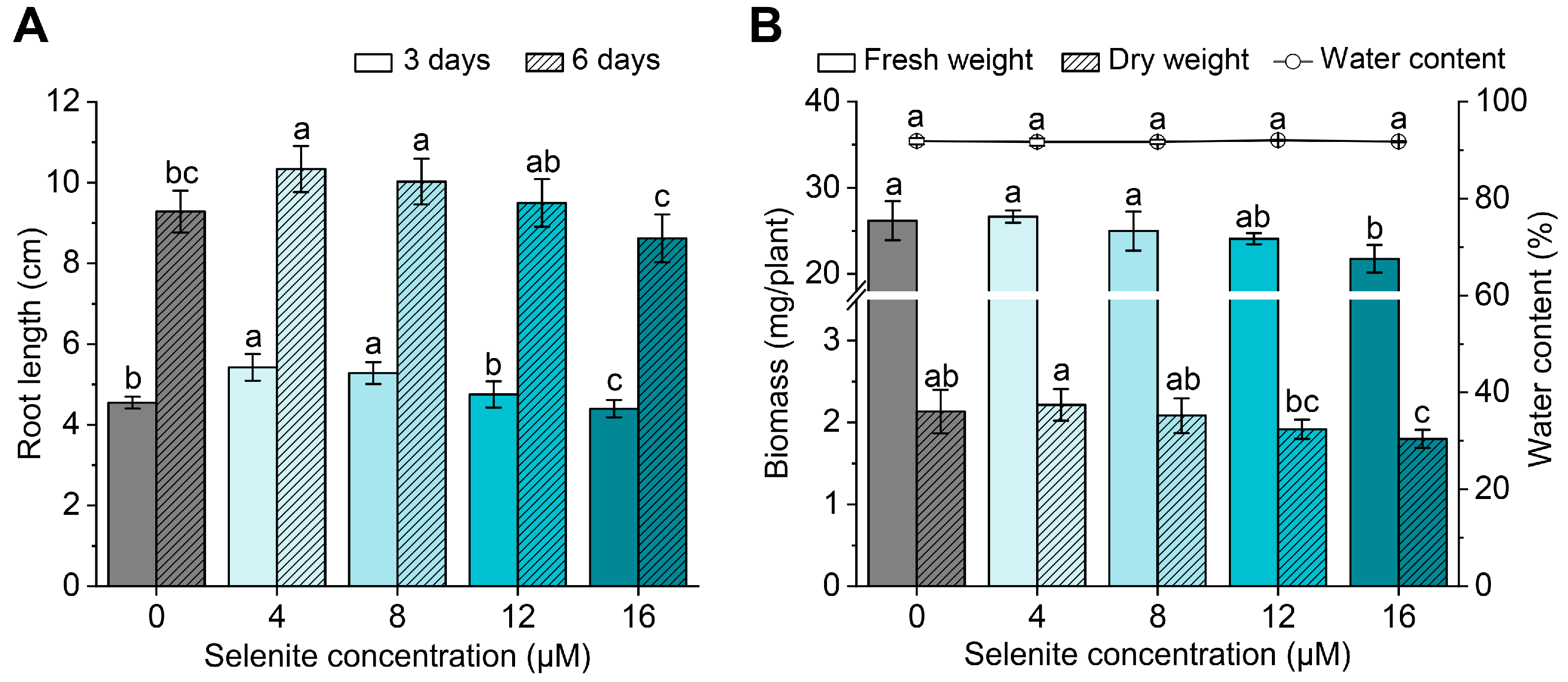
3.1. Effect of Exogenous Se Application on Seedling Growth

The germinated purple lettuce seedlings were treated with different concentrations of Se. Results showed that the root length first increased and then decreased with the increase of exogenous Se application (Figure 1A). Compared with CK, the root lengths of lettuce seedlings were significantly increased under the low concentrations of Se application (≤8 μM) after 3 and 6 days of treatment. However, the root lengths of lettuce seedlings were decreased under the high concentration of Se application (16 μM). Compared with CK, the FW and DW of lettuce seedlings were not significantly affected under the low concentrations of Se application (≤12 μM) (Figure 1B). However, the FW and DW of lettuce seedlings were significantly decreased under the high Se concentration (16 μM). Under 1~16 μM exogenous Se application, the water contents were unaffected. Overall, the young seedling growth of purple lettuce was enhanced under the low Se application concentration (≤8 μM) but inhibited under the high Se application concentration (16 μM).

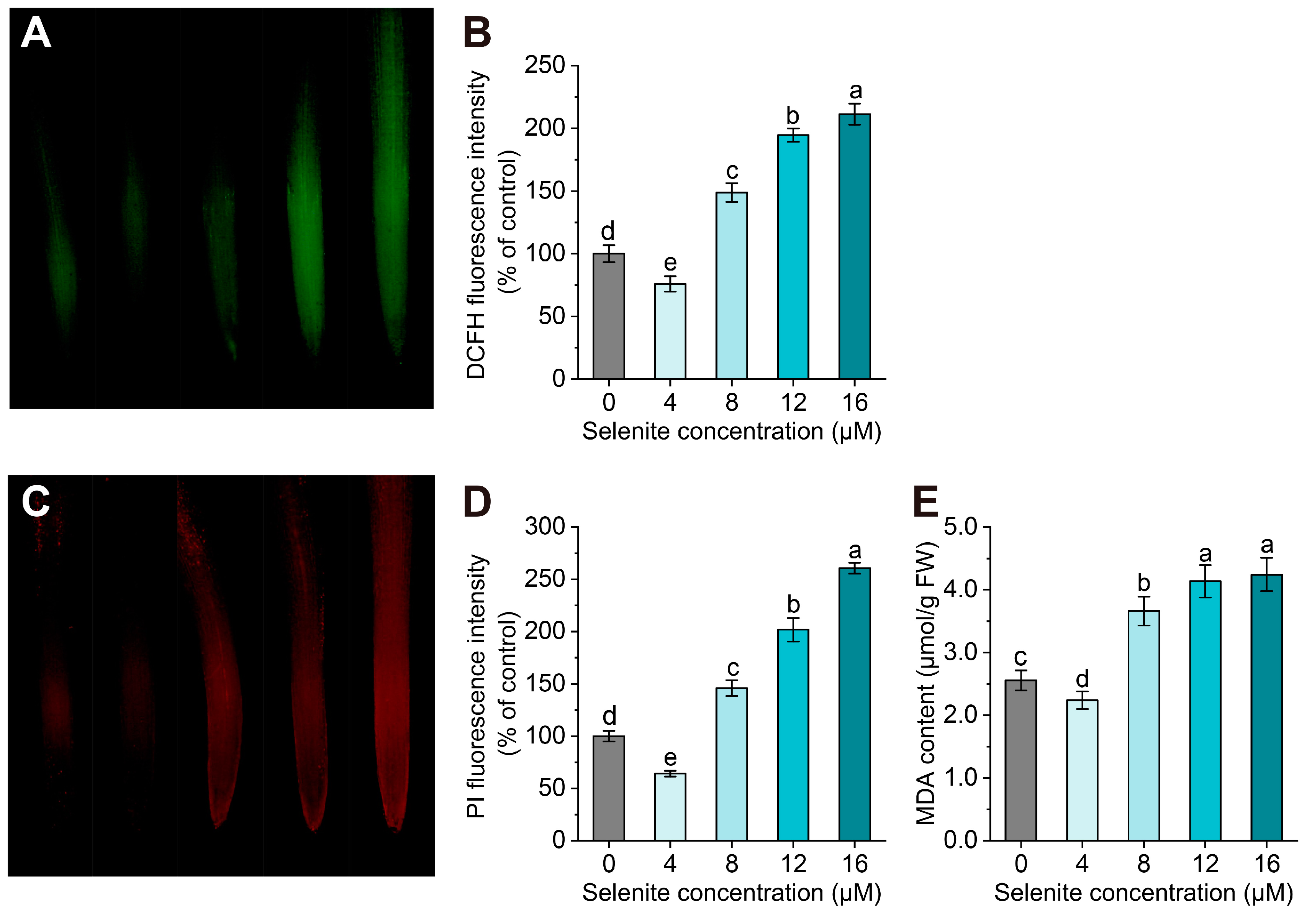
3.2. Oxidative Damage Induced by the High Se Application Concentration

Because high Se application inhibited the root length, we evaluated the oxidative state in the root of purple lettuce under different Se concentrations. High concentrations of selenite (≥8 μM) induced the accumulation of ROS in the root tips (Figure 2A,B). Moreover, the fluorescence intensity was significantly increased with the increased concentrations of selenite. However, the ROS accumulation under the 4 μM of Se application was lower than that under CK. These results indicated that the low Se application concentration inhibited the production of ROS, but the high Se application concentration induced the overproduction of ROS.
Further, we assessed the cell viability in root tips by analyzing the membrane integrity. Membrane integrity can be determined by intaking PI (reflected by red fluorescence), which can penetrate the damaged cell membrane [58]. The application of the high Se concentration (≥8 μM) caused damage to the membrane integrity (Figure 2C,D). Consistent with the changes in ROS, the application of the low Se concentration (4 μM) reduced the damage to membrane integrity.
MDA, a final product of lipid peroxidation in plants, reflects the degree of lipid peroxidation [51]. Consistent with the changes in ROS and membrane integrity, the application of the high Se concentration (≥8 μM) increased the MDA content in the roots of lettuce, compared with the CK (Figure 2E), whereas the low Se concentration (4 μM) significantly reduced the MDA level. These results indicated that the high Se application concentration induced overproduction of ROS in lettuce root tips, which causes oxidative damage to membrane lipids and cell death.

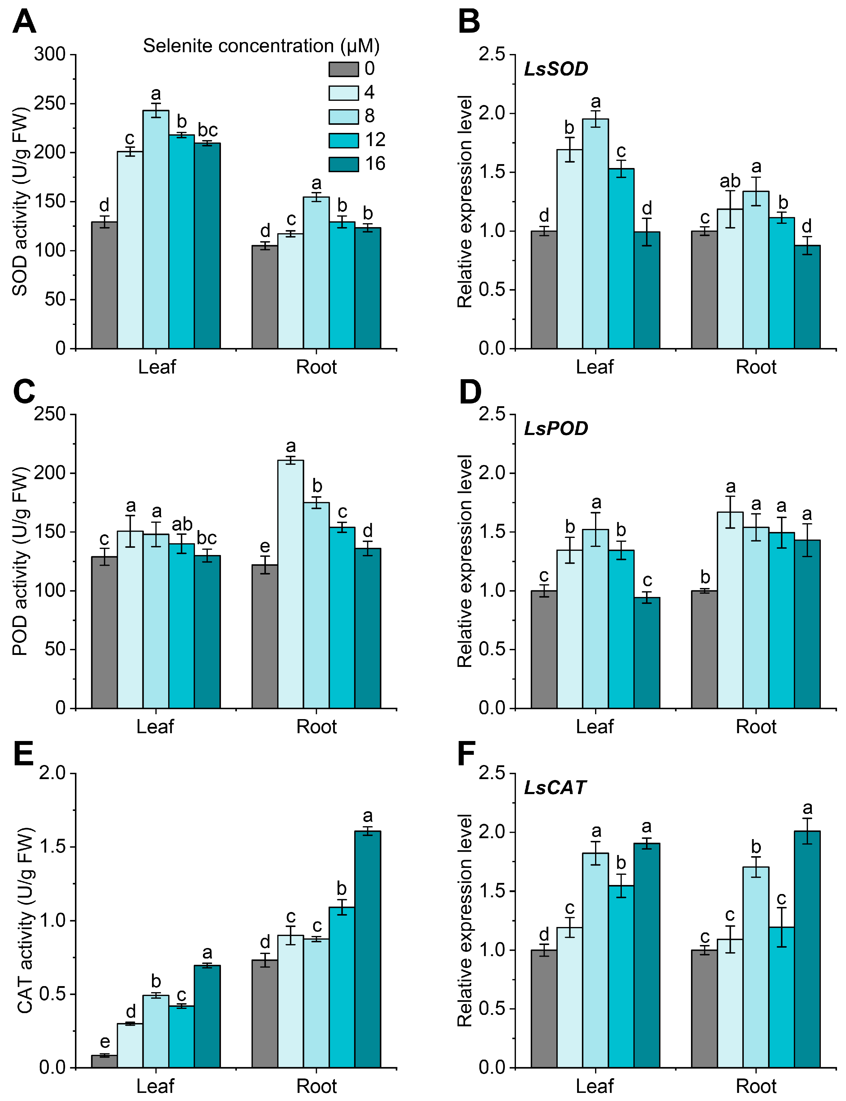
3.3. Effect of Exogenous Se Application on the Antioxidant Enzymes

The enzyme activities and relative expression of the antioxidant enzymes in the leaves and roots of purple lettuce under different Se concentrations were further determined (Figure 3). The activities of SOD, POD, and CAT in the leaves and roots of lettuce seedlings under exogenous Se application were significantly higher than those under CK. The activities of SOD and POD increased first and then decreased with the increased Se concentration. The activities of SOD and POD in the leaves and roots reached the highest values under 8 μM and 4 μM of Se application, respectively. At the same time, the CAT activities in leaves and roots were significantly increased with the increased Se concentration. The relative expression of SOD, POD, and CAT was almost consistent with the enzyme activities under different selenite concentrations. These indicated that the increased expression of these antioxidant enzymes resulted in high enzyme activities under exogenous Se application. These results suggest that Se application can significantly enhance the antioxidant capacity of lettuce seedlings.

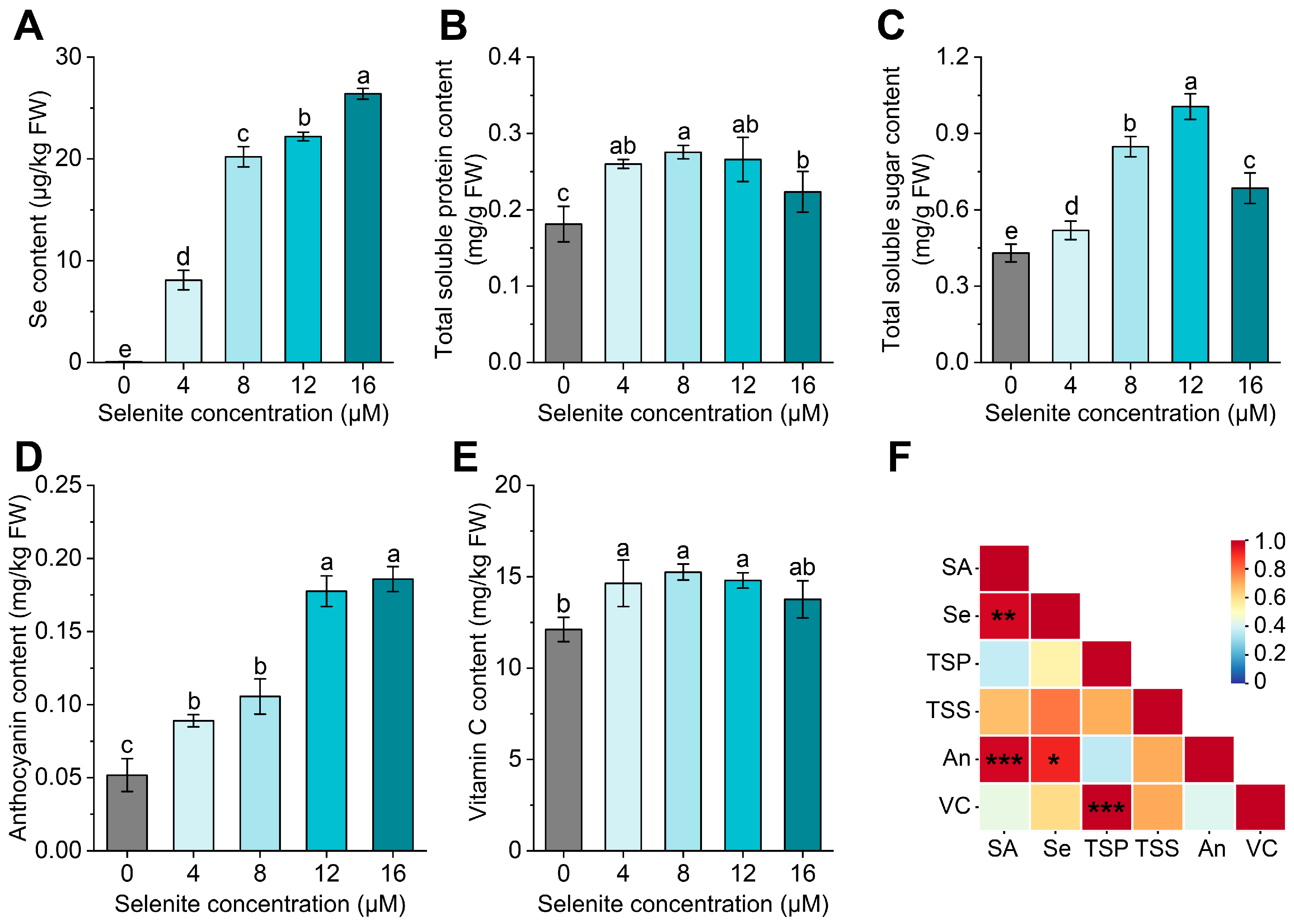
3.4. Effect of Exogenous Se Application on the Se and Nutrient Accumulation

We further determined the nutritional values of the purple lettuce seedlings under different Se applications. First, results showed that the Se contents significantly increased with the increased Se concentration applied (Figure 4A). The Se content reached the highest value under 16 μM of exogenous Se application. Under Se concentrations higher than 8 μM, the Se contents of the purple lettuce seedling were all in the high level of ≥20 μg/kg FW.
Compared with CK, the total soluble protein and total soluble sugar contents were significantly increased under exogenous Se application in the lettuce seedling (Figure 4B,C). Like the trend for the seedling growth, the total soluble protein and total soluble sugar contents were first increased and then declined with the increased Se concentration. The total soluble protein content reached its maximum values under 4~12 μM of Se concentration. Additionally, the total soluble sugar content reached its maximum value under 12 μM of Se concentration. Like the trend of Se accumulation in the lettuce seedling, the anthocyanin contents were also significantly increased with the increased Se concentration (Figure 2D). The anthocyanin content reached high values under 12 and 16 μM of exogenous Se application, almost 3.5-fold higher than that under CK. Furthermore, the vitamin C content only significantly increased by 20~25% under 4~8 μM of exogenous Se application (Figure 4E).
The Pearson correlation coefficients between the exogenous Se application concentration and these five nutritional values are shown in Figure 4F. Results showed that the exogenous Se application concentrations were significantly positively related to the Se content (p < 0.01) and anthocyanin content (p < 0.001) of the purple lettuce seedling. In addition, the vitamin C content was significantly positively related to the total soluble protein content (p < 0.001). Thus, the exogenous Se application greatly affected the Se and anthocyanin accumulation in the purple lettuce seedling. Considering the inhibition of growth under the high Se application concentration, the exogenous 8 μM of Se application is beneficial for both growth and nutrient accumulation, especially Se and anthocyanins.

3.5. The Anthocyanin Content was Positively Correlated with the Expression Level of LsCHS

For the close relationship between exogenous Se application concentration and anthocyanin contents, we determined the expression of a key gene involved in anthocyanin biosynthesis. CHS catalyzes the first step of the flavonoid biosynthesis pathway [40]. The results showed that the expression level of LsCHS was the highest in the stem, followed by the leaves, and the lowest in the roots (Figure 5A), indicating that the anthocyanin biosynthesis of purple lettuce in the seedling stage was primarily in the stems and leaves. The expression of LsCHS was significantly increased with the exogenous Se application concentrations (Figure 5B). Moreover, the expression of LsCHS was significantly positively correlated with the anthocyanin contents of purple lettuce (R2 = 0.87, p < 0.05) (Figure 5C). Therefore, Se treatment probably induces the expression of LsCHS, thus promoting anthocyanin biosynthesis and increasing the anthocyanin contents in purple lettuce.

4. Discussion

Se can trigger multiple effects in plants, including nutrition, toxicity, and detoxification, which greatly depends on the concentration of Se [10,59]. In this study, we demonstrated the effect of different exogenous Se application concentrations on the growth, antioxidative capacity, and nutritional quality of purple lettuce at the seedling stage. Similar to our previous research [23,24,26], low Se application concentrations (≤8 μM) in the hydroponic system can stimulate the growth of purple lettuce at the seedling stage, while high Se concentration (16 μM) inhibited its growth. Moreover, compared to long-term Se treatment (3 weeks) [23], it was revealed that the effect of Se treatment on growth increased with the increase of Se treatment time. In this hydroponic system, the Se concentrations between nutrition promotion and toxicity seem to be small, indicating the sensitive responses to Se concentration for purple lettuce. Nevertheless, lettuce probably has a higher tolerance to Se stress in soil, especially salt-affected soils. In the field experiments with salt-affected soils, soil and foliar application of Se lower than 100 ppm increased the head weight, leaf area, leaves’ dry weight, and chlorophyll content of lettuce [29]. In addition, Se toxicity also depends on the form of Se available [26]. Moreover, the effect of Se on the water content was related to the growth stage of lettuce. The water content of purple lettuce was not affected at the seedling stage but was significantly affected at the mature stage [23].
Se application has been reported to enhance the antioxidant metabolism in plants [10,11,31]. The application of selenite in lettuce induced the over-generation of H2O2 and MDA and a higher lipoxygenase activity in leaves [26]. Our study revealed that high Se stress (≥8 μM) results in oxidative damage (high ROS and MDA contents) in the lettuce roots. The inhibited root growth under a high Se concentration is probably caused by the oxidative damage induced by Se stress. At the same time, plants will increase the activities of antioxidant enzymes to alleviate oxidative damage. Previous studies have shown that Se-fertilized lettuce induced higher activity of the antioxidant enzymes, such as ascorbate peroxidase (APX) and glutathione (GSH) peroxidase [26]. Likewise, enhanced activities of antioxidant enzymes, SOD, POD, and CAT, were observed under Se application in leaves and roots of purple lettuce in this study. Moreover, the enhanced activities of these antioxidant enzymes were induced by the higher expression of these genes.
Lettuce is an advantageous candidate for Se biofortification [18,60]. Particularly, purple lettuce is popular for its various health benefits. Mice experiments revealed that purple lettuce can effectively decrease fat mass accumulation and increase energy expenditure to prevent body weight gain and reduce metabolic risk factors [19,32]. Our study confirmed that exogenous Se application not only greatly increased the Se content of purple lettuce but also greatly increased the contents of other nutrients, total soluble proteins and sugars, vitamin C, and anthocyanin. Particularly, anthocyanins, water-soluble pigments, can convert into various metabolites and interact with proteins and lipids [37]. These stable and tenacious compounds are involved in human health maintenance and chronic disease risk reduction. Anthocyanin content in plants is highly influenced by external and internal factors [38,61]. The increased accumulation of anthocyanins under exogenous Se application was also observed in other crops and vegetables, such as bread wheat [43,62], maize [63], radish sprouts [45], and sweet basil [46]. In this regard, varieties with high anthocyanin content generally have higher Se contents [43,44]. In addition, the anthocyanin content was also affected by the chemical form of Se and the pH of the nutrient solutions [46,63]. Researchers have attempted to explore the relationship between Se and anthocyanins. Recently, Chen et al. (2023) revealed that Se treatment increased the anthocyanin accumulation in radish sprouts through its regulation of photosynthesis and sucrose transport from cotyledon to hypocotyl induced by the up-regulation of sucrose transporters. Pu et al. (2021) revealed that the transcription factors R2R3-MYB and bHLH co-regulate anthocyanin biosynthesis and Se metabolism at the transcriptional level in wheat [44]. However, the underlying mechanisms between Se and anthocyanin accumulation in purple lettuce still need to be explored.
The synthesis pathway of anthocyanins has been elucidated in many plants [38,39,42,61]. Transcriptome analysis of red leaf lettuce revealed the main conserved genes in the anthocyanin biosynthesis pathway, including eight CHSs family, three chalone isomerases, and eight F3Hs [31,64]. Liu et al. (2017) revealed that the anthocyanin accumulation is probably due to the regulation of the expression of the UFGT LsF3H and LsUFGT in lettuce plants [23]. We revealed the increased anthocyanin content was probably induced by increased expression of LsCHS. The anthocyanin content in purple-head Chinese cabbage showed significant correlations with the expression of genes directly involved in anthocyanin biosynthesis, including BrCHSs, BrCHI2, BrF3Hs BrF3′H1, and BrUGTs. Thus, the synthesis of anthocyanins in lettuce is probably controlled by multiple genes. This LsCHS gene from Lactuca sativa var. crispa L. ‘Purple Rome’ is 1197 bp and encodes a 43.561 kDa protein. The putative protein sequence of LsCHS exhibits 100% identity to CHS from Lactuca sativa L. cultivar Salinas (XP_023735557). The LsCHS also shows high homologies with the CHSs from other plants in Asteraceae, such as Erigeron canadensis (97%, XP_043631312), Chrysanthemum boreale (96%, AGU91424), and Artemisia annua (95%, PWA65027). GhCHS1 from Gerbera hybrida (Asteraceae) was shown to contribute to flavonoid biosynthesis [40]. SmCHS1 and SmCHS3 probably play a key role in the anthocyanin and silymarin biosynthesis in milk thistle (Silybum marianum) [65]. The LsCHS in this study is probably a key gene in the synthesis of anthocyanins. Now, the function and regulatory mechanism of LsCHS are analyzed in progress for providing genetic resources for the biosynthesis of anthocyanins in purple lettuce.

5. Conclusions

Enough Se supplementation is crucial for meeting nutritional recommendations and requirements. In this study, we revealed that the exogenous Se application (≤8 μM) in the hydroponic system not only promoted the growth and antioxidant capacity of purple lettuce seedlings but also enhanced the accumulation of various nutrients, especially Se and anthocyanins. Thus, 8 μM sodium selenite was suggested as the appropriate application Se concentration for high growth and nutritional quality. Purple lettuce enriched in Se and anthocyanin has excellent potential as a functional food in preventing metabolic disorders, especially for Se-deficient populations.

Supplementary Materials

The following supporting information can be downloaded at: https://www.mdpi.com/article/10.3390/agronomy13071664/s1, Table S1: Primer sequences used in this study.

Author Contributions

Conceptualization, L.Y. and S.H.; methodology, Z.Y.; formal analysis, M.L.; investigation, Z.Y. and S.H.; resources, L.Y. and M.L.; writing—original draft preparation, M.L. and S.H.; writing—review and editing, M.L., S.H., Y.Y., J.Z., J.C. and L.Y.; supervision, J.Z. and L.Y.; project administration, J.Z. and L.Y.; funding acquisition, J.C. and L.Y. All authors have read and agreed to the published version of the manuscript.

Funding

This research was funded by the National Key R&D Program of China (2022YFD2100601), China Agriculture Research System (CARS-23-B16), and 2021 Youth Innovative Project in Jiangsu Coast Development Group Co., Ltd.

Institutional Review Board Statement

Not applicable.

Informed Consent Statement

Not applicable.

Data Availability Statement

The data for this study are included in the main document.

Acknowledgments

The authors thank Zhiwei Bian, Dandan Liu, Yongzhu Wang, and Neng Han from the Nanjing Agricultural University for their assistance with the experiments. The authors also thank Qizhi Shi and Yonglan Xi from the Jiangsu Academy of Agriculture Sciences for their help and discussion. We are also grateful to the reviewers for their useful comments and suggestions for improving the final manuscript.

Conflicts of Interest

The authors declare no conflict of interest.

References

  1. Hatfield, D.L.; Tsuji, P.A.; Carlson, B.A.; Gladyshev, V.N. Selenium and selenocysteine: Roles in cancer, health, and development. Trends Biochem. Sci. 2014, 39, 112–120. [Google Scholar] [CrossRef] [Green Version]
  2. Saito, Y. Selenium transport mechanism via selenoprotein P-its physiological role and related diseases. Front. Nutr. 2021, 8, 685517. [Google Scholar] [CrossRef] [PubMed]
  3. Rayman, M.P. The importance of selenium to human health. Lancet 2000, 356, 233–241. [Google Scholar] [CrossRef] [PubMed] [Green Version]
  4. Benstoem, C.; Goetzenich, A.; Kraemer, S.; Borosch, S.; Manzanares, W.; Hardy, G.; Stoppe, C. Selenium and its supplementation in cardiovascular disease—What do we know? Nutrients 2015, 7, 3094–3118. [Google Scholar] [CrossRef] [Green Version]
  5. Jones, G.D.; Droz, B.; Greve, P.; Gottschalk, P.; Poffet, D.; McGrath, S.P.; Seneviratne, S.I.; Smith, P.; Winkel, L.H. Selenium deficiency risk predicted to increase under future climate change. Proc. Natl. Acad. Sci. USA 2017, 114, 2848–2853. [Google Scholar] [CrossRef] [Green Version]
  6. Moreno-Reyes, R.; Suetens, C.; Mathieu, F.; Begaux, F.; Zhu, D.; Rivera, M.T.; Boelaert, M.; Nève, J.; Perlmutter, N.; Vanderpas, J. Kashin–Beck osteoarthropathy in rural Tibet in relation to selenium and iodine status. N. Engl. J. Med. 1998, 339, 1112–1120. [Google Scholar] [CrossRef] [Green Version]
  7. Liu, H.; Yu, F.; Shao, W.; Ding, D.; Yu, Z.; Chen, F.; Geng, D.; Tan, X.; Lammi, M.J.; Guo, X. Associations between Selenium content in hair and Kashin-Beck disease/Keshan disease in children in northwestern China: A prospective cohort study. Biol. Trace Elem. Res. 2018, 184, 16–23. [Google Scholar] [CrossRef] [PubMed]
  8. Finley, J.W. Increased intakes of selenium-enriched foods may benefit human health. J. Sci. Food Agric. 2007, 87, 1620–1629. [Google Scholar] [CrossRef]
  9. Newman, R.; Waterland, N.; Moon, Y.; Tou, J.C. Selenium biofortification of agricultural crops and effects on plant nutrients and bioactive compounds important for human health and disease prevention—A review. Plant Foods Hum. Nutr. 2019, 74, 449–460. [Google Scholar] [CrossRef]
  10. Lanza, M.; Reis, A.R.D. Roles of selenium in mineral plant nutrition: ROS scavenging responses against abiotic stresses. Plant Physiol. Biochem. 2021, 164, 27–43. [Google Scholar] [CrossRef] [PubMed]
  11. Jiang, C.; Zu, C.; Lu, D.; Zheng, Q.; Shen, J.; Wang, H.; Li, D. Effect of exogenous selenium supply on photosynthesis, Na+ accumulation and antioxidative capacity of maize (Zea mays L.) under salinity stress. Sci. Rep. 2017, 7, 42039. [Google Scholar] [CrossRef] [PubMed] [Green Version]
  12. Andrade, F.R.; da Silva, G.N.; Guimarães, K.C.; Barreto, H.B.F.; de Souza, K.R.D.; Guilherme, L.R.G.; Faquin, V.; Reis, A.R.D. Selenium protects rice plants from water deficit stress. Ecotoxicol. Environ. Saf. 2018, 164, 562–570. [Google Scholar] [CrossRef] [Green Version]
  13. Feng, R.; Wei, C.; Tu, S. The roles of selenium in protecting plants against abiotic stresses. Environ. Exp. Bot. 2013, 87, 58–68. [Google Scholar] [CrossRef]
  14. Fordyce, F.M. Selenium Deficiency and Toxicity in the Environment. In Essentials of Medical Geology: Revised Edition; Selinus, O., Ed.; Springer: Dordrecht, The Netherlands, 2013; pp. 375–416. [Google Scholar]
  15. Danso, O.P.; Asante-Badu, B.; Zhang, Z.; Song, J.; Wang, Z.; Yin, X.; Zhu, R. Selenium biofortification: Strategies, progress and challenges. Agriculture 2023, 13, 416. [Google Scholar] [CrossRef]
  16. Zhang, L.; Chu, C. Selenium uptake, transport, metabolism, reutilization, and biofortification in rice. Rice 2022, 15, 30. [Google Scholar] [CrossRef]
  17. Puccinelli, M.; Malorgio, F.; Pezzarossa, B. Selenium enrichment of horticultural crops. Molecules 2017, 22, 933. [Google Scholar] [CrossRef] [PubMed]
  18. Kim, M.J.; Moon, Y.; Tou, J.C.; Mou, B.; Waterland, N.L. Nutritional value, bioactive compounds and health benefits of lettuce (Lactuca sativa L.). J. Food Compost. Anal. 2016, 49, 19–34. [Google Scholar] [CrossRef]
  19. Nicolle, C.; Cardinault, N.; Gueux, E.; Jaffrelo, L.; Rock, E.; Mazur, A.; Amouroux, P.; Rémésy, C. Health effect of vegetable-based diet: Lettuce consumption improves cholesterol metabolism and antioxidant status in the rat. Clin. Nutr. 2004, 23, 605–614. [Google Scholar] [CrossRef]
  20. Sager, M. Selenium in agriculture, food, and nutrition. Pure Appl. Chem. 2006, 78, 111–133. [Google Scholar] [CrossRef]
  21. Businelli, D.; D’Amato, R.; Onofri, A.; Tedeschini, E.; Tei, F. Se-enrichment of cucumber ( Cucumis sativus L.), lettuce (Lactuca sativa L.) and tomato (Solanum lycopersicum L. Karst) through fortification in pre-transplanting. Sci. Hortic. 2015, 197, 697–704. [Google Scholar] [CrossRef]
  22. Hawrylak-Nowak, B. Comparative effects of selenite and selenate on growth and selenium accumulation in lettuce plants under hydroponic conditions. Plant Growth Regul. 2013, 70, 149–157. [Google Scholar] [CrossRef] [Green Version]
  23. Liu, D.; Li, H.; Wang, Y.; Ying, Z.; Bian, Z.; Zhu, W.; Liu, W.; Yang, L.; Jiang, D. How exogenous selenium affects anthocyanin accumulation and biosynthesis-related gene expression in purple lettuce. Pol. J. Environ. Stud. 2017, 26, 717–723. [Google Scholar] [CrossRef] [PubMed]
  24. Ríos, J.J.; Rosales, M.A.; Blasco, B.; Cervilla, L.M.; Romero, L.; Ruiz, J.M. Biofortification of Se and induction of the antioxidant capacity in lettuce plants. Sci. Hortic. 2008, 116, 248–255. [Google Scholar] [CrossRef]
  25. Pannico, A.; El-Nakhel, C.; Kyriacou, M.C.; Giordano, M.; Stazi, S.R.; De Pascale, S.; Rouphael, Y. Combating micronutrient deficiency and enhancing food functional quality through selenium fortification of select lettuce genotypes grown in a closed soilless system. Front. Plant Sci. 2019, 10, 1495. [Google Scholar] [CrossRef] [Green Version]
  26. Ríos, J.J.; Blasco, B.; Cervilla, L.M.; Rosales, M.A.; Sanchez-Rodriguez, E.; Romero, L.; Ruiz, J.M. Production and detoxification of H2O2 in lettuce plants exposed to selenium. Ann. Appl. Biol. 2009, 154, 107–116. [Google Scholar] [CrossRef]
  27. Smoleń, S.; Kowalska, I.; Czernicka, M.; Halka, M.; Keska, K.; Sady, W. Iodine and selenium biofortification with additional application of salicylic acid affects yield, selected molecular parameters and chemical composition of lettuce plants (Lactuca sativa L. var. capitata). Front. Plant Sci. 2016, 7, 1553. [Google Scholar] [CrossRef] [Green Version]
  28. Smoleń, S.; Kowalska, I.; Sady, W. Assessment of biofortification with iodine and selenium of lettuce cultivated in the NFT hydroponic system. Sci. Hortic. 2014, 166, 9–16. [Google Scholar] [CrossRef]
  29. Shalaby, T.; Bayoumi, Y.; Alshaal, T.; Elhawat, N.; Sztrik, A.; El-Ramady, H. Selenium fortification induces growth, antioxidant activity, yield and nutritional quality of lettuce in salt-affected soil using foliar and soil applications. Plant Soil 2017, 421, 245–258. [Google Scholar] [CrossRef]
  30. Abdalla, M.A.; Wick, J.E.; Famuyide, I.M.; McGaw, L.J.; Mühling, K.H. Selenium enrichment of green and red lettuce and the induction of radical scavenging potential. Horticulturae 2021, 7, 488. [Google Scholar] [CrossRef]
  31. Wang, Y.; Ye, X.; Yang, K.; Shi, Z.; Wang, N.; Yang, L.; Chen, J. Characterization, expression, and functional analysis of polyamine oxidases and their role in selenium-induced hydrogen peroxide production in Brassica rapa. J. Sci. Food Agric. 2019, 99, 4082–4093. [Google Scholar] [CrossRef]
  32. Han, Y.; Zhao, C.; He, X.; Sheng, Y.; Ma, T.; Sun, Z.; Liu, X.; Liu, C.; Fan, S.; Xu, W.; et al. Purple lettuce (Lactuca sativa L.) attenuates metabolic disorders in diet induced obesity. J. Funct. Foods 2018, 45, 462–470. [Google Scholar] [CrossRef]
  33. Drossard, C.; Alexy, U.; Bolzenius, K.; Kunz, C.; Kersting, M. Anthocyanins in the diet of infants and toddlers: Intake, sources and trends. Eur. J. Nutr. 2011, 50, 705–711. [Google Scholar] [CrossRef] [PubMed]
  34. Piberger, H.; Oehme, A.; Hofmann, C.; Dreiseitel, A.; Sand, P.G.; Obermeier, F.; Schoelmerich, J.; Schreier, P.; Krammer, G.; Rogler, G. Bilberries and their anthocyanins ameliorate experimental colitis. Mol. Nutr. Food Res. 2011, 55, 1724–1729. [Google Scholar] [CrossRef] [PubMed]
  35. Fernandes, I.; Pérez-Gregorio, R.; Soares, S.; Mateus, N.; de Freitas, V. Wine flavonoids in health and disease prevention. Molecules 2017, 22, 292. [Google Scholar] [CrossRef]
  36. Jamar, G.; Estadella, D.; Pisani, L.P. Contribution of anthocyanin-rich foods in obesity control through gut microbiota interactions. Biofactors 2017, 43, 507–516. [Google Scholar] [CrossRef] [PubMed]
  37. Lila, M.A.; Burton-Freeman, B.; Grace, M.; Kalt, W. Unraveling anthocyanin bioavailability for human health. Annu. Rev. Food Sci. Technol. 2016, 7, 375–393. [Google Scholar] [CrossRef]
  38. Koes, R.; Verweij, W.; Quattrocchio, F. Flavonoids: A colorful model for the regulation and evolution of biochemical pathways. Trends Plant Sci. 2005, 10, 236–242. [Google Scholar] [CrossRef]
  39. Broun, P. Transcriptional control of flavonoid biosynthesis: A complex network of conserved regulators involved in multiple aspects of differentiation in Arabidopsis. Curr. Opin. Plant Biol. 2005, 8, 272–279. [Google Scholar] [CrossRef]
  40. Deng, X.; Bashandy, H.; Ainasoja, M.; Kontturi, J.; Pietiainen, M.; Laitinen, R.A.E.; Albert, V.A.; Valkonen, J.P.T.; Elomaa, P.; Teeri, T.H. Functional diversification of duplicated chalcone synthase genes in anthocyanin biosynthesis of Gerbera hybrida. New Phytol. 2014, 201, 1469–1483. [Google Scholar] [CrossRef]
  41. Morata, A.; López, C.; Tesfaye, W.; González, C.; Escott, C. Anthocyanins as Natural Pigments in Beverages. In Value-Added Ingredients and Enrichments of Beverages; Elsevier: Amsterdam, The Netherlands, 2019; pp. 383–428. [Google Scholar]
  42. Zhang, Y.; Cheng, Y.; Ya, H.; Xu, S.; Han, J. Transcriptome sequencing of purple petal spot region in tree peony reveals differentially expressed anthocyanin structural genes. Front. Plant Sci. 2015, 6, 964. [Google Scholar] [CrossRef] [Green Version]
  43. Xia, Q.; Yang, Z.; Shui, Y.; Liu, X.; Chen, J.; Khan, S.; Wang, J.; Gao, Z. Methods of selenium application differentially modulate plant growth, selenium accumulation and speciation, protein, anthocyanins and concentrations of mineral elements in purple-grained wheat. Front. Plant Sci. 2020, 11, 1114. [Google Scholar] [CrossRef]
  44. Pu, Z.; Wei, G.; Liu, Z.; Chen, L.; Guo, H.; Li, Y.; Li, Y.; Dai, S.; Wang, J.; Li, W.; et al. Selenium and anthocyanins share the same transcription factors R2R3MYB and bHLH in wheat. Food Chem. 2021, 356, 129699. [Google Scholar] [CrossRef]
  45. Chen, J.; Chen, H.; Wang, H.; Zhan, J.; Yuan, X.; Cui, J.; Su, N. Selenium treatment promotes anthocyanin accumulation in radish sprouts (Raphanus sativus L.) by its regulation of photosynthesis and sucrose transport. Food Res. Int. 2023, 165, 112551. [Google Scholar] [CrossRef]
  46. Skrypnik, L.; Novikova, A.; Tokupova, E. Improvement of phenolic compounds, essential oil content and antioxidant properties of sweet basil (Ocimum basilicum L.) depending on type and concentration of selenium application. Plants 2019, 8, 458. [Google Scholar] [CrossRef] [PubMed] [Green Version]
  47. Kenchanmane Raju, S.K.; Barnes, A.C.; Schnable, J.C.; Roston, R.L. Low-temperature tolerance in land plants: Are transcript and membrane responses conserved? Plant Sci. 2018, 276, 73–86. [Google Scholar] [CrossRef] [PubMed]
  48. Nielsen, S.L.; Pedersen, M.F. Growth, photosynthesis and nutrient content of seedlings and mature plants of Cymodocea nodosa—The importance of clonal integration. Aquatic Botany 2000, 68, 265–271. [Google Scholar] [CrossRef]
  49. Zhou, W.; Liang, X.; Zhang, Y.; Li, K.; Jin, B.; Lu, L.; Jin, C.; Lin, X. Reduced nitrogen supply enhances the cellular antioxidant potential of phenolic extracts through alteration of the phenolic composition in lettuce (Lactuca sativa L.). J. Sci. Food Agric. 2019, 99, 4761–4771. [Google Scholar] [CrossRef]
  50. Iakimova, E.T.; Woltering, E.J. The wound response in fresh-cut lettuce involves programmed cell death events. Protoplasma 2018, 255, 1225–1238. [Google Scholar] [CrossRef] [Green Version]
  51. Zhang, S.; Sun, S.W.; Shi, H.L.; Zhao, K.; Wang, J.; Liu, Y.; Liu, X.H.; Wang, W. Physiological and biochemical mechanisms mediated by allelochemical isoliquiritigenin on the growth of lettuce seedlings. Plants 2020, 9, 245. [Google Scholar] [CrossRef] [Green Version]
  52. Ma, J.; Du, G.; Li, X.; Zhang, C.; Guo, J. A major locus controlling malondialdehyde content under water stress is associated with Fusarium crown rot resistance in wheat. Mol. Genet. Genom. 2015, 290, 1955–1962. [Google Scholar] [CrossRef]
  53. Gunes, A.; Turan, M.; Kitir, N.; Tufenkci, M.S.; Cimrin, K.M.; Yildirim, E.; Ercisli, S. Effects of bio-bor fertilizer applications on fruit yield, antioxidant enzyme activity and freeze injury of strawberry. Erwerbsobstbau 2016, 58, 177–184. [Google Scholar] [CrossRef]
  54. Livak, K.J.; Schmittgen, T.D. Analysis of relative gene expression data using real-time quantitative PCR and the 2−ΔΔCT Method. Methods 2001, 25, 402–408. [Google Scholar] [CrossRef] [PubMed]
  55. Verma, P.; Hiremani, N.S.; Gawande, S.P.; Sain, S.K.; Nagrale, D.T.; Narkhedkar, N.G.; Prasad, Y.G. Modulation of plant growth and antioxidative defense system through endophyte biopriming in cotton (Gossypium spp.) and non-host crops. Heliyon 2022, 8, e09487. [Google Scholar] [CrossRef]
  56. Sun, H.; Zhang, P.; Zhu, Y.; Lou, Q.; He, S. Antioxidant and prebiotic activity of five peonidin-based anthocyanins extracted from purple sweet potato (Ipomoea batatas (L.) Lam.). Sci. Rep. 2018, 8, 5018. [Google Scholar] [CrossRef] [Green Version]
  57. Santos, F.T.; Goufo, P.; Santos, C.; Botelho, D.; Fonseca, J.; Queirós, A.; Costa, M.S.; Trindade, H. Comparison of five agro-industrial waste-based composts as growing media for lettuce: Effect on yield, phenolic compounds and vitamin C. Food Chem. 2016, 209, 293–301. [Google Scholar] [CrossRef]
  58. Zhao, L.; Zhao, M.Y.; Phey, C.P.; Yang, H. Efficacy of low concentration acidic electrolysed water and levulinic acid combination on fresh organic lettuce (Lactuca sativa Var. Crispa L.) and its antimicrobial mechanism. Food Control 2019, 101, 241–250. [Google Scholar] [CrossRef]
  59. Moretti, C.; Bocchini, M.; Quaglia, M.; Businelli, D.; Orfei, B.; Buonaurio, R. Sodium selenate: An environmental-friendly means to control tomato bacterial speck disease. Agronomy 2022, 12, 1351. [Google Scholar] [CrossRef]
  60. Materska, M.; Olszówka, K.; Chilczuk, B.; Stochmal, A.; Pecio, Ł.; Pacholczyk-Sienicka, B.; Piacente, S.; Pizza, C.; Masullo, M. Polyphenolic profiles in lettuce (Lactuca sativa L.) after CaCl2 treatment and cold storage. Euro. Food Res. Technol. 2018, 245, 733–744. [Google Scholar] [CrossRef] [Green Version]
  61. He, Q.; Ren, Y.; Zhao, W.; Li, R.; Zhang, L. Low temperature promotes anthocyanin biosynthesis and related gene expression in the seedlings of purple head Chinese cabbage (Brassica rapa L.). Genes 2020, 11, 81. [Google Scholar] [CrossRef] [PubMed] [Green Version]
  62. Zhang, F.; Li, X.; Wu, Q.; Lu, P.; Kang, Q.; Zhao, M.; Wang, A.; Dong, Q.; Sun, M.; Yang, Z.; et al. Selenium application enhances the accumulation of flavones and anthocyanins in bread wheat (Triticum aestivum L.). Grains. J. Agric. Food Chem. 2022, 70, 13431–13444. [Google Scholar] [CrossRef]
  63. Hawrylak-Nowak, B. Changes in anthocyanin content as indicator of maize sensitivity to selenium. J. Plant Nutr. 2008, 31, 1232–1242. [Google Scholar] [CrossRef]
  64. Zhang, Y.Z.; Xu, S.Z.; Cheng, Y.W.; Ya, H.Y.; Han, J.M. Transcriptome analysis and anthocyanin-related genes in red leaf lettuce. Genet. Mol. Res. 2016, 15, 15017023. [Google Scholar] [CrossRef] [PubMed]
  65. Sanjari, S.; Shobbar, Z.S.; Ebrahimi, M.; Hasanloo, T.; Sadat-Noori, S.A.; Tirnaz, S. Chalcone synthase genes from milk thistle (Silybum marianum): Isolation and expression analysis. J. Genet. 2015, 94, 611–617. [Google Scholar] [CrossRef] [PubMed]
Figure 1. Effect of exogenous Se application on the growth of purple lettuce seedlings. (A) Root length and (B) biomass and water content. The germinated seeds of purple lettuce were grown in water for 3 days and then transferred to the water with five Se concentrations (0, 4, 8, 12, and 16 μM) for 3 and 6 days of Se treatment. The biomass and water content in B were determined after 6 days of Se treatment. Data are mean ± standard deviation from three independent experiments. Each value in A and B was determined from 10 seedlings and 6 seedlings, respectively. Different letters above the columns or dots represent significant differences among different Se concentrations using the least significant difference-based multiple range test at p < 0.05.
Agronomy 13 01664 g001
Figure 2. Effect of exogenous Se application on the oxidative state of purple lettuce roots. (A) Representative images of ROS staining in the root tips. The green signal indicates ROS generation (DCFH fluorescence). (B) Estimation of DCFH fluorescence intensity. (C) Representative images of PI staining in the root tips. The red signal indicates the cells with damaged membranes. (D) Estimation of PI fluorescence intensity. (E) MDA content in roots. The germinated seeds of purple lettuce were grown in water for 3 days and then transferred to the water with five Se concentrations (0, 4, 8, 12, and 16 μM) for 3 days. Roots were collected for histochemical analysis. Data are mean ± standard deviation from three independent experiments. Each value in (B,D,E) was determined from roots from 4 plants. Different letters above the columns represent significant differences among different Se concentrations using the least significant difference-based multiple range test at p < 0.05.
Agronomy 13 01664 g002
Figure 3. Effect of exogenous Se application on the antioxidant enzyme activities (A,C,E) and their relative expression levels (B,D,F) in the leaves and roots of purple lettuce. The germinated seeds of purple lettuce were grown in water for 3 days and then transferred to the water with five Se concentrations (0, 4, 8, 12, and 16 μM) for 3 days. Leaves and roots of seedlings were separated for analysis. Data are mean ± standard deviation from three independent experiments. Each value was determined from the mixtures of leaves or roots from 6 plants. Different letters above the columns represent significant differences among different Se concentrations using the least significant difference-based multiple range test at p < 0.05.
Agronomy 13 01664 g003
Figure 4. Effect of exogenous Se application on the nutrient accumulation of the purple lettuce seedling. (A) Se content, (B) total soluble protein (TSP) content, (C) total soluble sugar (TSS) content, (D) anthocyanin (An) content, (E) vitamin C (VC) content, and (F) heatmap depicting Pearson’s correlation coefficients between the exogenous Se application (SA) concentration and five nutritional values. The germinated seeds of purple lettuce were grown in water for 3 days and then transferred to the water with five Se concentrations (0, 4, 8, 12, and 16 μM) for 3 days for plant analysis. In (AE), data are mean ± standard deviation from three independent experiments. Each value was determined from 4 seedlings. Different letters above the columns represent significant differences among different Se concentrations using the least significant difference-based multiple range test at p < 0.05. In (F), *, **, and *** indicate significant correlations at the 0.05, 0.01, and 0.001 levels, respectively.
Agronomy 13 01664 g004
Figure 5. Gene expression of LsCHS. (A) The relative expression level of LsCHS in different organs. (B) The relative expression level of LsCHS in the seedlings under different exogenous Se application concentrations. (C) Correction analysis of the expression level of LsCHS and anthocyanin contents. The germinated seeds of purple lettuce were grown in water for 3 days and then transferred to the water with five Se concentrations (0, 4, 8, 12, and 16 μM) for 3 days for gene expression analysis. Data are mean ± standard deviation from three independent experiments. Each value was determined from different organs from 6 plants. Different letters above the columns in (A,B) represent significant differences using the least significant difference-based multiple range test at p < 0.05.
Agronomy 13 01664 g005
Disclaimer/Publisher’s Note: The statements, opinions and data contained in all publications are solely those of the individual author(s) and contributor(s) and not of MDPI and/or the editor(s). MDPI and/or the editor(s) disclaim responsibility for any injury to people or property resulting from any ideas, methods, instructions or products referred to in the content.

